Source
and
Purity
of
Dengue-Viral
Preparations
Impact
Requirement
for
Enhancing
Antibody
to
Induce
Elevated
IL-1
β
Secretion:
A
Primary
Human
Monocyte
Model
JustinB.Callaway1,2,ScottA.Smith3,4,DouglasG.Widman1¤,KarenP.McKinnon1,2, FrankScholle5,GregoryD.Sempowski6,DirkP.Dittmer1,2,JamesE.Crowe,Jr.3,7,8, AravindaM.deSilva1,JennyP.-Y.Ting1,2,9*
1DepartmentofMicrobiologyandImmunology,TheUniversityofNorthCarolinaatChapelHill,ChapelHill,
NC,UnitedStatesofAmerica,2TheLinebergerComprehensiveCancerCenter,TheUniversityofNorth
CarolinaatChapelHill,ChapelHill,NC,UnitedStatesofAmerica,3TheVanderbiltVaccineCenter,
VanderbiltMedicalCenter,Nashville,TN,UnitedStatesofAmerica,4DepartmentofMedicine,Vanderbilt
MedicalCenter,Nashville,TN,UnitedStatesofAmerica,5DepartmentofBiologicalSciences,North
CarolinaStateUniversity,Raleigh,NC,UnitedStatesofAmerica,6DukeHumanVaccineInstitute,Durham,
NC,UnitedStatesofAmerica,7DepartmentofPathology,MicrobiologyandImmunology,Vanderbilt
MedicalCenter,Nashville,TN,UnitedStatesofAmerica,8DepartmentofPediatrics,VanderbiltMedical
Center,Nashville,TN,UnitedStatesofAmerica,9DepartmentofGenetics,TheUniversityofNorthCarolina
atChapelHill,ChapelHill,NC,UnitedStatesofAmerica
¤ Currentaddress:DepartmentofEpidemiology,TheUniversityofNorthCarolinaatChapelHill,ChapelHill,
NC,UnitedStatesofAmerica
Abstract
Denguevirusisamajorglobalhealththreatandcanleadtolife-threateninghemorrhagic complicationsduetoimmuneactivationandcytokineproduction.Cross-reactiveantibodies toanearlierdenguevirusinfectionarearecognizedriskfactorforseveredisease.These antibodiesbindheterologousdengueserotypesandenhanceinfectioninto Fc-receptor-bearingcells,aprocessknownasantibody-dependentenhancementofinfection.One crucialcytokineseenelevatedinseveredenguepatientsisIL-1β,apotentinflammatory cytokinematuredbytheinflammasome.Weusedahighly-physiologicsystembystudying antibody-dependentenhancementofIL-1βinprimaryhumanmonocyteswithanti-dengue humanmonoclonalantibodiesisolatedfrompatients.Antibody-enhancementincreased viralreplicationinprimaryhumanmonocytesinoculatedwithsupernatantharvestedfrom Verocellsinfectedwithdenguevirusserotype2(DENV-2)16681.Surprisingly,IL-1β
secre-tioninducedbyinfectioussupernatantharvestedfromtwoindependentVerocelllineswas notenhancedbyantibody.Secretionofmultipleotherinflammatorycytokineswasalso independentofantibodysignaling.However,IL-1βsecretiondidrequireNLRP3and cas-pase-1activity.Immunodepletionofdenguevirionsfromtheinfectioussupernatant
con-firmedthatviruswasnotthemainIL-1β-inducingagent,suggestingthatasupernatant component(s)notassociatedwiththevirioninducedIL-1βproduction.WeexcludedRNA, DNA,contaminatingLPS,viralNS1protein,complement,andcytokines.Incontrast, Citation:CallawayJB,SmithSA,WidmanDG,
McKinnonKP,ScholleF,SempowskiGD,etal. (2015)SourceandPurityofDengue-Viral PreparationsImpactRequirementforEnhancing AntibodytoInduceElevatedIL-1βSecretion:A PrimaryHumanMonocyteModel.PLoSONE10(8): e0136708.doi:10.1371/journal.pone.0136708
Editor:XiaJin,UniversityofRochester,UNITED STATES
Received:July18,2014
Accepted:August7,2015
Published:August24,2015
Copyright:©2015Callawayetal.Thisisanopen accessarticledistributedunderthetermsofthe CreativeCommonsAttributionLicense,whichpermits unrestricteduse,distribution,andreproductioninany medium,providedtheoriginalauthorandsourceare credited.
DataAvailabilityStatement:Allrelevantdataare withinthepaper.
purified Vero-derived DENV-2 16681 exhibited antibody-enhancement of both infection and IL-1βinduction. Furthermore, C6/36 mosquito cells did not produce such an inflammatory component, as crude supernatant harvested from insect cells infected with DENV-2 16681 induced antibody-dependent IL-1βsecretion. This study indicates that Vero cells infected with DENV-2 16681 may produce inflammatory components during dengue virus propagation that mask the virus-specific immune response. Thus, the choice of host cell and viral purity should be carefully considered, while insect-derived virus represents a system that elicits anti-body-dependent cytokine responses to dengue virus with fewer confounding issues.
Introduction
With an estimated 390 million global infections per year, dengue virus (DENV) is the most burdensome arbovirus in the world [1]. The four distinct serotypes (DENV-1–DENV-4) are transmitted by the widespread, tropicalAedes aegyptiandAedes albopictusmosquitoes, and nearly half of the global population lives in DENV-endemic regions [2]. A first infection with any serotype may cause an asymptomatic infection or a mild to severe flulike illness referred to as dengue fever (DF) [3]. Patients typically recover without complication and develop long-term immunity to the same DENV serotype, but immunity to heterologous serotypes is tran-sient [4,5]. Upon later infection with a second serotype of DENV, a small percentage of patients progress to the life-threatening disease course of severe dengue [2,4]. During severe dengue, a reversible permeability develops in the vasculature, causing hemorrhagic manifesta-tions and potential hypovolemic shock [3,6]. There is no specific cure or vaccine, but support-ive therapy until the disease course passes can reduce mortality levels from greater than 20% to less than 1% [2,7].
It is now widely accepted that cross-reactive antibodies to a primary infection can increase disease severity during a heterologous DENV infection [8]. These antibodies may enhance infection of DENV into Fc-receptor-bearing cells by a mechanism known as antibody-depen-dent enhancement (ADE) of infection [3,9]. Circulating CD14+monocytes, which express high levels of Fc receptors, have been identified as the primary target of ADE among all periph-eral blood mononuclear cells (PBMCs), and increased activation of monocytes is associated with more severe dengue disease [10,11].
It is believed that a“cytokine storm,”a massive and aberrant upregulation of cytokine produc-tion, contributes to vascular permeability and hemorrhagic complications [3,7]. A substantial number of patient studies have identified the upregulation of a wide array of cytokines during DENV infection [7,12–17]. The lack of a consensus on the most damaging cytokines likely reflects the complicated nature of defining disease severity and achieving consistency between study parameters. As well, abundant evidence identifies the importance of the specific sequence of infecting serotypes, with a primary DENV-1 infection followed by a secondary DENV-2 infec-tion carrying a much higher risk of severe disease development than other combinainfec-tions [3].
One inflammatory cytokine that is elevated in many cytokine profiles of DENV patients is IL-1β. Higher levels of circulating IL-1βhave been detected in the sera of severe dengue patients compared to DF patients [12,18]. Also, 10-fold higher expression of the geneIL1B, which encodes the pro-IL-1βzymogen, has been measured in PBMCs of severe dengue patients compared to DF patients [15,18]. Further, monocytes infected in culture with DENV have been found to secrete IL-1β[19,20]. As IL-1βis a potent inflammatory cytokine, its mecha-nism of production during ADE may provide insight into severe disease pathogenesis.
2P50CA058223-19A1 (KPM). Susan G. Komen Breast Cancer Foundation (http://ww5.komen.org/): KG100307 (KPM). Supported in part by the University Cancer Research Fund (http:// unclineberger.org/ucrf/): KPM. The funders had no role in study design, data collection and analysis, decision to publish, or preparation of the manuscript.
The unique regulation of IL-1βproduction and secretion is generally controlled by a two-step process. The first two-step is transcriptional and translational stimulation (such as via NF-κB activation downstream of TLR signaling) leading to increased expression ofIL1Band transla-tion of the pro-IL-1βzymogen [21]. Alone, pro-IL-1βexpression is not sufficient to induce secretion of active IL-1β. Instead, the second step of the control mechanism is the activation of the inflammasome [22]. The inflammasome is a large, multi-protein complex typically con-taining one or more nucleotide-binding domain, leucine-rich repeat concon-taining (NLR) proteins which, upon assembly, recruit in multiple copies of the pro-caspase-1 zymogen [23–25]. Auto-catalytic cleavage occurs when pro-caspase-1 proteins come into close association with one another, forming active caspase-1 [24,26,27]. Active caspase-1 subsequently cleaves pro-IL-1β and pro-IL-18 into mature IL-1βand IL-18 [22,28]. More recent studies have revealed that other, non-NLR proteins, such as AIM2 and RIG-I can also form an inflammasome [29,30]. The stimuli that activate the inflammasome vary by specific NLR component. To date, the NLRP3 inflammasome is recognized to respond to the widest variety of pathogen- and danger-associated molecular patterns [23]. Additionally, DENV has been reported to activate the NLRP3 inflammasome in polarized human macrophages [31].
We have recently shown that ADE of DENV harvested from infected C6/36 mosquito cells enhances IL-1βsecretion by primary human monocytes in a mechanism dependent upon NLRP3 and caspase-1 [20]. However, DENV is also commonly propagated in Vero cells. Here, we employed a highly-relevant physiological system utilizing not only primary human mono-cytes, but also monoclonal antibodies (mAbs) isolated from DENV-immune patients [32,33], to study ADE-induced IL-1βusing DENV propagated in Vero cells. Interestingly, we found that inoculation with crude supernatant harvested from Vero cells infected with DENV-2 strain 16681 induced equivalent IL-1βsecretion by primary monocytes in the presence or absence of enhancing antibodies. Further studies indicated that the secretion of IL-1βwas completely independent of antibody signaling and viral replication. Removal of DENV-2 16681 virions from crude supernatant preparations indicated that a soluble component present in the supernatant was responsible for the induction of IL-1β. In contrast, purified Vero-derived DENV-2 16681 induced elevated IL-1βsecretion only in the presence of ADE. Simi-larly, crude supernatant harvested from C6/36 mosquito cells infected with DENV-2 16681 induced ADE-dependent IL-1βsecretion without the need to purify the viral preparation. Interestingly, crude supernatant from Vero cells infected with a second DENV strain induced ADE-dependent IL-1β. Collectively these data indicate that cell-line, strain, and purity selec-tions during DENV preparation should be carefully considered before undertaking a study investigating DENV-induced cytokine production.
Results
Primary human monocytes are the main blood cell type infected by
DENV
DENV-Infectious Vero-cell (DIV) crude supernatant. The human mAb 5G22, isolated from a DENV-immune patient, is known to potently enhance DENV infection [32].
At 24 hours post-inoculation (hpi), the inoculated PBMCs were analyzed by flow cytometry for both CD14 and DENV E-protein expression (Fig 1A). Intracellular DENV E protein detected by flow cytometry is a measure of viral replication [34]. Cells negative for DENV E
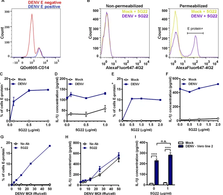
Fig 1. DIV crude supernatant induces IL-1βsecretion independent of ADE.(A) Flow-cytometric histogram overlay comparing CD14 expression levels in cells negative or positive for intracellular DENV E protein at 24 hpi. (B) Mobilized monocytes were inoculated with mock medium or DIV crude supernatant that had been incubated with 1μg/ml mAb 5G22. At 1 hpi, cells were washed and resuspended in fresh medium. At 24 hpi, cells were washed and stained for surface expression (left panel) or intracellular expression (right panel) of DENV E protein with mAb 4G2 conjugated to AlexaFluor 647 and analyzed by flow cytometry. (C) Cumulative percentages of mobilized monocytes positive for intracellular DENV E protein at 24 hpi with DIV crude supernatant in the presence of increasing concentrations of mAb 5G22. (D) Measurement of secreted IL-1βby ELISA using 24-hpi supernatants from 1C. (E) Repeat of 1C using fresh, non-mobilized monocytes (one value per point). (F) Measurement of secreted IL-1βusing supernatants from 1E (one value per point). (G) Measurement of DENV E-protein expression in mobilized monocytes at 24 hpi. Cells were inoculated with increasing doses of DIV crude supernatant with or without 1μg/ml mAb 5G22. (H) Measurement of secreted IL-1βusing 24-hpi supernatants from 1G. (I) Secreted IL-1βby mobilized monocytes at 24 hpi with mock
supernatant or DIV crude supernatant derived from a second line of Vero cells. For all figures:*= p<0.05,**= p<0.01,***= p<0.001,****= p<0.0001, and“n.s.”= not significant (p>0.05). Test used: Two-Way ANOVA with Tukey’s post-test (I).
protein expressed varying levels of CD14, while all cells positive for intracellular DENV E pro-tein also expressed high levels of CD14. These data confirm that CD14+monocytes are the tar-get of ADE in a mixed population of PBMCs. Thus, most of the subsequent experiments utilize cryopreserved CD14+mobilized monocytes that were purified using negative isolation to prevent activation through CD14. The advantage of mobilized, leukapheresed blood is that it provided a large source of cells from a single blood draw (with two separate donors total), elim-inating much of the variability associated with multiple blood draws from different volunteers over time. Key phenotypes were verified using CD14+monocytes purified from fresh draws of non-mobilized blood.
To verify that this flow-based assay was not detecting DENV adsorbed to the cell surface, we stained purified mobilized monocytes in the presence or absence of membrane permeabili-zation (Fig 1B). Inoculation with DIV crude supernatant and mAb 5G22 did not induce ele-vated DENV E-protein expression in non-permeabilized cells (Fig 1B, left panel). However, inoculation with DIV crude supernatant and mAb 5G22 induced a shift in DENV E-protein expression in permeabilized cells (Fig 1B, right panel). These data confirm that this flow-based assay does not detect surface-adsorbed virus.
DIV crude supernatant induces IL-1
β
secretion by primary monocytes
independent of enhancement with mAb 5G22
We next examined the induction of IL-1βin primary monocytes by DIV crude supernatant in the context of ADE. In the absence of mAb 5G22, no intracellular expression of DENV E pro-tein was detected at 24 hpi in mobilized monocytes (Fig 1C). In contrast, pre-incubation of DIV crude supernatant with increasing levels of mAb 5G22 caused a dose-dependent increase in intracellular E-protein expression. Unexpectedly, DIV crude supernatant induced IL-1β secretion by mobilized monocytes independent of the presence of mAb 5G22, as measured by ELISA on monocyte supernatants collected at 24 hpi (Fig 1D). An identical pattern was observed in freshly-isolated monocytes from a non-mobilized donor, with mAb 5G22 required for intracellular E-protein expression but not for induction of IL-1βsecretion (Fig 1E and 1F). We next inoculated mobilized monocytes with varying doses of DIV crude supernatant in the presence of a constant antibody dose or control condition. Increasing doses of DIV crude supernatant increased intracellular E-protein expression in the presence of 1μg/ml mAb 5G22
(Fig 1G). However, increasing doses of DIV crude supernatant induced elevated secretion of
IL-1βregardless of the presence of mAb 5G22 (Fig 1H). These data indicate a bifurcation in the dependence on anti-DENV antibody, with mAb 5G22 enhancing DENV replication but being dispensable for IL-1βsecretion in primary monocytes inoculated with DIV crude supernatant.
To rule out the possibility that these outcomes are unique to the Vero cell line we used, crude supernatant was harvested from a second, independent line of Vero cells infected with DENV-2 strain 16681. IL-1βsecretion induced by DIV crude supernatant harvested from this second Vero line also was independent of the presence of mAb 5G22 (Fig 1I). In total, these results indicate that anti-DENV antibodies significantly impact viral replication in primary monocytes but are completely dispensable for IL-1βsecretion induced by DIV crude supernatant.
DIV crude supernatant induces broad inflammatory cytokine secretion
independent of ADE
Enhancement of DENV infection with 0.1μg/ml mAb 2D22 and 2μg/ml mAb 1C17 (indicated by peaks with higher DENV E-protein expression in the histograms) both failed to enhance IL-1βsecretion induced by DIV crude supernatant (Fig 2A). In addition to enhancement of infec-tion, mAb 2D22 also strongly neutralizes DENV-2 infection at higher concentrations, as previ-ously reported [32]. The use of a neutralizing dose of mAb 2D22 at 2μg/ml (indicated by the loss of elevated DENV E-protein expression compared to 0.1μg/ml 2D22) also did not affect IL-1βsecretion. These data confirm that DIV crude supernatant induces IL-1βsecretion inde-pendent of antibody condition.
To assess if other cytokines are induced in a similar way, we inoculated mobilized mono-cytes with control medium, DIV crude supernatant alone, or DIV crude supernatant that had been incubated with 0.1μg/ml of mAbs 5G22, 1F4, 2D22, or 1C17. Monoclonal antibody 1F4 is specific to DENV-1 and serves as an isotype-matched control antibody [33]. At 24 hpi, supernatants were collected and assayed for a number of inflammatory cytokines by multiplex array (Fig 2B–2F). As expected, antibody against DENV did not increase IL-1βsecretion induced by DIV crude supernatant (Fig 2B). DIV crude supernatant also induced elevated secretion of inflammatory cytokines TNF (Fig 2C), IL-12 (Fig 2D), MIP-1α(Fig 2E), and
MIP-1β(Fig 2F). Various antibodies against DENV did not enhance secretion of these cytokines.
We next antagonized antibody binding by pre-incubating mobilized monocytes with an Fc-receptor binding inhibitor. Fc-Fc-receptor inhibition significantly reduced intracellular, ADE-induced E-protein expression (Fig 2G) but did not affect the secretion of IL-1β(Fig 2H). In sum, these data confirm that DIV crude supernatant induces a number of inflammatory cyto-kines independent of antibody signaling.
IL-1
β
secretion induced by DIV crude supernatant precedes viral replication
As enhancing viral replication had no effect on IL-1βsecretion, we determined if the kinetics of IL-1βrelease differ from the kinetics of viral replication. All previous experiments had assessed IL-1β production at 24 hpi, so we initiated a time course with sample collections at 2, 8, 16, and 24 hpi. Mobilized monocytes inoculated with DIV crude supernatant, with or without mAb 5G22, secreted significantly more IL-1βas early as 8 hpi compared to mock conditions (Fig 3A). However, viral replication only began to elevate significantly at 16 hpi (Fig 3B–3D). Intracellular DENV E-protein expression could not be detected until 16 hpi with DIV crude supernatant in the presence of mAb 5G22 (Fig 3B). To further assess replication of the virus, the amount of infectious virus present in the supernatant (Fig 3C) and intracellular presence of DENV genome copies (Fig 3D) were not sig-nificantly increased until 24 hpi with DIV crude supernatant and mAb 5G22. Importantly, none of these measures of DENV replication significantly increased when monocytes were inoculated with DIV crude supernatant in the absence of mAb 5G22. These data confirm that IL-1βsecretion induced by DIV crude supernatant precedes replication and is independent of ADE.
As monocytes are reported to secrete IL-1βwithin 4 hours of inoculation with DENV [19, 20], we next assessed IL-1βsecretion at 4 hpi. DIV crude supernatant induced IL-1βsecretion by 4 hpi in both mobilized monocytes (Fig 3E) and fresh, non-mobilized monocytes (Fig 3F). These data confirm that IL-1βsecretion occurs within 4 hours of inoculation with DIV crude supernatant. Interestingly, though ADE-enhanced IL-1βsecretion has been described at 4 hpi [19,20], ADE was dispensable for IL-1βsecretion even at 4 hpi in the current system.
IL-1
β
secretion induced by DIV crude supernatant is independent of viral
replication
we inactivated DIV crude supernatant by incubation with formalin or exposure to shortwave UV irradiation prior to inoculation onto mobilized monocytes. Both formalin and UV inacti-vation ablated intracellular DENV E-protein expression in mobilized monocytes (Fig 3G). Fig 2. DIV crude supernatant induces inflammatory cytokine secretion independent of antibody signaling.(A) Left: Flow-cytometric histograms of 24-hpi DENV E-protein expression in mobilized monocytes after inoculation with mock supernatant, DIV crude supernatant alone, or DIV crude supernatant that was pre-incubated with 0.1μg/ml mAb 2D22 (enhancing), 2μg/ml mAb 2D22 (neutralizing), or 2μg/ml mAb 1C17 (enhancing). Right: Measurement of secreted IL-1βat 24 hpi for corresponding samples. (B–F) Mobilized monocytes were inoculated with mock medium, DIV crude supernatant alone, or DIV crude supernatant that had been incubated with 0.1μg/ml of mAbs 5G22, 1F4, 2D22, or 1C17. At 1 hpi, cells were washed to remove inoculum and resuspended in fresh medium. At 24 hpi, supernatants were collected, and cytokine secretion was evaluated by multiplex array, including inflammatory cytokines IL-1β(B), TNF (C), IL-12 (D), MIP-1α(E), and MIP-1β(F). Dashed lines indicate mean concentration induced by mock medium, except for panel D, in which mock-induced values fell below the lower limit of detection. (G) Measurement of intracellular DENV E-protein at 24 hpi in mobilized monocytes that were incubated with PBS or Fc-receptor binding inhibitor prior to inoculation with DIV crude supernatant. (H) Secreted IL-1βat 24 hpi using supernatants from 2G. Tests used: One-Way ANOVA (within DENV treatment) with Tukey’s post-test (A), One-Way ANOVA with Dunnett’s post-test (B–F), and Two-Way ANOVA with Bonferroni’s post-test (G and H).
doi:10.1371/journal.pone.0136708.g002
Fig 3. DIV crude supernatant induces rapid IL-1βsecretion independent of viral replication.(A-D) Time course of mobilized monocytes inoculated with mock supernatant or DIV crude supernatant with or without mAb 5G22. Cells were washed at 1 hpi and resuspended in fresh medium. Samples were collected at 2, 8, 16, and 24 hpi. (A) Secreted IL-1β. (B) Intracellular DENV E-protein expression. (C) Infectious virus present in the supernatant, as measured by immunoassay on Vero cells. (D) Relative expression of DENV genome copies in mobilized monocytes, measured by real-time PCR. Values are
normalized to 2 hpi samples in the absence of mAb 5G22. For A-D, data are pooled from two independent experiments. (E) Secreted IL-1βby mobilized monocytes at 4 hpi. (F) Secreted IL-1βby fresh, non-mobilized monocytes at 4 hpi. (G) Intracellular DENV E-protein expression at 24 hpi with live DIV crude supernatant or DIV crude supernatant inactivated with formalin or UV exposure, all in the presence of mAb 5G22. (H) Secreted IL-1βat 24 hpi using supernatants from 3G. Tests used: Two-Way ANOVA with Dunnett’s post-test (A–D), One-Way ANOVA with Dunnett’s post-test (G, H), Two-Way ANOVA with Tukey’s post-test (E, F). For A and B, gray (A only) and blue asterisks compare DENV and DENV + 5G22, respectively, to mock within each time point. For C and D, blue asterisks compare DENV + 5G22 to the 2 hpi time point.
However, each inactivated infectious supernatant induced significant elevation of IL-1β secre-tion compared to mock condisecre-tions (Fig 3H). These data indicate that the replication compe-tency of DENV is dispensable for IL-1βinduction by DIV crude supernatant.
DIV crude supernatant induces
IL1B
and pro-IL-1
β
expression in primary
monocytes
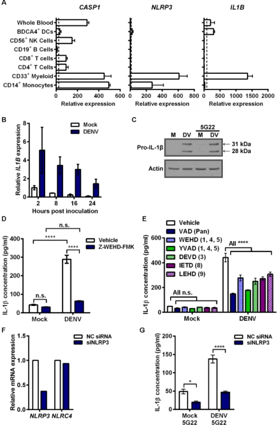
Elevated secretion of IL-1βcan be caused by increased pro-IL-1βexpression, increased inflam-masome activation, or both. To assess the mechanism of IL-1βinduction by DIV crude super-natant, we first considered known cellular expression of key genes by accessing the online bioinformatics database BioGPS (Fig 4A). CD14+monocytes express high baseline levels of
CASP1andNLRP3, which encode the inflammasome components caspase-1 and NLRP3,
respectively. However, basal expression ofIL1Bis low, indicating it likely needs induction. Thus, we measuredIL1Bexpression after inoculation of mobilized monocytes with DIV crude supernatant (Fig 4B).IL1Bexpression increased rapidly 2 hours after inoculation with DIV crude supernatant, compared to mock conditions, and gradually reduced over time. Corre-spondingly, we detected a strong induction of 31-kDa pro-IL-1βexpression in the cell lysates of mobilized monocytes collected 4 hours after inoculation with DIV crude supernatant (Fig 4C). As expected from the ELISA results, mAb 5G22 did not enhance the pro-IL-1βexpression induced by DIV crude supernatant.
Caspase activity and NLRP3 are required for IL-1
β
secretion induced by
DIV crude supernatant
As mobilized monocytes processed IL-1βafter inoculation with DIV crude supernatant, we next sought to assess inflammasome involvement. We were unable to reproducibly and reliably detect active caspase-1, a notoriously difficult protein to detect in human cells. However, pre-treatment of monocytes with the irreversible caspase-1 inhibitor Z-WEHD-FMK at 80μM sig-nificantly reduced the IL-1βsecretion induced by DIV crude supernatant (Fig 4D). As caspase inhibitor peptides can be prone to cross-reactivity, we next expanded our caspase studies to uti-lize low (1μM) doses of a broad panel of caspase inhibitors (Fig 4E). Inhibitors targeting all caspases (Z-VAD-FMK), caspases-1, -4, and -5 (Z-WEHD-FMK and Z-YVAD-FMK), cas-pase-3 (Z-DEVD-FMK), caspase-8 (Z-IETD-FMK), and caspase-9 (Z-LEHD-FMK) all signifi-cantly reduced IL-1βsecretion induced by DIV crude supernatant. This suggests that either cell death plays an important role in this IL-1βinduction or other caspases contribute to inflamma-some activation. For example, caspase-8 has been found to be important for inflammainflamma-some activation [35,36].
To test for inflammasome involvement more specifically, we genetically interfered with
NLRP3expression, as the NLRP3 inflammasome is activated by a wide array of stimuli. We
Depletion of antibody-bound virions does not reduce IL-1
β
secretion
induced by DIV crude supernatant
We next sought to determine the reason that anti-DENV antibodies can enhance DENV repli-cation without altering the secretion of IL-1β. We considered the possibility that a soluble fac-tor not associated with the virion could induce signaling in the monocytes. To test this, we developed a method of depleting antibody-bound virions from DIV crude supernatant (Fig 5A). Antibodies targeting several DENV epitopes were incubated with DIV crude supernatant individually or in combination. Un-depleted control tubes (fraction C in schematic) received an equal volume of PBS in lieu of beads. Magnetic protein G beads were incubated in depletion tubes. Subsequently, depletion tubes were placed on a magnet, sequestering beads, and all bead-bound components, to the side of the tube. Residual supernatant (fraction R) could then be collected free of bead-bound components. Finally, the bead-bound fraction (fraction B) was resuspended to the original volume for analysis. Depleting monoclonal antibodies had specific-ity for prM, EDI/II, or EDIII, three main antibody targets found on the surface of the DENV virion, as described previously [37]. Control, residual, and bead-bound fractions were then assessed by immunoblot for efficiency of DENV depletion (Fig 5B). Under mock conditions with all three antibodies, DENV E and prM antigens were not detected, as expected. Human IgG was not detected in the residual fraction depleted of bead-bound components (lane R), while there was strong detection of human IgG Fc in the bead-bound fraction (lane B). This indicates that the protein G beads efficiently removed all human IgG. In a second control con-taining DIV crude supernatant in the absence of antibodies (DENV + PBS), DENV E protein and prM were only detected in the residual fractions (lane R). This indicates that beads did not non-specifically bind and deplete DENV virions. Next, each antibody was used individually at 3μg/ml with DIV crude supernatant. This resulted in varying levels of DENV depletion, as assessed by the depletion of DENV E or prM proteins from the residual fractions (lane R). Importantly, the mAb targeting DENV EDI/II removed nearly all detectable virus from the residual supernatant. Similar results were obtained when all three antibodies were added at 1μg/ml each with DIV crude supernatant. By contrast, anti-prM and anti-EDIII mAbs only partially removed DENV virions when compared to their corresponding control supernatants.
Subsequently, both the un-depleted control supernatants (lane C) and antigen-depleted supernatants (lane R) were inoculated onto mobilized monocytes at an approximate pre-deple-tion MOI of 50 (Fig 5C). No depletion condition altered IL-1βsecretion compared to its respective un-depleted control supernatant. These data suggest that DENV virions are not responsible for the IL-1βsecretion induced by DIV crude supernatant.
values per condition) are normalized to 2 hpi mock samples. (C) Immunoblot assessing intracellular pro-IL-1β (31 kDa) in 4 hpi lysates of mobilized monocytes after inoculation with mock supernatant or DIV crude supernatant in the presence or absence of mAb 5G22. (D) Secreted IL-1βby mobilized monocytes at 5 hpi with DIV crude supernatant. Cells were pre-treated with DMSO vehicle or 80μM of Z-WEHD-FMK caspase-1 inhibitor for 30 minutes prior to inoculation. (E) Secreted IL-1βby mobilized monocytes at 4 hpi with DIV crude supernatant. At time of inoculation, monocytes were treated with DMSO vehicle or 1μM of various caspase inhibitors: Z-VAD-FMK (pan), Z-WEHD-FMK (1, 4, 5), Z-YVAD-FMK (1, 4, 5), Z-DEVD-FMK (3), Z-IETD-FMK (8), or Z-LEHD-FMK (9). (F) Assessment ofNLRP3(target) andNLRC4(control) expression by real-time PCR, 24 hours after mobilized monocytes were transfected with a control siRNA or an siRNA targetingNLRP3. (G) Secreted IL-1βby knockdown and control cells described in 4F at 6 hpi with mock supernatant or DIV crude supernatant in the presence of 1μg/ml 5G22. Monocytes were inoculated 24 hours after siRNA transfection. Tests used: Two-Way ANOVA with Tukey’s post-test (D), Two-Way ANOVA with Dunnett’s post-test (E), Two-Way ANOVA with Bonferroni’s post-test (G).
Fig 5. Inflammatory component produced by Vero cells during DENV propagation is responsible for IL-1βinduction.(A) Schematic depicting the different depletion and control fractions for depleting antibody-bound virions from DIV crude supernatant by using magnetic protein G beads. (B) Immunoblot for human IgG Fc, and two DENV structural proteins, E protein and prM, to assess efficiency of antibody-mediated DENV depletion from DIV crude
supernatant.“*”denotes all 3 antibodies were used in combination at 1μg/ml each. 3μg/ml of each antibody was used for individual antibody conditions. (C) Control supernatants (Lane C) and residual supernatants (Lane R) from panel B were inoculated onto mobilized monocytes. Monocyte supernatants were collected at 24 hpi, and secreted IL-1βwas measured by ELISA. (D) Mock and DIV crude supernatants were incubated with PBS or anti-DENV NS1 antibody prior to addition of protein G beads and subsequent depletion. Top: Residual supernatants (R) and bead-bound fractions (B) were assessed for efficiency of NS1 depletion by immunoblot. Bottom: Residual supernatants were inoculated onto mobilized monocytes. At 4 hpi, monocyte supernatants were collected and assessed for secreted IL-1β. (E) Mock and DIV crude supernatants were incubated with PBS, DNase, or Riboshredder prior to inoculation onto mobilized monocytes. At 4 hpi, monocyte supernatants were collected and assessed for secreted IL-1β. (F) Mobilized monocytes were inoculated with mock
supernatant, live DIV crude supernatant, or DIV crude supernatant that had been incubated for 30 minutes at 56°C to heat inactivate (HI) it. At 24 hpi, monocyte supernatants were collected and assessed for secreted IL-1β. (G) DIV crude supernatant was centrifuged in an Amicon centrifugal filtration unit with a 100-kDa molecular-weight cutoff to generate fractions smaller and larger than 100 kDa. Fractions were brought to equal volumes and inoculated onto mobilized monocytes. At 24 hpi, monocyte supernatants were collected, and IL-1βsecretion was measured. Tests used: Two-Way ANOVA with Bonferroni’s post-test (D), One-Way ANOVA with Dunnett’s post-test (F).
Several potential factors eliminated as IL-1
β
-inducing component of DIV
crude supernatant
To identify the component in DIV crude supernatant that could induce IL-1βsecretion, we investigated several possibilities. First, though most DENV nonstructural proteins are neither present in an infectious virion nor secreted from infected cells, nonstructural protein 1 (NS1) is secreted from infected cells as a soluble hexamer [38]. To determine if soluble NS1 present in DENV supernatant is responsible for the induction of IL-1βsecretion, we depleted NS1 from viral supernatant using antibody-mediated depletion with protein G beads, which resulted in the near-complete removal of NS1 from DIV crude supernatant (Fig 5D). However, inoculat-ing mobilized monocytes with NS1-depleted DIV crude supernatant had no impact on the secretion of IL-1β.
We next verified that DIV crude supernatants were not contaminated with LPS, a potent IL-1βagonist. Two independent DIV crude supernatant samples were sent to the UNC Tissue Culture Facility for LPS screening by a limulus amebocyte lysate assay. Both samples were reported to contain less than or equal to 0.10 Endotoxin Units/ml, thus excluding LPS contam-ination as a factor.
To investigate the possibility that DENV-infected Vero cells release inflammatory RNA or DNA, we treated DIV crude supernatant with DNase or RNase prior to inoculation onto mobi-lized monocytes (Fig 5E). Neither nuclease prevented IL-1βsecretion induced by DIV crude supernatant. Complement has been associated with the response to DENV infection [39]. To assess if complement or another heat-sensitive component had a role, DIV crude supernatant was heated for 30 minutes at 56°C prior to inoculation onto mobilized monocytes (Fig 5F). Heat-inactivated DIV crude supernatant still induced elevated IL-1βsecretion over mock conditions.
To test if cytokines secreted by infected Vero cells, which originated from African green monkey kidney, could induce IL-1βsecretion by primary human monocytes, we filtrated the DIV crude supernatant. Ultrafiltration of human plasma using a pore with a 60-kDa molecu-lar-weight cutoff (MWCO) has been shown to effectively remove IL-1β, IL-6, TNFα, IL-10, and IL-8, as these cytokines are smaller and able to pass through the pore [40]. Thus, we chose to employ centrifugal filtration with a MWCO of 100 kDa to allow flow-through of cytokines but not DENV or other larger components. The flow-through fraction, containing everything less than 100 kDa (approximately), and the concentrated retentate (components above 100 kDa) were collected and brought to equal volumes. Subsequently, equal volumes of both frac-tions and a mock condition were inoculated onto mobilized monocytes (Fig 5G). Only the frac-tion containing components larger than 100 kDa induced the secrefrac-tion of IL-1βby primary monocytes. This excluded many key cytokines present in Vero-derived supernatant as a con-tributing factor to IL-1βinduction.
Antibody promotes DENV-induced IL-1
β
secretion by primary
monocytes infected with purified virus
DENV-2 16681 derived from Vero cells induced significant elevation of IL-1βsecretion only in the presence of mAb 5G22 (Fig 6B). Interestingly, the level of IL-1βinduced by purified DENV is lower than that induced by DIV crude supernatant shown in earlier experiments, consistent with the presence of an additional inflammatory component in the crude supernatant.
IL-1
β
induction varies by DENV strain and cell-type used to propagate
virus
We next assessed whether a different DENV strain propagated in Vero cells would similarly induce ADE-independent IL-1βsecretion. In addition to DENV-2 16681, monocytes were also inoculated with DIV crude supernatant harvested from Vero cells infected with DENV-1 West Pac 74, in the presence or absence mAb 5G22. At 24 hpi, both DENV strains induced Fig 6. ADE-induced IL-1βsecretion varies by DENV purity, strain, and cell-type used for propagation.(A) Mobilized monocytes were inoculated with mock conditions or purified DENV-2 16681 derived from Vero cells with or without the addition of mAb 5G22. Intracellular DENV E-protein expression was measured at 24 hpi. (B) Secreted IL-1βdetected in 24-hpi monocyte supernatants from 6A. (C) Monocytes were inoculated with DIV crude supernatants harvested from Vero cells infected with DENV-2 16681 or DENV-1 West Pac 74 with or without mAb 5G22. Cells were washed at 1 hpi, and intracellular DENV E-protein expression was measured at 24 hpi. (D) IL-1βin 24-hpi supernatants from 6C was measured by ELISA. (E) Vero cells were inoculated with MOI 0.5 of DENV-2 16681 or DENV-1 West Pac 74. At 1 hpi, inoculum was aspirated and fresh medium was added. Supernatant samples were collected at days 1, 3, 5, and 7 post-inoculation and assessed for adenylate kinase release by ToxiLight assay. Values are normalized to the 1 dpi mock condition. (F) Mobilized monocytes were inoculated with control supernatant from uninfected C6/36 cells or supernatant derived from C6/36 cells infected with DENV-2 16681, with or without 1μg/ml mAb 5G22. Cells were washed at 1 hpi, and intracellular DENV E-protein expression was analyzed at 24 hpi. (G) Secreted IL-1βdetected in 24-hpi monocyte supernatants from 6E. Tests used: Two-Way ANOVA with Bonferroni’s post-test (B, D, G).
intracellular E-protein expression only when inoculated in the presence of mAb 5G22 (Fig 6C). Interestingly, the induction of IL-1βvaried dramatically by strain (Fig 6D). As before, DENV-2 16681 DIV crude supernatant induced ADE-independent IL-1βsecretion. However, DENV-1 West Pac 74 DIV crude supernatant induced IL-1βonly under ADE conditions. These data indicate that IL-1βinduced by DIV crude supernatant can vary by DENV strain.
Though grown in Vero cells under the same conditions, we found DENV-1 West Pac 74 to grow to titers approximately three-fold higher than DENV-2 16681. Thus, inoculating mono-cytes with equal MOIs of the two strains requires a lower volume of DIV crude supernatant for DENV-1 West Pac 74. To see if these DENV strains induce cell death in Vero cells, we inocu-lated Vero cells with equal MOIs of both DENV strains and monitored adenylate kinase (AK) release over time compared to mock inoculation (Fig 6E). Both DENV strains induced a similar degree of cell death in Vero cells compared to the mock condition. Thus, increased cell death alone cannot account for the masking phenotype seen when inoculating monocytes with DENV-2 16681. However, the lower volume of DIV crude supernatant required to inoculate monocytes with DENV-1 West Pac 74 may reveal ADE-induced IL-1βby exposing monocytes to a reduced amount of the inflammatory components.
Other studies describing ADE-induced IL-1βsecretion in primary monocytes utilized crude supernatant harvested from DENV-infected C6/36 mosquito cells [19,20]. We next verified that crude supernatant harvested from C6/36 cells infected with DENV-2 16681 induced ADE-dependent IL-1βsecretion. Crude supernatant harvested from C6/36 cells infected with DENV-2 16681 was inoculated onto mobilized monocytes in the presence or absence of mAb 5G22. Similar to Vero-derived DENV-2 16681, mAb 5G22 enhanced the intracellular expres-sion of DENV E protein in mobilized monocytes 24 hours after inoculation with crude infec-tious supernatant from C6/36 cells (Fig 6F). As expected based on our previous study [20], ADE with mAb 5G22 significantly enhanced IL-1βsecretion when mobilized monocytes were inoculated with crude supernatant from C6/36 cells infected with DENV-2 16681 (Fig 6G). Importantly, the presence of mAb 5G22 did not impact IL-1βsecretion induced by crude supernatant from uninfected C6/36 cells. These data suggest that, unlike Vero cells, C6/36 mos-quito cells do not produce a potent inflammatory component during the propagation of DENV-2 16681 in culture. Instead, DENV-2 16681 propagated in mosquito cells displayed ADE of both infection and IL-1βsecretion.
Discussion
This work shows that while all DENV preparations display an identical requirement for ADE to enhance viral replication, the source and purity of DENV preparations greatly impact the induction of IL-1βsecretion by primary human monocytes. DENV-2 16681 propagation in Vero cells produced not only high titers of infectious virus, but also inflammatory moieties that induced IL-1βsecretion. Initial experiments utilized debris-cleared supernatant derived from DENV-infected Vero cells, a common method of viral preparation to analyze the immune response to DENV [42–46]. This Vero-derived supernatant displayed a strong IL-1β-inducing activity that was unaltered by ADE, despite concurrent enhancement of infection by antibody. Crude supernatant harvested from a second, independent line of Vero cells infected with DENV-2 16681 similarly induced ADE-independent IL-1βsecretion. Induction of other inflammatory cytokines was also independent of ADE. This indicates that an unknown factor produced by DENV-infected Vero cells may mask the DENV-induced production of numer-ous cytokines.
Vero cells induced significantly more IL-1βsecretion by monocytes in an ADE-dependent fashion. ADE similarly enhanced IL-1βsecretion when inoculating monocytes with crude supernatant from C6/36 mosquito cells infected with DENV-2 16681. This indicates that the inflammatory moiety in Vero-derived supernatant that caused IL-1βsecretion is not produced by mosquito cells. As inoculation with crude supernatant from DENV-infected Vero cells is commonly used in the field, precaution should be exercised in using such a preparation to study the DENV-induced inflammatory response in immune cells. However, this phenotype was not universal amongst DENV strains tested. Crude supernatants harvested from Vero cells infected with DENV-1 West Pac 74 induced ADE-dependent IL-1β. This is possibly because this virus grows to high titers and thus less virus-containing supernatant was added to cells.
Inoculation of primary mobilized monocytes with crude supernatant from Vero cells infected with DENV-2 16681 rapidly induced the expression ofIL1Band pro-IL-1β. Thus, a key step leading to elevated IL-1βsecretion after inoculation with this supernatant appears to be the enhancement of pro-IL-1βexpression. Although bioinformatic analysis with BioGPS shows thatCASP1andNLRP3transcripts are constitutively expressed by human monocytes, inhibition of caspase-1 with a pharmacologic inhibitor or NLRP3 with RNA interference reduced the secretion of IL-1β, indicating involvement of the NLRP3 inflammasome.
Although much effort was devoted to identifying the IL-1β-inducing moiety or moieties in DIV crude supernatant, the nature of the inducer(s) remained elusive. Could it be a cell-culture artifact that needs to be eliminated from conventional crude viral preparations? Or is this a physiologically-relevant component (either viral- or host-derived) that merits further study-ing? Our data suggests that the factor is larger in size than many key cytokines, is heat stable at 56°C, and is not RNA, DNA, or LPS contamination. Further, immunodepletion studies con-firm that NS1 is not responsible for the IL-1βinduction. Of note, one study has shown that Vero cells infected with DENV undergo more apoptosis than infected C6/36 mosquito cells [47]. It is possible that by-products of increased cell death may alter the inflammatory pheno-type of crude supernatant from DENV-infected Vero cells.
The IL-1β-inducing factor present in crude supernatant from Vero cells infected with DENV-2 16681 likely masked the response induced by DENV and anti-DENV antibody in the current system. This is an important point to consider, as it can generate misleading results. Consistent with our results using DENV-1 West Pac 74, successful achievement of ADE-induced cytokines by other groups using crude supernatant derived from DENV-infected Vero cells indicates that this inflammatory phenotype is not universally produced by all Vero cells or in all systems [43,45]. However, a masking component should be considered when disjointed results between infection and cytokine production are found, particularly under ADE condi-tions. We have previously shown [20] and confirmed here that crude supernatant from DENV-infected C6/36 cells does not exhibit these confounding issues and represents a more straightforward system to study the ADE-induced inflammatory response in human immune cells. Finally, though the use of purified virus effectively removed the inflammatory moiety from DIV crude supernatant, this approach is much more labor-intensive and unlikely to be used routinely.
Materials and Methods
Ethics Statement
blood of leukapheresed patients enrolled in Study #05–2860 approved by University of North Carolina at Chapel Hill Institutional Review Board and Office of Human Research Ethics after providing written informed consent. Samples were anonymized and provided as de-identified samples prior to use in the described studies. The University of North Carolina at Chapel Hill Office of Human Research Ethics determined that the use of the de-identified samples does not constitute human subjects research as defined under federal regulations [45 CFR 46.102 (d or f) and 21 CFR 56.102(c)(e)(l)] and does not require further Institutional Review Board approval.
PBMC isolation and cell culture
Due to the large number of cells required for this study, we mostly employed cryopreserved, primary mobilized PBMCs from two separate patients injected with G-CSF, which greatly increases the number of circulating leukocytes, for further studies [48]. This established a large stock of monocytes isolated in one day that was capable of supplying months of experiments, reducing the inherent variability of human studies. We then verified key phenotypes with PBMCs from freshly-isolated, non-mobilized blood.
PBMCs were isolated from the blood using a 1.073 g/ml Ficoll-Hypaque Premium gradient (GE Healthcare) to enhance monocyte isolation. The manufacturer’s suggested protocol was followed for buffy coat isolation. Negative isolation was done using the Dynabeads Untouched Human Monocytes kit (Invitrogen) by following manufacturer’s protocol. Antibodies against CD3, CD7, CD16 (specific for CD16a and CD16b), CD19, CD56, CDw123, and CD235a depleted cells expressing these markers. Mixed PBMCs and purified monocytes were cryopre-served in 90% heat-inactivated FBS (FBS-HI) with 10% DMSO. On experimental days, cells were thawed, washed twice, and placed at 37°C for 2 hours in PBMC cell culture media consist-ing of RPMI with 10% FBS-HI, 1% L-glutamine, 1% NEAA, 1% penicillin/streptomycin, and 30 units/ml DNase to prevent cell clumping from the release of DNA by dying granulocytes. Cells were counted and resuspended in PBMC medium without DNase for use in experiments.
The majority of experiments used DENV propagated in the Vero 76 cell line (ATCC CRL-1587). Confirmation of the inflammatory phenotype with an independent line of Vero cells was done using DENV propagated in the original Vero cell line, acquired from the UNC Line-berger Comprehensive Cancer Center Tissue Culture Facility (ATCC CCL-81). TheAedes
albopictusC6/36 cell line was acquired from ATCC (CRL-1660).
DENV-specific antibodies
Crude supernatant of mAb D14G2 (4G2), a pan-flavivirus E-protein specific mouse IgG2a mAb, for use in IHC viral titrations was kindly provided by Dr. Mariano Garcia-Blanco of Duke University. Purified mAb 4G2 was generated by the UNC Antibody Core Facility. The isolation and purification of human mAbs 5G22 (α-prM used for enhancement and depletion), 2D22 (α-DENV-2 used for enhancement and neutralization), 6B22 (α-EDI/II used for deple-tion), 1C17 (α-EDIII used for enhancement and depletion), 1F4 (α-DENV-1 used for isotype-matched control) and 2H21 (α-prM used for immunoblots) were described in detail previously [32,33].
Virus stock growth
All experiments used DENV-2 strain 16681, kindly provided by Dr. Robert Tesh of UTMB-Galveston, unless otherwise noted. Vero cells were cultured at 37°C with 5% CO2in MEM
16681 or DENV-1 West Pac 74 at an MOI of 0.5 in low-volume, low-serum conditions, placed in the incubator, and rocked every 15 minutes. At 2 hpi, culture medium was added. For viral stocks grown for purification, 1% FBS-HI was used so as not to clog the centrifugal filters dur-ing the concentration step. At days 3, 7, and 10, medium was collected from the flasks and cen-trifuged at 4,000 RPM for 10 minutes to clarify the supernatants. Infectious crude supernatants were then aliquoted into tubes for freezing at -70°C. Fresh medium replaced the collected supernatants. C6/36 cells were cultured at 29.5°C with 5% CO2in MEM + 10% FBS-HI, 1%
NEAA, 1% penicillin/streptomycin, and 20 mM HEPES. Propagation of DENV in C6/36 cells was done as with Vero cells except for listed differences in medium composition.
We modified a previously described protocol to quantitate the infectious titer of viral stocks [49]. Briefly, near-confluent Vero cell monolayers in flat-bottom, 96-well plates were inocu-lated with 50μl of sequential 10-fold dilutions of DENV stocks. At 2 hpi, an overlay of 150μl of 1.6% carboxymethylcellulose (diluted 1:1 in 2X MEM and supplemented with 1% FBS-HI, 10 mM HEPES and 1X antibiotics) was added to limit spread of virus. At 72 hpi, cells were fixed using a 1:1 mixture of acetone and methanol. Fixed cells were blocked with 2% normal horse serum in PBS and subsequently stained for 1 hour using mAb 4G2 as the primary anti-body (1:500 dilution of crude supernatant into blocking solution) and 1:1000 goat anti-mouse IgG-HRP (KPL) as the secondary antibody. Viral foci were visualized using Vector VIP Peroxi-dase Substrate kit (Vector Laboratories, Inc.) and counted under a dissecting microscope, with titers calculated as Vero focus forming units per ml (ffu/ml).
Inoculation of PBMCs or monocytes
One hour pre-inoculation, dilutions of enhancing antibodies or control medium were plated into 96-well, round-bottom, non-tissue-culture treated plates. Crude infectious supernatants or purified DENV preparations were diluted to the appropriate multiplicity of infection (MOI) and mixed with antibodies or control medium. DENV and antibody mixtures were incubated for 1 hour at 37°C with 5% CO2to allow immune complex formation. For mock infection
wells, spent culture medium from uninfected Vero cell or C6/36 cultures was used instead of infectious supernatant. MEM with 1% FBS-HI, 1% penicillin/streptomycin, and 20 mM HEPES was used as the mock condition for purified virus. After the 1 hour incubation for com-plex formation, mixed PBMCs or purified primary monocytes were added. At 1–2 hours post-inoculation (hpi), cells were washed at least 2 times with PBS and resuspended in fresh culture medium. At collection, cells were resuspended and pelleted by centrifugation. After two washes with 1X PBS (four for genome copy studies), cells were fixed with Cytofix/Cytoperm (BD Bio-sciences) for 20 minutes at 4°C for flow cytometry, lysed with RLT Lysis Buffer (Qiagen) for RNA analysis, or lysed with 1X RIPA buffer (Boston BioProducts) with protease inhibitors (Roche) for western blot analysis. Monocyte supernatants were centrifuged to clear cells and debris, recollected, and stored at 4°C until analysis by ELISA. Unless otherwise noted, all monocyte inoculations were done with an MOI of 50.
Flow cytometry
Purified mAb 4G2 was conjugated to Alexa Fluor 647 using the Alexa 647 Protein Labeling Kit (Life Technologies) according to manufacturer’s instructions. After labeling, the labeled anti-body was titrated by staining known DENV-positive monocytes to identify the best dilution for detection of DENV E protein without high levels of non-specific background in mock-inoc-ulated cells.
(eBiosciences) diluted 1:5 in Perm/Wash Buffer for 15 minutes at 4°C. Cells were then incu-bated with 25μl of mAb 4G2 conjugated to Alexa Fluor 647 diluted 1:250 in Perm/Wash buffer for 30 minutes at 4°C. Cells were then washed twice with Perm/Wash buffer and finally re-sus-pended in 200μl of Perm/Wash buffer for analysis on a Cyan ADP flow cytometer (Dako). Cells were initially gated on FSC-area vs. SSC-area, with single cells positively selected for by gating cells on the diagonal of FSC-height vs. FSC-area. Cells positive for DENV E-protein were detected on the APC channel, with positive gates set based on the mock-inoculated controls.
For surface detection of DENV E protein, cells at 24 hpi were washed with PBS and then resuspended in 25μl of Human Fc Receptor Binding Inhibitor diluted 1:5 in FACS buffer (2% FBS in PBS) and incubated for 15 minutes at 4°C. Cells were then incubated with 25μl of mAb 4G2 conjugated to Alexa Fluor 647 diluted 1:250 in FACS buffer for 30 minutes at 4°C. Cells were then washed twice with FACS buffer and resuspended in 50μl of fresh PBS. Cells were then resuspended with 50μl of 2% formalin diluted into PBS and allowed to incubate at room temperature for 20 minutes protected from light. Cells were then washed twice with FACS buffer and resuspended with 200μl fresh FACS buffer. Stained cells were stored at 4°C pro-tected from light until analysis by flow cytometry.
For staining of surface CD14 expression in conjunction with intracellular DENV E-protein expression, cells were washed twice with PBS and incubated at 4°C with Human Fc Receptor Binding Inhibitor in eFluor NC Flow Cytometry Staining Buffer (eBiosciences) supplemented with 2% FBS-HI for 15 minutes at 1 test per well. Cells were then stained with anti-human CD14 eFluor 605NC mouse mAb (eBiosciences) at 1 test size per well in 25μl 1X staining buffer and incubated for 30 minutes in the dark at 4°C. Cells were washed twice with staining buffer, fixed, permeabilized, and stained with mAb 4G2 conjugated to Alexa Fluor 647, as described above. An unstained control and single-stained controls were set up for gating analy-sis. For compensation, antibodies were added to anti-mouse BD CompBeads at same dilution as sample staining. Cells and beads were run on a LSRII flow cytometer (Becton Dickinson), using the APC channel for the anti-DENV antibody and QDot605 for the anti-CD14 antibody. Compensation calculations and analysis were done using FlowJo.
Real-time PCR
After lysis with RLT buffer, cell lysates were passed through QIAshredder columns (QIAGEN) to homogenize lysates. Homogenized lysates were then added to RNA isolation columns from RNeasy Mini kits (QIAGEN), and all steps were followed as detailed in the manufacturer’s pro-tocol. RNA was eluted in a volume of 30μl RNase/DNase free water. To generate cDNA, 1μl Random Primers (3μg/μl; Invitrogen) and 1μl dNTP Mix (10 mM; Invitrogen) was added to 10μl of eluted RNA and heated at 65°C for 5 minutes in a PTC-225 Peltier Thermal Cycler (MJ Research). Tubes were chilled on ice and 4μl Invitrogen 5X First Strand Buffer, 2μl Invi-trogen 0.1M DTT, and 1μl Promega RNasin RNase Inhibitor (40 units/μl) were added to each tube and incubated at 37°C for 2 minutes. Then 1μl Invitrogen M-MLV Reverse Transcriptase (200 units/μl) was added per tube, and tubes were incubated at 25°C for 10 minutes and 37°C for 50 minutes, with an inactivation step of 70°C for 15 minutes.
applicable, all biological replicates (including their corresponding pipetting triplicates) were combined and outliers greater than 2 standard deviations away from the mean were excluded. For all samples, 18s rRNA was used as the housekeeping-gene control.
The following ABI TaqMan Gene Expression Assays were used for real-time PCR analysis of gene expression: assay Hs01555410_m1 was used forIL1B, Hs00918082_m1 was used for
NLRP3, Hs00368367_m1 was used forNLRC4, and Hs03928985_g1 was used forRN18S1. For
DENV genome copies, a custom TaqMan Gene Expression Assay was designed using nucleo-tides 10635–10708, a portion of the 30UTR of DENV conserved between all 4 serotypes, identi-fied and described by Gurukumar,et al. [50].
Caspase inhibition
For inhibition of caspase-1 only, Z-WEHD-FMK Caspase-1 Inhibitor (R&D Systems) was diluted in sterile DMSO and cells were pre-treated with 80μM Z-WEHD-FMK or DMSO vehi-cle 30 minutes prior to inoculation with DENV. Dilutions were made into cell culture medium.
For the caspase inhibition panel, Z-VAD-FMK (pan caspase inhibitor), Z-WEHD-FMK and Z-YVAD-FMK (caspases-1, -4, and -5 inhibitors), Z-DEVD-FMK (caspase-3 inhibitor), Z-IETD-FMK (caspase-8 inhibitor), and Z-LEHD-FMK (caspase-9 inhibitor) (all from R&D Systems) were diluted in sterile DMSO and given to the cells at a final concentration of 1μM each at the time of inoculation with DIV crude supernatant.
Bioinformatic analysis
The publicly available BioGPS expression database was used for all bioinformatic analysis [51]. The U133A, gcrma dataset was used for analysis of the following genes:CASP1(probeset 206011_at),NLRP3(probeset 207075_at), andIL1B(probeset 205067_at) [52].
Gene knockdown
Knockdown ofNLRP3was achieved using QIAGEN’s HiPerFect Transfection Reagent and FlexiTube GeneSolution GS114548 for NLRP3. QIAGEN’s AllStars Negative Control siRNA served as a negative control forNLRP3knockdown. Briefly, based upon manufacturer’s instructions, concentrations of both transfection reagent and siRNAs were optimized in a pilot experiment. Knockdown was analyzed by decrease ofNLRP3expression as analyzed by real-time PCR compared to control. For the experimental knockdown, 4 x 10⁶cells per condition were plated in 7 ml of growth medium in a 150 mm dish. 8μl of each 10 mM stock of NLRP3 siRNAs was added to 1 total ml of serum-free RPMI (16μl of 20 mM negative control siRNA). Then, 20μl of HiPerFect transfection reagent was added to each tube, vortexed, and allowed to incubate at room temperature. After 10 minutes, complexes were dripped onto the cells, plates were swirled, and allowed to incubate for 24 hours. At 24 hours post-transfection, cells were collected, counted, and immediately used for infection assays.
SDS-PAGE and immunoblots
For viral protein analysis by immunoblot, NuPAGE LDS Sample Buffer without DTT was added directly to DENV-containing supernatant to a final concentration of 1X. Samples were vortexed and heated at 97°C for 10 minutes. DTT was added to samples at above concentra-tions for detection of DENV NS1 by immunoblot.
Samples were loaded in NuPAGE 4–12% Bis-Tris pre-cast gels (Life Technologies) and sep-arated by SDS-PAGE using 1X NuPAGE MES SDS running buffer (Life Technologies). Gels were transferred onto 0.2μm nitrocellulose membranes (Bio-Rad) under wet transfer condi-tions using 1X Transfer Blotting Buffer (Boston BioProducts) with 30% methanol for 45 min-utes at a constant 100V. Membranes were blocked in a 10% milk solution in TBS-T for 1 hour at room temperature. Primary antibodies were incubated overnight at 4°C with rotation at the following dilutions in blocking buffer: 1:1,000 rabbit polyclonal antibody anti-IL-1β(Santa Cruz sc-7884), 1μg/ml purified mouse mAb 4G2 to detect DENV E protein, 1μg/ml human mAb 2H21 to detect DENV prM, 1:1000 goat anti-human Fc conjugated to HRP (Bethyl Laboratories) to detect human IgG Fc, or 1:1000 rabbit anti-DENV NS1 polyclonal antibody (Genetex GTX103346). After 4 washes with TBS-T, the membranes were incubated with the following secondary antibodies diluted into blocking buffer for 2 hours at room temperature: 1:5000 goat anti-rabbit HRP-conjugate (Santa Cruz) for IL-1βand NS1, 1:5000 goat anti-mouse HRP-conjugate (Santa Cruz) for DENV E protein, or 1:5000 goat anti-human HRP con-jugate (Bethyl) for DENV prM. Human IgG in depletion studies was detected directly after pri-mary antibody incubation because of HRP conjugate. Membranes were then washed 4–5 times with TBS-T and subsequently developed after 5 minute incubations with Thermo Scientific SuperSignal West Pico Chemiluminescent Substrate.
Depletion of antibody-bound virions and NS1
Mock and DIV crude supernatants were incubated with indicated concentrations of antibodies (or equal volume of PBS control) for 1 hour on ice. Subsequently, either 50μl of pre-washed Dynabeads Protein G beads (Life Technologies) or equal volume of PBS were added to samples, and placed on a rotator at 4°C for 1 hour. Tubes were then placed onto a DynaMag-2 magnet for 1 minute, and bead-free supernatants were collected and transferred to a new tube. Place-ment on the magnet with collection of the supernatant was repeated two more times to ensure complete removal of beads. The bead-free supernatants beads were considered the residual fractions, while the resuspended beads (for protein analysis only) were considered the bead-bound fractions. Tubes that had PBS added in lieu of beads were considered the control frac-tions, as equal volumes were maintained but no antigens were depleted. Depletion of NS1 was done identically by using mouse anti-NS1 monoclonal antibody clone DN1 (Abcam #ab41490) to immunoprecipitate NS1 with protein G beads. Monoclonal antibody DN1 was washed through an Amicon filter to remove preservatives, resuspended in PBS at a 6-fold increase in concentration, and then syringe filtered to purify. This was then used at a final dilution of 1:2 for successful depletion of NS1.
DNase and RNase treatment of viral supernatant
Virus purification
To purify virus, a previously described protocol was used [41]. Briefly, crude supernatants from DENV-infected Vero cells were concentrated by centrifugation at 1500 x g for 20–25 minutes in 15 ml Millipore Amicon Centrifugal Filter Units with a 100-kDa cutoff, allowing components<100 kDa to pass through but not virus. The final concentrated volume was approximately 2 ml, which was gently layered on top of an 8-ml 10% -40% continuous sucrose gradient in an ultracentrifuge tube. The gradient was centrifuged in a Beckman Coulter Optima L-90K Ultracentrifuge using rotor SW40TI for 2.5 hours at 35,000 RPM and 4°C with no brake. Sequential 0.5 ml fractions were collected from the bottom of the ultracentrifuge tube into microfuge tubes. Fractions 4–8 were pooled and washed to remove sucrose using another 15 ml Amicon tube. After 2 washes, the virus was resuspended in MEM with 1% FBS-HI, 20 mM HEPES, and 1% penicillin/streptomycin. The pooled fractions were then titered as described earlier.
Virus inactivation
DIV crude supernatant was inactivated using formalin or shortwave UV exposure. For forma-lin inactivation, 0.25% formaforma-lin (final concentration) was added to an aliquot of virus stock and allowed to incubate at room temperature for 2.5 hours. For shortwave UV inactivation, 100μl/well of virus stock was added to each well of a 24-well plate, placed on ice, and exposed to shortwave UVB (254 nm) irradiation for 2 min at a distance of approximately 5 cm. After inactivation, the inactivated supernatants, and an equal volume of live virus, were added into separate 15 ml Amicon tubes and washed twice using fresh medium to remove formalin (or as control for sample loss for supernatants not treated with formalin). After washing, the superna-tants were resuspended in fresh culture medium to equal volumes and inoculated onto
monocytes.
Cytokine detection
Detection of human IL-1βin the supernatant was done using BD Biosciences’BD OptEIA Human IL-1beta ELISA Set II. The kit was followed as per manufacturer’s instructions, and the absorbance at 450 nm was read on a PerkinElmer EnSpire Multimode Reader 2300. Wave-length correction at 570 nm was used.
Detection of human IL-1β, IL-12, TNF, MIP-1α, and MIP-1βforFig 2B–2Fwas done by assaying 50μl of 24 hpi supernatants in the Invitrogen Human Cytokine 25-Plex Luminex Bead Panel according to manufacturer’s instructions in the Regional Biocontainment Labora-tory at Duke Immunology Unit (Duke University, Durham, NC).
Adenylate kinase release
Adenylate kinase release into culture supernatants was assessed by ToxiLight (Lonza). Super-natants were stored at -70°C until the assay was run. Then, 20μl of culture supernatant was mixed with 100μl of adenylate kinase detection reagent. After 5 minutes, luminescence was measured by plate reader. Cell-free media controls collected at each time point were subtracted from each sample, and values were normalized to the mock condition at day 1.
Illustration
Data presentation and statistical analysis
Graphs are presented as mean ± SEM of 3 or more biological replicates, unless otherwise noted. All statistical analyses were done with 3 or more replicate values per group. Student’s unpaired, two-tailed t tests were used when making comparison between only 2 means. One-way ANOVAs were employed to compare the means of 3 or more groups within one indepen-dent variable. Two-way ANOVAs were used for comparisons between two indepenindepen-dent vari-ables (e.g. viral treatment vs. antibody treatment). Asterisks for comparisons done by One-Way or Two-One-Way ANOVA represent the multiplicity-adjusted p values from multiple compar-isons tests. Multiple comparcompar-isons tests were selected as follows: Dunnett’s post-test for compar-ing all means to a scompar-ingle control value, Bonferroni’s post-test for comparing only means within one independent variable, and Tukey’s post-test for comparing all means for all independent variables. Choice of post-test was dependent on desired comparisons and is indicated in figure legends. Analyses were done using GraphPad Prism 6.0. Statistical outliers (p<0.05) were excluded from graphs and analysis using Grubbs’test on the GraphPad website. For all figures, statistical significance was defined as p<0.05 (), but all comparisons were also tested for p<0.01 (), p<0.001 (), and p<0.0001 ().“n.s.”= not significant.
Acknowledgments
The authors would like to thank Shannon M. Reisdorf for her efforts and assistance in isolating the mobilized PBMCs used in this study, and Gregory Robbins, PhD, for his advice in the development and modification of the immunodepletion protocols. The UNC Flow Cytometry Core Facility is supported in part by an NCI Center Core Support Grant (P30CA06086) to the UNC Lineberger Comprehensive Cancer Center. Multiplex cytokine profiling was performed in the Regional Biocontainment Laboratory at Duke: Immunology Unit (Durham, NC) under the direction of Dr. Heather E. Lynch. The Regional Biocontainment Laboratory at Duke received partial support for construction from the NIH/NIAID (UC6AI058607).
Author Contributions
Conceived and designed the experiments: JBC DGW SAS KPM FS GDS DPD JEC AMD JPYT. Performed the experiments: JBC DGW GDS. Analyzed the data: JBC DGW FS DPD JEC AMD JPYT. Contributed reagents/materials/analysis tools: SAS DGW KPM FS GDS JEC AMD. Wrote the paper: JBC DGW SAS KPM FS DPD JEC AMD JPYT.
References
1. Bhatt S, Gething PW, Brady OJ, Messina JP, Farlow AW, Moyes CL, et al. The global distribution and burden of dengue. Nature. 2013; 496(7446):504–7. doi:10.1038/nature12060PMID:23563266; PubMed Central PMCID: PMCPMC3651993.
2. WHO. Dengue and severe dengue WHO Media Centre2012 [cited 2012 April 10]. Fact sheet N°117]. Available:http://www.who.int/mediacentre/factsheets/fs117/en/.
3. Guzman MG, Alvarez M, Halstead SB. Secondary infection as a risk factor for dengue hemorrhagic fever/dengue shock syndrome: an historical perspective and role of antibody-dependent enhancement of infection. Arch Virol. 2013; 158(7):1445–59. doi:10.1007/s00705-013-1645-3PMID:23471635.
4. Rothman AL. Immunity to dengue virus: a tale of original antigenic sin and tropical cytokine storms. Nat Rev Immunol. 2011; 11(8):532–43. doi: nri3014 [pii] doi:10.1038/nri3014PMID:21760609.
5. Murphy BR, Whitehead SS. Immune response to dengue virus and prospects for a vaccine. Annu Rev Immunol. 2011; 29:587–619. doi:10.1146/annurev-immunol-031210-101315PMID:21219187.
7. Pang T, Cardosa MJ, Guzman MG. Of cascades and perfect storms: the immunopathogenesis of den-gue haemorrhagic fever-denden-gue shock syndrome (DHF/DSS). Immunol Cell Biol. 2007; 85(1):43–5. doi:10.1038/sj.icb.7100008PMID:17130899.
8. Halstead SB, O'Rourke EJ. Dengue viruses and mononuclear phagocytes. I. Infection enhancement by non-neutralizing antibody. J Exp Med. 1977; 146(1):201–17. PMID:406347; PubMed Central PMCID: PMCPMC2180729.
9. Beltramello M, Williams KL, Simmons CP, Macagno A, Simonelli L, Quyen NT, et al. The human immune response to Dengue virus is dominated by highly cross-reactive antibodies endowed with neu-tralizing and enhancing activity. Cell Host Microbe. 2010; 8(3):271–83. doi: S1931-3128(10)00279-9 [pii] doi:10.1016/j.chom.2010.08.007PMID:20833378.
10. Kou Z, Quinn M, Chen H, Rodrigo WW, Rose RC, Schlesinger JJ, et al. Monocytes, but not T or B cells, are the principal target cells for dengue virus (DV) infection among human peripheral blood mononu-clear cells. J Med Virol. 2008; 80(1):134–46. doi:10.1002/jmv.21051PMID:18041019.
11. Durbin AP, Vargas MJ, Wanionek K, Hammond SN, Gordon A, Rocha C, et al. Phenotyping of periph-eral blood mononuclear cells during acute dengue illness demonstrates infection and increased activa-tion of monocytes in severe cases compared to classic dengue fever. Virology. 2008; 376(2):429–35. doi: S0042-6822(08)00199-2 [pii] doi:10.1016/j.virol.2008.03.028PMID:18452966; PubMed Central PMCID: PMCPMC2546568.
12. Bozza FA, Cruz OG, Zagne SM, Azeredo EL, Nogueira RM, Assis EF, et al. Multiplex cytokine profile from dengue patients: MIP-1beta and IFN-gamma as predictive factors for severity. BMC Infect Dis. 2008; 8:86. doi: 1471-2334-8-86 [pii] doi:10.1186/1471-2334-8-86PMID:18578883; PubMed Central PMCID: PMCPMC2474613.
13. Srikiatkhachorn A, Green S. Markers of dengue disease severity. Curr Top Microbiol Immunol. 2010; 338:67–82. doi:10.1007/978-3-642-02215-9_6PMID:19802579.
14. Nguyen TH, Lei HY, Nguyen TL, Lin YS, Huang KJ, Le BL, et al. Dengue hemorrhagic fever in infants: a study of clinical and cytokine profiles. J Infect Dis. 2004; 189(2):221–32. doi:10.1086/380762PMID:
14722886.
15. Ubol S, Masrinoul P, Chaijaruwanich J, Kalayanarooj S, Charoensirisuthikul T, Kasisith J. Differences in global gene expression in peripheral blood mononuclear cells indicate a significant role of the innate responses in progression of dengue fever but not dengue hemorrhagic fever. J Infect Dis. 2008; 197 (10):1459–67. doi:10.1086/587699PMID:18444802.
16. Priyadarshini D, Gadia RR, Tripathy A, Gurukumar KR, Bhagat A, Patwardhan S, et al. Clinical findings and pro-inflammatory cytokines in dengue patients in Western India: a facility-based study. PLoS One. 2010; 5(1):e8709. doi:10.1371/journal.pone.0008709PMID:20090849; PubMed Central PMCID: PMCPMC2806829.
17. Rathakrishnan A, Wang SM, Hu Y, Khan AM, Ponnampalavanar S, Lum LC, et al. Cytokine expression profile of dengue patients at different phases of illness. PLoS One. 2012; 7(12):e52215. doi:10.1371/ journal.pone.0052215PMID:23284941; PubMed Central PMCID: PMCPMC3527385.
18. Jaiyen Y, Masrinoul P, Kalayanarooj S, Pulmanausahakul R, Ubol S. Characteristics of dengue virus-infected peripheral blood mononuclear cell death that correlates with the severity of illness. Microbiol Immunol. 2009; 53(8):442–50. doi: MIM148 [pii] doi:10.1111/j.1348-0421.2009.00148.xPMID:
19659928.
19. Chang DM, Shaio MF. Production of interleukin-1 (IL-1) and IL-1 inhibitor by human monocytes exposed to dengue virus. J Infect Dis. 1994; 170(4):811–7. PMID:7930722.
20. Callaway JB, Smith SA, McKinnon KP, de Silva AM, Crowe JE, Ting JP. Spleen Tyrosine Kinase (Syk) Mediates IL-1βInduction by Primary Human Monocytes During Antibody-Enhanced Dengue Virus Infection. J Biol Chem. 2015. doi:10.1074/jbc.M115.664136PMID:26032420.
21. Goto M, Katayama KI, Shirakawa F, Tanaka I. Involvement of NF-kappaB p50/p65 heterodimer in acti-vation of the human pro-interleukin-1beta gene at two subregions of the upstream enhancer element. Cytokine. 1999; 11(1):16–28. doi: S1043-4666(98)90390-8 [pii] doi:10.1006/cyto.1998.0390PMID:
10080875.
22. Martinon F, Burns K, Tschopp J. The inflammasome: a molecular platform triggering activation of inflammatory caspases and processing of proIL-beta. Mol Cell. 2002; 10(2):417–26. doi: S1097276502005993 [pii]. PMID:12191486.
23. Davis BK, Wen H, Ting JP. The inflammasome NLRs in immunity, inflammation, and associated dis-eases. Annu Rev Immunol. 2011; 29:707–35. doi:10.1146/annurev-immunol-031210-101405PMID:
21219188.