The handle
http://hdl.handle.net/1887/73640
ho
lds various files of this Leiden University
dissertation.
Author: Melia, C.E.
Title:
Endomembrane mutiny: how picornaviruses hijack host organelles to support their
replication
Endomembrane mutiny
How picornaviruses hijack host organelles
to support their replication
Charlotte E. Melia
Ho
w picornavirus
es hijack host or
ganelle
s t
o supp
or
t their r
eplic
ation
C
harlott
e E
. M
ENDOMEMBRANE MUTINY:
HOW PICORNAVIRUSES HIJACK
HOST ORGANELLES TO
SUPPORT THEIR REPLICATION
van Kuppeveld.
Cover design by Charlotte E. Melia
Printed by:
ISBN:
Copyright © 2019, Charlotte E. Melia, all rights reserved
978-94-6332-497-7
ENDOMEMBRANE MUTINY:
HOW PICORNAVIRUSES HIJACK
HOST ORGANELLES TO
SUPPORT THEIR REPLICATION
Proefschrift
ter verkrijging van
de graad van Doctor aan de Universiteit Leiden, op gezag van Rector Magnificus prof.mr. C.J.J.M. Stolker,
volgens besluit van het College voor Promoties te verdedigen op dinsdag 21 mei 2019
klokke 13.45 uur
door
Charlotte Ellen Melia
Prof. dr. A. Akhmanova, Utrecht University Prof. dr. R.C. Hoeben
Escaping Host Factor PI4KB Inhibition: Enterovirus Genomic RNA Replication in the Absence of Replication Organelles
The Emergence of Enterovirus Replication Organelles Captured by Whole-Cell Electron Microscopy
The Origin, Dynamic Morphology, and PI4P-Independent Formation of Encephalomyocarditis Virus Replication Organelles
General Discussion
Samenvatting
Curriculum Vitae
Publications
Frequently used abbreviations
CHAPTER 3
CHAPTER 4
CHAPTER 5
CHAPTER 6
47
75
93
115
126
128
128
GENER
AL INTR
ODUC
TION
1
CHAPTER
GENERAL INTRODUCTION AND THESIS OUTLINE
Charlotte E. Melia1,2
1Department of Cell and Chemical biology, Section Electron Microscopy, Leiden University
Medical Center, 2300 RC Leiden, The Netherlands.
2Sir William Dunn School of Pathology, University of Oxford, Oxford, OX1 3RE, United
Kingdom.
ODUC
TION
Perhaps the most infamous picornavirus is poliovirus, a member of the enterovirus genus and causative agent of poliomyelitis. Pictorial evidence of the disease is believed to date as far back as 2000 BCE, etched into an Egyptian stele (3) (Figure 1), suggesting that the virus has circulated in human populations for many thousands of years. Fortunately, extensive immunisation campaigns have seen a dramatic reduction in polio cases in more recent decades. This success has been in large part thanks to the development of the oral polio vaccine (OPV) which confers intestinal immunity, inhibiting viral multiplication in subsequent infections and limiting the risk of person-to-person spread (4). Nonetheless, efforts to eradicate poliovirus have met with resistance in some areas, where regional conflicts obstruct the distribution of vaccines, and poverty, overcrowding and poor sanitation contribute to viral spread (5). These challenges are further compounded
by the reversion of OPV to virulence, which can stimulate new outbreaks in partially vaccinated populations (6).
The success story of poliovirus is a relatively isolated case, as the development and deployment of antiviral drugs and vaccines for other enteroviruses is largely wanting. The task of developing vaccines against some picornaviruses is complicated by the number of viral serotypes in circulation; for instance, rhinovirus serotypes number in the hundreds (7), while for poliovirus only three serotypes are known. Vaccines to counter enterovirus EV-A71, a type of enterovirus A and causative agent of hand foot and mouth disease, are available in China (8). However, strategies to prevent or treat countless other enteroviruses, including the coxsackie A and B viruses, which can cause acute febrile illness, meningitis, encephalitis and myocarditis in young children, remain to be developed. The challenge of managing outbreaks where vaccines or treatments are not available is exemplified by the 2014 epidemic of EV-D68, an enterovirus capable of causing major respiratory illness, which developed across the United States, and spread as far as Canada, Europe and Asia (9) (10). Antiviral strategies to tackle cardioviruses, another genus of picornaviridae, are also limited. Diseases resulting from encephalomyocarditis virus (EMCV) infection are largely limited to swine, manifesting as acute focal myocarditis and sudden death, or reproductive failure in pregnant sows (11, 12). The recently discovered Saffold virus is more closely related to Theiler’s murine encephalitis virus (TMEV), which can cause chronic demyelination in the mice it infects (13). Saffold virus has
Figure 1. Putative evidence for early poliovirus.
GENER
AL INTR
ODUC
TION
1
been linked to disease in humans, and has been found predominantly as a co-infection with other viruses. As such the diseases caused by Saffold virus are difficult to untangle, but it may cause or contribute to pathologies as varied as non-polio acute flaccid paralysis, encephalitis, myocarditis and meningitis.
Picornavirus replication
Picornaviruses utilise receptor-mediated endocytosis as a general mechanism for internalisation into cells (14), representing the first step in the viral life-cycle (Figure 2). The viral genome is then released from its capsid and must cross the endocytic membrane to enter the cell cytosol. While the mechanism underlying this process remains unclear, genome release may be mediated by the VP4 capsid protein multimerising to form a pore within the endocytic membrane (15). Like all +RNA viral genomes, picornavirus genomic RNA, which resembles cellular mRNA, can be directly translated by host-cell ribosomes once within the cell. In the absence of a 5’cap, picornavirus genomes include an internal ribosome entry site (IRES) to facilitate translation. The single polyprotein produced during translation is subsequently cleaved by viral proteases into a handful of viral proteins and stable precursors, while the maturation of some capsid proteins (i.e. VP0)
Figure 2. Model of the picornavirus life cycle.
ODUC
TION
cleavages of precursor proteins, but are also responsible for the cleavage of host proteins. Cellular innate immune sensors, like MDA5 and PKR, are cleaved by 2A and 3C proteases during infection to limit a type I interferon-mediated innate immune response (20). Cleavage of eIF4G by the 2A protease, which is important for (cap-dependent) cellular mRNA translation, results in the shut-down of host protein production (21, 22). While lacking obvious protease activity, the cardiovirus leader protein is also able to contribute to host translational shut-off (23). A critical step during infection, viral RNA synthesis, is mediated by the viral 3D RNA-dependent RNA polymerase, but supported by many of the other non-structural proteins and precursors, including 2C, 3AB, 3B and 3CD (24). The viral 3B protein, or viral protein genome-linked (VPg), associates with the 5’end of the genome and acts as a primer for synthesis first of negative-sense viral RNA, which in turn serves as a template for positive-sense RNA synthesis. The 2BC and 3A proteins also contribute to another intriguing process during replication; the induction of endomembrane rearrangements (25). Genome replication in all eukaryote-infecting +RNA viruses occurs in conjunction with cellular endomembranes, whose compositions are necessarily modified by the accumulation of factors required for viral RNA synthesis. These changes are accompanied by striking alternations to the membrane morphology, resulting in novel membrane architectures that are believed to provide dedicated platforms for efficient viral RNA replication. For this reason, these structures can be referred to as viral ‘replication organelles’ (ROs).
THE REPLIC ATION ORG ANELLE S OF POSITIVE-SENSE RNA VIRUSE S
A spectrum of replication organelle morphologies
GENER
AL INTR
ODUC
TION
1
Secondary functions of replication organelles
Although supporting RNA synthesis is likely their primary function, additional roles have been ascribed to or hypothesised for ROs, which may confer distinct advantages during replication. ROs could act as replication hubs in the cell, by compartmentalising or segregating viral products and processes. Concentrating viral factors within ROs may serve to increase the efficacy of processes like viral RNA synthesis, but could also facilitate the coupling of ostensibly different processes, like genome synthesis and genome encapsidation, as has been suggested for enteroviruses (39, 40). Whether enterovirus RNA synthesis and viral translation are coupled or segregated remains uncertain. While both processes are essential for successful infection, translation itself precludes viral RNA synthesis (41), and thus, the timing and balance of both processes must be carefully controlled. Another proposed role of ROs is to segregate viral replication intermediates from sensors of the host innate immune system. These cellular sensors, like RIG-I and MDA-5 present in the cytosol, recognise viral dsRNA and trigger the production of type 1 interferons, thus promoting an antiviral state in infected and neighbouring cells (42-45). For viruses that induce invaginated spherules there is strong evidence to suggest that vRNA replication occurs within the spherule lumen, well separated from the remainder of the cytosol (31). Thus, the notion that spherules can shield dsRNA from cellular sensors is largely intuitive. For viruses that induce the formation of replication membranes with a DMV morphology the site of replication is less clear. While replication could also occur within the DMVs, the interior of DMVs often appears to be closed to the remaining cytosol. vRNA replication the DMV interior would be limited, and any replication products would be effectively trapped within the DMV. If replication happens instead on the DMV surface then, with regard to innate immune evasion, the topology of DMVs is less favourable than that of spherules, as cytosol-facing positive-curvature vesicles offers no physical barrier to cytosolic proteins.
Figure 3. The transforming morphologies of picornavirus replication organelles. At early replication stages
ODUC
TION
Picornavirus replication organelles are an unusual case. Enteroviruses, the best-studied genus of picornaviridae, have been found to produce ROs with a two-phase morphology. Early in infection clusters of single-membrane tubules (SMTs) are found, interspersed with DMVs. Despite an abundance of suggestive data, the ultimate origin of these structures remains contentious (46). SMTs appear to transform into DMVs over the course of infection via membrane pairing, bending and enwrapping (1, 36) (Figure 3). These studies made use of electron tomography, highlighting the power of this 3D electron microscopy approach for the characterisation of RO membrane topologies, but also the importance of monitoring potentially dynamic structures throughout the infectious cycle. Our understanding of RO morphology in other picornavirus genera, like aphthoviruses and cardioviruses, is based on more limited thin-section electron microscopy analyses. Studies of EMCV infection ultrastructure thus far have reported vesicle clusters (47, 48) or vesicle clusters accompanied by DMVs (38). Similar structures are induced by the aphthovirus foot-and-mouth disease virus (FMDV), where single-membrane structures interspersed with occasional double-membrane structures have been reported (37). While these structures could closely reflect the RO morphologies formed in enterovirus infection, confirmation of an equivalent 3D RO architecture is lacking. Moreover, in the absence of in-depth time courses analyses, or reported intermediate structures, it remains unclear whether the transition from SMTs to DMVs characteristic of enterovirus infection is more broadly conserved amongst picornaviruses.
The origin of picornavirus replication organelles
Minimal systems to induce the formation of ROs based on the expression of a subset of viral proteins have been successfully applied across many virus families, for example, coronaviruses, arteriviruses and HCV (49-52). While this approach has yet to be applied to other picornaviruses, expression of the enterovirus 2BC and 3A proteins in cells was found to be sufficient to generate structures that, in 2D EM cell-sections, are morphologically similar to ROs found in infection (53).
GENER
AL INTR
ODUC
TION
1
more suggestive than demonstrative, RO formation is also concomitant with Golgi apparatus disassembly during infection (1, 56), a process which could be driven in part by the transformation of Golgi membranes into ROs. Similarly, the host factors GBF1 and PI4KB (described below), essential for enterovirus replication, are Golgi-resident proteins in uninfected cells, which may point to the Golgi as a suitable target organelle for RO formation. Altogether, these studies present a somewhat fragmented array of evidence, and interpreting these data is problematic regardless. Sites of early vRNA synthesis may not necessarily correspond to sites utilised for RO formation, and cellular marker proteins may be actively recruited to novel sites by viral proteins, rendering them poor indicators of host-membrane utilisation. Thus, supporting ultrastructural evidence of the transition from cellular organelle to RO is still ultimately required. While for spherule-inducing viruses and many DMV-inducing viruses, connections between the cellular organelle and RO are readily found, direct connections between a cellular organelle and enterovirus ROs have not been reported. It may be that the transition between host membrane and enterovirus RO is rapid, or that the transition foci are small or rare. While uncertainty remains over the origins of picornavirus ROs, it is likely that the specific endomembranes targeted reflect compositional requirements either for other viral replication processes, or for RO biogenesis itself.
Essential host factors for picornavirus replication
ODUC
TION
cryo-transmission electron microscopy (cryo-TEM), as sensitive detectors, rapid data collection and streamlined image processing have burst the established limits of structure determination by sub-tomogram averaging (74). While cryo-electron tomography (cryo-ET) can also provide unparalleled insights into the ultrastructure of cellular features, it is technically highly demanding. This renders it unsuitable in practice for addressing ultrastructural questions that require extensive sampling of cells and tissues. Room temperature TEM of resin-embedded sections provides a practical imaging solution at the level of cellular ultrastructure, particularly when combined with advanced sample preparation procedures (i.e. high-pressure freezing and freeze substitution) to preserve morphology. While this approach can provide a detailed snapshot of cellular morphology, it cannot be used to monitor processes in cells. For capturing dynamic events, light microscopy is the method of choice. Proteins of interest can now be routinely tagged using
genetically-encoded fluorophores, or specific lipids or organelles stained with fluorophores, which can be then be visualised in live cells using fluorescence microscopy (reviewed in (75) and (76)). In this way important cellular or viral processes can be monitored, providing a route to unravel the functions of proteins and structures involved. Correlative light and electron microscopy (CLEM) workflows couple these approaches, wherein features of interest are first imaged by (live-cell) light microscopy and then electron microscopy (77). Cells can be monitored to follow specific processes and then immobilised at a critical moment by chemical fixation or rapid freezing. Foci of interest highlighted by light microscopy can then serve further as a guide to exploring the sample in EM, where structures underlying the fluorescent signal and their surrounding cellular context are revealed.
Figure 4. Schematic of Serial block-face Scanning Electron Microscope chamber.
GENER
AL INTR
ODUC
TION
1
Serial block-face scanning electron microscopy
While CLEM overcomes the hurdle of pinpointing small, rare or transient foci of interest in a dynamic and structure-dense cell, further benefits can be gained by considering the electron microscopy method utilised. Correlating optical sections from light microscopy and the EM sections is challenging, as diffraction-limited light microscopy imaging results in a large z-resolution discrepancy in images from the two modalities. This discrepancy typically necessitates laborious and error-prone serial sectioning to find the most appropriate EM section. Furthermore, thin-section TEM provides only limited contextual information, as each thin-section includes <100 nm of the cell depth. One approach to resolve more information is to use electron tomography, where images of thicker sections are collected at different tilt angles and reconstructed to produce a 3D volume (78). However, electron tomography is unsuitable for imaging very large volumes, as section thickness is limited to a few hundred nanometres.
The alternative to electron tomography is to produce serial thin sections through the entire sample volume for conventional 2D imaging. However, visualizing serial sections is laborious, error-prone and often impractical. To image complete eukaryotic cells or even larger volumes, a number of techniques have recently emerged. Rather than TEM, these approaches utilise scanning electron microscopy (SEM), where a focused electron beam scans the sample surface. One of these approaches, array tomography, involves the collection of ultrathin sections on glass slides or silicon wafers, which can then be repeatedly stained and imaged by LM to gain functional information within large cellular volumes. These sections are subsequently scanned in the SEM to obtain high-resolution ultrastructural information (79). Another approach for volume imaging is focussed ion beam SEM (FIB-SEM). FIB-SEM utilises a secondary beam within the SEM column, typically of gallium ions, to etch away the sample surface and reveal the material below. Etching and imaging can be performed iteratively to gain 3D information about the sample, although this two-step process results in long collection times (80). Another approach is serial block-face SEM (SBF-SEM) (81). For this, samples are prepared and mounted within an SEM chamber that incorporates a diamond knife. Once the sample surface is imaged, the diamond knife slices away a specified thickness to reveal the material beneath, and in this way semi-automated imaging of sample volumes can be iteratively collected to produce stacks of TEM-like images (Figure 4). While the xy resolution is in the nm range (somewhat lower than in TEM), the z-resolution is limited by the minimum sectioning thickness (~25-50 nm), and in all cases a compromise between resolution, field of view and data collection time must be reached. Nonetheless, SBF-SEM provides more complete contextual information than thin section TEM imaging, eliminating the need for extensive serial sectioning for CLEM.
THE SIS OUTLINE
ODUC
TION
Chapter 2 explores the generation and applications of a genetically-tagged enterovirus, which uti-lises a split-GFP approach. This system was used to monitor RO formation concomitant with Golgi apparatus disassembly during infection by live-cell confocal microscopy, and to visualise the ac-cumulation of the 3A protein at ROs using CLEM. This latter application was utilised in Chapter 3, which examines the functions of PI4P in enterovirus replication. By exploiting a mutant coxsacki-evirus capable of replication under PI4KB inhibition, we explore the effects of low PI4P conditions on RO formation. Using a range of approaches including TEM, CLEM and autoradiography, a role for PI4P in expediting RO formation was established. The intriguing ability of this virus to replicate extensively in the absence of ROs was further exploited to investigate the functions of enterovirus ROs, the results of which cast doubt on the notion that enterovirus ROs are essential for innate immune evasion.
In Chapter 2 and Chapter 3, tantalising hints regarding the origin of enterovirus ROs were un-covered. Accumulation of the 3A protein at the Golgi apparatus was found to result in rapid Golgi disintegration and, under low PI4P conditions that slow RO formation, substantial vRNA synthesis occurred at a morphologically intact Golgi apparatus. While these data support the hypothesis that enterovirus ROs are derived from Golgi membranes, in Chapter 4 this possibility is addressed di-rectly by visualising nascent ROs. Using the split-GFP system, we pinpoint the early phase of RO for-mation in live cells for CLEM. Given that RO biogenesis may occur at small foci, CLEM was coupled to SBF-SEM to ensure the whole-cell volume was captured. Remarkably, membrane continuities were found between both ER and trans-Golgi network and ROs, suggesting that both organelles are capable of supporting RO biogenesis, and uniting seemingly conflicting hypotheses regarding the origin of enterovirus ROs. SBF-SEM data were probed further, revealing more about the cellular context at this time point, and later in infection.
To date, our understanding of ROs in picornaviruses other than enteroviruses is based upon evalu-ations of 2D EM cell-sections. In Chapter 5 we pursue an in-depth investigation of cardiovirus ROs, using electron tomography to probe the 3D morphology across a range of infection time points. This investigation reveals striking morphological similarities and differences in the origin of cardio-virus and enterocardio-virus ROs. Furthermore, we found that any role for PI4P in cardiocardio-virus replication, including RO formation, can be bypassed. These data highlight important similarities between the ROs of these closely related viruses, which may well constitute hallmarks of picornavirus infection.
GENER
AL INTR
ODUC
TION
1
REFERENCE S
1.
2.
3.
4.
5.
6.
7. 8.
9.
10.
11. 12.
13.
14.
15.
Limpens RWAL, van der Schaar HM, Kumar D, Koster AJ, Snijder EJ, van Kuppeveld FJM, Barcena M. 2011. The Transformation of Enterovirus Replication Structures: a Three-Dimensional Study of Single- and Double-Membrane Compartments. mBio 2.
Whitton JL, Cornell CT, Feuer R. 2005. Host and virus determinants of picornavirus pathogenesis and tropism. Nat Rev Microbiol 3:765-76.
Galassi FM, Habicht ME, Ruhli FJ. 2017. Poliomyelitis in Ancient Egypt? Neurological Sciences 38:375-375.
Ghendon Y, Robertson SE. 1994. Interrupting the Transmission of Wild Polioviruses with Vaccines - Immunological Considerations. Bulletin of the World Health Organization 72:973-983.
Nnadi C, Etsano A, Uba B, Ohuabunwo C, Melton M, Nganda GW, Esapa L, Bolu O, Mahoney F, Vertefeuille J, Wiesen E, Durry E. 2017. Approaches to Vaccination Among Populations in Areas of Conflict. Journal of Infectious Diseases 216:S368-S372.
Parker EPK, Grassly NC. 2016. Polio vaccination: preparing for a change of routine. Lancet 388:107-108.
McLean GR. 2014. Developing a vaccine for human rhinoviruses. J Vaccines Immun 2:16-20. Mao QY, Wang YP, Bian LL, Xu M, Liang ZL. 2016. EV71 vaccine, a new tool to control outbreaks of hand, foot and mouth disease (HFMD). Expert Review of Vaccines 15:599-606. Midgley CM, Watson JT, Nix WA, Curns AT, Rogers SL, Brown BA, Conover C, Dominguez SR, Feikin DR, Gray S, Hassan F, Hoferka S, Jackson MA, Johnson D, Leshem E, Miller L, Nichols JB, Nyquist AC, Obringer E, Patel A, Patel M, Rho B, Schneider E, Schuster JE, Selvarangan R, Seward JF, Turabelidze G, Oberste MS, Pallansch MA, Gerber S, Grp E-DW. 2015. Severe respiratory illness associated with a nationwide outbreak of enterovirus D68 in the USA (2014): a descriptive epidemiological investigation. Lancet Respiratory Medicine 3:879-887.
Holm-Hansen CC, Midgley SE, Fischer TK. 2016. Global emergence of enterovirus D68: a systematic review. Lancet Infectious Diseases 16:E64-E75.
Carocci M, Bakkali-Kassimi L. 2012. The encephalomyocarditis virus. Virulence 3:351-367. Koenen F, Vanderhallen H, Castryck F, Miry C. 1999. Epidemiologic, pathogenic and molecular analysis of recent encephalomyocarditis outbreaks in Belgium. Journal of Veterinary Medicine Series B-Infectious Diseases and Veterinary Public Health 46:217-231. Lipton HL. 1975. Theilers Virus-Infection in Mice - Unusual Biphasic Disease Process Leading to Demyelination. Infection and Immunity 11:1147-1155.
Tuthill TJ, Groppelli E, Hogle JM, Rowlands DJ. 2010. Picornaviruses. Cell Entry by Non-Enveloped Viruses 343:43-89.
ODUC
TION
19.
20.
21.
22.
23.
24.
25.
26.
27.
28.
29.
30.
31.
32.
mouth disease viruses in suppressing host antiviral responses. Vet Res 46:127.
Paul S, Michiels T. 2006. Cardiovirus leader proteins are functionally interchangeable and have evolved to adapt to virus replication fitness. Journal of General Virology 87:1237-1246. Dotzauer A, Kraemer L. 2012. Innate and adaptive immune responses against picornaviruses and their counteractions: An overview. World J Virol 1:91-107.
Novoa I, Carrasco L. 1999. Cleavage of eukaryotic translation initiation factor 4G by exogenously added hybrid proteins containing poliovirus 2A(pro) in HeLa cells: Effects on gene expression. Molecular and Cellular Biology 19:2445-2454.
Glaser W, Skern T. 2000. Extremely efficient cleavage of eIF4G by picornaviral proteinases L and 2A in vitro. FEBS Lett 480:151-5.
Zoll J, Galama JMD, vanKuppeveld FJM, Melchers WJG. 1996. Mengovirus leader is involved in the inhibition of host cell protein synthesis. Journal of Virology 70:4948-4952.
Lin JY, Chen TC, Weng KF, Chang SC, Chen LL, Shih SR. 2009. Viral and host proteins involved in picornavirus life cycle. J Biomed Sci 16:103.
Suhy DA, Giddings TH, Kirkegaard K. 2000. Remodeling the endoplasmic reticulum by poliovirus infection and by individual viral proteins: an autophagy-like origin for virus-induced vesicles. Journal of Virology 74:8953-8965.
Cortese M, Goellner S, Acosta EG, Neufeldt CJ, Oleksiuk O, Lampe M, Haselmann U, Funaya C, Schieber N, Ronchi P, Schorb M, Pruunsild P, Schwab Y, Chatel-Chaix L, Ruggieri A, Bartenschlager R. 2017. Ultrastructural Characterization of Zika Virus Replication Factories. Cell Reports 18:2113-2123.
Welsch S, Miller S, Romero-Brey I, Merz A, Bleck CKE, Walther P, Fuller SD, Antony C, Krijnse-Locker J, Bartenschlager R. 2009. Composition and Three-Dimensional Architecture of the Dengue Virus Replication and Assembly Sites. Cell Host & Microbe 5:365-375.
Grimley PM, Berezesky IK, Friedman RM. 1968. Cytoplasmic Structures Associated with an Arbovirus Infection - Loci of Viral Ribonucleic Acid Synthesis. Journal of Virology 2:1326-+. Kujala P, Ahola T, Ehsani N, Avuinen P, Vihinen H, Kaariainen L. 1999. Intracellular distribution of rubella virus nonstructural protein P150. Journal of Virology 73:7805-7811.
Short JR, Speir JA, Gopal R, Pankratz LM, Lanman J, Schneemann A. 2016. Role of Mitochondrial Membrane Spherules in Flock House Virus Replication. Journal of Virology 90:3676-3683.
Kopek BG, Perkins G, Miller DJ, Ellisman MH, Ahlquist P. 2007. Three-dimensional analysis of a viral RNA replication complex reveals a virus-induced mini-organelle. Plos Biology 5:2022-2034.
GENER
AL INTR
ODUC
TION
1
de Wilde AH, Raj VS, Oudshoorn D, Bestebroer TM, van Nieuwkoop S, Limpens RWAL, Posthuma CC, van der Meer Y, Barcena M, Haagmans BL, Snijder EJ, van den Hoogen BG. 2013. MERS-coronavirus replication induces severe in vitro cytopathology and is strongly inhibited by cyclosporin A or interferon-alpha treatment. Journal of General Virology 94:1749-1760.
Knoops K, Barcena M, Limpens RW, Koster AJ, Mommaas AM, Snijder EJ. 2012. Ultrastructural characterization of arterivirus replication structures: reshaping the endoplasmic reticulum to accommodate viral RNA synthesis. J Virol 86:2474-87.
Romero-Brey I, Merz A, Chiramel A, Lee JY, Chlanda P, Haselman U, Santarella-Mellwig R, Habermann A, Hoppe S, Kallis S, Walther P, Antony C, Krijnse-Locker J, Bartenschlager R. 2012. Three-Dimensional Architecture and Biogenesis of Membrane Structures Associated with Hepatitis C Virus Replication. Plos Pathogens 8.
Belov GA, Nair V, Hansen BT, Hoyt FH, Fischer ER, Ehrenfeld E. 2012. Complex Dynamic Development of Poliovirus Membranous Replication Complexes. Journal of Virology 86:302-312.
Monaghan P, Cook H, Jackson T, Ryan M, Wileman T. 2004. The ultrastructure of the developing replication site in foot-and-mouth disease virus-infected BHK-38 cells. J Gen Virol 85:933-46.
Amako K, Dales S. 1967. Cytopathology of Mengovirus infection. II. Proliferation of membranous cisternae. Virology 32:201-15.
Nugent CI, Johnson KL, Sarnow P, Kirkegaard K. 1999. Functional coupling between replication and packaging of poliovirus replicon RNA. Journal of Virology 73:427-435. Liu Y, Wang CL, Mueller S, Paul AV, Wimmer E, Jiang P. 2010. Direct Interaction between Two Viral Proteins, the Nonstructural Protein 2C(ATPase) and the Capsid Protein VP3, Is Required for Enterovirus Morphogenesis. Plos Pathogens 6.
Gamarnik AV, Andino R. 1998. Switch from translation to RNA replication in a positive-stranded RNA virus. Genes Dev 12:2293-304.
Hensley LE, Fritz EA, Jahrling PB, Karp CL, Huggins JW, Geisbert TW. 2004. Interferon-beta 1a and SARS coronavirus replication. Emerging Infectious Diseases 10:317-319.
Liu ML, Lee YP, Wang YF, Lei HY, Liu CC, Wang SM, Su IJ, Wang JR, Yeh TM, Chen SH, Yu CK. 2005. Type I interferons protect mice against enterovirus 71 infection. Journal of General Virology 86:3263-3269.
Yu GY, He GB, Li CY, Tang M, Grivennikov S, Tsai WT, Wu MS, Hsu CW, Tsai Y, Wang LHC, Karin M. 2012. Hepatic Expression of HCV RNA-Dependent RNA Polymerase Triggers Innate Immune Signaling and Cytokine Production. Molecular Cell 48:313-321.
Nikonov A, Molder T, Sikut R, Kiiver K, Mannik A, Toots U, Lulla A, Lulla V, Utt A, Merits A, Ustav M. 2013. RIG-I and MDA-5 Detection of Viral RNA-dependent RNA Polymerase Activity Restricts Positive-Strand RNA Virus Replication. Plos Pathogens 9.
Richards AL, Soares-Martins JAP, Riddell GT, Jackson WT. 2014. Generation of Unique Poliovirus RNA Replication Organelles. Mbio 5.
Masek-Hammerman K, Miller AD, Lin KC, MacKey J, Weissenbock H, Gierbolini L, Burgos A, Perez H, Mansfield KG. 2012. Epizootic Myocarditis Associated With Encephalomyocarditis Virus in a Group of Rhesus Macaques (Macaca mulatta). Veterinary Pathology 49:386-392. 33.
34.
35.
36.
37.
38.
39.
40.
41.
42.
43.
44.
45.
46.
ODUC
TION
Associated with Coronaviral RNA Replication. MBio 8.
Angelini MM, Akhlaghpour M, Neuman BW, Buchmeier MJ. 2013. Severe acute respiratory syndrome coronavirus nonstructural proteins 3, 4, and 6 induce double-membrane vesicles. MBio 4.
Snijder EJ, van Tol H, Roos N, Pedersen KW. 2001. Non-structural proteins 2 and 3 interact to modify host cell membranes during the formation of the arterivirus replication complex. J Gen Virol 82:985-94.
Romero-Brey I, Berger C, Kallis S, Kolovou A, Paul D, Lohmann V, Bartenschlager R. 2015. NS5A Domain 1 and Polyprotein Cleavage Kinetics Are Critical for Induction of Double-Membrane Vesicles Associated with Hepatitis C Virus Replication. MBio 6:e00759. Suhy DA, Giddings TH, Jr., Kirkegaard K. 2000. Remodeling the endoplasmic reticulum by poliovirus infection and by individual viral proteins: an autophagy-like origin for virus-induced vesicles. J Virol 74:8953-65.
Dorobantu CM, Albulescu L, Harak C, Feng Q, van Kampen M, Strating JRPM, Gorbalenya AE, Lohmann V, van der Schaar HM, van Kuppeveld FJM. 2015. Modulation of the Host Lipid Landscape to Promote RNA Virus Replication: The Picornavirus Encephalomyocarditis Virus Converges on the Pathway Used by Hepatitis C Virus. Plos Pathogens 11.
Bienz K, Egger D, Pasamontes L. 1987. Association of Polioviral Proteins of the P2-Genomic Region with the Viral Replication Complex and Virus-Induced Membrane Synthesis as Visualized by Electron-Microscopic Immunocytochemistry and Autoradiography. Virology 160:220-226.
Hsu NY, Ilnytska O, Belov G, Santiana M, Chen YH, Takvorian PM, Pau C, van der Schaar H, Kaushik-Basu N, Balla T, Cameron CE, Ehrenfeld E, van Kuppeveld FJM, Altan-Bonnet N. 2010. Viral Reorganization of the Secretory Pathway Generates Distinct Organelles for RNA Replication. Cell 141:799-811.
Lai JKF, Sam IC, Chan YF. 2016. The Autophagic Machinery in Enterovirus Infection. Viruses-Basel 8.
Schlegel A, Giddings TH, Jr., Ladinsky MS, Kirkegaard K. 1996. Cellular origin and ultrastructure of membranes induced during poliovirus infection. J Virol 70:6576-88. Rust RC, Landmann L, Gosert R, Tang BL, Hong WJ, Hauri HP, Egger D, Bienz K. 2001. Cellular COPII proteins are involved in production of the vesicles that form the poliovirus replication complex. Journal of Virology 75:9808-9818.
Wessels E, Duijsings D, Niu TK, Neumann S, Oorschot VM, de Lange F, Lanke KHW, Klumperman J, Henke A, Jackson CL, Melchers WJG, van Kuppeveld FJM. 2006. A viral protein that blocks Arf1-mediated COP-I assembly by inhibiting the guanine nucleotide exchange factor GBF1. Developmental Cell 11:191-201.
Tang WF, Yang SY, Wu BW, Jheng JR, Chen YL, Shih CH, Lin KH, Lai HC, Tang P, Horng JT. 50.
51.
52.
53.
54.
55.
56.
57.
58.
59.
60.
GENER
AL INTR
ODUC
TION
1
2007. Reticulon 3 binds the 2C protein of enterovirus 71 and is required for viral replication. Journal of Biological Chemistry 282:5888-5898.
Diaz A, Wang XF, Ahlquist P. 2010. Membrane-shaping host reticulon proteins play crucial roles in viral RNA replication compartment formation and function. Proceedings of the National Academy of Sciences of the United States of America 107:16291-16296.
Diaz A, Zhang JT, Ollwerther A, Wang XF, Ahlquist P. 2015. Host ESCRT Proteins Are Required for Bromovirus RNA Replication Compartment Assembly and Function. Plos Pathogens 11. Lanke KHW, van der Schaar HM, Belov GA, Feng Q, Duijsings D, Jackson CL, Ehrenfeld E, van Kuppeveld FJM. 2009. GBF1, a Guanine Nucleotide Exchange Factor for Arf, Is Crucial for Coxsackievirus B3 RNA Replication. Journal of Virology 83:11940-11949.
Belov GA, Altan-Bonnet N, Kovtunovych G, Jackson CL, Lippincott-Schwartz J, Ehrenfeld E. 2007. Hijacking components of the cellular secretory pathway for replication of poliovirus RNA. Journal of Virology 81:558-567.
Arita M. 2016. Mechanism of Poliovirus Resistance to Host Phosphatidylinositol-4 Kinase III beta Inhibitor. Acs Infectious Diseases 2:140-148.
Lyoo H, Dorobantu CM, van der Schaar HM, van Kuppeveld FJM. 2017. Modulation of proteolytic polyprotein processing by coxsackievirus mutants resistant to inhibitors targeting phosphatidylinositol-4-kinase IIIbeta or oxysterol binding protein. Antiviral Res 147:86-90.
Ishikawa-Sasaki K, Sasaki J, Taniguchi K. 2014. A Complex Comprising Phosphatidylinositol 4-Kinase III beta, ACBD3, and Aichi Virus Proteins Enhances Phosphatidylinositol 4-Phosphate Synthesis and Is Critical for Formation of the Viral Replication Complex. Journal of Virology 88:6586-6598.
Berger KL, Kelly SM, Jordan TX, Tartell MA, Randall G. 2011. Hepatitis C Virus Stimulates the Phosphatidylinositol 4-Kinase III Alpha-Dependent Phosphatidylinositol 4-Phosphate Production That Is Essential for Its Replication. Journal of Virology 85:8870-8883.
Reiss S, Harak C, Romero-Brey I, Radujkovic D, Klein R, Ruggieri A, Rebhan I, Bartenschlager R, Lohmann V. 2013. The lipid kinase phosphatidylinositol-4 kinase III alpha regulates the phosphorylation status of hepatitis C virus NS5A. PLoS Pathog 9:e1003359.
Reghellin V, Donnici L, Fenu S, Berno V, Calabrese V, Pagani M, Abrignani S, Peri F, De Francesco R, Neddermann P. 2014. NS5A inhibitors impair NS5A-phosphatidylinositol 4-kinase IIIalpha complex formation and cause a decrease of phosphatidylinositol 4-phosphate and cholesterol levels in hepatitis C virus-associated membranes. Antimicrob Agents Chemother 58:7128-40.
Mesmin B, Bigay J, Moser von Filseck J, Lacas-Gervais S, Drin G, Antonny B. 2013. A four-step cycle driven by PI(4)P hydrolysis directs sterol/PI(4)P exchange by the ER-Golgi tether OSBP. Cell 155:830-43.
Strating JRPM, van der Linden L, Albulescu L, Bigay J, Arita M, Delang L, Leyssen P, van der Schaar HM, Lanke KHW, Thibaut HJ, Ulferts R, Drin G, Schlinck N, Wubbolts RW, Sever N, Head SA, Liu JO, Beachy PA, De Matteis MA, Shair MD, Olkkonen VM, Neyts J, van Kuppeveld FJM. 2015. Itraconazole Inhibits Enterovirus Replication by Targeting the Oxysterol-Binding Protein. Cell Reports 10:600-615.
Callaway E. 2015. The revolution will not be crystallized: a new method sweeps through 62
63.
64..
65.
66.
67.
68.
69.
70.
71.
72.
73.
74.
ODUC
TION
78.
79.
80.
81.
Koster AJ, Grimm R, Typke D, Hegerl R, Stoschek A, Walz J, Baumeister W. 1997. Perspectives of molecular and cellular electron tomography. Journal of Structural Biology 120:276-308. Micheva KD, Smith SJ. 2007. Array tomography: A new tool for Imaging the molecular architecture and ultrastructure of neural circuits. Neuron 55:25-36.
Subramaniam S, Zhang PJ, Lefman J, Juliani J, Kessel M. 2003. Electron tomography: a powerful tool for 3D cellular microscopy. Asm News 69:240-+.
ILL
UMINA
TING THE SITE
S OF ENTER
O
VIR
US INFEC
TION USING A SPLIT
-GFP
-T
A
GGED PR
O
TEIN
2
CHAPTER
ILLUMINATING THE SITES OF ENTEROVIRUS
REPLICATION IN LIVING CELLS BY USING A
SPLIT-GFP-TAGGED VIRAL PROTEIN
H.M. van der Schaar1,$, C.E. Melia2,$, J.A.C. van Bruggen1,*, J.R.P.M. Strating1,
M.E.D. van Geenen1, A.J. Koster2, M. Bárcena2,& and F.J.M. van Kuppeveld1,&
Published in mSphere 2016. 1(4):e00104-16 DOI: 10.1128/mSphere.00104-16
1 Department of Infectious Diseases & Immunology, Virology Division, Faculty of Veterinary
Medicine, Utrecht University, Utrecht, The Netherlands
2 Department of Molecular Cell Biology, Section Electron Microscopy, Leiden University
Medical Center, Leiden, The Netherlands
*Current address: Department of Hematology, Lymphoma and Myeloma Center, Academic Medical Center Amsterdam, University of Amsterdam, Amsterdam, The Netherlands
$,&These authors contributed equally
ILL
UMINA
TING THE SITE
S OF ENTER
O
VIR
US INFEC
TION USING A SPLIT
-GFP
-T
A
GGED PR
O
TEIN
reporters have to date not been reported. To overcome this limitation, we used a split-GFP system, comprising a large fragment (S1-10) and a small fragment (S11) of only 16 residues. The GFP(S11) fragment was inserted into the 3A protein of the enterovirus coxsackievirus B3 (CVB3), while the large fragment was supplied by transient or stable expression in cells. The introduction of GFP(S11) did not affect the known functions of 3A when expressed in isolation. Using correlative light electron microscopy (CLEM) we showed that GFP fluorescence was detected at ROs, whose morphologies are essentially identical to those previously observed for wild-type CVB3, indicating that GFP(S11)-tagged 3A proteins assemble with GFP(S1-10) to form GFP for illumination of bona fide ROs. It is well established that enterovirus infection leads to Golgi disintegration. Through live-cell imaging of infected cells expressing an mCherry-tagged Golgi marker, we monitored RO development and revealed the dynamics of Golgi disassembly in real time. Having demonstrated the suitability of this virus for imaging ROs, we constructed a CVB3 encoding GFP(S1-10) and GFP(S11)-tagged 3A to bypass the need to express GFP(S1-10) prior to infection. These tools will have multiple applications in future studies on the origin, location, and function of enterovirus ROs.
INTRODUCTION
The Enterovirus genus of the Picornaviridae family comprises many human pathogens, such as poliovirus, coxsackievirus A and B, enterovirus-68, enterovirus-71, and rhinovirus, which can cause a wide spectrum of illnesses (1). Being obligate intracellular parasites, enteroviruses rely on the machineries of their host cell for propagation. Like all other viruses that carry a positive-sense, single-stranded RNA genome, enteroviruses redecorate the cell’s interior to form new membranous structures that serve as a platform for viral RNA replication (2–6). These structures may aid in concentrating as well as in conferring the proper topology of all required components for genome replication. Furthermore, it has been suggested that they can shield viral RNA products from degradation by cellular RNAses or from detection by sensors of the innate immune system (7, 8).
ILL
UMINA
TING THE SITE
S OF ENTER
O
VIR
US INFEC
TION USING A SPLIT
-GFP
-T
A
GGED PR
O
TEIN
2
synthesis. Later in infection, the tubular ROs morph into double-membrane vesicles (DMVs) and multilamellar structures, a phenomenon that is reminiscent of autophagy. For poliovius it was shown that newly-synthesized viral RNA localizes not only to the tubular structures, but also to the DMVs, implying that the DMVs may also facilitate genome replication (17). In addition, DMVs have been proposed to mediate non-lytic release of progeny virions (19–21).
While EM analyses have provided insight into the structure of the enterovirus ROs, fluorescence microscopy studies have focused on unraveling their origin by investigating the presence of essential host factors or marker proteins on the ROs. These studies have been performed in cells that had been fixed at various time points post-infection, usually at 1 to 2 hour intervals, which gives limited insight into the dynamics of RO formation. ROs are mostly visualized by immunolabeling using antibodies directed against viral proteins that are anchored in the RO membrane, i.e. 2B, 2C, or 3A. With this approach, ROs were shown to colocalize with several proteins involved in ER-to-Golgi transport (22–25) as well as with LC3, a protein involved in the autophagy pathway (11, 13, 19). However, ROs are not mere remnants of the early secretory pathway or constituents of the autophagy pathway. Instead, enteroviruses seem highly selective in hijacking components from these pathways to create completely new organelles with a unique protein and lipid composition optimized for genome replication (reviewed in (5)).
Although we are learning more and more about the morphology and the origin of enterovirus ROs by three-dimensional EM studies and by fluorescence microscopy of fixed cells, it has not been possible thus far to directly visualize ROs in living cells. Live-cell imaging can provide unprecedented insights into the dynamics of biological processes, including RO formation and viral effects on other cellular structures or organelles, and may capture rare or rapid events that may be missed in the analysis of fixed cells. Moreover, observations by immunolabeling of fixed cells may be compromised by artifacts induced during sample preparation, and should be complemented by live-cell imaging (26). To date, enterovirus ROs have only been monitored indirectly in living cells that expressed a fluorescently labeled cellular protein, the Golgi-resident Arf1-RFP, as an RO marker (22). Yet, the use of a cellular protein as a RO marker may complicate investigations examining the transition from Golgi to early enterovirus ROs. A better strategy for direct visualization of ROs in living cells is to label a viral protein involved in their formation. The enterovirus genome can accept coding sequences of foreign proteins (GFP (27, 28), proLC3 (16), Timer (29), and luciferase (27, 30) among others) at the start of the open-reading frame before the capsid coding region. In addition, a poliovirus encoding a viral proteinase (i.e. the 2A protein) tagged with dsRed was generated (31). However, 2A is not a bona fide RO-marker as it does not localize exclusively to ROs. Attempts to generate enteroviruses that encode fluorescently labeled, membrane-anchored viral proteins to illuminate ROs have thus far been futile, most likely because fusion of a fluorescent protein to an RO-anchored viral protein impaired its function or liberation from the polyprotein. The use of small genetically-encoded tags may overcome this limitation, as evidenced by the successful generation of recombinant polioviruses that encode small epitope tags (such as HA, FLAG, and c-myc) in their 3A protein (32) and a recombinant CVB3 encoding an HA-tag in its 2B protein (33).
ILL
UMINA
TING THE SITE
S OF ENTER
O
VIR
US INFEC
TION USING A SPLIT
-GFP
-T
A
GGED PR
O
TEIN
the suitability of CVB3 encoding this split-GFP-tagged 3A for live-cell imaging by monitoring the development of ROs and the disintegration of the Golgi, as visualized using GM130-mCherry, in infected cells in real time. This new tool simplifies the visualization of enterovirus ROs, avoiding immunolabeling of viral proteins in fixed cells, and will have multiple applications in future studies on the origin, location, and function of enterovirus ROs.
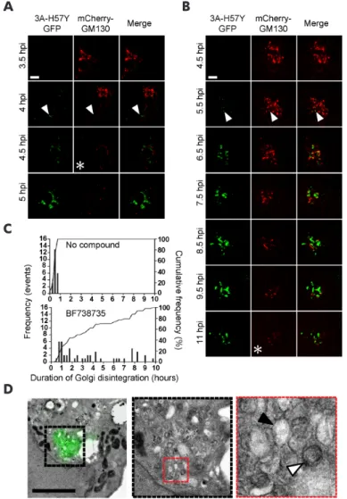
RE SULTS
3A tagged with GFP(S11) assembles with GFP(S1-10) to yield GFP fluorescence
In order to visualize the enterovirus ROs in living cells, one of the non-structural proteins that is anchored in the membranes of the ROs should be labeled with a fluorescent tag. A transposon-based insertion mutagenesis study revealed that the N-terminal region of poliovirus 3A tolerates small insertions (32, 37). Specifically, viable insertions were found after residues 2, 6, 9, 10, or 11. Since the 3A proteins of coxsackievirus B3 (CVB3) and poliovirus are highly homologous, these positions were good candidates for introduction of a small tag in CVB3 3A. We chose to insert GFP(S11) after amino acid 2 in CVB3 3A without using any linker residues, yielding 3A(S11aa2) (Fig. 1A).
Before introducing GFP(S11) into the viral genome, we first tested whether inserting the tag into 3A at this position would generate GFP fluorescence upon co-expression with GFP(S1-10). Staining with an anti-3A antibody demonstrated that the expression pattern of 3A(S11aa2) resembled that of untagged 3A (i.e. lacking GFP(S11)) in HeLa (Fig. S1A) and in BGM cells (data not shown), indicating that the GFP(S11)-tag does not alter the localization of 3A. Co-expression of GFP(S1-10) with 3A(S11aa2), but not with untagged 3A, resulted in a GFP signal that greatly overlapped with the 3A-pattern generated with the anti-3A antibody (Fig. S1B, HeLa cells not shown). Furthermore, 3A(S11aa2) was not GFP fluorescent in the absence of GFP(S1-10) (Fig. S1A). Thus, GFP(S1-10) can assemble with 3A(S11aa2) to yield GFP fluorescence at the sites where 3A is localized in the cell.
Introduction of GFP(S11) into 3A does not impair its known functions
Next, we investigated whether 3A(S11aa2) is able to perform the same functions as untagged 3A. Enteroviruses hijack the Golgi-resident PI4KB (phosphatidylinositol 4-kinase III beta) via their 3A protein to ROs to enrich these membranes in PI4P (phosphatidylinositol 4-phosphate), an essential lipid for viral RNA replication (22). In line with previous observations (22, 38, 39), PI4KB was present in a faint Golgi-like pattern in untransfected HeLa cells, whereas in 3A-transfected cells an intese PI4KB signal was detected at membranes containing untagged 3A (Fig. 1B). Likewise, in cells co-expressing 3A(S11aa2) and GFP(S1-10), PI4KB recruitment was observed to the membranes containing fluorescent 3A (Fig. 1B).
ILL
UMINA
TING THE SITE
S OF ENTER
O
VIR
US INFEC
TION USING A SPLIT
-GFP
-T
A
GGED PR
O
TEIN
2
in turn recruits the COP-I complex to Golgi membranes upon activation (40). The COP-I coat initiates budding of transport vesicles that ferry cargo at the ER-Golgi interface and in the Golgi (41). Expression of 3A in isolation leads to the perturbation of ER to Golgi transport (30, 42–44), as
demonstrated by the dissociation of COP-I from Golgi membranes in BGM cells (44), presumably as a result of the interaction of 3A with GBF1 (30, 44). Similar to untagged 3A, 3A(S11aa2) visualized by co-expression of GFP(S1-10) clearly caused COP-I dissociation (Fig. 1C).
In addition to the interaction with GBF1, enterovirus 3A also directly binds to the host factor ACBD3 (acyl-CoA-binding protein domain 3), although the role of this protein in enterovirus
Figure 1. 3A(S11aa2) recruits PI4KB and dissociates COP-I from Golgi membranes. A) The CVB3-3A(S11aa2)
protein. The GFP(S11)-tag (bold, underlined) is inserted after amino 2 of 3A without any linker residues. The hydrophobic domain, predicted by the Kyte and Doolittle method (67), is indicated as well as the location of the helical hairpin predicted by NMR analysis of the truncated poliovirus 3A protein (68). B, C) HeLa R19 (B) or BGM (C) cells were co-transfected with the plasmid encoding GFP(S1-10) (i.e. pGFP(S1-10)) and either p3A-myc or p3A(S11aa2)-myc. The next day, cells were fixed and subjected to immunofluorescence analysis. The 3A protein was visualized with an antibody staining for p3A-myc (a primary anti-3A antibody and a secondary Alexa488-labeled antibody), while p3A(S11aa2) was visualized by the GFP fluorescence as a result of assembly with GFP(S1-10). PI4KB (A) or COP-I (B) were detected with immunofluorescence using a secondary Alexa594-labeled antibody. Untransfected cells are indicated with an asterisk. Nuclei were stained with DAPI. Wide-field images were acquired with an Olympus BX60 fluorescence microscope. Scale bars equal 10 µm.
A
B
ILL
UMINA
TING THE SITE
S OF ENTER
O
VIR
US INFEC
TION USING A SPLIT
-GFP
-T
A
GGED PR
O
TEIN
CVB3.
Tagging 3A with GFP(S11) results in a replication-competent CVB3 that generates fluorescent replication organelles
Introduction of the GFP(S11)-tag into the infectious clone of CVB3 yielded viable virus, i.e. CVB3-3A(S11aa2). Sequence analysis of the viral genome confirmed that the tag was retained in the 3A protein without mutations. In addition, we generated a replication-competent CVB3 with the short affinity tag StrepII of 10 residues in 3A (i.e. CVB3-3A(StrepIIaa2)). Insertion of the StrepII-tag also did not affect the known functions of 3A (data not shown), and the tag was retained in 3A for five passages (data not shown), further demonstrating that residue 2 is an amenable site for introduction of a small tag.
Subsequently, we tested whether the GFP(S11)-tag in 3A affects the replication kinetics of the virus. Since cells that have been transfected with plasmid DNA are less susceptible to picornavirus infection (48), we generated single cell clones of HeLa and BGM cells that stably express GFP(S1-10). Next, we compared the growth kinetics of CVB3-3A(S11aa2) to wild-type (wt) CVB3 in the presence and in the absence of GFP(S1-10). Samples were subjected to endpoint titration to determine the production of infectious virus, or quantitative PCR to measure viral RNA levels. Replication of wt CVB3 in BGM cells and BGM(GFPS1-10) cells was nearly identical, showing that the presence of GFP(S1-10) did not alter CVB3 replication kinetics (Fig. 2A, 2B). The replication kinetics of tagged CVB3 in BGM(GFPS1-10) also resembled that of BGM cells, indicating that binding of GFP(S1-10) to 3A(S11aa2) does not impede replication (Fig. 2A, 2B). Both infectious virus production and viral RNA levels of the tagged CVB3 were delayed compared to wt CVB3 (Fig. 2B). This delay in replication has also been observed for recombinant polioviruses that encode 3A proteins with small epitope tags (32). Together, these findings showed that replication of CVB3-3A(S11aa2) is similar to other enteroviruses that encoding small tags in a viral protein.
ILL
UMINA
TING THE SITE
S OF ENTER
O
VIR
US INFEC
TION USING A SPLIT
-GFP
-T
A
GGED PR
O
TEIN
2
the anti-GFP antibody has a higher affinity for full-length, assembled GFP.
To ensure that GFP fluorescence is exclusively emitted from 3A(S11aa2)-containing sites, cells from the same infection were stained with an anti-3A antibody. Figure 3B shows that GFP fluorescence was detected in a pattern that greatly overlapped with 3A visualized by immunofluorescence. However, the immunofluorescence with the 3A-antibody generated a slightly more extensive 3A-pattern, most likely as a consequence of not all 3A(S11aa2) proteins being bound by GFP(S1-10). This could be due to partial inaccessibility of 3A(S11aa2) proteins to their GFP(S1-10) counterparts as 3A proteins localize to densely-packed ROs.
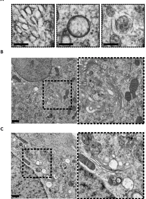
Next, we set out to confirm that the emerging GFP fluorescence is emitted from ROs that contain 3A(S11aa2). To this end, we applied correlative light electron microscopy (CLEM) (49, 50), which provides a direct link between fluorescent signals and the virus-induced structures that underlie them at high resolution. The emerging fluorescence signal in BGM (GFPS1-10) cells infected with CVB3-3A(S11aa2) and stained with Mitotracker Deep Red FM was monitored by live-cell imaging. Cells were imaged and fixed at an intermediate time point in infection. Following sample processing and EM imaging, the Mitotracker signal and corresponding mitochondria were used to orient fluorescence and EM images with an independent marker. Using this method, 3A(S11aa2) was found to localize to ROs, evident as both tubules and clusters of DMVs and multilamellar structures (Fig. 4). Furthermore, the CLEM results not only show that GFP fluorescence was emitted from bona fide ROs, they also demonstrate that the GFP(S11) insertion in 3A does not affect the development of the different RO morphologies typically observed during enterovirus infections.
Figure 2. Single-cycle growth curve analysis of CVB3-3A(S11aa2). BGM cells (A) or BGM cells that stably express
GFP(S1-10) (B) were infected with wt CVB3 or CVB3-3A(S11aa2) for 30 min at MOI 1. At the indicated time points, cells were subjected to titration analysis after freeze-thawing cycles to determine the amount of infectious virus particles. Alternatively, cells were lysed to determine the amount of viral RNA by quantitative PCR. The results are expressed as fold induction relative to the quantities determined directly after removing the inoculum.
A
ILL
UMINA
TING THE SITE
S OF ENTER
O
VIR
US INFEC
TION USING A SPLIT
-GFP
-T
A
GGED PR
O
TEIN
Live-cell imaging reveals the dynamics of Golgi disassembly in live infected cells
Enterovirus infections trigger the disassembly of the Golgi apparatus, which coincides with the formation of early ROs (18, 22). Here, we used live-cell imaging to visualize this process in living cells. To this end, BGM cells stably expressing GFP(S1-10) were transduced with MLV particles encoding mCherry-GM130 as a traceable marker for the Golgi. Live-cell confocal imaging was first carried out with a narrow pinhole (95.56 µm) to detect the first local changes in Golgi structure during infection. As expected, GM130 was observed as a condensed perinuclear signal in uninfected cells (Fig. 5A) or early in infection with CVB3-3A(S11aa2) when fluorescent 3A (which will be further referred to as 3A-GFP) could not yet be detected (Fig. 5B, Movies S1 and S2). Strikingly, the first 3A-GFP signal detected in infected cells was rarely associated with GM130, but could instead be observed as distinct cytoplasmic punctae. Localization of 3A-GFP signal to the Golgi region typically occurred
Figure 3. GFP fluorescence is only emitted from sites where 3A(S11aa2) is localized in cells. A, B) BGM cells
stably expressing GFP(S1-10) were infected with CVB3-3A(S11aa2). At 5 and 6 h p.i., cells were fixed and subjected to immunofluorescence analysis. The detected GFP fluorescence is the result of GFP(S1-10) assembling with 3A(S11aa2). Nuclei were visualized with DAPI. Images were acquired with a Leica SPE-II DMI-4000 confocal laser scanning microscope. Scale bars equal 10 µm. A) GFP(S1-10) was detected with a primary polyclonal antibody directed against GFP and a secondary Alexa594-labeled antibody. The uninfected cell is marked with an asterisk. B) The 3A(S11aa2) protein was stained with a primary antibody directed against 3A and a secondary Alexa594-labeled antibody. An uninfected cell and typical examples of cells early in infection (5 h p.i.) and later in infection (6 h p.i.) are shown.
ILL
UMINA
TING THE SITE
S OF ENTER
O
VIR
US INFEC
TION USING A SPLIT
-GFP
-T
A
GGED PR
O
TEIN
2
within 25 minutes (n=17, range 0-45 minutes) of initial detection. This was followed by a sharp increase in the intensity and number of Golgi-adjacent 3A-GFP punctae and a local perturbation of Golgi morphology, characterized by the onset of GM130 signal fragmentation (Fig. 5B, asterisk GM130 channel). Interestingly, while this rapidly increasing 3A-GFP signal was clearly detected in the Golgi region, it rarely co-localized directly with the GM130 marker (Fig. 5B inset, arrowhead), which is in agreement with previous observations in fixed cells (22). This suggests that RO membranes originate from a compartment of the Golgi not labeled by the cis-Golgi marker GM130, or that 3A resides there only transiently before accumulating in the ROs.
The first signs of disruption of Golgi morphology largely began during or even preceding the accumulation of visible 3A signal (Fig. 5B asterisk 3A-GFP channel, Movie S1), suggesting that local changes to morphology occur rapidly and may be triggered by 3A accumulation in regions of the Golgi outside the imaging plane. To facilitate the detection of the whole Golgi and to monitor large scale changes in Golgi morphology, imaging was carried out using a wider confocal
Figure 4. 3A(S11aa2) localizes to ROs that have tubule and DMV morphologies. A, B) BGM GFP(S1-10) cells were
infected with CVB3-3A(S11aa2), stained with Mitotracker Deep Red FM and monitored by live cell imaging. Full-depth z-stacks of cells emitting GFP fluorescence were taken and cells were processed for EM. LM-EM overlays were made using the Mitotracker as an orientation guide, and 3A-GFP signal was aligned to the corresponding EM image in this manner first at low magnification (A), and then within individual cells of interest (B, which is the boxed area in A). 3A-GFP signal was found at the typical structures that develop during CVB3 infection, including single-membrane tubules (B, red box, enlargement shown on the right) and double-membrane vesicles (B, black box, enlargement shown on the right). Scale bars equal 30 µm (A) and 5 µm (B).
A
B
ILL
UMINA
TING THE SITE
S OF ENTER
O
VIR
US INFEC
TION USING A SPLIT
-GFP
-T
A
GGED PR
O
TEIN
pinhole (600 µm). Global Golgi fragmentation began typically 10-30 minutes after 3A started to accumulate in high amounts (Fig. 6) (n=8, range 0-45 min), which presumably reflects the time taken for the cumulative local changes to become apparent within the entire structure, and was completed 75-105 min after the initial detection of 3A-GFP signal (n=8). Together, these data show that 3A accumulation coincides with the local disruption of Golgi morphology, leading to
Figure 5. Live-cell imaging reveals the association between 3A accumulation and local perturbation to Golgi morphology. A, B) BGM GFP(S1-10) cells transduced with mCherry-GM130 MLV particles were mock-infected (A) or
infected with CVB3-3A(S11aa2) (B), and imaged by live-cell microscopy with images taken at 5 minute intervals. The frames given as time ‘0’ represent either an arbitrary time point (A) or the moment of appearance of 3A-GFP signal (B). Images are presented at 180 (A) or 10 to 20 (B) min intervals as indicated. The confocal pinhole was adjusted for analysis of local effects (95.56 µm confocal pinhole). While Golgi integrity was maintained for the duration of imaging in mock cells (A), local fragmentation of GM130 was evident in infected cells (B, asterisk GM130 channel). The onset of fragmentation was associated with a marked increase in the intensity and number of 3A-GFP punctae at the Golgi (B, asterisk 3A-GFP channel) although, while 3A signal and GM130 were proximal at this stage, there was no clear colocalization (B inset). Scale bar equals 10 µm.
ILL
UMINA
TING THE SITE
S OF ENTER
O
VIR
US INFEC
TION USING A SPLIT
-GFP
-T
A
GGED PR
O
TEIN
2
global fragmentation and eventual disassembly of the Golgi apparatus. After complete Golgi fragmentation, the 3A-GFP signal expands throughout the cell and eventually occupies the entire cytoplasm before the cell goes into demise, demonstrating that 3A-GFP signals can be imaged to the point of cell lysis (Movies S1 and S2).
Construction of CVB3 encoding both GFP(S1-10) and 3A(S11aa2)
Studying ROs induced by CVB3 encoding split-GFP-tagged 3A in distinct cell types requires the cellular expression of GFP(S1-10). To bypass the need for delivering the GFP(S1-10) gene via retroviral transduction or by generating stable cell lines, we constructed a CVB3 that encodes not
Figure 6. Global disruption to Golgi morphology during CVB3 infection visualized by live-cell imaging. Cells
ILL
UMINA
TING THE SITE
S OF ENTER
O
VIR
US INFEC
TION USING A SPLIT
-GFP
-T
A
GGED PR
O
TEIN only 3A(S11aa2) but also GFP(S1-10) (Fig. 7A). For this, the gene encoding GFP(S1-10) was inserted upstream of the capsid coding region (P1) in the infectious clone containing 3A(S11aa2). GFP(S1-10)
was followed by an artificial 3CD cleavage site to release the protein from the P1 region upon translation. Viable virus (i.e. CVB3-GFP(S1-10)-3A(S11aa2) was obtained upon transfection of BGM cells with RNA transcripts of the infectious clone. After harvesting CVB3-GFP(S1-10)-3A(S11aa2), we compared its replication kinetics in BGM cells to CVB3-3A(S11aa2) in BGM(GFPS1-10), so that in both cases the 3A(S11aa2) protein would bind to GFP(S1-10). The level of viral RNA replication was nearly identical for both viruses (Fig. 7B), suggesting that the generation of ROs during the course of infection occurs similarly. However, the virus encoding both GFP fragments (i.e. CVB3-GFP(S1-10)-3A(S11aa2)) was delayed in production of infectious progeny in comparison to CVB3-3A(S11aa2) (Fig. 7B). In line with a previous study (51), we found that the processing of the artificial 3CD cleavage site
Figure 7. Construction of CVB3 that encodes both GFP(S1-10) and 3A(S11aa2). A) A schematic diagram of the
genome organization of CVB3 showing the insertion of GFP(S1-10) before the capsid coding region and GFP(S11) in 3A after amino acid 2. B) Growth curve analysis of CVB3-GFP(S1-10)-3A(S11aa2) in BGM cells and CVB3-3A(S11aa2) in BGM(GFPS1-10) cells. Cells were infected for 30 min at MOI 1. At the indicated time points, cells were subjected to titration analysis after freeze-thawing cycles to determine the amount of infectious virus particles. Alternatively, cells were lysed to determine the amount of viral RNA with quantitative PCR. The results are expressed as fold induction relative to the quantities determined directly after removing the inoculum. C) BGM cells were infected with CVB3 GFP(S1-10) 3A(S11aa2). At 5 and 6 h p.i., cells were fixed and subjected to immunofluorescence analysis. The detected GFP fluorescence resulted from the assembly of GFP(S1-10) with 3A(S11aa2). Nuclei were visualized with DAPI. Images were acquired with a Leica SPE-II DMI-4000 confocal laser scanning microscope. Typical examples of cells early in infection (top panel) and later in infection (lower panel) are shown. Scale bars equal 10 µm.
B
ILL
UMINA
TING THE SITE
S OF ENTER
O
VIR
US INFEC
TION USING A SPLIT
-GFP
-T
A
GGED PR
O
TEIN
2
between a foreign protein and P1 is suboptimal (Fig. S3), which may explain the delay in progeny virion production.
Next, we tested whether BGM cells became GFP fluorescent upon infection with CVB3-GFP(S1-10)-3A(S11aa2). Figure 7C shows that GFP fluorescence colocalized with 3A, visualized with an anti-3A antibody, both early (upper panel) and later (lower panel) in infection. Yet, the GFP fluorescence was substantially dimmer compared to BGM(GFPS1-10) cells infected with CVB3-3A(S11aa2). It is plausible that the dimmer signal is a result of the equimolar ratios of 3A(S11aa2) and GFP(S1-10) generated by this virus, while BGM(GFPS1-10) cells produce an excess of GFP(S1-10). Another explanation for the dim GFP fluorescence is that not all GFP(S1-10) is released from P1 due to suboptimal cleavage of the artificial cleavage site. When GFP(S1-10) is still fused to P1, it might be unable to assemble with 3A(S11aa2) and/or become fluorescent. Nevertheless, these findings suggest that the virus containing both GFP fragments is suitable for live-cell imaging.
DISCUSSION
Live-cell imaging is a powerful technology to gain insight into the dynamics of biological processes. In the field of virology, this method has mostly been applied to visualize the entry and egress pathways of viruses by following the fate of fluorescently labeled, individual virus particles in living cells and monitoring their interactions with cellular structures (52). Imaging replication structures during infection has so far only been reported for a few viruses, including hepatitis C virus (53, 54), vaccinia virus (55), turnip mosaic virus (56), mouse hepatitis virus (57, 58) and equine arteritis virus (59). In most of these studies, replication structures are illuminated by a viral protein that is fused to GFP. Recombinant enteroviruses that encode an RO-anchored viral protein fused to GFP (or another fluorescent reporter) have to date not been reported. Small epitope tags on the other hand were successfully introduced in the 3A protein of poliovirus (32), which prompted us to test whether small tags suitable for fluorescent labeling are accepted in the 3A protein of CVB3. The smallest tag for fluorescent labeling of a protein is the tetracysteine tag of 6-20 residues (depending on the version), but it requires visualization with biarsenical dyes FlAsH and ReAsH in extra labeling steps, and results in fluorescence with a relatively poor quantum yield (60, 61). The split-GFP system uses a tag of 16 residues, GFP(S11), which has the advantage that it does not require staining but becomes directly fluorescent upon assembly with the large GFP(S1-10) fragment in living cells (34). This system has been successfully applied before in the context of a virus to study the intracellular trafficking of ribonucleoproteins of Influenza A virus, where the PB2 polymerase subunit was tagged with GFP(S11) (62). In our study, we incorporated GFP(S11) into the RO-anchored 3A protein of CVB3. We show that the introduction of GFP(S11) after the second residue does not affect the localization or function of 3A when it is expressed in isolation. Whether binding of GFP(S1-10) affects the function of 3A(S11aa2) proteins remains unknown, as it is possible that not all 3A(S11aa2) proteins are bound by the GFP(S1-10) counterparts. Therefore, the subset of unbound 3A(S11aa2) proteins could be solely responsible for exerting the 3A functions.
ILL
UMINA
TING THE SITE
S OF ENTER
O
VIR
US INFEC
TION USING A SPLIT
-GFP
-T
A
GGED PR
O
TEIN
bona fide ROs, which took the form of both tubular structures and DMVs resembling those observed previously with wt CVB3 in Vero cells (18). Split-GFP CLEM thus allows the unambiguous identification of RO morphologies underlying the 3A signal, and opens up new possibilities for a better understanding of the requirements for their development. This method also circumvents limitations inherent in other, similar approaches. For instance, while immuno-gold labeling of proteins is a popular technique that couples an electron-dense gold particle to the protein of interest, allowing it to be visualized by EM, its success depends largely upon the antibody used and the resilience of epitopes during EM sample preparation. In our experience, sample preparation procedures for immunolabeling of CVB3 3A that retain RO membranes unfortunately do so at the expense of viral epitope integrity, resulting in insufficient labeling of 3A (unpublished data). Enterovirus ROs were imaged in living BGM(GFPS1-10) cells upon infection with CVB3-3A(S11aa2). As a proof of concept, we also expressed GM130-mCherry in these cells to visualize Golgi disassembly in real time. The onset of Golgi fragmentation was found to correspond to a period of rapid 3A accumulation in the Golgi region. Under the experimental conditions used, Golgi disruption was completed within 75-105 min after the first detection of 3A-GFP. Interestingly, while 3A accumulation occurred adjacent to the GM130 signal, 3A rarely co-localized with GM130-mCherry before and during Golgi disassembly. While this does not preclude the transient localization of 3A to the cis-Golgi, our findings suggest that ROs may be generated from another Golgi compartment not labeled by GM130, which is in line with previous observations that suggest that RO formation is initiated at the trans-Golgi (22).
Enteroviruses can infect both polarized and non-polarized cells. The use of CVB3 encoding split-GFP-tagged 3A in distinct cell types relies on the cellular expression of GFP(S1-10). As an alternative to delivering the GFP(S1-10) gene via transduction, we introduced the coding sequence of GFP(S1-10) into the viral genome together with 3A(S11aa2). This new recombinant CVB3 encoding both GFP fragments also induced GFP fluorescent foci that colocalized with 3A. While progeny virion production by this virus was delayed, the viral RNA levels were very similar to CVB3-3A(S11aa2) infection of BGM(GFPS1-10) cells, implying that the “two-fragment virus” retained its ability to form ROs. Hence, this virus would be suitable for studying enterovirus ROs in physiologically more relevant cell types, including pancreatic cells (14) and 3D-cultured CaCo-2 cells (63), without the need for ectopic expression of GFP(S1-10).