Cover Page
The handle
http://hdl.handle.net/1887/35971
holds various files of this Leiden University
dissertation.
Author: Bai, Yani
Copy right©2015, Yani Bai
This dissertation or parts there of, may be reproduced freely for scientific and eductioanl purposes as long as the source of the material is acknowledged. The study described in theis thesis was perfomed at the institute of Biology of Leiden University; the practical work was performed at the Netherland Institite of Ecology, NIOO-KNAW.
The financial support of this study was provided by the NIOO-KNAW and China Scholarship council (CSC).
Cover pictures were taken by Yani Bai.
Ecological functioning of bacterial chitinases
in soil
Proefschrift
ter verkrijging van
de graad van Doctor aan de Universiteit Leiden,
op gezag van Rector Magnificus Prof. mr. C.J.J.M. Stolker,
volgens besluit van het College voor Promoties
te verdedigen op dinsdag 28 October 2015
te klokke 15:00 uur
door
Yani Bai
Promotiecommissie
Promotores: Prof. dr. J.A. van Veen
Prof. dr. W. de Boer, Wageningen Universiteit
Overige leden: Prof. dr H.P. Spaink
Prof. dr P.G.L. Klinkhamer
Prof. dr J.D. van Elsas, Rijks Universiteit Groningen
Contents
Chapter 1 General introduction 7
Chapter 2 Genomic comparison of chitinolytic enzyme systems from terrestrial and aquatic bacteria 17
Chapter 3 Chitin degrading abilities and antifungal activity of bacterial strains differing in complexity of their chitinolytic systems and growth form 39
Chapter 4 Composition of chitinolytic soil bacterial community colonizing different chitin resources 55
Chapter 5 Impact of plant growth stage and fungal biomass on the composition of the chitinolytic bacterial community in the potato rhizosphere 85
Chapter 6 General Discussion 105
Summary 113
Samenvatting 117
摘要 121
Bibliography 123
Supplementary tables 143
Acknowledgement 149
Curriculum Vitae 151
7
Chapter 1
General Introduction
Chitin is the second most abundant polymer in nature and it plays an essential role in aquatic and terrestrial ecosystems. In soils, chitin is mainly found in fungal cell walls and in the outer shields, exoskeleton, of arthropods. Because of the relatively high nutritional value of chitin, including both C and N
components, many primary decomposers such as bacteria and fungi contain chitin degrading enzymes, among which chitinases.
The possession of chitinases is widely distributed among bacteria (and fungi). As there is a fierce competition between bacteria and fungi in the
oligotrophic environment of the soil for the scarce nutrients, bacterial chitinases are also used as tools to attack fungi by destroying the chitin in fungal cell walls. The main target for this activity is the hyphal tip where the chitin is still in a native form, i.e. where chitin is present as single strands and not yet cross-polymerized and linked with other cell-wall components, so that it can easily be attacked by chitinases. Previous studies showed that bacteria differ in their ability to use chitinases for chitin degradation (De Boer et al 2008, De Boer et al 1999, De Boer et al 1998). Some are good degraders of solid chitin particles and such bacteria might primarily be involved in the decomposition of dead hyphal (and arthropod) remnants (Igarashi et al 2014). Others can only degrade pre-treated (colloidal) chitin and several of such bacteria have shown to have additional antifungal properties and they may use their chitinases as part of the machinery to compete with or defense against fungi (Aktuganov et al 2008).
Although the importance of bacterial chitinases was recognized in
General introduction
living fungal hyphae. Until now, most of the studies on bacterial chitinases were performed using chitin resources from the exoskeleton of arthropods. However, chitin of arthropods is different chemically from the chitin of fungal cell wall. Thus, for a deeper understanding of the functioning of bacterial chitinases in soil
ecosystems, where fungi are in far greater abundance than arthropods, it is necessary to focus on the chitin from fungi in relation to the particular conditions in which bacteria and fungi share the available niches and compete for nutrients. In this thesis I will provide novel information on the functioning of bacterial chitinases depending on a variety of bacterial-fungal interactions.
Chitin: natural polymers with important ecological function
Chitin is a polymer of N-acetyl-D-glucosamine. In 1811, chitin has been described for the first time by the French chemist H. Broconnot. The word “chitin” originates from the Greek word “envelope”, which, to some extent, describes its function. It is an important structural compound of the exterior of different organisms, e.g. fungi, crustaceans and insects (Synowiecki & Al-Khateeb 2003). Arthropods (mainly crustaceans) are the major chitin producing organisms in aquatic ecosystems and their annual global chitin production has been estimated to range from 2.8 × 10⁷ T yr-1 for freshwater ecosystems to 1.3 × 10⁹ T yr-1 for marine ecosystems (Cauchie 2002). The majority of chitin in terrestrial ecosystems is present in fungal propagules (hyphae, spores etc.) or in remainders of fungi (Smrz & Catska 2010). Although soil arthropods also contain chitin, their biomass and consequently their contribution to the chitin mass in soil is far lower than that of fungi (Holtkamp et al 2008). So far, no reliable estimates exist of the annual fungal chitin production in terrestrial ecosystems. Moreover, it is speculated that also certain bacteria, such as the Actinobacteria, among which Streptomyces
9 According to the orientation of the poly-N-acetylglucosamine chains, chitin can be divided into three different forms. The most common form is α-chitin, in which the polymer chains are in antiparallel arrangement. The two other, less common, forms are β- and γ-chitin, in which the polymer chains are in parallel and mixed of parallel and antiparallel arrangements, respectively (Gooday 1990). Crustaceans contain mostly α-chitin; squid pens possess β- chitin. Fungal chitin is comparable to crustacean chitin, due to the uniform composition of the chitinous material, which is both of the alpha-chitin form (Agullo et al 2003).
Chitin is a common structural component of fungal cell walls. It is located in the innermost layer of the cell wall as α-form microfibrils, linked to mannans, glucans and proteins (Figure 1). Generally, the chitin content of fungal cell walls ranges from 22-40 % (Muzzarelli 1977).
Figure 1 Structure of fungi cell wall (Selitrennikoff 2001)
General introduction
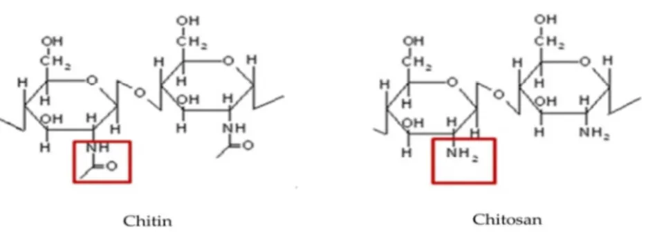
Figure 2 Structure of chitin and chitosan (deacetylation of chitin)
Although the natural forms of chitin are cross-linked with other organic polymers, purified chitin is often used in research, mostly as crystal chitin purified from crab cuticles. To make the chitin more accessible for enzymes, colloidal chitin is prepared from crystal chitin by dissolving it in strong acid followed by precipitation in water (Hirano & Nagao 1988). It is the most used form of chitin to obtain chitinolytic bacteria on agar plates. When chitinolytic bacteria grow on top of colloidal chitin plates, a halo will be produced due to the activity of chitinase (De Boer et al 1998). This method has commonly been used to identify and enumerate chitinolytic bacteria.
Enzymatic Chitin degradation
Chitin is insoluble in water and resistance to moderate acid, alkaline and many organic solvents and therefore microbial degradation is the most important degradation pathway (Hoell et al 2010). Both fungi and bacteria are able to decompose chitin (Ghosh & Chakraborty 2010). In fungi, nearly 80 % of the species are chitinolytic, but it is not clear whether all these fungi do actually degrade chitin since fungal chitinases play also a role in branching of chitin polymers in the fungal cell wall. In different environments 1 – 20 % of culturable bacterial species are chitinolytic (Gooday 1990). The chitinolytic ability is
11 Chitin can be degraded enzymatically in two ways. First chitin degradation can be achieved by deacetylation by deacetylases. This degradation will result in the production of chitosan. Through the activity of chitosanase, chitosan can be degraded further into chitobiose and glucosamine.
The main degradation pathway of chitin is soil is through chitinases (EC 3.2.1.14) resulting in the hydrolysis of the 1, 4-β- glycosidic bonds between the N-acetyl-D-glucosamine units. The products of this enzyme activity are monomers of N-acetyl-D-glucosamine (GlcNAc) (Beier & Bertilsson 2013). This may be followed by degradation of the N-acetyl-D-glucosamine by N-acetylglucosaminidases, producing glucosamine.
Bacterial chitinases can be divided into two families according to their amino acid sequence homology: family 18 and 19 of glycoside hydrolases. The majority of the known bacterial chitinases belong to family 18. The catalytic domains of family 18 chitinases, have (β/α)8 barrel folds whereas those of family 19 chitinases have a high α-helical content and share structural similarity with chitosanases and lysozymes (Svitil & Kirchman 1998, Watanabe et al 2001). Glycoside hydrolase family 18 bacterial chitinases can be divided into different types. In 1993, bacterial chitinases were divided into group I to IV, with among group I, a further division of the chitinases in A, B and C types (Watanabe et al 1993). Later on, another classification method was suggested (Karlsson & Stenlid 2008, Suzuki et al 1999), in which the family 18 chitinases was divided in types A, B and C with most of the bacterial chitinases belonging to the A type. This
nomenclature is referring to the enzyme catalytic domains (CD). The ChiA catalytic domain consists a (β/α) 8- barrel with a small α+β domain inserted between the
seventh and eighth β- strands of (β/α) 8- barrel (Perrakis et al 1994). Most of the
General introduction
Kirchman 1998). Fibronectin type III domains are thought to maintain an optimal distance and orientation between the catalytic and binding domains (Folders et al 2001). It is reported that chitin binding and fibronectin type III domains are not necessary for the hydrolysis of colloid chitin, but play an important role in hydrolysis of chitin–glucan complex of fungal cell walls (Dehestani et al 2010).
According to their functional patterns, chitinases can also be classified into endo and exo-enzymes (Henrissat & Davies 2000). Endochitinases cleave chitin randomly resulting in chitin-oligomers of different size, such as chitobiose, chitotriose, and chitotetraose; exochitinases include chitobiosidases and β -N-acetylhexosaminidase; the former produces GlcNAc dimers (chitobiose) by cleaving the non-reducing end of chitin while the latter hydrolyzes chitobiose, chitotriose, and chitotetraose, resulting in the release of N-acetylglucosamine (Bhattacharya et al 2007, Tronsmo & Harman 1993).
In order to function, bacterial chitinases need to be associated with the outer parts of bacteria or be secreted as extracellular enzymes (LeCleir et al 2004). The extracellular activity of chitinases suggests that they must be adapted to function under the physicochemical conditions prevailing in the surrounding environment. This may imply that different chitinases will be secreted depending on different situations. Other compounds that cooperate with chitinase during chitin degradation are also secreted by bacteria. For instance, Serretia marcescens
was found to produce a protein CBP21, which binds to and disrupts the chitin polymer, and so increases the effect of chitinases by making chitin more accessible (Suzuki et al 1998, Vaaje-Kolstad et al 2005a).
Factors affecting the function of bacterial chitinases
13 ability of these bacteria to degrade fungal chitin resources in soil as the chitin in media is often derived from marine arthropods like crabs and shrimps.
Furthermore, chitin in artificial media is often made colloidal to make it more accessible to chitinases. It has been shown that several bacteria that are able to hydrolyse colloidal chitin are very slow degraders of chitin particles (De Boer et al 1999). This observation has led to the suggestion that such bacteria may use their chitinases for different purposes. The bacterial genus Collimonas is an example of chitinolytic soil bacteria with limited chitin-degrading abilities (Leveau et al 2010). This bacterial genus is mycophagous, which implies that it uses living fungi as a source of nutrients. It has been proposed that chitinases form part of the machinery with which Collimonas attack fungal hyphae. Chitinases could help to destabilize the hyphal tip. Destabilization of the hyphal tip can provide Collimonas
with the ability to get access to the nutrients in the fungal cytoplasm. In addition to the predatory strategy of Collimonas, bacterial chitinases may also be involved in other antagonistic interactions with fungi such as competition and defence against bacteriolysis by fungi (De Boer et al 2005, De Boer et al 1998, Suma & Podile 2013).
The complexity of the bacterial chitinolytic enzyme system is large and there is a large variation in the chitinolytic systems of different bacterial species. In addition, other enzymes, such as β-1, 3-glucosidases, as well as secondary metabolites may be important for the ecological functioning of chitinases. Other cell wall lytic enzymes may be needed to make the chitin of the fungal cell walls accessible to chitinase (Figure 1) (Arlorio et al 1992) and the combination of chitinases and secondary metabolites may be effective to suppress fungal growth. Therefore, it is the combination of chitinases and other enzymes and/or other compounds that may be indicative for a particular ecological function.
General introduction
rhizosphere (the soil in the immediate surroundings of the plant root that is under the influence of the roots). In these two compartments, soil characteristics and nutrient resources are different. This is reflected by the composition and distribution of microorganisms. Plant roots secrete exudates such as sugars, organic acids, amino acids, etc, which are energy resources for many
microorganisms (Meng et al 2005, Rengel & Marschner 2005). Therefore, in the rhizosphere many bacteria and fungi live and feed on these exudates and other root derived products such as sloughed off root cells (Norton & Firestone 1991). There is a fierce competition between bacteria and fungi for these nutrients (Couteaudier 1992). So in the rhizosphere, bacterial chitinases may primarily be used as tools in the competition between bacteria and fungi (De Boer et al 2008). However, in the bulk soil, bacteria have to grow primarily on dead organic matter among which fungal hyphal remnants containing chitin and here bacterial
chitinases may mainly be used to degrade fungal chitin as source of C and N. Additional factors which may be important for the functioning of chitinases are the morphological characteristics and motility properties of bacteria. There is evidence that hyphal forming bacteria, e.g. Actinomycetes, are better able to degrade chitin particles than most non-hyphal bacteria (De Boer et al 1999). Actinomycetes do often become the dominant chitin-degrading bacteria when the soil is mixed with solid chitinous material (Krsek & Wellington 2001, Sato et al 2010). The reason for the good chitin degrading ability of Actinomycetes
15
Aims and research questions
The research described in this thesis aimed to improve the understanding of the ecological function of bacterial chitinases in soil. The following research questions have been addressed:
1) Is there a relationship between the composition of chitinases genes, as well as other bacterial capabilities, such as the production of antibiotics and fungal cell wall lytic enzymes and the ecological function for which chitinases are used in different habitats?
2) Is there a relationship between the composition of the chitinolytic bacterial community, chitinase genes and the abundance of saprotrophic fungi in situations of competition between bacteria and fungi such as in the plant rhizosphere? 3) What is the response of soil borne bacteria upon the addition of different chitin resources and will there be a selection for different chitinolytic bacteria
depending on the type of the available chitin source?
4) What are the main factors in the bacterial chitinolytic system determining the functioning of chitinases, i.e. chitin degradation and/or antifungal activity and what are the effects of morphological characteristics, such as hyphaeformation on the functioning of bacterial chitinases?
In order to get a better understanding of the ecological functions of bacterial chitinases and answering above formulated questions, I applied both a “in silico” genomic comparison of the bacterial chitinolytic system and
General introduction
Outline of thesis:
In Chapter 2 an “in silico” approach was applied to compare the chitinolytic systems of aquatic and terrestrial bacteria, including chitinase numbers, domains within chitinases and other assisting proteins on the basis of information present in publically available bacterial genomes. These
characteristics were connected to possible ecological functions of bacterial chitinolytic systems in different environments.
Chapter 3 provides results of an experiment in which a selection of 13 chitinolytic bacterial species was exposed to different chitin sources. Bacteria were selected from different phyla, having different numbers of chitinases and also differing in morphologic properties. Here, I wanted to find out which of these properties are most important for chitin degradation as well as for the antifungal activity of these bacteria; the latter was tested in confrontation plate assays.
In Chapter 4 I report on the selection of chitinolytic bacteria in soil upon the addition of different chitinous sources, namely crystal chitin, fungal cell walls and exoskeletons of mealworms. The result will reveal effects of chitin resources on soil-borne bacteria.Chapter 5 provides insight in the abundance, composition and diversity of bacterial chitinase genes and the chitinase harboring bacterial community in the rhizosphere as a response to fungal biomass dynamics during potato crop growth in the field.
17
Chapter 2
Genomic comparison of chitinolytic enzyme systems from
terrestrial and aquatic bacteria
Yani Bai, Vincent G.H. Eijsink, Anna M. Kielak, Johannes A. van Veen, Wietse de Boer
Genomic comparison of bacterial chitinolytic system
Abstract
19
Introduction
Chitin, a polymer of N-acetyl-D-glucosamine, is one of the most abundant biopolymers in nature. It is an important structural compound of the exterior of different organisms, e.g. fungi, crustaceans and insects (Synowiecki and Al-Khateeb, 2003). Arthropods (mainly crustaceans) are the major chitin producing organisms in aquatic ecosystems and their annual global chitin production has been estimated to range from 2.8 × 10⁷ Mgyr-1 for freshwater ecosystems to 1.3 × 10⁹ Mg yr-1 for marine ecosystems (Cauchie 2002). The majority of chitin in terrestrial ecosystems is present in fungal propagules (hyphae, spores etc.) or in remainders of fungi (Smrz & Catska 2010). Although soil arthropods do also contain chitin, their biomass and consequently, their contribution to soil chitin is far lower than that of fungi (Holtkamp et al., 2008). Decomposition of chitin is an important component of carbon and nitrogen cycling in both terrestrial and aquatic ecosystems (Gooday 1990). Bacteria, which do not produce chitin
themselves, have an important role in chitin decomposition (Beier and Bertilsson, 2013; Metcalfe et al., 2002). Chitinolytic bacteria occur in both aquatic and terrestrial habitats and produce enzymes, chitinases that hydrolyze chitin to chito-dextrins. Most bacterial chitinases belong to family 18 of the glycoside hydrolases, whereas occurrence of chitinases belonging to the other main chitinase family, glycoside hydrolases 19, is less common (Cantarel et al., 2009). Possession of chitinases is taxonomically widespread among bacteria: chitinolytic
representatives are, amongst others, known from the phyla Actinobacteria,
Firmicutes, Acidobacteria and Proteobacteria (Hunt et al., 2008; Williamson et al., 2000). Yet, the rate and efficiency of chitin degradation appears to differ strongly among chitinolytic bacterial species. Streptomyces species were shown to degrade solid chitin particles rapidly and completely in soils and sand microcosms,
Genomic comparison of bacterial chitinolytic system
Boer et al., 1999; Leveau et al., 2010). Therefore, it is uncertain if such bacteria play a major role in the decomposition of natural chitin resources.
A possible alternative function of soil bacterial chitinases is the
involvement in antagonistic interactions with fungi (De Boer et al., 2001; De Boer and van Veen, 2001). In soil, co-existence of bacteria and fungi is ubiquitous. Since metabolic abilities of fungi and bacteria overlap, competition for organic
21
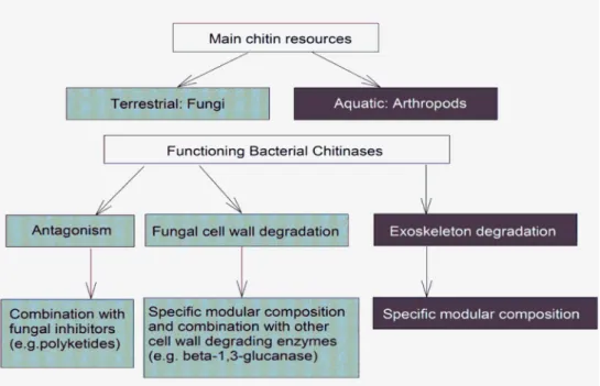
Figure 1 Schematic overview of the hypothesized differences in ecological functioning of chitinases for terrestrial and aquatic bacteria
Based on the aforementioned consideration we hypothesized that the composition of chitinolytic enzyme systems may differ between terrestrial and aquatic bacteria (Figure 1). In particular, we hypothesize that the function of terrestrial bacterial chitinases is strongly related to the decomposition of fungal remainders and/or antagonistic interactions with fungi. To address these hypotheses, we performed an “in silico” study by screening and comparing publicly accessible aquatic and terrestrial bacterial genomes with respect to the characteristics of their chitinolytic systems and, at a lower level of detail, with respect to the occurrence of genes encoding other components with possible anti-fungal activities. In addition to addressing our hypotheses, the data presented provide a comprehensive overview of the composition of bacterial chitinolytic enzyme systems, including catalytic hydrolase domains, carbohydrate-binding modules, and the recently discovered lytic polysaccharide
Genomic comparison of bacterial chitinolytic system
referred to as CBM33 or, more recently, Auxilliary Activity family 10 (AA10) (Levasseur et al., 2013).
Methods
In order to obtain information on the composition of bacterial chitinolytic enzyme systems, we examined genomes of bacteria that are available in public databases, mainly the NCBI, JGI and GOLD (http://www.ncbi.nlm.nih.gov,
http://www.jgi.doe.gov and
http://www.genomesonline.org/cgi-bin/GOLD/index.cgi, based on information available in June 2013). The genome protein annotations were screened for the presence of chitinases by searching for the term “chitinase” in the full description of the genome annotation at NCBI websites. Obtained amino acid sequences were first checked for possession of conserved domains characteristic for chitinases using the search tool of The Conserved Domain Database, NCBI
(http://www.ncbi.nlm.nih.gov/Structure/cdd/wrpsb.cgi). Conserved domain names containing the term “chitinase”, including “chitinase-like”,
“chitinase_glyco_hydro_19” etc., were selected. A further check was done using the Pfam database (http://pfam.sanger.ac.uk) (Punta et al., 2012) in which PF00704 corresponds to family 18 glycoside hydrolases and PF00182 to the family 19 glycoside hydrolases.
23 We also examined whether adaptations to aquatic and terrestrial chitin resources may be mirrored by the modular composition of the 18 and 19 chitinases. With respect to the latter, the majority of chitinases contain other domains next to the catalytic domains. In particular domains classified in CAZy as carbohydrate binding modules (CBMs) and fibronectin type III (FnIII) domains. To identify CBMs and relevant FnIII domains (see Vaaje-Kolstad et al., 2013 and below), amino acid sequences of the selected chitinases were submitted to the Pfam database and screened (http://pfam.sanger.ac.uk). Domain names were retrieved from CAZy using the links provided in Pfam.
For identification of chitinases and additional domains, the threshold value for sequence similarity was set to 10-6 expect value (E-value). Although
usually values smaller than 10-20 are considered as good similarity scores, it has
been shown repeatedly that comparison of the sequences of functional chitinases may yield higher E-values. For instance, a chitinase from Ewingella americana
(Inglis et al., 2000), accession number CAA62151.1 in the NCBI database, yields an E-value for the catalytic domain of 2.1-7 when searching the Pfam database.
Another example is a chitinase from an antarctic Arthrobacter sp. strain TAD20 (Lonhienne et al., 2001), accession number CAB62382.1 in NCBI database, which has a functional carbohydrate binding module (CBM_5_12) yielding an E-value of 3.7-7 when using the Pfam database.
Genomes encoding confirmed chitinases were further analyzed for the presence of other proteins possibly involved in chitin and/or fungal cell wall degradation. This included the CBM33-type LPMO (PF03067 in the Pfam database), also known as chitin-binding proteins, and currently classified in the CAZy
database as AA10 (Auxilliary Activity family 10; Levasseur et al., 2013). The best known member of this family is the 21 kDa chitin-binding proteins CBP21 from
Genomic comparison of bacterial chitinolytic system
(PF03067) applying a threshold of E-value of 10-6. Additional screens were
performed, focusing on potential fungal cell wall degrading β -1.3-glucanases and polyketide synthases (PKS) which may produce antifungal secondary metabolites. The full genome annotation texts were screened for the presence of names of these two proteins. The presence of different carbohydrate-binding modules, CBM33 proteins, β -1.3-glucanases and polyketide synthases in chitinase-containing genomes was quantified as the fraction of total chitinase-chitinase-containing genomes containing at least one of the respective genes. Differences in fractions were statistically analyzed using a Chi square test (P < 0.05) after construction of 2 × 2 contingency tables (http://math.hws.edu/javamath/ryan/ChiSquare.html).
Results and Discussion
Phylogenetic distribution of terrestrial and aquatic chitinase-containing
bacterial genomes
25
Table 1 Number of publicly available chitinase-containing bacterial genomes from terrestrial and aquatic habitats in June 2013
Habitat Numbers of chitinase containing bacterial genomes
Total bacterial genomes
Fraction of chitinase-containing genomes
Terrestrial 74 256 0.29
Soil 68 195 0.35
Other terrestrial
6 61 0.10
Aquatic 36 401 0.09
sediment 4 86 0.05
marine 15 229 0.07
Other aquatic 15 58 0.26
fresh water 2 28 0.07
The difference in relative abundance of chitinase-containing genomes between aquatic and terrestrial bacteria could indicate that the possession of chitinase genes is more common for terrestrial bacterial species than for aquatic ones. However, the difference may well be due to a bias in the availability of annotated bacterial genomes. For instance, almost half of the terrestrial chitinase-containing bacterial genomes belonged to the phylum Actinobacteria (Table 2).
The availability of many annotated genomes of Actinobacteria is due to the interest in these microbes as producers of valuable secondary metabolites (Worrall and Vijgenboom, 2010). Earlier studies had already indicated the widespread occurrence of chitinases in terrestrial Actinobacteria (Metcalfe et al., 2002; Williamson et al., 2000). The order Vibrionales (ɣ-subdivision of the phylum
Proteobacteria) was strongly represented among the chitinase-containing genomes of aquatic bacteria. The order Vibrionales is for the largest part
Genomic comparison of bacterial chitinolytic system
Table 2 Taxonomic distribution of chitinase-containing bacterial genomes and average number of chitinases per genome
Phylum Class/order Genome numbers in terrestrial habitats
Genome numbers in aquatic habitats
Average numbers of chitinase-genes per genome
Terrestrial Aquatic GH18 GH19 GH18 GH19
Actinobacteria Actinomycetales 29 - 5.38 1.07 - -
Bacteroidetes 4 3 4 0.25 2.67 0
Firmicutes 22 9 3.45 0.05 2.55 0
Bacillales 19 5 3.53 0.05 1.6 0
Clostridiales 3 4 3 0 2.25 0
Proteobacteria 14 24 2.36 0.79 3.67 1
Beta subdivision 7 1 1.86 0.29 4 1
Beta/Burkholderiales 7 - 1.86 0.29 - -
Beta/Neisseriales - 1 - - 4 1
Delta subdivision 1 1 5 2 1 0
Gamma subdivision 6 22 2.5 1.17 3.78 1.04
Gamma/Xanthomonadales - 1 - - 2 0
Gamma/Pseudomonadales 3 - 1.33 1 - -
Gamma/Vibrionales - 15 - - 4.33 1.27
Gamma/Oceanospirillales - 1 - - 6 1
Gamma/Enterobacteriaceae 3 1 3.67 1.33 3 2
27
terrestrial habitats aquatic habitats genome
Terrestrial Aquatic
Chloroflexi 1 - 5 0 - -
Chlamydiae/Verrucomi crobia
2 - 2 0 - -
Cyanobacteria Nostocales 1 - 1 0 - -
Genomic comparison of bacterial chitinolytic system
Distribution of family 18 and 19 chitinases
Bacterial chitinases belong to the glycoside hydrolase (GH) families 18 and 19 with the majority belonging to family 18 (Cantarel et al., 2009; Henrissat, 1991). The enzymes of these families do not share sequence similarity and have different catalytic mechanisms (Eijsink et al., 2010). Generally, family 18 chitinases are much more abundant in bacteria than family 19 chitinases (Table A1 and A3).
With the exception of three genomes of terrestrial Mycobacteriumspp.
that only contain one single gene encoding a family 19 chitinase, all selected genomes contained family 18 chitinases. The average number of family 18 chitinase genes per selected genome was 3.95 for terrestrial bacteria and 3.14 for aquatic bacteria. The higher numbers in terrestrial bacteria are due to the
Actinobacteria which have an average number of 5.38 family 18 genes per genome, and in particular to the genus Streptomyces that has an average number of 5.82 family 18 genes per genome. Among the aquatic bacteria, the highest numbers of family 18 encoding genes were observed for strains of the order
Vibrionales (4.31).
Family 19 chitinases are mostly known from plants, but have been detected in several bacterial genera after the first description in 1996 of a family 19 chitinase in Streptomyces griseus (Ohno et al., 1996; Prakash et al., 2010). Family 19 chitinases in plants are thought to be involved in defense against plant-pathogenic fungi (Kasprzewska, 2003). Therefore, the possible role of bacterial family 19 chitinases in antagonistic interactions with fungi has received attention. In a comparative study of the antifungal activities of the family 18 and 19
chitinases of Streptomyces coelicolor, only a family 19 chitinase showed an
29 More generally, it is well known that family 18 chitinases can have anti-fungal activity as well (Prasanna et al., 2013).
The fraction of chitinase-containing bacterial genomes with family 19 chitinases did not differ significantly between terrestrial and aquatic genomes and was 42 % and 50 % (Table A1 and A3), respectively. The number of family 19 chitinases per genome was mostly one, sometimes two, and in three cases three (Streptomyces sp. SirexAA-E, Streptomyces clavuligerus ATCC 27064and Vibrio orientalis CIP 102891). Most of the terrestrial bacterial genomes containing family 19 chitinases belonged to the Actinobacteria, whereas for aquatic bacteria it is mostly found in genomes of ɣ- Proteobacteria, in particular Vibrionales. With the exception of one Paenibacillus genome, the genomes of Firmicutes did not contain family 19 chitinases.
Carbohydrate binding modules and fibronectin type III domains within
bacterial chitinases
Next to the catalytic domains, most of the chitinases contain other
domains, in particular domains classified as carbohydrate binding modules (CBMs) and fibronectin type III (FnIII) domains. It is well known that CBMs increase chitinase affinity for chitin, whereas other functions for CBMs have also been proposed, including facilitation of enzyme movement along a chitin chain during processive action and stimulation of local decrystallization of the substrate (Eijsink
et al., 2008; Nimlos et al., 2012; Svitil and Kirchman, 1998; Watanabe et al., 1994). Notably, chitinases acting on heteropolymeric structures such as fungal cell walls may benefit from CBMs that create affinity to other polysaccharides nearby (Hervé et al., 2010). Less information is available for FnIII domains nevertheless a role in enhancement of hydrolysis of chitin has been suggested (Uchiyama et al., 2001; Watanabe et al., 1994).
In order to obtain a better understanding of differences between
Genomic comparison of bacterial chitinolytic system
in composition of CBMs in chitinases. We found four dominating types of CBMs, with Pfam IDs PF02839, PF03422, PF02018 and PF00553, respectively. Domain PF02839 (CBM5/12 in CAZy) is characterized by the presence of conserved exposed tryptophans that interact with the substrate (Vaaje-Kolstad et al., 2013). This domain is generally thought to be rather chitin-specific and there are reports which confirm its contribution to chitinase efficiency, in particular in the
degradation of crystalline chitin (Hashimoto et al., 2000; Uni et al., 2012). Domain PF03422 (CBM6 in CAZy) has been shown to have a cellulose-binding function, whereas some also bind to β -1, 3-glucans, β -1, 4-glucans and xylan (Henshaw et al., 2004; van Bueren et al., 2005). Domain PF00553 (CBM2 in CAZy) has been reported to bind to soluble and insoluble forms of cellulose, whereas binding to xylan and chitin also has been reported (Simpson et al., 1999; Xu et al., 1995). Domain PF02018 covers four CBM families in CAZy (CBM4, CBM9, CBM16 and CBM22) comprising domains known to bind to cellulose, β -glucans, and xylan (Brun et al., 2000; Winterhalter et al., 1995).
Chitinases contain two families of FnIII domains. One of the families, PF08329, includes the N-terminal domain of the well-known chitinase A from S. marcescens, which contains exposed aromatic residues that were found to enhance the hydrolysis of chitin (Uchiyama et al., 2001). The role of the other FnIII domain, PF00041, is not clear. The limited available information indicates that this domain plays a functional role in the hydrolysis of chitin but is not directly
involved in the binding to chitin (Watanabe et al., 1994). For this latter domain, a more structural role, e.g. in ensuring correct positioning of other domains relative to each other, has been proposed (Watanabe et al., 1994).
31
Figure 2 Fraction of bacterial genomes that contain at least one of the indicated carbohydrate binding modules in a chitinase gene. (A) For all genomes; (B) For selected phyla (only those for which a larger number of chitinase-containing genomes were available; Actino-: Actinobacteria; T:
terrestrial habitat; A: aquatic habitat)
Figure 2 showed some interesting trends. The chitin-specific CBM5/12 domain (PF02839) commonly occurs in chitinases of all bacterial phyla and its occurrence is significantly higher in genomes derived from aquatic habitats than in terrestrial genomes (p< 0.05). A small number of CBM5/12 domains group in another Pfam entry (PF14600, referred to as CBM_5_12_2). These CBMs only occurred in bacterial chitinases from aquatic Proteobacteria (Table A3). Most interestingly, aquatic bacteria hardly contained any other CBMs, whereas many additional CBMs occur in the chitinases of terrestrial bacteria. CBM2 (PF00553) is abundant in the terrestrial bacterial chitinases, where they, according to available exprimental data, may contribute to binding of chitin, but also could interact with other polysaccharides. The other CBMs found in terrestrial chitinases, belonging to CBM families 4, 6, 9, 16 and/or 22 (PF02018 and PF03422), are not likely to bind to chitin but rather to β -glucans. These domains, only found in chitinases from actinobacterial genomes, may endorse the chitinases with the ability to interact with β -glucan containing co-polymeric structures which are present in fungal cell walls (Bowman and Free, 2006). The general impression of the
Genomic comparison of bacterial chitinolytic system
of chitin requires more complex modular chitinase architectures in terrestrial habitats than in aquatic habitats. This might be a general adaptation to larger variation in substrate composition or a more specific adapatation to the occurrenc of fungi.
Some chitinases from terrestrial Actinobacteria and Proteobacteria
contained a CBM13 domain, which have Pfam IDs PF00652 or PF14200, and are named Ricin_B_lectin and RicinB_lectin_2, respectively. Both of them are related to a lectin (a carbohydrate-binding protein) produced in the seeds of the castor oil plant Ricinus communis (Lord et al., 1994). There is no evidence that this domain is involved in the functioning of chitinases. Due to their unknown function and limited numbers, PF00652 and PF14200 were not included in our analysis.
Figure 3 Fraction of bacterial genomes that contain one or more of the indicated fibronectin type III domains in at least one of their chitinases (only family 18 enzymes) (A) for all genomes (B) for selected phyla (only those for which a larger number of chitinase-containing genomes were available; Actino-: Actinobacteria; T: terrestrial habitat; A: aquatic habitat)
33
Actinobacteria and Firmicutes and, to a lesser extent, Proteobacteria. The
prominent presence of the FnIII domain PF00041 in terrestrial bacterial chitinases may point at a role in the degradation of fungal chitin resources. As mentioned before very little is known about the function of PF00041; if this domain indeed has a structural role (see above) its presence in chitinases from terrestrial origin may be linked to the higher modular complexity in these enzymes, relative to those from aquatic habitats. The other FnIII domain, PF08329, most likely being directly involved in chitin-binding, is highly present in bacterial chitinases of aquatic bacteria (Figure 3), in particular in aquatic Proteobacteria (around 80%). Consequently, in aquatic habitats, PF08329 is the more common FnIII domain within bacterial chitinases, whereas PF00041 is the by far most common FnIII domain in chitinases from terrestrial habitats. Further research on the functioning of these domains, especially the PF00041 domains, is warranted.
The presence of FnIII domains was restricted to family 18 chitinases. None of the family 19 chitinases contain FnIII domains. The only CBM present in family 19 chitinases is CBM5/12 (PF02839). CBM5/12 domains are particularly frequent in family 19 chitinases of aquatic Vibrionales (71 %) (Table A3).
Occurrence of lytic polysaccharide monoxygenases- CBM33 proteins
Genomic comparison of bacterial chitinolytic system
chitinases (Horn et al., 2012; Vaaje-Kolstad et al., 2010). In addition, CBM33s from
Streptomycestendae Tu901 and Bacillusthuringiensis are known to possess antifungal activity (Bormann et al., 1999; Mehmood et al., 2011).
Our analyses showed that genes encoding CBM33s are widely distributed among containing bacterial genomes. The fraction of
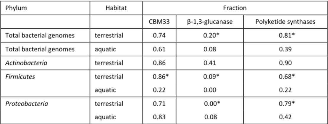
chitinase-containing genomes that contained CBM33s did not differ significantly between terrestrial (74 %) and aquatic (61 %) genomes (Table 3). CBM33s are
monooxygenases and require oxygen for their functioning (Vaaje-Kolstad et al., 2010). Accordingly, none of the genomes of strictly anaerobic bacteria contained CBM33s (Table A2 and A4).
Occurrence of other proteins possibly related to anti-fungal activity
Next to chitin, fungal cell walls contain different polysaccharides, such as β -1, 3-glucan. Therefore β -1, 3- glucanases are involved in the degradation of fungal cell walls and may provide chitinolytic bacteria with an additional defense mechanism against fungi (Aono et al., 1992; Arora et al., 2007; Hoshikawa et al., 2012). Yet, the frequency of β -1, 3- glucanases was low in both aquatic and terrestrial chitinase-containing bacterial genomes with the exception of genomes of terrestrial Actinobacteria (Table 3). Several studies have suggested the
35 Bacterial polyketides are secondary metabolites that often have
antifungal activities (Staunton and Weissman, 2001; Zirkle et al., 2004). Polyketide synthases (PKSs) are a family of multi-domain enzymes or enzyme complexes that are involved in the production of these secondary metabolites. The fraction of PKSs positive bacterial genomes was significantly higher for terrestrial bacteria (81 %) than for aquatic ones (39 %) (Table 3). Therefore, PKSs do appear to be more common for terrestrial chitinolytic bacteria. This observation supports the assumption that the combination of chitinases and antibiotics may be important in antagonistic activities of soil bacteria against fungi (De Boer et al., 2001; De Boer and van Veen, 2001). Interestingly, it has been shown that induction of chitinase gene expression coincided with upregulation of genes encoding secondary metabolites in Streptomyces (Światek et al., 2012; Nazari et al., 2013).
Table 3 Fraction of chitinase-containing bacterial genomes containing at least one gene encoding CBM33-type LPMOs, β-1, 3-glucanase or polyketide synthase
Phylum Habitat Fraction
CBM33 β-1,3-glucanase Polyketide synthases Total bacterial genomes terrestrial 0.74 0.20* 0.81* Total bacterial genomes aquatic 0.61 0.08 0.39
Actinobacteria terrestrial 0.86 0.41 0.90
Firmicutes terrestrial 0.86* 0.09* 0.68* aquatic 0.22 0.00 0.22
Proteobacteria terrestrial 0.71 0.00* 0.79*
aquatic 0.83 0.08 0.42
*: significant difference between terrestrial and aquatic habitat according to chi square test (P< 0.05)
Conclusions and perspectives:
The occurrence of multiple chitinases is thought to reflect the flexibility of bacteria to deal with variability in chitin structures, e.g. parallel (α) and anti-parallel (β) orientations of chitin-chains and different degrees of acetylation (Hunt
et al., 2008; Svitil et al., 1997). In addition, synergistic actions of different
Genomic comparison of bacterial chitinolytic system
Goodman, 1999). Hence, the numbers of chitinases in a bacterium could affect the efficiency of its chitin degradation ability (Beier and Bertilsson, 2013). Our study reveals clear differences in the complexities of bacterial chitinolytic machineries regardless of the difference between terrestrial and aquatic ecosystems.
Our screening of the modular composition of chitinases in bacterial genomes indicates differences between aquatic and terrestrial bacteria that could be related to the different chitinous resources they utilize. Arthropods
exoskeleton and fungal cell walls form the major source of chitin in aquatic and terrestrial habitats, respectively. Whereas both arthropod exoskeletons and fungal cell walls can be considered as multiphase composite material, their composition is very different. In arthropod exoskeletons α-chitin fibers are embedded in a matrix of proteins that can also contain minerals, such as calcite (Moussian et al., 2005; Raabe et al., 2006). The composition of fungal cell walls is completely different. The latter are mainly composed of α-chitin, glucans, mannans and glycoproteins with chitin, glucans and glycoproteins being
covalently cross-linked (Bowman and Free, 2006). In addition, the ultrastructure of chitin fibres differs between arthropods and fungi (Bowman and Free, 2006; Raabe et al., 2006). It is to be expected that such differences between arthropod exoskeletons and fungal cell walls are reflected in adaptations of bacterial chitin-degrading enzyme systems. The present study indeed reveals such adaptations. In particular, whereas the CBMs connected to chitinases from aquatic bacteria seem to be targeting chitin only, chitinases from terrestrial bacteria, in particular
Actinomycetes, have a CBM repertoire indicating binding to composite
37 The high abundance of polyketide synthases in chitinase-containing genomes of terrestrial Firmicutes and Proteobacteria comparing to aquatic ones may indicate that several non-filamentous terrestrial bacteria use a combination of chitinases and antibiotics in antagonistic interactions with fungi. In addition, the low abundance of β -1, 3-glucanases in genomes of these groups of non-filamentous bacteria does point at the inability to degrade the glucans in the fungal cell walls that are cross-linked with chitin polymers. This cross-linking has not yet occurred in the hyphal tip of fungi, which are therefore most vulnerable to destabilization by chitinases and which may be the prime target for Firmicutes and
Proteobacteria.
An issue of great interest is the role of the recently discovered LPMOs, which, in bacteria, are of the CBM33-type. Their role in chitin-degradation is indisputable but their possible effect as an anti-fungal agent remains largely unexplored. Certain bacteria, such as Streptomycetes contain several CBM33-type LMPOs and it is conceivable that these have different substrate specificities (Horn
et al., 2012). They might act on different substrates (e.g. chitin and cellulose) (Forsberg et al., 2011), on different forms of the same substrate (e.g. α-chitin vs
β-chitin), or on the same substrate in different composite materials. This is a novel area of research and so far only few CBM33-type LPMOs have been
experimentally characterized.
It should be noted that our screening of chitinase-containing genomes has several limitations. For instance, for both the terrestrial and aquatic chitinase-containing genomes there is an over-representation of certain bacterial groups (Actinomycetales in terrestrial habitats and Vibrionales in aquatic habitats). In addition, as we included genes annotated as “chitinase-like” and “chitinase-like super family”, there is a possibility that some of the enzymes counted as chitinases are not functional. On the other hand, it is possible that a few
Genomic comparison of bacterial chitinolytic system
an E-value exceeding 10-6 and has been excluded from the current research.
Furthermore, bacteria which are difficult to culture are not present in this paper because of the lack of publicly available genomes. Despite these minor errors, our analyses provide an important first step to unravelling the compositions of and the differences between terrestrial and aquatic chitinolytic systems, which again are linked to different degradative capabilities and ecological roles. Our data reveal clear trends in how chitinases in different habitats are composed. The observed differences in modular structure and the rather clear correlations of the occurrence of certain CBMs and FnIII domains with bacterial habitats give
directions for further studies on the functional roles of these domains.
39
Chapter 3
Chitin degrading abilities and antifungal activity of bacterial
strains differing in complexity of their chitinolytic systems and
growth form
Bacteria strains differ in chitinolytic activity
Abstract
Chitinolytic bacteria have different abilities with respect to chitin degradation and antifungal properties. This might be partially explained by differences in numbers of chitinases and/or possession of additional enzymes. In order to get a deeper understanding of the factors determining the use of chitinases for chitin degradation and antifungal activity, we selected 13 bacteria from different phyla and tested their chitin degrading ability in liquid cultures and the antifungal activity on agar plates. Next we compared the degradation rates and antifungal activity with the composition of the chitinolytic system of the strains as based on their annotated genomes. Degradation rates of chitin
41
Introduction
Chitin is one of the most abundant biopolymers in nature (Chater et al 2010, Gooday 1990). Chitin degradation, which is mainly a microbial process, is important in global carbon and nitrogen cycling. Chitinases are the major chitin decomposing enzymes (Bhattacharya et al 2007). Bacterial chitinases are typically divided into two families according to their amino acid sequence homology: family 18 and 19 of glycoside hydrolases (Henrissat & Davies 2000). The majority of the known bacterial chitinases belong to family 18. Chitinases of both families have also been reported to exhibit antifungal activity (Kawase et al 2006, Prasanna et al 2013).
The ability to hydrolyze chitin is taxonomically widespread in bacteria and is found for both Gram-negative and Gram-positive genera (Gooday 1990). Yet, bacteria differ strongly in their ability to degrade chitin. This has been ascribed to differences in chitinolytic systems, i.e. different numbers of chitinases and different types of chitinases (Bai et al 2015). In addition, Vaaje-Kolstad et al (2005a) showed that chitin binding proteins, also called CBM33 proteins, contribute to the efficiency of chitinases. These proteins were reported to catalyze oxidative cleavage of glycosidic bonds in crystalline chitin, thereby making the chitin fibers more accessible for chitinases (Vaaje-Kolstad et al 2005b). Hence, these proteins can contribute to the chitinolytic performance of bacteria.
Furthermore, next to physiological properties also morphological properties can influence chitin degrading capabilities. Hyphal forming bacteria, such as Actinomycetes, are reported to be better capable of degrading chitin particles than most non-hyphal bacteria (De Boer et al 1999). Filamentous bacteria were often found to be the dominant chitin-degrading bacteria when chitinous material was added to soil (Krsek & Wellington 2001, Sato et al 2010). One of the possible reasons for the good chitin degrading abilities of
Bacteria strains differ in chitinolytic activity
thereby extending the area where chitinases are in close contact with the chitin polymers.
The overall hypothesis of this study is that the chitin degrading
performance and antifungal activity of chitinolytic bacteria is determined by the number of chitinases (including family 18 and 19 chitinase), the number of chitin binding proteins and the bacterial growth form. In the current study this was tested using a selection of 13 bacterial chitinolytic bacterial strains for which genome information is available. These bacteria were exposed to different forms of chitin in liquid media to test their chitin degrading abilities. In addition, their antifungal activity capabilities were tested in an in-vitro confrontation assay with different fungi. The aim of the study was to indicate the possible importance of the chitinolytic system complexity and morphological characteristics for bacterial chitin degrading capacities and antifungal activities.
Method
Strains
43
Strain name Gram Order Chitinase number Filamentous Chitin binding proteins number
family 18 family 19
Streptomyces coelicolor A3 (2) + Actinomycetales 8 2 + 6
Streptomyces lividans TK24 + Actinomycetales 9 2 + 7
Streptomyces clavuligerus ATCC 27064 + Actinomycetales 13 3 + 6
Streptomyces viridochromogenes DSM 40736 + Actinomycetales 4 1 + 5
Kribbella flavida DSM 17836 + Actinomycetales 7 2 + 1
Conexibacter woesei DSM 14684 + Solirubrobacterales 1 0 - 0
Micromonospora aurantiaca ATCC 27029 + Actinomycetales 4 0 + 5
Catenulispora acidiphila DSM 44928 + Actinomycetales 10 2 + 1
Chitinophaga pinensis DSM 2588 - Sphingobacteriales 6 1 - 0
Flavobacterium johnsoniae UW101 - Flavobacteriia 4 0 - 0
Ktedonobacter racemifer DSM 44963 + Ktedonobacterales 5 0 + 0
Bacillus mycoides DSM 2048 + Bacillales 2 0 - 3
Bacteria strains differ in chitinolytic activity
Chitin resources and bacterial growth
We used four different chitin sources: N-acetylglucosamine (chitin monomer), colloidal chitin, small particles of crystal chitin (<0.5 mm) and big particles of crystal chitin (> 2 mm). Purified crystal chitin (poly-N-acetyl-1, 4-β- D-glucosamine, Sigma-Aldrich) was manufactured from shrimp shells. For use in this experiment it was sieved through 2 (big size) or 0.5 (small size) mm mesh filters. Colloidal chitin was prepared from crystal chitin according to Hsu & Lockwood (1975). Their procedure includes the dissolution of bleached chitin in
concentrated HCl, suspending the dissolved chitin by adding water, and removing the HCl in several washing steps until the pH of the suspension was between 2.5 to 3.0.
The chitin containing liquid growth medium was prepared by mixing either 0.1 g colloid chitin, 0.5 g N-acetylglucosamine, 1 g big (>2.0 mm) or small (< 0.5 mm) size chitin powder in 1 L medium containing 40 mg MgSO4‧7 H2O, 20 mg
CaCl2‧2 H2O, 1 g KH2PO4. The chitin sources were the only carbon- and energy
substrates for bacterial growth. The final pH of media was adjusted to 6.5. The medium was sterilized by heating at 121 0C for 15 minutes.
Bacterial inocula were prepared by transferring bacterial biomass from agar plates to 1 ml sterilized phosphate buffer to make suspensions with an OD of 0.01 (approximately 107 cells/ml), 0.2 ml of these bacterial suspensions was transferred to 100 ml of liquid media with different chitin resources. After inoculation, flasks were put on a shaker (100 rpm) and incubated at 20 oC. Every two days 1.5 ml samples were taken from the flask and centrifuged, after which the supernatant was stored at – 20 oC. We took samples for a total period of 24
days.
Chitin degradation was quantified by measuring the amount of NH4+-N
45 using an auto analyzer QUAATRO (SEAL). A control treatment, without
N-acetylglucosamine or chitin in the growth medium, was included.
Antifungal activity
Antifungal activity of the selected bacteria was tested by an in vitro
confrontation assay as shown in Figure 1. Bacteria were grown on water-yeast agar for one week in area B of the plate. Next, a PDA agar block of 1 cm
ø
from the growing margin of a fungal colony was placed in area A. As a control fungal strains were transferred to agar without the presence of bacteria in area B. After sealing the plates were incubated at 20°C for 2 weeks.Figure 1 Design of antifungal activity test of bacteria. See text for explanation of the letters
Antifungal activity of bacteria was determined by comparing the fungal mycelium development in area C of control plates with that of bacteria-containing plates. The fungal strains used in the test were: Mucor hiemalis (zygomycete) isolated from Dutch coastal dunes, Apergillus niger (ascomycete)obtained from the Centraalbureau voor Schimmelcultures (CBS, Utrecht, The Netherlands), and
Rhizoctonia solani (basidiomycete) obtainedfrom the Instituut voor Rationele Suikerproductie (IRS), Bergen op Zoom, The Netherlands.
Data analysis
Bacteria strains differ in chitinolytic activity
Correlation between chitin degradation rates or antifungal activity and chitinase numbers, chitin binding protein numbers of the bacteria was calculated in PAST (Hammer et al 2001). Antifungal activity was classified in 5 different levels, namely 1 to 5 (see Table 5). These classification levels were used to calculate correlations with other parameters. Hyphal-forming morphology bacteria was categorized by 1(presence of hyphae) and 0 (no hyphal formation). Effects of hyphal morphology were tested for significance using the non-parametric Kruskall-Wallis test in PAST.
Results
Bacterial chitin degradation in liquid media
The release of NH4+ from the different chitin resources was determined at
12 time points, every other day of the total incubation period. In Table 2 NH4+-N
concentration are given at day 1, 12 and 24. For colloidal chitin and crystal chitin we calculated the NH4+-N production for two periods namely the accumulation of
NH4+-N from day 1 to day 12, and from day 12 to day 24. For both periods, we
calculated the release rate of NH4+-N on a daily basis. Several bacteria showed an
increase of NH4+-N production during the first 24 h followed by a slow
accumulation during the next 11 or 23 days. We attributed this initial quick release to decomposition of easily accessible chitin on the outside of the particles or of soluble chitin oligomers present in the sieved chitin fractions. Therefore, we used the NH4+-N release rate during the subsequent period (day 1 – day 12 and
day 12 – day 24) as proxy for chitin degrading rate of these bacteria. Since rapid decomposition is to be expected for N-acetylglucosamine the NH4+-N production
47
N-acetylglucosamine Colloidal chitin Small size crystal chitin Big size crystal chitin Day 1 Day 12 Day 24 Day 1 Day 12 Day 24 Day 1 Day 12 Day 24 Day 1 Day 12 Day 24
Control 0,163 - - 0 - - 0 - - 0 - -
Streptomyces coelicolor A3 (2) 0,148 0,641 1,394 0,026 0,303 0,801 0,045 0,568 2,492 0,041 0,281 1,337
Streptomyces lividans TK24 0,178 0,282 0,57 0,047 0,127 0,492 0,033 0,077 0,601 0,03 0,073 0,467
Streptomyces clavuligerus ATCC 27064 0,885 1,122 1,419 0,043 0,149 0,292 0,38 0,898 2,083 0,184 0,313 0,932
Streptomyces viridochromogenes DSM 40736 0,941 1,7 2,853 0,058 0,376 0,942 0,337 0,914 2,149 0,163 0,327 0,659
Kribbella flavida DSM 17836 0,272 0,742 0,985 0,036 0,192 0,306 0,029 0,245 0,859 0,033 0,178 0,263
Conexibacter woesei DSM 14684 1,262 1,326 1,36 0,031 0,049 0,228 0,19 0,373 0,682 0,139 0,199 0,256
Micromonospora aurantiaca ATCC 27029 0,839 0,909 0,909 0,04 0,107 0,254 0,282 0,486 1,45 0,159 0,234 0,477
Catenulispora acidiphila DSM 44928 0,157 0,335 0,624 0,022 0,082 0,263 0 0,071 0,128 0 0,052 0,077
Chitinophaga pinensis DSM 2588 1,493 1,546 1,571 0 0,039 0,034 0,222 0,223 0,417 0 0,291 0,552
Flavobacterium johnsoniae UW101 0,748 2,205 2,685 0,056 0,717 2,353 0,168 0,291 0,722 0,093 0,296 0,682
Ktedonobacter racemifer DSM 44963 - 0,903 1,172 0 0,033 0,102 0,332 0,368 0,477 0,12 0,147 0,26
Bacillus mycoides DSM 2048 0,159 1,948 2,71 0 0,044 0,05 0,112 0,202 0,246 0,082 0,118 0,376
Bacteria strains differ in chitinolytic activity
Table 3 Release rate of NH4 +
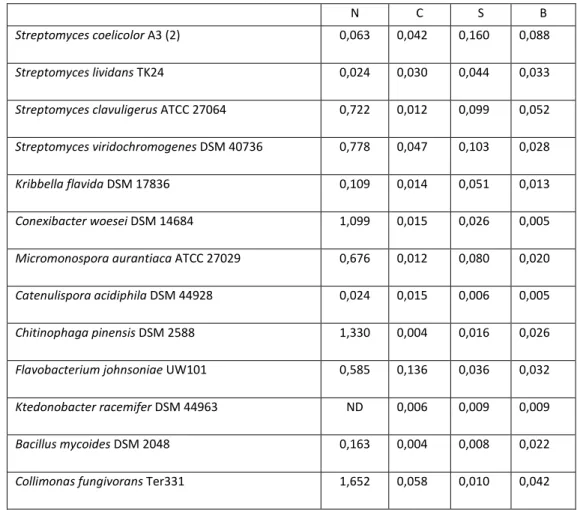
-N from chitinous substrates in liquid media. Data represent the amount of NH4+-N (mmol/l) released per day over the periods day 1 – day 12 or day 12 – day 24 (highest rate of the two periods is presented). For N (N-acetylglucosamine) substrate, data represent the highest rate for the periods of day 0- day 1, day 1- day 12 and day 12- day 24
N C S B
Streptomyces coelicolor A3 (2) 0,063 0,042 0,160 0,088
Streptomyces lividans TK24 0,024 0,030 0,044 0,033
Streptomyces clavuligerus ATCC 27064 0,722 0,012 0,099 0,052
Streptomyces viridochromogenes DSM 40736 0,778 0,047 0,103 0,028
Kribbella flavida DSM 17836 0,109 0,014 0,051 0,013
Conexibacter woesei DSM 14684 1,099 0,015 0,026 0,005
Micromonospora aurantiaca ATCC 27029 0,676 0,012 0,080 0,020
Catenulispora acidiphila DSM 44928 0,024 0,015 0,006 0,005
Chitinophaga pinensis DSM 2588 1,330 0,004 0,016 0,026
Flavobacterium johnsoniae UW101 0,585 0,136 0,036 0,032
Ktedonobacter racemifer DSM 44963 ND 0,006 0,009 0,009
Bacillus mycoides DSM 2048 0,163 0,004 0,008 0,022
Collimonas fungivorans Ter331 1,652 0,058 0,010 0,042 N: N-acetylglucosamine; B: big size crystal chitin; S: small size crystal chitin; C: colloidal chitin
For most of the bacterial strains the degrading rates of N
-acetylglucosamine were much higher than for the other substrates (Table 3). The highest rate of N-acetylglucosamine degradation was seen for Collimonas
49 degradation of crystal chitin were observed for Streptomyces spp. Degardation rates of small and big crystal chitin were correleted (Table 4).
Table 4 Pearson correlation coefficients for degradation rates of different substrates. Significant correlations (P < 0.05) are indicated in bold
Degrading rate
N C S B
Degrading rate
N - 0.18 -0.17 0.016
C - - 0.10 0.29
S - - - 0.74
N: N-acetylglucosamine; B: big size crystal chitin; S: small size crystal chitin; C: colloidal chitin
Antifungal activities
The scores for antifungal activities of the chitinolytic bacterial strains are given in Table 5. The results indicated that bacterial strains showed different antifungal activities in the in vitro assay. Overall, most of the Streptomyces strains showed a broader and stronger antifungal activity than other bacteria.
Flavobacterium johnsomiae UW 101, Ktedonobacter racefmifer DSM 44963,
Bacillus mycoides DSM 2048 and Collimonas fungivoras Ter331 showed also strong antifungal activity against one or two fungi.
Bacteria strains differ in chitinolytic activity
Table 5 Classification of in vitro antifungal activities of the selected chitinolytic bacteria
Strains M A R Extra information
Streptomyces coelicolor A3 (2) 5 5 2
Streptomyces lividans TK24 5 5 4
Streptomyces clavuligerus ATCC 27064 2 5 5
Streptomyces viridochromogenes DSM 40736 4 2 3
Kribbella flavida DSM 17836 1 5 3
Conexibacter woesei DSM 14684 1 1 1 Did not grow well
Micromonospora aurantiaca ATCC 27029 1 1 2
Catenulispora acidiphila DSM 44928 1 1 1
Chitinophaga pinensis DSM 2588 1 1 1 Did not grow well
Flavobacterium johnsoniae UW101 2 3 3
Ktedonobacter racemifer DSM 44963 1 5 2
Bacillus mycoides DSM 2048 4 2 2
Collimonas fungivorans Ter331 2 1 5
M: Mucor hiemalis; A: Aspergillus niger; R: Rhizoctonia solani. (Classification of antifungal activity: 1: no inhibition; 2: weak inhibition at only one time point; 3: weak inhibition at both time points; 4: weak inhibition at one time point and strong inhibition at another time point; 5: strong inhibition at both time points)
Correlation analysis
The degradation rates of crystal chitin particles, both big and small size were significantly positive related with the number of genes encoding chitin binding proteins but not with the number of chitinase genes. Except this, there was no significant correlation between other parameters and degrading rates of different substrates (Table 6).
51
Table 6 Pearson correlation coefficients for the relationship between degradation rates and in vitro antifungal activities with number of chitinases and chitin-binding proteins. Significant correlations (P < 0.05) are indicated in bold
Degrading rate Antifungal activity
N C S B M A R
GH 18 chitinases numbers -0.46 -0.21 0.37 0.30 0.10 0.56 0.21
GH 19 chitinases numbers -0.22 -0.17 0.42 0.48 0.24 0.48 0.43
Chitin binding protein numbers -0.29 -0.11 0.72 0.58 0.71 0.38 0.41
N: N-acetylglucosamine; B: big size crystal chitin; S: small size crystal chitin; C: colloidal chitin; M:
Mucor Hiemali;A: Aspergillus niger; R: Rhizoctonia solani
Table 7 Non-parametric Kruskall Wallis test Effects of growth morphology (hyphal growth versus unicellular growth) on degradation rates and in vitro antifungal activity (Non-parametric Kruskall Wallis test). Given values are Bonferroni corrected P-values
Degrading rate Antifungal activity
N C S B M A R
Hyphal versus non-hyphal 0.04 0.94 0.11 0.94 0.81 0.07 0.55
N: N-acetylglucosamine; B: big size crystal chitin; S: small size crystal chitin; C: colloidal chitin; M:
Mucor Hiemali;A: Aspergillus niger; R: Rhizoctonia solani
The impact of hyphal morphology on degradation of the chitin substrates was only significant for N-acetylglucosamine, for which filamentous bacteria had a lower degradation rate then non-filamentous ones (Table 7).
Table 8 Pearson correlation coefficients for the relationships between degradation rates and in vitro antifungal activities. Significant correlations (P < 0.05) are indicated in bold
Degradation rate
N C S B
Antifungal activity
M -0.33 0.19 0.49 0.62
A -0.60 0.01 0.41 0.40
R 0.19 0.32 0.20 0.41
N: N-acetylglucosamine; B: big size crystal chitin; S: small size crystal chitin; C: colloidal chitin; M:
Bacteria strains differ in chitinolytic activity
The in vitro antifungal activity against fungal strain Mucor hiemalis was significantly positively correlated with degradation rates of big crystal chitin particles but not with degradation rates of small size crystal chitin particles or colloidal chitin. Antifungal activity of chitinolytic bacteria against Aspergillus niger
was significantly negatively correlated with their degrading rates of N -acetylglucosamine (Table 8).
Discussion
Bacteria play an important role in the degradation of chitinous resources via hydrolysis of chitin polymers by chitinases (Hoell et al 2010). The chitinolytic system (number of chitinases, modular composition, additional proteins) differs considerably among bacteria and these differences may point at differences in ecological functioning (Bai et al 2015). Next to degradation of chitin, chitinases can be part of an antagonistic system involved in competition with fungi or in defense against bacteriolytic fungi. This antagonistic activity is based on the fact that chitinases can contribute to destabilization of the fungal exterior (cell wall and cell membrane) as chitin is an important structural component of the fungal cell wall (Bowman & Free 2006). In this study we tested, via correlation analysis, whether there is a relationship between the assembly of the chitinolytic system and chitin-degrading abilities or antifungal activities for a set of bacteria.
The results showed that the abilities to degrade the different chitin resources and the chitin monomer N-acetylglucosamine differed strongly among the bacterial strains. The positive correlation between degradation of small and big crystal chitin indicates that bacteria use similar mechanisms to degrade these substrates. In particular, strains from the genus Streptomyces, showed high decomposition activity with respect to both big and small chitin particles. It has been suggested that the formation of hyphae by these bacteria is important for the degradation of crystal chitin as it allows for penetration of chitin layers (Gooday 1994). Yet, several other hyphal Actinomycetes (such asKribbella flavida