Germline mutations in
PMS2
and
MLH1
in individuals with solitary loss
of PMS2 expression in colorectal
carcinomas from the Colon Cancer
Family Registry Cohort
Christophe Rosty,1,2,3Mark Clendenning,3Michael D Walsh,4Stine V Eriksen,3 Melissa C Southey,3Ingrid M Winship,5,6Finlay A Macrae,7Alex Boussioutas,8,9,10 Nicola K Poplawski,11,12Susan Parry,13,14Julie Arnold,14Joanne P Young,15,16,17 Graham Casey,18Robert W Haile,19Steven Gallinger,20Loïc Le Marchand,21 Polly A Newcomb,22,23John D Potter,22,23,24Melissa DeRycke,25
Noralane M Lindor,26Stephen N Thibodeau,27John A Baron,28Aung Ko Win,29 John L Hopper,29Mark A Jenkins,29Daniel D Buchanan,3,29on behalf of the Colon Cancer Family Registry Cohort
To cite:Rosty C, Clendenning M, Walsh MD, et al. Germline mutations in PMS2andMLH1in individuals with solitary loss of PMS2 expression in colorectal carcinomas from the Colon Cancer Family Registry Cohort.BMJ Open 2016;6:e010293.
doi:10.1136/bmjopen-2015-010293
▸ Prepublication history and additional material is available. To view please visit the journal (http://dx.doi.org/ 10.1136/bmjopen-2015-010293).
Received 20 October 2015 Revised 12 January 2016 Accepted 29 January 2016
For numbered affiliations see end of article.
Correspondence to
Dr Christophe Rosty; [email protected]
ABSTRACT
Objectives:Immunohistochemistry for DNA mismatch repair proteins is used to screen for Lynch syndrome in individuals with colorectal carcinoma (CRC). Although solitary loss of PMS2 expression is indicative of carrying a germline mutation inPMS2, previous studies reportedMLH1mutation in some cases. We determined the prevalence ofMLH1germline mutations in a large cohort of individuals with a CRC demonstrating solitary loss ofPMS2expression.
Design:This cohort study included 88 individuals affected with a PMS2-deficient CRC from the Colon Cancer Family Registry Cohort. GermlinePMS2 mutation analysis (long-range PCR and multiplex ligation-dependent probe amplification) was followed byMLH1mutation testing (Sanger sequencing and multiplex ligation-dependent probe amplification).
Results:Of the 66 individuals with complete mutation screening, we identified a pathogenicPMS2mutation in 49 (74%), a pathogenicMLH1mutation in 8 (12%) and aMLH1variant of uncertain clinical significance predicted to be damaging by in silico analysis in 3 (4%); 6 (9%) carried variants likely to have no clinical significance. Missense point mutations accounted for most alterations (83%; 9/11) inMLH1. TheMLH1 c.113A> G p.Asn38Ser mutation was found in 2 related individuals. One individual who carried theMLH1 intronic mutation c.677+3A>G p.Gln197Argfs*8 leading to the skipping of exon 8, developed 2 tumours, both of which retained MLH1 expression.
Conclusions:A substantial proportion of CRCs with solitary loss of PMS2 expression are associated with a deleteriousMLH1germline mutation supporting the screening forMLH1in individuals with tumours of this immunophenotype, when noPMS2mutation has been identified.
INTRODUCTION
Lynch syndrome is an autosomal-dominant inherited condition defined by the identifi ca-tion of a germline mutaca-tion in a DNA mis-match repair (MMR) gene (MLH1, MSH2, PMS2 or MSH6), or in the EPCAM gene, leading to constitutional epigenetic silencing of the downstream MSH2 gene.1 Individuals who carry a MMR gene mutation are at an increased risk of developing cancers at mul-tiple sites, most notably colorectal and endo-metrial carcinomas, but also carcinomas from the upper urinary tract, pancreas, hepa-tobiliary tract, stomach, small intestine and ovaries.2
The current diagnostic approach for the identification of individuals with an MMR
Strengths and limitations of this study
▪ Largest reported sample of colorectal cancers with solitary loss of PMS2 expression.
▪ Most comprehensive approaches used for testing for germlinePMS2mutations.
▪ Multicentre setting which may affect the consist-ency in the formalin fixation conditions of tissue blocks and lead to immunostaining artefacts.
▪ No mutation screening data available for 20 cases (24%).
gene mutation is a multistep process in which patholo-gists play an instrumental role. Tumours arising in indivi-duals with an MMR gene mutation demonstrate high levels of microsatellite instability (MSI) secondary to altered DNA MMR mechanisms in tumour cells. Immunohistochemistry for DNA MMR proteins is widely used to identify MMR deficiency in colorectal carcin-omas (CRCs) as a screen for MMR gene carriers.3Of all abnormal patterns of immunohistochemical results, loss of expression of MLH1 and PMS2 is the most common. MLH1 and PMS2 function as a stable heterodimer that, along with MSH2, MSH6 and EXO1, corrects small errors involving mispaired nucleotides which are intro-duced by DNA polymerase during DNA replication. A functional defect inMLH1 results in the degradation of both MLH1 and PMS2, whereas a defect inPMS2 results only in the degradation of PMS2. Consequently, loss of expression of MLH1 and PMS2 in CRC generally indi-cates an alteration in MLH1, either by somatic methyla-tion of the MLH1 promoter region (sporadic cases) or by a MLH1 germline mutation (Lynch syndrome), and solitary loss of PMS2 expression generally indicates an underlying germline defect inPMS2.
Inconsistent immunohistochemical results have been reported, in particular the retained expression of MLH1 in tumours from individuals with a germline MLH1 mutation.4–8 This phenomenon can be misleading if PMS2 immunostaining is not performed. We sought to confirm that germline mutations inMLH1may underlie a substantial proportion of CRC with solitary loss of PMS2 expression. To address this question, we per-formed mutation analysis of theMLH1andPMS2 genes in individuals from the Colon Cancer Family Registry Cohort whose tumours showed solitary loss of PMS2.
MATERIALS AND METHODS Study participants
Participants were probands and relatives from families recruited between 1997 and 2012 to the Colon Cancer Family Registry Cohort via both population-based recruitment and clinic-based recruitment in Australasia and North America.9 All CRC cases were reviewed by specialist gastrointestinal pathologists for histological type and grade.10 Tumours from the caecum, ascending colon, hepaticflexure and transverse colon were consid-ered proximal tumours. Immunohistochemistry for DNA MMR proteins MLH1, PMS2, MSH2 and MSH6 was per-formed as previously described.3 A subset of tumours were analysed for MSI status from formalin-fixed paraffin-embedded tissue as previously described.3 Individuals were eligible for this study if they had a histo-logically confirmed diagnosis of CRC with an immuno-histochemical profile of the DNA MMR proteins, demonstrating presence of expression of the MLH1 protein and concomitant loss of expression of the PMS2 protein. The somatic T> A mutation at nucleotide 1799 in exon 15 of theBRAF gene (BRAFV600Emutation) was
detected using fluorescent allele-specific PCR.11 MLH1 promoter methylation was analysed using the MLH1-M2 methylight reaction using an Arthobacter luteus (ALU) control reaction to normalise for bisulfite-converted input DNA.12 Informed consent was obtained from all partici-pants to collect a blood sample and tumour pathology materials (tumour blocks and slides). Ethics approval was obtained from the relevant institutional Human Research Ethics Committees at recruiting centres.
Family history of cancer
Information on personal and family history of CRC and other cancers in first-degreeand second-degree relatives was obtained via standardised questionnaires at the time of baseline recruitment. Cancer diagnoses were verified, where possible, using pathology reports, medical records, cancer registry reports and death certificates. Probands and relatives were either actively or passively followed up approximately every 5 years from baseline enrolment, including the collection of updated informa-tion by linkage to tumour registries and death indices on the number, sex and birthdates of first-degree rela-tives, their cancer history, vital status and, if deceased, date of death. All cancers, except for non-melanoma skin cancers, were recorded with dates of diagnosis. The present study was based on all available baseline and follow-up data. Family history of cancer that fulfilled either the Amsterdam I or II criteria were determined.13
Germline mutation testing
Germline mutation testing for the individuals in this study primarily involved testing forPMS2gene mutations and when aPMS2mutation was not identified, germline mutation testing of the MLH1 gene was conducted. PMS2 was screened for germline mutations using a DNA-based, best practice, approach combining long-range PCR and multiplex ligation-dependent probe amplification (MLPA). Briefly, for point mutation ana-lysis, parts of the PMS2 gene (exons 1–5, 9 and 11–15) were specifically targeted, while avoiding pseudogene sequences, via a set of three long-range PCRs (TaKaRa LA Taq; TaKaRa Bio Inc, Shiga, Japan). These long-range products are then used as the template for a set of PMS2-specific exonic PCRs (see online supplementary table S1 for primer sequences). To assess for large-scale (whole exon) deletions, we used the P008-B1 MLPA kit according to the manufacturer’s instructions (MRC-Holland; Amsterdam, The Netherlands). To accurately call PMS2 mutations at the 3′ end of the gene, the MLPA kit contains probes targeted to
paralogous sequence variants which requires
variants within the MLH1 and PMS2 genes were
classi-fied for pathogenicity based on the InSiGHT database classifications15 (http://insight-group.org/variants/ classifications/). If no classification was available, the predicting effect of an unclassified variant (UV) to the protein function was assessed in silico using the ‘Sorting Tolerant From Intolerant’ (SIFT) and the ‘Polymorphism Phenotyping v2’ (PolyPhen-2) web-based algorithms.16 17
Statistical analysis
Statistical analyses were performed with SPSS statistics software V.17.0 (SPSS Inc, Chicago, Illinois, USA). Comparisons for categorical variables were performed using Pearson’sχ2test or Fisher’s exact test where appro-priate. Student t test was used for continuous variables. A two-tailed p value was used for all analyses and values less than 0.05 were considered to be significant.
RESULTS
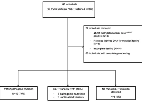
The study included 90 CRCs from 88 individuals demon-strating loss of PMS2 expression and normal retained MLH1 expression by immunohistochemistry. They had a mean age at CRC diagnosis of 51.7±SD 12.4 years and included 57% males. MSI status was available for 46/90 CRCs (51%), with high levels of MSI observed in 42/46 (91%) cases. MLH1 methylation and/or a BRAFV600E mutation were present in 4 of the 90 CRCs that were excluded from the study. Six CRCs (7%) also showed loss of MSH6 protein expression. Four individuals were not tested for PMS2 and MLH1 mutations due to the unavailability of blood-derived DNA, and complete gene testing was not possible for a further 14 individuals (figure 1). The final study group consisted of 66 indivi-duals with complete screening for germline mutations in the PMS2 and MLH1 genes. A pathogenic PMS2 germline mutation was identified in 49 individuals (74%; see online supplementary table S2), some of which were reported previously.18 Variants in theMLH1 gene were identified in 11 individuals (17%). In eight individuals, the variants were classified as pathogenic mutations (class 5); in the other three individuals, var-iants were unclassified but predicted to be damaging by SIFT and PolyPhen-2 algorithms (table 1andfigure 2).
Immunostained slides were reviewed in 5 of these 11 cases, confirming the retained expression of MLH1 and the loss of PMS2 expression in carcinoma cells. No mutation within PMS2 or MLH1 could be found in the remaining six individuals (9%). The clinicopathological characteristics of thePMS2 mutation carriers, theMLH1 pathogenic mutation and UV carriers and those indivi-duals tested but found not to have a mutation inPMS2 orMLH1are shown intable 2.
The mean age at CRC diagnosis of the individuals with a MLH1 mutation or UV was significantly younger than those individuals with aPMS2mutation ( p=0.046). Amsterdam criteria I or II were less frequently found in
PMS2 mutation carriers compared with MLH1 variant carriers ( p=0.001).
Missense variants were the most commonMLH1 alter-ation identified, in eight individuals (83%). The MLH1 c.113A>G p.Asn38Ser variant was found in two related individuals (cases 2 and 3). One individual who carried the intronic MLH1 germline mutation c.677+3A>G p. Gln197Argfs*8, which leads to the skipping of exon 8, developed two CRCs both of which retained MLH1 expression (cases 5 and 6). One individual carried a splice site mutation leading to an in-frame deletion of two exons (case 4) and one individual carried a small insertion resulting in a frameshift mutation (case 7; table 1).
DISCUSSION
To assess the possible role of MLH1 mutations in CRCs showing solitary loss of PMS2 expression by immunohis-tochemistry, we studied a series of 90 CRCs from 88 indi-viduals from the Colon Cancer Family Registry Cohort with this immunophenotype. Among the 66 individuals with complete germline mutation analysis, we identified a pathogenic PMS2 mutation in 49 cases (74%) and a pathogenic MLH1 mutation in 8 cases (12%). A further three cases (4%) had a variant of uncertain clinical sig-nificance in MLH1 predicted to be damaging, and six cases (9%) had no identifiable variant likely to have clin-ical significance in either gene. Moreover, a high pro-portion of the MLH1 variants identified resulted in missense changes, suggesting that a non-functional MLH1 protein that retains its MLH1 antigenicity is a conceivable explanation.
Immunohistochemistry for the DNA MMR proteins MLH1, PMS2, MSH2 and MSH6 in CRC is a highly sen-sitive test to screen for Lynch syndrome, with 93–100% concordance with MSI testing.3 4However, false-negative results for MLH1 immunohistochemistry have been reported in small series. In a study evaluating the benefit of adding PMS2 to MLH1 staining, de Jonget al4 found eight MLH1 mutations (42%) compared with only three PMS2mutations (16%) out of 19 CRCs dem-onstrating solitary loss of PMS2 expression. When con-sidering all the MLH1 mutations identified in their study, a high proportion (8/35; 23%) showed loss only of PMS2 expression while retaining expression of MLH1. A large deletion of exons 14–19 of MLH1 was also reported in 2 of 8 (25%) CRC with solitary PMS2 loss of expression in a separate study.5A recent study of 16 CRCs and 16 endometrial carcinomas from 31 indivi-duals, all with solitary loss of PMS2 expression, explored the frequency of MLH1 mutations in this group.19 Of the 17 individuals who subsequently had germline muta-tion testing of the MLH1 and PMS2 genes, six had pathogenic mutations in PMS2 (35%), two had variants of uncertain clinical significance in PMS2 (12%), four
had MLH1 pathogenic mutations (24%) whereas
When restricted to patients with a CRC, a deleterious germline mutation inMLH1was reported in two of nine tested patients (22%). Compared with these studies, our rate of PMS2 mutation in 66 tested individuals was higher at 74% and the rate of MLH1 deleterious muta-tion slightly lower at 12%. Two cousins (tumours 2 and 3) who carried the sameMLH1mutation both had CRC with solitary PMS2 loss. Similarly, one individual, who carried the MLH1 c.677+3A>G p.Gln197Argfs*8 muta-tion, developed two CRCs with solitary PMS2 loss. Both these examples suggest that it is the nature of the muta-tion rather than a technical anomaly associated with tissuefixation or staining quality that is the cause of this differential staining pattern. In support of this, Zighelboim et al20 described two sisters who carried the
same MLH1 mutation: one developed endometrial cancer at 48 years and the other CRC at 45 years and endometrial cancer at 53 years; all tumours showed soli-tary loss of PMS2 expression and the presence of MLH1 expression.
A trend towards universal CRC tumour immunohisto-chemistry will increase the detection of abnormal stain-ing patterns that require interpretation. This allows the most probable cause to be decided and thus the most appropriate management instituted. A solitary loss of PMS2 expression is suggestive of Lynch syndrome with a primary defect in the PMS2gene. Interestingly, we iden-tifiedMLH1methylation or the somaticBRAFV600E muta-tion in four cases, indicating that isolated PMS2 loss of expression can occur outside Lynch syndrome. It may
Figure 1 Flow diagram of the study. CRC, colorectal carcinoma.
Table 1 Characteristics of the 11 individuals with a germlineMLH1variant from 12 colorectal carcinomas with loss of PMS2 expression and retained MLH1 expression
Tumour
# Gender
Age, years
Amsterdam criteria
Tumour
location Variant Protein
InSiGHT classification
1 Female 40 None Descending c.230G>A p.Cys77Tyr Class 5
2* Male 44 None Descending c.113A>G p.Asn38Ser Class 5
3* Male 40 I Rectum c.113A>G p.Asn38Ser Class 5
4 Female 51 I Descending c.790+1G>A p.Glu227_Ser295del Class 5 5† Male 34 II Cecum c.677+3A>G p.Gln197Argfs*8 Class 5 6† Male 34 II Rectum c.677+3A>G p.Gln197Argfs*8 Class 5 7 Male 63 I Caecum c.2195_2198dup p.His733Glnfs*14 Class 5
8 Male 49 None Unknown c.230G>A p.Cys77Tyr Class 5
9 Female 33 None Rectum c.199G>A p.Gly67Arg Class 5
10 Male 62 II Transverse c.374C>A p.Ala125Glu UV
11 Male 24 None Ascending c.187G>C p.Asp63His UV
12 Male 38 I Cecum c.187G>C p.Asp63His UV
UV: unclassified variant by InSiGHT. These UVs were predicted to be damaging through in silico analysis. *Cousins.
therefore be useful to test PMS2-deficient CRC for BRAFV600E mutation or MLH1 methylation to exclude
sporadic tumour. Screening for PMS2 mutations has been problematic due a large number of homologous sequences within pseudogenes that closely flank the functional gene and most likely accounts for the lower proportion of PMS2 mutations reported in previous studies. The recent development of new methods incorporating long-range PCR and MLPA has eliminated most of the previous problems, such that the identifi ca-tion of large-scale deleca-tions of exons 3 and/or 4 are now the only difficulty. The results from this study, represent-ing the largest number of CRC with solitary loss of PMS2, support germline mutation screening of MLH1 when no mutation inPMS2has been found. However, a substantial proportion of MMR-deficient CRCs with no evidence of MLH1 methylation or BRAFV600E mutation remain unexplained and are referred to as Lynch-like or suspected Lynch syndrome. A number of potential causes for the underlying loss of PMS2 protein expres-sion in these cases, including biallelic somatic mutations and cryptic mutations, have been described in a recent review.21 In a large population-based study of the Colon Cancer Family Registry Cohort, 5.6% (271/4 853) of all CRCs were classified as Lynch-like syndrome, represent-ing 56% of all MMR-deficient CRCs not secondary to
MLH1 methylation. In our study, six CRCs showed con-current loss of MSH6 and PMS2. The most likely explan-ation for the loss of MSH6 expression in these six cases is the somatic frameshift mutation in the (C)8 microsat-ellite in exon 5 of the MSH6gene secondary to the loss of MMR function resulting from the PMS2 defect.22The use of panel testing rather than a single-gene approach would be useful; this is of particular interest clinically, where the PMS2 gene has lower penetrance than other MMR genes23 and family history is a suboptimal way of
finding potentially high-risk families, where risk assess-ment and risk manageassess-ment has improved outcomes. However,PMS2testing remains challenging even by next generation sequencing due to its complex structure.
Our study included the largest reported sample of CRCs with solitary loss of PMS2 to date. Testing for germline PMS2 mutations used in this study employed the most up-to-date and comprehensive approaches described,18 24as demonstrated by the high rate of iden-tifiedPMS2mutations. One limitation of this study is the multicentre setting which may affect the consistency in the formalin fixation conditions of tissue blocks and lead to immunostaining artefacts. Other limitations include the absence of other Lynch syndrome-associated tumours, and the lack of mutation screening data for 20 (24%) cases. Moreover, our results may not reflect the
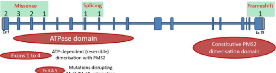
Figure 2 Graphical overview of the location of the 11MLH1
mutations identified. Numbers above the gene schematic denote the amount of mutations identified in the corresponding exons. Mutation subtypes are boxed in green and the predicted functional domains of the MLH1 protein are displayed below the gene schematic.
Table 2 Characteristics of 66 individuals with 68 colorectal carcinomas (CRCs) showing loss of PMS2 expression and retained MLH1 expression
Group
All (68 CRCs from 66 individuals)
PMS2mutations (N=50 CRCs from 49 individuals)
MLH1variant (N=12 CRCs from 11 individuals)
P valuePMS2 mutations vs
MLH1variant
NoPMS2or
MLH1
mutation (N=6)
Mean age at diagnosis±SD (range) in years
51.7±11.7 (24–80)
52.1±11.3 (35–80)
43.5±12.0 (24–63)
0.046 52.2±11.7 (33–69) Gender male, N (%) 36 (54.5) 26/49 (53.1) 8/11 (72.7) 0.32 2 (33.3) Amsterdam criteria I, N (%) 6 (9.1) 0 (0) 6 (54.5) 0.001* 0 (0) Amsterdam criteria II, N (%) 10 (15.2) 4 (8.2) 6 (54.5) 0 (0) Proximal CRC location, N (%) 42/64 (65.6) 35/47 (74.5) 5/10 (50) 0.14 2/6 (33.3)
Histological type, N (%) 1
Adenocarcinoma 50/63 (79.4) 36/47 (76.6) 9/11 (81.8) 6/6 (100) Mucinous carcinoma 13/63 (20.6) 11/47 (23.4) 2/11 (18.2) 0/6 (0)
Histological grade, N (%) 0.024
Well/moderate 42/62 (67.7) 36/46 (78.3) 4/10 (40) 1/5 (20)
Poor 20/62 (32.3) 10/46 (21.7) 6/10 (60) 4/5 (80)
actual rate of PMS2-deficient CRC in the general popu-lation and the mutation rates in PMS2 and MLH1, as these cases were selected in young individuals with strong family history of CRC.
In conclusion, the findings from this study suggest that CRCs inMLH1mutation carriers can demonstrate a normal pattern of MLH1 expression and justify the testing for MLH1germline mutation in individuals with a CRC showing solitary loss of PMS2 expression when a PMS2mutation is not identified.
Author affiliations
1Envoi Pathology, Brisbane, Queensland, Australia
2The School of Medicine, The University of Queensland, Brisbane,
Queensland, Australia
3Genetic Epidemiology Laboratory, Department of Pathology, The University of
Melbourne, Parkville, Victoria, Australia
4Department of Histopathology, Sullivan Nicolaides Pathology, Brisbane,
Queensland, Australia
5Department of Medicine, The University of Melbourne, Parkville, Victoria,
Australia
6Genetic Medicine and Family Cancer Clinic, Royal Melbourne Hospital,
Parkville, Victoria, Australia
7Colorectal Medicine and Genetics, The Royal Melbourne Hospital, Parkville,
Victoria, Australia
8Department of Medicine, Royal Melbourne Hospital, The University of
Melbourne, Parkville, Victoria, Australia
9Cancer Genomics and Predictive Medicine, Peter MacCallum Cancer Centre,
East Melbourne, Victoria, Australia
10Sir Peter MacCallum Department of Oncology, The University of Melbourne,
Parkville, Victoria, Australia
11South Australian Clinical Genetics Service, SA Pathology at the WCH, North
Adelaide, South Australia, Australia
12University Department of Paediatrics, University of Adelaide, Adelaide, South
Australia, Australia
13New Zealand Familial Gastrointestinal Cancer Registry, Auckland City
Hospital, Auckland, New Zealand
14Department of Gastroenterology, Middlemore Hospital, Auckland, New
Zealand
15Department of Haematology and Oncology, The Queen Elizabeth Hospital,
Woodville, South Australia, Australia
16School of Medicine, University of Adelaide, Adelaide, South Australia,
Australia
17SAHMRI Colorectal Node, Basil Hetzel Institute for Translational Research,
Woodville, South Australia, Australia
18Department of Preventive Medicine, Keck School of Medicine and Norris
Comprehensive Cancer Center, University of Southern California, Los Angeles, California, USA
19Department of Medicine, Division of Oncology, Stanford Cancer Institute,
Stanford University, Stanford, California, USA
20Lunenfeld Tanenbaum Research Institute, Mount Sinai Hospital, University
of Toronto, Toronto, Ontario, Canada
21University of Hawaii Cancer Center, Honolulu, Hawaii, USA
22Public Health Sciences Division, Fred Hutchinson Cancer Research Center,
Seattle, Washington, USA
23School of Public Health, University of Washington, Seattle, Washington,
USA
24Centre for Public Health Research, Massey University, Wellington, New
Zealand
25Departments of Health Sciences Research, Biomedical Statistics and
Informatics, Laboratory Medicine and Pathology, Medical Genetics, Medical Genomics Technology and Advanced Genomics Technology Center, Mayo Clinic College of Medicine, Rochester, Minnesota, USA
26Department of Health Science Research, Mayo Clinic Arizona, Scottsdale,
Arizona, USA
27Molecular Genetics Laboratory, Department of Laboratory Medicine and
Pathology, Mayo Clinic, Rochester, Minnesota, USA
28Department of Medicine, University of North Carolina, Chapel Hill, North
Carolina, USA
29Centre for Epidemiology and Biostatistics, Melbourne School of Population
and Global Health, The University of Melbourne, Parkville, Victoria, Australia
Acknowledgements The authors thank all study participants of the Colon Cancer Family Registry and staff for their contributions to this project, in particular Sue Preston, Allyson Templeton, Teresa Selander and Maggie Angelakos.
Contributors CR, MC, DDB were involved in design of the study, acquisition, analysis and interpretation of data, drafting the manuscript and final approval of the version to be published. MDW, SVE, JPY were involved in acquisition, analysis and interpretation of data, revising the manuscript critically and final approval of the version to be published. MCS, IMW, FAM, AB, NKP, SP, JA, GC, RWH, SG, LLM, PAN, JDP, MD, NML, SNT, JAB, AKW, JLH, MAJ were involved in data acquisition, revising the manuscript critically and final approval of the version to be published.
Funding This work was supported by grant UM1 CA167551 from the National Cancer Institute and through cooperative agreements with the following Colon Cancer Family Registry centres: Australasian Colorectal Cancer Family Registry (U01 CA074778 and U01/U24 CA097735), Mayo Clinic Cooperative Family Registry for Colon Cancer Studies (U01/U24 CA074800), Ontario Familial Colorectal Cancer Registry (U01/U24 CA074783), Seattle Colorectal Cancer Family Registry (U01/U24 CA074794), University of Hawaii Colorectal Cancer Family Registry (U01/U24 CA074806), USC Consortium Colorectal Cancer Family Registry U01/U24 CA074799). AKW is an Australian National Health and Medical Research Council (NHMRC) Early Career Fellow. MCS is a NHMRC Senior Research Fellow. JLH is a NHMRC Senior Principal Research Fellow and Distinguished Visiting Professor at Seoul National University, Korea. MAJ is a NHMRC Senior Research Fellow. DDB is a University of Melbourne Research at Melbourne Accelerator Program (R@MAP) Senior Research Fellow. This project was conducted under Colon Cancer Family Registry approval C-CP-0309-01-A1.
Disclaimer The content of this manuscript does not necessarily reflect the views or policies of the National Cancer Institute or any of the collaborating centres in the CFRs, nor does mention of trade names, commercial products, or organisations imply endorsement by the US Government or the CFR. Authors had full responsibility for the design of the study, the collection of the data, the analysis and interpretation of the data, the decision to submit the manuscript for publication, and the writing of the manuscript.
Patient consent Obtained.
Ethics approval Multi-institutional Human Research Ethics Committees.
Provenance and peer review Not commissioned; externally peer reviewed.
Data sharing statement No additional data are available.
Open Access This is an Open Access article distributed in accordance with the Creative Commons Attribution Non Commercial (CC BY-NC 4.0) license, which permits others to distribute, remix, adapt, build upon this work non-commercially, and license their derivative works on different terms, provided the original work is properly cited and the use is non-commercial. See: http:// creativecommons.org/licenses/by-nc/4.0/
REFERENCES
1. Lynch PM. Hyperplastic polyposis: semantics, biology, and endoscopy.Gut2010;59:1019–21.
2. Win AK, Young JP, Lindor NM,et al. Colorectal and other cancer risks for carriers and noncarriers from families with a DNA mismatch repair gene mutation: a prospective cohort study.J Clin Oncol 2012;30:958–64.
3. Lindor NM, Burgart LJ, Leontovich O,et al. Immunohistochemistry versus microsatellite instability testing in phenotyping colorectal tumors.J Clin Oncol2002;20:1043–8.
5. Halvarsson B, Lindblom A, Rambech E,et al. The added value of PMS2 immunostaining in the diagnosis of hereditary nonpolyposis colorectal cancer.Fam Cancer2006;5:353–8.
6. van Riel E, Ausems MG, Hogervorst FB,et al. A novel pathogenic MLH1 missense mutation, c.112A > C, p.Asn38His, in six families with Lynch syndrome.Hered Cancer Clin Pr2010;8:7.
7. Thibodeau SN, French AJ, Roche PC,et al. Altered expression of hMSH2 and hMLH1 in tumors with microsatellite instability and genetic alterations in mismatch repair genes.Cancer Res
1996;56:4836–40.
8. Salahshor S, Koelble K, Rubio C,et al. Microsatellite instability and hMLH1 and hMSH2 expression analysis in familial and sporadic colorectal cancer.Lab Invest2001;81:535–41.
9. Newcomb PA, Baron J, Cotterchio M,et al. Colon Cancer Family Registry: an international resource for studies of the genetic epidemiology of colon cancer.Cancer Epidemiol Biomark Prev 2007;16:2331–43.
10. Rosty C, Young JP, Walsh MD,et al. Colorectal carcinomas with KRAS mutation are associated with distinctive morphological and molecular features.Mod Pathol2013;26:825–34.
11. Buchanan DD, Win AK, Walsh MD,et al. Family history of colorectal cancer in BRAF p.V600E-mutated colorectal cancer cases.Cancer Epidemiol Biomark Prev2013;22:917–26.
12. Poynter JN, Siegmund KD, Weisenberger DJ,et al. Molecular characterization of MSI-H colorectal cancer by MLHI promoter methylation, immunohistochemistry, and mismatch repair germline mutation screening.Cancer Epidemiol Biomark Prev
2008;17:3208–15.
13. Vasen HF, Watson P, Mecklin JP,et al. New clinical criteria for hereditary nonpolyposis colorectal cancer (HNPCC, Lynch syndrome) proposed by the International Collaborative group on HNPCC.Gastroenterology1999;116:1453–6.
14. Walsh MD, Buchanan DD, Cummings MC,et al. Lynch syndrome-associated breast cancers: clinicopathologic
characteristics of a case series from the colon cancer family registry. Clin Cancer Res2010;16:2214–24.
15. Thompson BA, Spurdle AB, Plazzer JP,et al. InSiGht. Application of a 5-tiered scheme for standardized classification of 2,360 unique mismatch repair gene variants in the InSiGHT locus-specific database.Nat Genet2014;46:107–15.
16. Kumar P, Henikoff S, Ng PC. Predicting the effects of coding non-synonymous variants on protein function using the SIFT algorithm.Nat Protoc2009;4:1073–81.
17. Adzhubei I, Jordan DM, Sunyaev SR. Predicting functional effect of human missense mutations using PolyPhen-2.Curr Protoc Hum Genet2013;Chapter 7:Unit7.20.
18. Clendenning M, Macrae FA, Walsh MD,et al., Colorectal Cancer Family Registry. Absence of PMS2 mutations in colon-CFR participants whose colorectal cancers demonstrate unexplained loss of MLH1 expression.Clin Genet2013;83:591–3.
19. Dudley B, Brand RE, Thull D,et al. Germline MLH1 mutations are frequently identified in Lynch syndrome patients with colorectal and endometrial carcinoma demonstrating isolated loss of PMS2 immunohistochemical expression.Am J Surg Pathol 2015;39:1114–20.
20. Zighelboim I, Powell MA, Babb SA,et al. Epitope-positive truncating MLH1 mutation and loss of PMS2: implications for IHC-directed genetic testing for Lynch syndrome.Fam Cancer 2009;8:501–4.
21. Buchanan DD, Rosty C, Clendenning M,et al. Clinical problems of colorectal cancer and endometrial cancer cases with unknown cause of tumor mismatch repair deficiency (suspected Lynch syndrome).Appl Clin Genet2014;7:183–93.
22. Shia J, Holck S, Depetris G,et al. Lynch syndrome-associated neoplasms: a discussion on histopathology and
immunohistochemistry.Fam Cancer2013;12:241–60.
23. Senter L, Clendenning M, Sotamaa K,et al. The clinical phenotype of Lynch syndrome due to germ-line PMS2 mutations.
Gastroenterology2008;135:419–28.