Maternally
Acquired
Zika
Antibodies
Enhance
Dengue
Disease
Severity
in
Mice
AngelaM.Fowler,1,5WilliamW.Tang,1,5MatthewP.Young,1AnilaMamidi,1KarlaM.Viramontes,1MelanieD.McCauley,1 AaronF.Carlin,2RobertT.Schooley,2JesicaSwanstrom,3RalphS.Baric,3JenniferGovero,4MichaelS.Diamond,4and SujanShresta1,2,6,*
1Inflammation Biology, La Jolla Institute for Allergy and Immunology, La Jolla, CA 92037, USA 2DepartmentofMedicine,UniversityofCaliforniaSanDiego,LaJolla,CA92093,USA
3DepartmentofEpidemiology,UniversityofNorthCarolina,ChapelHill,NC27599,USA
4Departments of Medicine, Molecular Microbiology, Pathology & Immunology, Washington University School of Medicine, St. Louis, MO 63110,USA
5Theseauthorscontributedequally
6LeadContact
*Correspondence:[email protected]
https://doi.org/10.1016/j.chom.2018.09.015
SUMMARY
Antibody (Ab)-dependent enhancement can
exacer-bate dengue virus (DENV) infection due to
cross-reac-tive Abs from an initial DENV infection, facilitating
replication of a second DENV. Zika virus (ZIKV)
emerged in DENV-endemic areas, raising questions
about whether existing immunity could affect these
related flaviviruses. We show that mice born with
circulating maternal Abs against ZIKV develop severe
disease upon DENV infection. Compared with pups of
naive mothers, those born to ZIKV-immune mice
lacking type I interferon receptor in myeloid cells
(LysMCre
+Ifnar1
fl/fl) exhibit heightened disease and
viremia upon DENV infection. Passive transfer of IgG
isolated from mice born to ZIKV-immune mothers
re-sulted in increased viremia in naive recipient mice.
Treatment with Abs blocking inflammatory cytokine
tumor necrosis factor linked to DENV disease or Abs
blocking DENV entry improved survival of
DENV-in-fected mice born to ZIKV-immune mothers. Thus, the
maternal Ab response to ZIKV infection or vaccination
might predispose to severe dengue disease in infants.
INTRODUCTION
Since the emergence of Zika virus (ZIKV) in dengue virus (DENV) endemic areas, a question that remains unanswered is how ZIKV and DENV immunity reciprocally affect each other in the context of sequential infections. DENV is the leading mosquito-trans-mitted viral infection globally (Guzman et al., 2010), with an esti-mated 390 million infections per year, and results in clinical symptoms ranging from unapparent to life threatening (Bhatt
et al., 2013). DENV disease creates a substantial burden on
pub-lic health resources, with more than 3 billion people at risk for infection worldwide (Shepard et al., 2016). DENV is a flavivirus and circulates as four different serotypes (DENV1–4) that vary by 25%–40% at the amino acid level. Although primary DENV
infection usually manifests as a self-limiting febrile illness, sec-ondary infections with a heterotypic serotype can result in dengue hemorrhagic fever/dengue shock syndrome (DHF/ DSS), also referred to as severe dengue, which is associated with vascular leakage, hemodynamic shock, and death. One model for the pathogenesis of severe dengue involves the phe-nomenon of antibody (Ab)-dependent enhancement (ADE), where circulating cross-reactive Abs from the first DENV infec-tion bind to the second DENV and facilitate its entry and replica-tion in Fcgreceptor-expressing cells (Halstead, 2007).
ZIKV, the causal agent of congenital Zika syndrome (World
Health Organization, 2016), is genetically and antigenically
similar to DENV, with56% amino acid identity (Chang et al., 2017), and cross-reactivity between the two viruses at the Ab epitope level has been documented extensively (Bardina et al., 2017; Charles and Christofferson, 2016; Dejnirattisai et al., 2016; Kawiecki and Christofferson, 2016; Priyamvada et al.,
2016; Stettler et al., 2016; Swanstrom et al., 2016). Indeed,
studies have begun to evaluate the impact of the cross-reactive Ab response in protection against or pathogenesis of ZIKV and DENV infections. Although some cross-reactive monoclonal Abs generated against DENV protect against ZIKV (
Barba-Spaeth et al., 2016; Fernandez et al., 2017), others generated
against ZIKV can enhance DENV infection (Stettler et al., 2016). In the context of polyclonal Ab responses, prior ZIKV infection resulted in increased peak DENV viremia in macaques
(George et al., 2017) and DENV-immune plasma enhanced ZIKV
infection and disease severity inStat2 / mice (Bardina et al., 2017). These studies suggest that ADE can occur in different ZIKV and DENV infection scenarios.
In humans, maternal Abs from DENV-immune mothers can pro-vide protection, enhancement, or no effect when passively trans-ferred to an infant (Chau et al., 2009; Elong Ngono and Shresta,
2018; Halstead et al., 2002; Simmons et al., 2007). As levels of
Improved Clinical Phenotypes and Decreased ZIKV Burden in Pups Born to ZIKV-Immune Compared with Naive or DENV-Immune Mothers
As we observed a negative impact of maternal ZIKV immunity in pups upon challenge with DENV, we next examined the recip-rocal conditions by testing whether maternal ZIKV or DENV im-munity influenced the outcome of subsequent challenge of pups with ZIKV. When pups born to ZIKV-immune mothers were challenged with ZIKV, they had significantly less infectious ZIKV in the serum, spleen, liver, brain, and eyes compared with pups born to naive or DENV2-immune mothers (Figure S2A). Similar levels of infectious ZIKV were detected in pups born to DENV2-immune and naive mothers, with the exception of the liver where maternal DENV immunity had a protective effect (
Fig-ure S2A). Consistent with these data, pups born to ZIKV-immune
mothers and challenged with ZIKV had better clinical scores and less weight loss than those born to naive mice (Figures S2B, S2C, and S2F). In contrast, pups born to DENV2-immune mothers and challenged with ZIKV had clinical scores similar to those born to naive mice but had a slightly different weight change (Figures S2D, S2E, and S2G). These data suggest that ZIKV maternal immunity protects against ZIKV challenge in in-fancy whereas DENV maternal immunity has a more neutral ef-fect. As expected (Ng et al., 2014), pups born to DENV2-immune mothers and then challenged with DENV2 exhibited better clin-ical scores and no weight loss compared with pups born to naive mothers (Figures S3A and S3B), indicating that maternal DENV immunity protects against homologous DENV challenge. Thus, at least under the conditions tested, the enhanced pathogenesis observed in pups from ZIKV-immune mothers that are chal-lenged with DENV2 is unique and does not occur when pups born to ZIKV-immune or DENV2-immune mothers are chal-lenged with ZIKV.
Maternally Acquired ZIKV Abs Bind but Do Not Neutralize DENV2
To begin defining the mechanism of maternal ZIKV Ab-mediated DENV pathogenesis, we tested maternal ZIKV Abs from the serum of 4- to 5-week-old pups from ZIKV-immune mothers for their capacity to bind and neutralize DENV2. Sera of pups born to immune but not naive mothers contained ZIKV-and DENV2-reactive Abs (Figures 2A and 2B). However, these sera neutralized ZIKV but not DENV2 infection in U937-DC-SIGN cells and even appeared to enhance DENV2 infection (
Fig-ures 2C and 2D). Levels of ZIKV-binding Abs in pups born to
short-term (2 months) mothers was lower than the levels observed in pups born to long-term ZIKV-immune mothers, whereas DENV2-binding Ab levels were similar between the two groups of mice (Figures 2E and 2F). Thus, maternal Abs in 4- to 5-week-old pups born to ZIKV-immune mothers cross-react with but do not cross-neutralize DENV2in vitro.
To understand the lack of enhancement of ZIKV pathogenesis in pups with maternally acquired DENV2 Abs, we analyzed the binding and neutralization capacity of sera from 4- to 5-week old pups born to DENV2-immune mothers. Serum samples from mice born to DENV2-immune mothers bound to DENV2, but not ZIKV (Figures S3C and S3D). Additionally, these sera could neutralize DENV2 (Figure S3E). Thus, the absence of enhanced ZIKV pathogenesis in pups born to DENV2-immune dengueinmiceborntoZIKV-immunemothersusingestablished
LysMCre+Ifnar1fl/fl mouse modelsof ZIKV and DENVinfection
(ElongNgonoetal.,2017;Pintoetal.,2015;Tangetal.,2016).
OurresultsdemonstratethatmaternallyacquiredZIKVAbscan enhanceDENVinfectionanddiseaseseverityinyoungmice.
RESULTS
DecreasedSurvivalofPupsBorntoZIKV-Immune MothersafterDENV2Infection
To investigate whether maternal Abs from ZIKV-immune mothers conferred protection or promoted severe dengue disease in young mice, we inoculated 4- to 5-week-old
LysMCre+Ifnar1fl/flpupsborntoZIKV-immuneornaivemothers
withDENV2-S221andmonitoredthemforweightloss,clinical signs, and survival. Pups bornto ZIKV-immune mothershad similarimmune cell numbers and frequency inthe spleen as thoseborntonaivemothers(FigureS1).Animalsbornto long-term(8–12months)ZIKV-immunemothersandchallengedwith DENV2 had increased clinical scores compared with naive
LysMCre+Ifnar1fl/flmice,althoughbothgroupsexhibitedsimilar
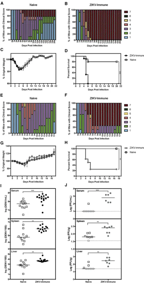
weightloss(Figures1A–1C).Mostofthemiceinthenaivecontrol grouprecoveredfromDENVinfection,whereas100%miceborn tolong-term(8–12months)ZIKV-immunemothersdiedbyday6 post infection (p.i.) (Figures 1A, 1B, and 1D). To determine whether short-term ZIKV infection period in mothers also affectedtheoutcomeofDENVinfectionintheirpups,we as-sessedclinicalscore,weightloss,andsurvivalinpupsbornto short-term(2months)ZIKV-immunemothers(Figures1E–1H). Bothnaiveand ZIKV-immunegroupsexhibitedsimilarweight lossafterDENV2challenge(Figure1G),yetmicebornto ZIKV-immunemothershadincreasedclinicalscoresanddecreased survivalcomparedwith those born to naivemothers(Figures 1E,1F,and1H).Theseresultsdemonstratethat,at4–5weeks ofage,pupsborntoZIKV-immunebutnotnaivemicedevelop lethaldiseaseuponchallengewithDENV2.
IncreasedDENV2BurdeninPupsBornto ZIKV-ImmuneMice
Todeterminewhetherenhanceddiseaseseverityinpupsbornto long-term(6–13months)ZIKV-immunemotherswasassociated with increased levels of DENV2infection, wecompared viral RNAlevelsinpupsborntoZIKV-immuneversusnaivemothers atday3p.i.DENV2RNAlevelswereincreasedsignificantlyin theserum (5-fold, ***p < 0.001), spleen (13-fold, **p< 0.01), and liver (8-fold, **p < 0.01) in pups born to ZIKV-immune mothersrelativetonaivepups(Figure1I).Thus,severedengue diseasemanifestationscorrelatedwithincreasedDENV2tissue burdeninmiceborntoZIKV-immunemothers.
Figure 1. Mice Born to ZIKV-Immune Mothers Have Increased Dengue Disease Severity and DENV Burden
Four- to 5-week-oldLysMCre+
Ifnar1fl/fl
mice born to mothers previously infected with ZIKV strain SD001 (106
focus-forming units [FFU] via retro-orbital route) or to naive mothers were challenged with DENV2 strain S221 (106
FFU via tail vein).
(A) Clinical scores of infected mice (n = 11) from naive mothers (n = 2).
(B) Clinical scores of infected mice (n = 15) from ZIKV-immune mothers infected for 8–12 months (n = 3).
(C and D) Weight loss and survival data of infected mice from naive (open squares) versus ZIKV-immune mothers infected for 8–12 months (black circles). (E) Clinical scores of infected mice (n = 8) from naive mothers (n = 2).
(F) Clinical scores of infected mice (n = 8) from ZIKV-immune mothers infected for 2 months (n = 2). (G and H) Weight loss and survival data of infected mice from naive (open squares) versus ZIKV-immune mothers infected for 2 months (gray circles). To determine DENV viral burden, we euthanized mice at 3 days p.i. and harvested serum, spleens, and livers. (I) The levels of DENV RNA from each tissue were measured via qRT-PCR. Viral RNA levels in the serum, spleen, and liver of DENV2-infected pups (open squares, n = 11) from naive mothers (n = 2) were compared with pups (black circles, n = 13) from ZIKV-immune mothers infected for 6–13 months (n = 4).
(J) The levels of infectious DENV2 were measured via focus-forming assay in the serum, spleen, and liver of DENV2-infected pups (open squares, n = 7) from naive mothers (n = 2) and pups (gray circles, n = 7) from ZIKV-immune mothers infected for 2 months (n = 2).
TNF Levels Are Increased in Mice with Passively Transferred ZIKV IgG upon DENV2 Challenge
We next determined whether tumor necrosis factor (TNF) levels were increased in mice that received IgG from pups born to ZIKV-immune pups relative to mice that received IgG from naive pups. Human studies have shown that patients with severe dengue have higher levels of several pro-inflammatory cyto-kines, including TNF, compared with individuals with mild dengue (Green et al., 1999; Hober et al., 1993; Kittigul et al.,
2000; Wang et al., 2007), and in a small observational study
indi-viduals on anti-TNF Ab therapy did not develop DHF/DSS (
De-ligny et al., 2014). Consistent with several studies demonstrating
that the lethal ADE-mediated dengue disease in mice is TNF dependent (Ng et al., 2014; Phanthanawiboon et al., 2016; Shresta et al., 2006; Watanabe et al., 2015; Zellweger et al., 2010), TNF levels were increased in ZIKV IgG recipient mice compared with naive IgG recipient mice (Figure S4E), implying that ZIKV IgG treatment potentiates TNF induction.
TNF Blockade Decreases DENV2-Induced Lethality in Mice Born to ZIKV-Immune Mothers
To assess the significance of TNF induction in our mouse model, we determined whether DENV2-induced lethal disease in pups born to ZIKV-immune mice was mediated by TNF by performing blocking experiments with a neutralizing anti-TNF Ab. Mice born to ZIKV-immune mothers were inoculated with DENV2 and administered 100mg of an anti-TNF or an isotype control Ab on days 1, 2, and 3 p.i. The anti-TNF Ab-treated group exhibited improved clinical scores, weight gain, and increased survival
Figure 2. Sera from Mice Born to ZIKV-Im-mune Mothers Neutralize ZIKV but Not DENV2 Infection
Serum was collected from 4- to 5-week-old
LysMCre+
Ifnar1fl/fl
mice born to mothers previously infected with ZIKV strain SD001 (106
FFU via retro-orbital route) or naive mothers. Serum of pups (open squares, n = 10) from naive mothers (n = 2) were compared with pups (black circles, n = 13) from ZIKV-immune mothers infected for 6–13 months (n = 4).
(A–D) Anti-ZIKV IgG (A) and anti-DENV IgG (B) were detected via ELISA. Neutralization capacity was assessed against (C) ZIKV SD001 and (D) DENV2 S221 via U937-DC-SIGN cells and a flow cytometry-based assay (Wen et al., 2017). Dotted line indicates limit of detection for ELISA and 50% or 0% neutral-ization line for the neutralneutral-ization assay. Serum of pups (black circles, n = 13) from ZIKV-immune mothers infected for 6–13 months (n = 4) were compared with serum of pups (open squares, n = 10) from ZIKV-immune mothers infected for 2 months (n = 3).
(E and F) Anti-ZIKV IgG (E) and anti-DENV2 IgG (F) were detected via ELISA.
Data are pooled from three independent experi-ments and are expressed as mean ± SEM.
mothersislikelyduetoalackofZIKVbindingbymaternally ac-quiredDENV2Abs.
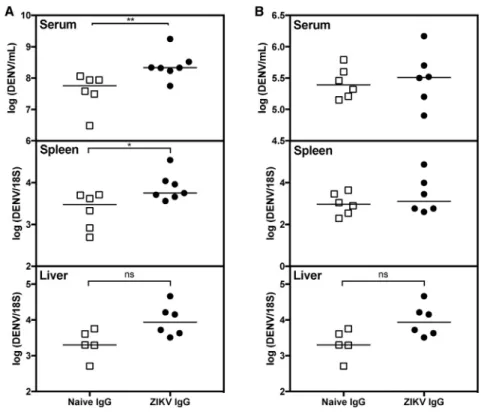
IncreasedViralBurdeninDENV2-InfectedMicewith PassivelyTransferredIgGfromPupsBornto ZIKV-ImmuneMothers
ToconfirmthatmaternalZIKVAbswereresponsibleforenhanced DENV2infection,weisolatedserumimmunoglobulinG(IgG)from 4-to5-week-oldpupsborntoZIKV-immuneornaivemothersand thenpassivelytransferreditintoage-matchednaive4-to5-week
oldLysMCre+Ifnar1fl/flrecipientmiceimmediatelypriorto
compared with isotype control Ab-treated mice (Figures 4A–4D). This result is consistent with the hypothesis that DENV2-infected pups born to ZIKV-immune mothers have increased disease severity through ADE and overexuberant production of pro-in-flammatory cytokines.
Monoclonal Ab Treatment Decreases DENV2-Induced Lethality in Mice Born to ZIKV-Immune Mothers
The original studies demonstrating ADE-mediated lethal dengue disease in mice showed that neutralizing DENV monoclonal Abs (mAbs) that blocked fusion could abrogate ADE (Balsitis et al.,
2010; Zellweger et al., 2010). Given these data, we assessed
whether the DENV2-induced lethal disease in pups born to ZIKV-immune mothers could also be prevented via treatment with neutralizing mAbs. We tested two human EDE1 mAbs (C8 and C10) that cross-neutralize different DENV serotypes and ZIKV (Dejnirattisai et al., 2015; Fernandez et al., 2017;
Swan-strom et al., 2016) for their ability to reduce disease severity in
DENV2-infected pups born to ZIKV-immuneLysMCre+Ifnar1fl/fl
mothers. Administration of 100mg of EDE1 C8 or EDE1 C10 Ab on days 1, 2, and 3 p.i. decreased dengue disease compared with treatment with PBS alone, based on clinical scores (Figures 4E–4G), weight loss (Figure 4H), and survival (Figure 4I). Thus, administration of EDE1 mAbs can prevent severe dengue dis-ease in mice born to ZIKV-immune mothers.
DISCUSSION
Currently, little is known about how prior ZIKV immunity affects DENV pathogenesis and infectionin vivo. One study showed that previous ZIKV exposure increased peak DENV viremia in macaques (George et al., 2017), and another reported enhanced DENV pathogenesis in mice administered a ZIKV
Figure 3. Passive Transfer of IgG Isolated from Mice Born to ZIKV-Immune Mothers into Naive Mice Increases Viral Burden upon DENV2 Challenge
(A and B) IgG was isolated from serum of 36 pups born to ZIKV-immune mothers infected for 6–12 months (n = 8) or from 32 age-matched naive mice from naiveLysMCre+
Ifnar1fl/fl
mothers (n = 7). 145mg (A) or 14.5mg (B) of purified IgG isolated from pups born to ZIKV-immune mothers (ZIKV IgG) or naive mothers (naive IgG) was passively transferred into naive 4- to 5-week-old LysMCre+
Ifnar1fl/fl
recipient mice, followed by challenge of the passively transferred recipient mice with 23105 FFU of DENV2 strain S221 via tail vein injection. Tissues were harvested 3 days p.i. and DENV2 RNA levels in the serum, spleen, and liver were quantified by qRT-PCR. n = 6–7 ZIKV IgG recipient mice and n = 6 naive IgG recipient mice. Mann-Whitney test (**p < 0.01, *p < 0.05).
mAb (Stettler et al., 2016). Consistent with these findings, our data show increased viral burden, worsened clinical signs, and decreased survival in mice born to ZIKV-immune mothers relative to those born to naive mothers. Our study demonstrates maternal Ab-mediated infant DHF/DSS in the context of pre-existing anti-flavivirus immune sera other than anti-DENV immune sera (Martinez Gomez et al., 2016; Ng et al., 2014). In compar-ison, maternal DENV Abs exerted mainly neutral effects against ZIKV in our mouse model, in agreement with the observation that prior DENV immunity (1–3 years long) did not influence subsequent ZIKV infection in macaques (McCracken et al.,
2017; Pantoja et al., 2017) and consistent with reports that
the anti-DENV Ab response in humans becomes less cross-neutralizing against ZIKV over time (Collins et al., 2017;
Montoya et al., 2018). However, our result contrasts with a
published study demonstrating increased ZIKV infection and pathogenesis in Stat2 / mice that were passively transferred with DENV-immune human plasma (Bardina et al., 2017). The disparity in results may be related to the magnitude and quality of Ab responses (e.g., binding, neutralization, isotype, speci-ficity, and avidity) and perhaps the use of LysMCre+Ifnar1fl/fl
versusStat2 / mice.
Figure 4. Administration of Anti-TNF Ab or EDE1 C8 and C10 Abs, which Recognize EDE1 Epitopes, Prevents Lethal Dengue Dis-ease in Mice Born to ZIKV-Immune Mothers Four- to 5-week-oldLysMCre+
Ifnar1fl/fl
mice born to mothers infected with ZIKV strain SD001 (106
FFU via retro-orbital route) for 7–9 months (n = 3) were treated via an intraperitoneal injection with 100mg of isotype control Ab (clone HPRN) or anti-TNF Ab (clone XT3.11) on days 1, 2, and 3 following inocu-lation with 106
FFU of DENV2 strain S221 via tail vein injection.
(A–D) Clinical scores of isotype control Ab-treated mice (n = 7) (A), clinical scores of anti-TNF Ab-treated mice (n = 8) (B), and weight loss (C) and survival rates (D) of isotype control Ab-treated mice (black circles) and anti-TNF Ab-treated mice (open circles). Administration of EDE1 C8 or EDE1 C10 Abs was performed in 4- to 5-week-old LysMCre+Ifnar1fl/fl
mice born to mothers infected with ZIKV strain SD001 (106
FFU via retro-orbital route) for 12–13 months (n = 4). These pups were injected via an intraperitoneal route with PBS, EDE1 C8 Ab (100mg), or EDE1 C10 Ab (100mg) on days 1, 2, and 3 following challenge with DENV2 strain S221 (106
FFU via tail vein).
(E–H) Clinical scores of PBS control mice (n = 9) (E), clinical scores of EDE1 C8 Ab-administered mice (n = 10) (F), clinical scores of EDE1 C10 Ab-admin-istered mice (n = 8) (G), and weight loss (H) and survival rates (I) of PBS-treated (black circles), EDE1 C8-treated (open circles), and EDE1 C10-treated (red circles) mice.
Data are pooled from two independent experiments and are expressed as mean ± SEM. Unpaired Stu-dent’s t test of groups for each day was used in (C) and (H). Log-rank test (*p<0.05, **p < 0.01, ***p<
0.001, ****p<0.0001) was used in (D) and (I).
infants born to DENV-immune women
(Balsitis et al., 2010; Halstead, 2007;
Katzel-nick et al., 2017; Ng et al., 2014; Zellweger
et al., 2010). Our results suggest that if
women are infected with ZIKV or potentially immunized with a ZIKV vaccine that elicits a cross-reactive Ab response, their infants might have an increased risk of developing DHF/DSS. A ZIKV vaccine with fusion loop mutations in the E protein has been ported to reduce cross-reactive Ab re-sponses and minimize enhancement of DENV infection and pathogenesis in mice
(Richner et al., 2017), suggesting one potential avenue for
designing ZIKV vaccines that avoid ZIKV maternal Ab-mediated severe DENV disease in infants. Thus, the emergence of ZIKV immunity in DENV-endemic regions may create an added risk for ADE-mediated severe dengue disease. Our mouse model may be useful for testing the effects of ZIKV and DENV (including different serotypes) on infant DENV and ZIKV infections and for evaluating the effects of ZIKV and DENV vaccine candidates that are designed for deployment in DENV-endemic regions. As T, B, and most dendritic cell responses are normal in flavivirus-naive versus immune status of mothers (Collins
et al., 2017; Montoya et al., 2018; Yu et al., 2017), thereby
affectingthewindowofbothpotentiallyprotectiveand path-ogenicperiodsininfants.
LysMCre+Ifnar1fl/flmice, this mouse model may also be useful for investigating mechanisms of DENV pathogenesis and immunity.
STAR+METHODS
Detailed methods are provided in the online version of this paper and include the following:
d KEY RESOURCES TABLE
d CONTACT FOR REAGENT AND RESOURCE SHARING
d EXPERIMENTAL MODEL AND SUBJECT DETAILS
B Viruses
B Cell Lines
B Mice
d METHOD DETAILS
B Disease Scoring
B Flow Cytometric Analysis of Immune Cell Populations
B Infectious Virus Quantification
B Viral RNA Quantification
B ZIKV and DENV ELISA
B Neutralization Assays
B TNF Blockade
B Passive Transfer of IgG
B TNF ELISA
B Monoclonal Ab Treatment
d QUANTIFICATION AND STATISTICAL ANALYSIS
B Flow Cytometric Analysis of Immune Cell Populations
B Infectious Virus Quantification
B Viral RNA Quantification
B ZIKV and DENV ELISA
B Neutralization Assays
B Statistical Analysis
SUPPLEMENTAL INFORMATION
Supplemental Information includes four figures and can be found with this article online athttps://doi.org/10.1016/j.chom.2018.09.015.
ACKNOWLEDGMENTS
This research was funded by NIH grants AI116813 and NS100477 to S.S., AI104972 and HD091218 to M.S.D., and AI106695 and AI125198 to R.S.B. and by the NIH/La Jolla Institute for Allergy and Immunology Training grant T32AI125179 to A.M.F.
AUTHOR CONTRIBUTIONS
W.W.T. and S.S. conceived the project. A.M.F., W.W.T., M.P.Y., A.M., K.M.V., and J.G. performed and analyzed experiments. M.D.M., A.F.C., and R.T.S. ob-tained the ZIKV clinical isolate SD001. J.S. and R.S.B. were responsible for generating and testing EDE1-C8 and -C10 neutralizing antibodies. A.M.F. and S.S. wrote the manuscript. M.S.D., R.S.B., and S.S. conceived experi-ments and edited the manuscript.
DECLARATION OF INTERESTS
M.S.D. is a consultant for Inbios and is on the Scientific Advisory Board of Moderna. R.S.B. has consulted with Takeda and Sanofi Pasteur. All other au-thors declare no competing interests.
Received: March 22, 2018 Revised: June 21, 2018 Accepted: September 20, 2018 Published: November 14, 2018
REFERENCES
Balsitis, S.J., Williams, K.L., Lachica, R., Flores, D., Kyle, J.L., Mehlhop, E., Johnson, S., Diamond, M.S., Beatty, P.R., and Harris, E. (2010). Lethal anti-body enhancement of dengue disease in mice is prevented by Fc modification. PLoS Pathog.6, e1000790.
Barba-Spaeth, G., Dejnirattisai, W., Rouvinski, A., Vaney, M.C., Medits, I., Sharma, A., Simon-Loriere, E., Sakuntabhai, A., Cao-Lormeau, V.M., Haouz, A., et al. (2016). Structural basis of potent Zika-dengue virus antibody cross-neutralization. Nature536, 48–53.
Bardina, S.V., Bunduc, P., Tripathi, S., Duehr, J., Frere, J.J., Brown, J.A., Nachbagauer, R., Foster, G.A., Krysztof, D., Tortorella, D., et al. (2017). Enhancement of Zika virus pathogenesis by preexisting antiflavivirus immu-nity. Science356, 175–180.
Bhatt, S., Gething, P.W., Brady, O.J., Messina, J.P., Farlow, A.W., Moyes, C.L., Drake, J.M., Brownstein, J.S., Hoen, A.G., Sankoh, O., et al. (2013). The global distribution and burden of dengue. Nature496, 504–507.
Chang, H.H., Huber, R.G., Bond, P.J., Grad, Y.H., Camerini, D., Maurer-Stroh, S., and Lipsitch, M. (2017). Systematic analysis of protein identity between Zika virus and other arthropod-borne viruses. Bull. World Health Organ.95, 517–525I.
Charles, A.S., and Christofferson, R.C. (2016). Utility of a dengue-derived monoclonal antibody to enhance Zika infection in vitro. PLoS Curr.8,https:// doi.org/10.1371/currents.outbreaks.4ab8bc87c945eb41cd8a49e127082620.
Chau, T.N., Hieu, N.T., Anders, K.L., Wolbers, M., Lien le, B., Hieu, L.T., Hien, T.T., Hung, N.T., Farrar, J., Whitehead, S., et al. (2009). Dengue virus infections and maternal antibody decay in a prospective birth cohort study of Vietnamese infants. J. Infect. Dis.200, 1893–1900.
Clausen, B.E., Burkhardt, C., Reith, W., Renkawitz, R., and Forster, I. (1999). Conditional gene targeting in macrophages and granulocytes using LysMcre mice. Transgenic Res.8, 265–277.
Collins, M.H., McGowan, E., Jadi, R., Young, E., Lopez, C.A., Baric, R.S., Lazear, H.M., and de Silva, A.M. (2017). Lack of durable cross-neutralizing an-tibodies against Zika virus from dengue virus infection. Emerg. Infect. Dis.23, 773–781.
Dejnirattisai, W., Wongwiwat, W., Supasa, S., Zhang, X., Dai, X., Rouvinski, A., Jumnainsong, A., Edwards, C., Quyen, N.T.H., Duangchinda, T., et al. (2015). A new class of highly potent, broadly neutralizing antibodies isolated from viremic patients infected with dengue virus. Nat. Immunol.16, 170–177.
Dejnirattisai, W., Supasa, P., Wongwiwat, W., Rouvinski, A., Barba-Spaeth, G., Duangchinda, T., Sakuntabhai, A., Cao-Lormeau, V.M., Malasit, P., Rey, F.A., et al. (2016). Dengue virus sero-cross-reactivity drives antibody-dependent enhancement of infection with Zika virus. Nat. Immunol.17, 1102–1108.
Deligny, C., de Bandt, M., Dehlinger, V., Numeric, P., Cabie, A., Lombard, F., Polomat, K., JeanBaptiste, G., and Arfi, S. (2014). Dengue fever in patients un-der biologics. J. Clin. Virol.61, 442–443.
Diamond, M.S., Kinder, M., Matsushita, H., Mashayekhi, M., Dunn, G.P., Archambault, J.M., Lee, H., Arthur, C.D., White, J.M., Kalinke, U., et al. (2011). Type I interferon is selectively required by dendritic cells for immune rejection of tumors. J. Exp. Med.208, 1989–2003.
Elong Ngono, A., and Shresta, S. (2018). Immune response to dengue and Zika. Annu. Rev. Immunol.36, 279–308.
Elong Ngono, A., Vizcarra, E.A., Tang, W.W., Sheets, N., Joo, Y., Kim, K., Gorman, M.J., Diamond, M.S., and Shresta, S. (2017). Mapping and role of the CD8+ T cell response during primary Zika virus infection in mice. Cell Host Microbe21, 35–46.
Fernandez, E., Dejnirattisai, W., Cao, B., Scheaffer, S.M., Supasa, P., Wongwiwat, W., Esakky, P., Drury, A., Mongkolsapaya, J., Moley, K.H., et al. (2017). Human antibodies to the dengue virus E-dimer epitope have ther-apeutic activity against Zika virus infection. Nat. Immunol.18, 1261–1269.
heparan sulfate causes severe disease in mice by establishing increased sys-temic viral loads. J. Virol.82, 8411–8421.
Priyamvada, L., Quicke, K.M., Hudson, W.H., Onlamoon, N., Sewatanon, J., Edupuganti, S., Pattanapanyasat, K., Chokephaibulkit, K., Mulligan, M.J., Wilson, P.C., et al. (2016). Human antibody responses after dengue virus infec-tion are highly cross-reactive to Zika virus. Proc. Natl. Acad. Sci. U S A113, 7852–7857.
Richner, J.M., Himansu, S., Dowd, K.A., Butler, S.L., Salazar, V., Fox, J.M., Julander, J.G., Tang, W.W., Shresta, S., Pierson, T.C., et al. (2017). Modified mRNA vaccines protect against Zika virus infection. Cell168, 1114–1125.e10.
Shepard, D.S., Undurraga, E.A., Halasa, Y.A., and Stanaway, J.D. (2016). The global economic burden of dengue: a systematic analysis. Lancet Infect. Dis.
16, 935–941.
Shresta, S., Sharar, K.L., Prigozhin, D.M., Beatty, P.R., and Harris, E. (2006). Murine model for dengue virus-induced lethal disease with increased vascular permeability. J. Virol.80, 10208–10217.
Simmons, C.P., Chau, T.N., Thuy, T.T., Tuan, N.M., Hoang, D.M., Thien, N.T., Lien le, B., Quy, N.T., Hieu, N.T., Hien, T.T., et al. (2007). Maternal antibody and viral factors in the pathogenesis of dengue virus in infants. J. Infect. Dis.196, 416–424.
Stettler, K., Beltramello, M., Espinosa, D.A., Graham, V., Cassotta, A., Bianchi, S., Vanzetta, F., Minola, A., Jaconi, S., Mele, F., et al. (2016). Specificity, cross-reactivity, and function of antibodies elicited by Zika virus infection. Science
353, 823–826.
Swanstrom, J.A., Plante, J.A., Plante, K.S., Young, E.F., McGowan, E., Gallichotte, E.N., Widman, D.G., Heise, M.T., de Silva, A.M., and Baric, R.S. (2016). Dengue virus envelope dimer epitope monoclonal antibodies isolated from dengue patients are protective against Zika virus. MBio7,https://doi. org/10.1128/mBio.01123-16.
Tang, W.W., Young, M.P., Mamidi, A., Regla-Nava, J.A., Kim, K., and Shresta, S. (2016). A mouse model of Zika virus sexual transmission and vaginal viral replication. Cell Rep.17, 3091–3098.
Wang, L., Chen, R.F., Liu, J.W., Yu, H.R., Kuo, H.C., and Yang, K.D. (2007). Implications of dynamic changes among tumor necrosis factor-alpha (TNF-alpha), membrane TNF receptor, and soluble TNF receptor levels in regard to the severity of dengue infection. Am. J. Trop. Med. Hyg.77, 297–302.
Watanabe, S., Chan, K.W., Wang, J., Rivino, L., Lok, S.M., and Vasudevan, S.G. (2015). Dengue virus infection with highly neutralizing levels of cross-reactive antibodies causes acute lethal small intestinal pathology without a high level of viremia in mice. J. Virol.89, 5847–5861.
Wen, J., Tang, W.W., Sheets, N., Ellison, J., Sette, A., Kim, K., and Shresta, S. (2017). Identification of Zika virus epitopes reveals immunodominant and pro-tective roles for dengue virus cross-reactive CD8+ T cells. Nat. Microbiol.
2, 17036.
World Health Organization. (2016). Zika Virus, Microcephaly and Guillain-Barre´ Syndrome (World Health Organization).
Yauch, L.E., Zellweger, R.M., Kotturi, M.F., Qutubuddin, A., Sidney, J., Peters, B., Prestwood, T.R., Sette, A., and Shresta, S. (2009). A protective role for dengue-virus specific CD8+ T cells. J. Immunol.182, 4865–4873.
Yu, L., Wang, R., Gao, F., Li, M., Liu, J., Wang, J., Hong, W., Zhao, L., Wen, Y., Yin, C., et al. (2017). Delineating antibody recognition against Zika virus during natural infection. JCI Insight2,https://doi.org/10.1172/jci.insight.93042.
Zellweger, R.M., Prestwood, T.R., and Shresta, S. (2010). Enhanced infection of liver sinusoidal endothelial cells in a mouse model of antibody-induced se-vere dengue disease. Cell Host Microbe7, 128–139.
Zust, R., Toh, Y.X., Valdes, I., Cerny, D., Heinrich, J., Hermida, L., Marcos, E., Guillen, G., Kalinke, U., Shi, P.Y., et al. (2014). Type I interferon signals in mac-rophages and dendritic cells control dengue virus infection: implications for a new mouse model to test dengue vaccines. J. Virol.88, 7276–7285.
Green,S.,Vaughn,D.W.,Kalayanarooj,S.,Nimmannitya,S.,Suntayakorn,S., Nisalak,A.,Lew,R.,Innis,B.L.,Kurane,I.,Rothman,A.L.,etal.(1999).Early immuneactivationinacutedengueillnessisrelatedtodevelopmentofplasma leakageanddiseaseseverity.J.Infect.Dis.179,755–762.
Guzman,M.G.,Halstead,S.B.,Artsob,H.,Buchy,P.,Farrar,J.,Gubler,D.J., Hunsperger, E., Kroeger, A., Margolis, H.S., Martinez, E., et al. (2010). Dengue:acontinuingglobalthreat.Nat.Rev.Microbiol.8,S7–S16.
Halstead,S.B.(2007).Dengue.Lancet370,1644–1652.
Halstead,S.B.,Lan,N.T.,Myint,T.T.,Shwe,T.N.,Nisalak,A.,Kalyanarooj,S., Nimmannitya,S.,Soegijanto,S.,Vaughn,D.W.,andEndy,T.P.(2002).Dengue hemorrhagicfeverininfants:researchopportunitiesignored.Emerg.Infect. Dis.8,1474–1479.
Hober,D.,Poli,L.,Roblin,B.,Gestas,P.,Chungue,E.,Granic,G.,Imbert,P., Pecarere,J.L.,Vergez-Pascal,R.,Wattre,P.,etal.(1993).Serumlevelsof tumornecrosisfactor-alpha(TNF-alpha),interleukin-6(IL-6),and interleukin-1beta(IL-1beta)indengue-infectedpatients.Am.J.Trop.Med.Hyg.48, 324–331.
Katzelnick,L.C.,Gresh,L.,Halloran,M.E.,Mercado,J.C.,Kuan,G.,Gordon, A.,Balmaseda,A.,andHarris,E.(2017).Antibody-dependentenhancement ofseveredenguediseaseinhumans.Science358,929–932.
Kawiecki,A.B.,andChristofferson,R.C.(2016).Zikavirus-inducedantibody responseenhancesdenguevirusserotype2replicationinvitro.J.Infect. Dis.214,1357–1360.
Kittigul,L.,Temprom,W.,Sujirarat,D.,andKittigul,C.(2000).Determination oftumor necrosisfactor-alphalevelsindenguevirusinfectedpatientsby sensitivebiotin-streptavidinenzyme-linkedimmunosorbentassay.J. Virol. Methods90,51–57.
MartinezGomez,J.M.,Ong,L.C.,Lam,J.H.,BinteAman,S.A.,Libau,E.A., Lee,P.X.,StJohn,A.L.,andAlonso,S.(2016).Maternalantibody-mediated diseaseenhancementintypeIinterferon-deficientmiceleadstolethaldisease associatedwithliverdamage.PLoSNegl.Trop.Dis.10,e0004536.
McCracken,M.K.,Gromowski,G.D.,Friberg,H.L.,Lin,X.,Abbink,P.,DeLa Barrera,R.,Eckles,K.H.,Garver,L.S.,Boyd,M.,Jetton,D.,etal.(2017). ImpactofpriorflavivirusimmunityonZikavirusinfectioninrhesusmacaques. PLoSPathog.13,e1006487.
Montoya,M.,Collins,M.,Dejnirattisai,W.,Katzelnick,L.C.,Puerta-Guardo,H., Jadi,R.,Schildhauer,S.,Supasa,P.,Vasanawathana,S.,Malasit,P.,etal. (2018).Longitudinalanalysisofantibodycross-neutralizationfollowingZika anddengue virusinfectioninAsiaandtheAmericas.J. Infect.Dis.218, 536–545.
Ng,J.K.,Zhang,S.L.,Tan,H.C.,Yan,B.,Martinez,J.M.,Tan,W.Y.,Lam,J.H., Tan,G.K.,Ooi,E.E.,andAlonso,S.(2014).Firstexperimentalinvivomodelof enhanceddenguediseaseseveritythroughmaternallyacquiredheterotypic dengueantibodies.PLoSPathog.10,e1004031.
Pantoja,P.,Perez-Guzman,E.X.,Rodriguez,I.V.,White,L.J.,Gonzalez,O., Serrano,C.,Giavedoni,L.,Hodara,V.,Cruz,L.,Arana,T.,etal.(2017).Zika vi-ruspathogenesisinrhesusmacaquesisunaffectedbypre-existingimmunity todenguevirus.Nat.Commun.8,15674.
Phanthanawiboon,S.,Limkittikul,K.,Sakai,Y.,Takakura,N.,Saijo,M.,and Kurosu,T.(2016).Acutesystemicinfectionwithdenguevirusleadstovascular leakageanddeaththroughtumornecrosisfactor-alphaandTie2/angiopoietin signalinginmicelackingtypeIandIIInterferonreceptors.PLoSOne11, e0148564.
Pinto,A.K.,Brien, J.D.,Lam, C.Y., Johnson, S.,Chiang,C., Hiscott, J., Sarathy,V.V.,Barrett,A.D.,Shresta,S.,andDiamond,M.S.(2015).Defining newtherapeutics usingamoreimmunocompetent mousemodelof anti-body-enhanceddenguevirusinfection.MBio6,e01316–15.
STAR
+
METHODS
KEY RESOURCES TABLE
REAGENT or RESOURCE SOURCE IDENTIFIER
Antibodies
4G2 ATCC Cat. #HB-112
RRID: CVCL_J890
PerCP/Cy5.5 anti-mouse CD3e (clone: 145-2C11) TONBO Biosciences Cat. #65-0031-U100 RRID: AB_394599
Brilliant Violet 510 anti-mouse CD8a (clone: 53-6.7) BioLegend Cat. #100751 RRID: AB_2561389
CD4 Monoclonal Antibody APC-eFluor 780 (clone: GK1.5), eBioscience
Thermo Fisher Scientific Cat. #47-0041-82 RRID: AB_11218896
CD19 Monoclonal Antibody PE (eBio1D3 (1D3)), eBioscience
Thermo Fisher Scientific Cat. #12-0193-83 RRID: AB_657660
PerCp/Cy5.5 anti-mouse CD138 (Syndecan-1) (clone: 281-2)
BioLegend Cat. #142510
RRID: AB_2561601
BD Pharmingen FITC rat anti-mouse IgD (clone: 11-26c.2a)
BD Biosciences Cat. #553439
RRID: AB_394859
EDE1 C8 Ralph Baric (Swanstrom et al., 2016) N/A
EDE1 C10 Ralph Baric (Swanstrom et al., 2016) N/A
In vivoMAb rat IgG1 isotype control, anti-horseradish peroxidase (clone: HPRN)
BioXCell Cat. #BE0088
RRID: AB_1107775
BD Pharmingen PE-labeled anti-human CD209 (clone: DCN46)
BD Biosciences Cat. #551265
RRID: AB_394123
In vivoMab anti-mouse TNFa(clone: XT3.11) BioXCell Cat. #BE0058 RRID: AB_1107764
Peroxidase conjugated affini-pure goat anti-mouse IgG F(ab’)2fragment
Jackson ImmunoResearch Cat. #115-035-072 RRID: AB_2338507
Peroxidase conjugated affini-pure goat anti-mouse IgG Fcg
Jackson ImmunoResearch Cat. #115-035-008 RRID: AB_2313585
Bacterial and Virus Strains
ZIKV (SD001) A.F.C. et al., unpublished data N/A
DENV2 (S221) Yauch et al., 2009 N/A
Chemicals, Peptides, and Recombinant Proteins
Cytofix/Cytoperm BD Biosciences Cat. #554714
Qiamp Viral Mini Kit Qiagen Cat. #52904
RNAlater Thermo Fisher Scientific Cat. #AM7021
QuantaBio qScript one-step qRT-PCR kit VWR Cat. #101414-172
eBioscience TMB solution Thermo Fisher Scientific Cat. #00-4201-56
Experimental Models: Cell Lines
Aedes albopicticus: C6/36 ATCC Cat. #ATCC: CRL-1660
RRID: CVCL_Z230
Baby Hamster Kidney (BHK)-21 ATCC Cat. #ATCC: CCL-10
RRID: CVCL_1915
U937-DC SIGN cells ATCC Cat. #CRL-3253
RRID: CVCL_2Z95
Experimental Models: Organisms/Strains
Mouse:LysMCre+Ifnar1fl/flC57BL/6 Michael S. Diamond (Clausen et al., 1999) N/A
Oligonucleotides
DENV2 forward primer: CATATTGACGCTGGGAAAGA Prestwood et al., 2008 N/A
DENV2 reverse primer: AGAACCTGTTGATTCAAC Prestwood et al., 2008 N/A
Continued
REAGENT or RESOURCE SOURCE IDENTIFIER
18S forward primer: CGGCTACCACATCCAAGGAA Prestwood et al., 2008 N/A
18S reverse primer: GCTGGAATTACCGCGGCT Prestwood et al., 2008 N/A
DENV2 probe – [Fam]-TGCTGGCCTC – [TamraQ] Eurofins N/A
18S probe – [Fam] – CTGTCTGGCA – [TamraQ] Eurofins N/A
Software and Algorithms
FlowJo version 10 FlowJo https://www.flowjo.com/
Graphpad Prism 7 Graphpad Prism Software https://www.graphpad.com/
Other
Pierce FITC antibody labelling kit Thermo Fisher Scientific Cat. #53027
Nab Protein G Spin Kit Thermo Fisher Scientific Cat. #89979
Slide-A-Lyzer Thermo Fisher Scientific Cat. #66212
Mouse TNF-aQuantikine ELISA kit R&D Systems Cat. #MTA00B
ZIKV E protein Suriname strain Native Antigen Company Cat. #ZIKVSU-ENV
KPL True Blue SeraCare Cat. #5510-0030
CONTACTFORREAGENTANDRESOURCESHARING
FurtherinformationandrequestsforresourcesandreagentsshouldbedirectedtoandwillbefulfilledbytheLeadContact,Sujan Shresta([email protected]).
EXPERIMENTALMODELANDSUBJECTDETAILS
Viruses
SD001is a ZIKV clinicalisolateobtained froman adultfemale traveler infectedinCaracas, Venezuelain 2016(A.F.C. etal., unpublisheddata).InfectiousviruswasisolatedfromafilteredurinesamplefrompatientSD001andpropagatedinC6/36Aedes
albopictus cells(ATCC, cat. # ATCC: CRL1660). Mouse-adapted DENV2strain S221 was propagated inC6/36 cells.
ZIKV-SD001andDENV2-S221weretitratedbyfocus-formingassayusingbabyhamsterkidney(BHK-21)(ATCC,cat.#ATCC:CCL10) cellsasdescribedinthevirusquantificationsection.
CellLines
C6/36mosquitocellswerepropagatedinLeibovitz’sL-15(ThermoFisher Scientific,cat. #11415064)supplemented with10% fetalbovine serum(FBS) (ThermoFisher Scientific, cat. #16000044),1% penicillin/streptomycin (ThermoFisher Scientific cat. #15140-122),and 1%HEPES(ThermoFisher Scientific,cat.#15630080) at28C.BHK-21cellsweregrown inMEMa(Fisher, cat.#12-561-072)supplementedwith10%FBS,1%penicillin/streptomycin,and1%HEPESat37Cina5%CO2atmosphere.
U937-DC-SIGNcellswerepropagatedinRPMI1640(ThermoFisher Scientific,cat.#11875093) supplementedwith10%FBS, 1%penicillin/streptomycin,and1%HEPESat37Cina5%CO2atmosphere.
Mice
LysMCre+Ifnar1fl/flC57BL/6micelacktypeIinterferon(IFN)receptorsinasubsetofmyeloidcells(Clausenetal.,1999;Diamond
etal.,2011)andwerecharacterizedpreviouslyasamodelforDENV(Pintoetal.,2015;Zustetal.,2014)andZIKV(ElongNgono
etal.,2017;Tangetal.,2016)infection.Femalemicewereinoculatedviaretro-orbitalroutewith106focus-formingunits(FFU)of
ZIKV-SD001dilutedin10%FBS/PBS(100mLtotalvolume)orwith5x105FFUofDENV2-S221viatailveininjection.Atfourweeks
post-ZIKVorDENVinfection,femaleswerebredwithnaive6-to8-weekoldmaleLysMCre+Ifnar1fl/flmice.Maleandfemalepups borntoZIKV-orDENV-infectedLysMCre+Ifnar1fl/flfemaleswerechallengedat4-to5-weeksofage.Analyseswerenotperformed
onwhetherornotthesexofthemouseinfluencedtheoveralloutcomeastheoutcomedidnotdifferbasedonthesexofthemouse. Four-to5-week-oldage-matchedpups(bothmalesandfemales)borntonaiveLysMCre+Ifnar1fl/flmotherswereusedascontrols.
METHOD DETAILS
Disease Scoring
Pups born to ZIKV-immune or naive mothers at 4- to 5-weeks of age were inoculated with 1 x 10⁶FFU of DENV2 S221 diluted in 10% FBS/PBS (200mL total volume) per mouse via tail vein injection. Mice were monitored daily for weight and clinical scores from day 0 to day 20 p.i. Clinical scores ranged from 1 - 7: 1, heathy mice with a smooth coat and bright, alert eyes; 2, mice are slightly ruffled around the head and neck, but active and alert; 3, mice have a ruffled coat throughout the body, but still active and alert; 4, mice have a very ruffled coat and slightly closed eyes, they walk slowly, and they have mild lethargy; 5, mice have a very ruffled coat and closed inset eyes, slow to no movement but will return to the upright position if put on the side; 6, mice have a very ruffled coat and closed inset eyes, are moribund, they have no movement or uncontrollable spastic movements, will not return to upright position if put on its side, and completely unaware or in noticeable distress and require humane euthanasia; 7, mice are deceased. Mice were humanely euthanized if weight loss was greater than or equal to 20% of their body mass or if their clinical score was a 6.
Flow Cytometric Analysis of Immune Cell Populations
Spleens were harvested from pups born to ZIKV-immune or naive mothers at 4-5 weeks of age after being humanely euthanized with CO2. Splenocytes were plated into 96-well round-bottom plates at 1 x 10⁶cells/well in RPMI 1640 medium supplemented with 10%
FBS, 1% penicillin/streptomycin, and 1% HEPES. Splenocytes were washed with PBS and stained with anti-CD3 PerCP-Cy5.5 (Tonbo Biosciences, cat. #65-0031-U100), anti-CD4 APC eflour780 (Thermo Fisher Scientific, cat. #47-0041-82), and anti-CD8 BV510 (BioLegend, cat. #100751) or with anti-CD19 PE (Thermo Fisher Scientific, cat. #12-0193-83), anti-CD138 PerCP-Cy5.5 (BioLegend, cat. #142510), and anti-mouse IgD FITC (BD Biosciences, cat. #553439). Cells were incubated with these Abs (each at 1:200 dilution) for 30 min, followed by washing for 3 times with FACs buffer. The cells were then fixed and permeabilized with Cytofix/Cytoperm (BD Biosciences, cat. #554714), washed and resuspended in FACs buffer.
Infectious Virus Quantification
Four- to 5-week oldLysMCre+Ifnar1fl/flmice born to ZIKV-immune or naive mothers were inoculated with 2 x 10⁵FFU DENV2 S221 diluted in 10% FBS/PBS via tail vein injection (200mL total volume) or with 1 x 10⁵FFU ZIKV in 10% FBS/PBS retro-orbitally (100mL total volume). Mice were humanely euthanized with CO23 days p.i. Blood was obtained via cardiac puncture, centrifuged (16,363 x g
for 15 min at 4C), and serum was stored at -80C. Mice were perfused with PBS. Spleen and livers were harvested and put in pre-weighed tubes containing complete MEMacontaining a metal bead and stored at -80C. Viral titers were measured using a BHK-21 cell-based focus forming assay (FFA). BHK-21 cells were plated at 2 x 10⁵cells per well in a 24-well plate and incubated overnight in
complete MEMamedium supplemented with 10% FBS, 1% penicillin/streptomycin, and 1% HEPES at 37C in a 5% CO2
atmo-sphere. Cells were infected with serial dilutions of virus for 1.5 hr with gentle shaking every 15 min. The medium was then aspirated and replaced with fresh MEMasupplemented with 1% carboxymethyl cellulose (Sigma, cat. #419273), 10% FBS, 1% penicillin/ streptomycin, and 1% HEPES. Cells were then cultured for 3 days. At 3 days p.i., cells were fixed with 4% formalin (Fisher Scientific, cat. #SF98-4) for 30 min, washed 3 times with PBS, permeabilized with 1% Triton X-100 (Sigma, cat. #X100-100ML) for 30 min, washed 3 times with PBS, and blocked by 10% FBS in PBS for 30 min. ZIKV or DENV was detected by incubation of cells with 4G2, a pan-flavivirus E protein-specific monoclonal Ab (ATCC, cat. # D1-4G2-4-15 (ATCC HB-112)) (1mg/mL in 1% FBS/PBS) for 1.5 h at room temperature or overnight at 4C. Cells were washed 3 times with PBS and incubated for 1.5 h with horseradish perox-idase (HRP)-conjugated goat anti-mouse IgG (Jackson ImmunoResearch, cat. #115-035-072) (diluted 1:1000 in 1% bovine serum albumin (BSA)/PBS), followed by washing 3 times with PBS. Finally, foci were detected by incubation with KPL True Blue substrate (SeraCare, cat. #5510-0030) for 20 min and rinsed in diH2O.
Viral RNA Quantification
Four- to 5-week-oldLysMCre+Ifnar1fl/flmice born to ZIKV-immune or naive mothers were inoculated with 2 x 10⁵FFU DENV2 S221
diluted in 10% FBS/PBS (200mL total volume) via tail vein injection. Mice were humanely euthanized 3 days p.i. Blood was obtained via cardiac puncture, centrifuged (16,363 x g for 15 min at 4C), and serum was stored at -20C. Mice were perfused with PBS, and their spleens and livers were harvested and stored in RNAlater (Thermo Fisher Scientific, cat. #AM7021) at 4C. Tissues were homog-enized prior to RNA extraction. RNA was isolated via Qiagen QIAmp viral mini kit (Qiagen, cat. #52904) and qRT-PCR was performed as previously described (Prestwood et al., 2008) using the QuantaBio qScript one-step qRT-PCR kit (VWR, cat. #101414-172) and probes and primers described in theKey Resources Table. PCR mixtures were pre-incubated at 50C for 2 min, then 95C for 10 min followed by 40 cycles of two-step incubations at 95C for 15 s and 60C for 1 min for DENV2. DENV2 samples were compared to a standard curve of 103– 107copies of DENV2 RNA. 18S samples were diluted and compared to a standard of 18S rRNA that was derived from a mouse spleen and diluted to 102– 10418S RNA. 18S RNA was ran for 10 min at 48C, 5 min at 98C, and 39 cycles
of 15 seconds at 95C and 1 min at 60C.
ZIKV and DENV ELISA
Serum samples from mice born to ZIKV- or DENV-immune mothers were tested for ZIKV- and DENV-binding antibodies using a direct ELISA. To detect DENV antibodies, sucrose purified DENV2 S221 virions were used at a concentration of 1 x 10⁶FFU/well as the
4Covernight.Wellswerewashed3timeswithELISAwashingbuffer(0.05%Tween20inPBS)andthenblockedwith5%caseinin PBSforanhouratroomtemperature.Serumwasfirstdiluted1:10in10%FBS/PBSandthen1:3forsubsequentdilutionsandwas incubatedinwellsfor1.5hratroomtemperature.Wellswerewashed3timeswithELISAwashingbuffer.Wellswerethenincubated withperoxidaseconjugatedAffini-PureGoatanti-mouseIgGFcg(JacksonImmunoResearch,cat.#115-035-008)diluted1:5000in 1%BSA/PBSatroomtemperature.Wellswerewashed3timeswithELISAwashingbuffer.100mLofTMBsubstratesolutionwas addeduntilbluecolorchange,andreactionwasstoppedwith50mLof2Nsulfuricacid(Sigma,cat.#339741).TodetectZIKV Abs,ZIKVEprotein(Surinamestrain,NativeAntigenCompany,#ZIKVSU-ENV)wasadsorbedto96-wellplatesataconcentration of1mg/mLincoatingbuffer(0.1MNaHCO3inPBS)overnightat4CandtheremainingstepswerethesameastheDENVELISA. NeutralizationAssays
SerumsamplesfromnaivepupsborntoZIKV-orDENV-immunemotherswereusedinastandardflowcytometry-based neutrali-zationassayusingU937-DC-SIGNcells(Wenetal.,2017).Mouseserumwasinactivatedat56Cfor30min.Serawerediluted at1:10andthenat1:3forsubsequentdilutionsupto1:7290andthenincubatedwith105FFUofDENV2S221orwith6x104
FFUZIKV-SD001inRPMI1640supplementedwith1%penicillin/streptomycinand1%HEPESina96-wellroundbottomplate.Virus andserawereincubatedtogetherat37Cwith5%CO2for1hr.U937-DC-SIGNcellswerethenseededintoeachwell(105cellsper
wellina96-wellroundbottomplate),followedbyadditionofthevirus/serummixturetocells.Plateswereincubatedat37Cwith5% CO2for2hr,rockingevery15min.Plateswerethencentrifugedat300xgfor5minandmediawasreplacedwithRPMI1640
(supplementedwith10%FBS,1%penicillin/streptomycin,and1%HEPES).Sixteenhourslater,cellswerestainedwithPE-labeled anti-humanCD209diluted1:100(BDPharmingen,cat.#551265),fixedandpermeabilizedwithCytofix/Cytoperm(BDBiosciences, cat.#554714),andthenstainedwithFITC-labeledanti-flavivirusEproteinantibody(clone4G2)diluted1:100.4G2wasconjugatedto PierceFITCusinganantibodylabellingkit(ThermoFisherScientific,cat.#53027).
TNFBlockade
Atdays1,2,and3postchallengewithDENV,pupsborntoZIKV-immunemothers(n=3mothersinfectedfor7-9months)were treatedviaintraperitonealinjectionwith100mgofanti-TNF(BioXcell,cloneXT3.11,RatIgG1,cat.#BE0058)oranisotypecontrol (BioXcell,cloneHRPN,RatIgG1,cat.#BE0088)AbthatwasdilutedinPBS(200mLtotalvolumepermouse).Micewereclinically scored,weighed,andmonitoredforsurvival,onadailybasis,untilday20p.i.
PassiveTransferofIgG
SerumIgGwaspurifiedfrom4-to5-week-oldmicethatwereborntonaiveorZIKV-immunemothers.ForthefirstIgGpreparation, serumwasisolatedfrom234-to5-weekoldmicefrom3differentZIKV-immunemothersand20naivemicefrom2differentmothers. ForthesecondIgGpreparation,serumwasisolatedfrom134-to5-week-oldmicefrom2differentZIKV-immunemothersand9 naivemicefrom2differentmothers.IgGwasisolatedfromthepooledserumusingtheNAbproteinGspincolumns,1mL(Thermo FisherScientific,cat.#89957).BufferwasexchangedwithPBSbytheSlide-a-lyzerdialysiscassettes,2Kmolecularweightcut-off, 12mL(ThermoFisherScientific,cat.#66212)permanufacturer’sdirections.TheisolatedIgGswereinjectedviaintraperitonealroute into4-to5-weekoldnaiveLysMCre+Ifnar1fl/flmice30minpriortoinoculationwith2x105FFUofDENV2S221viathetailvein.Three daysp.i.,micewerehumanelyeuthanized.Bloodwasdrawn,serumwasisolated,andspleensandliverswereharvested.
TNFELISA
SerumsamplesfrommicethatreceivedIgGwasassessedforTNFlevelsusingaR&DSystemsquantikineELISAkit(R&DSystems, cat.#MTA00B).52mLoftheserumfromeachrecipientmousethatreceived145mgofIgGwasassessedtodeterminelevelsofTNF usingthekitprotocol.
MonoclonalAbTreatment
Atdays1,2,and3postchallengewithDENV2,pupsborntoZIKV-immunemothers(n=4mothersinfectedfor12-13months)were treatedviaintraperitonealinjectionwith100mgofmonoclonalAb(C8orC10)recognizingtheDENVE-dimerepitope(EDE)(
Dejnir-attisaietal.,2015)dilutedinPBStoatotalof200mLtotalvolumepermouse.BothEDE1-C8andEDE1-C10Abswereproduced
recombinantly (Swanstrom etal.,2016).Micewereclinically scored,weighed,and monitoredfor survivalona dailybasis for 20daysp.i.
QUANTIFICATIONANDSTATISTICALANALYSIS
FlowCytometricAnalysisofImmuneCellPopulations
FollowingAbstaining,splenocyteswereresuspendedinFACsbufferandanalyzedontheLSRIIflowcytometer.Datawereanalyzed usingFlowJosoftwareX10.0.7(TreeStar).
InfectiousVirusQuantification
Viral RNA Quantification
DENV2 RNA was quantified using the CFX96 Touch real-time PCR detection system (Bio-Rad CFX Manager 3.1) and normalized to volume for serum and to 18S RNA levels for spleens and livers.
ZIKV and DENV ELISA
ZIKV and DENV specific IgG were detected by TMB reaction. Signals were quantified using a Spectramax M2E at 450 nm.
Neutralization Assays
After Ab staining, infected U937-DC-SIGN cells were resuspended in FACS buffer and analyzed on the LSRII flow cytometer. Data were analyzed using FlowJo software X 10.0.7 (Tree Star). Percent inhibition was calculated by taking the value of % infection of the control with no serum - % infection of sample and dividing all by the value of % infection of the control with no serum.
Statistical Analysis
n demonstrates the number of mice used per experiment. For Figures 1A–1D: 11 male and female pups were used from 2 naive mothers and 15 male and female pups were used from 3 ZIKV-immune mothers that were infected for 8-12 months;Figures 1E–1H: 8 male and female mice were used from 2 naive mothers and 8 male and female mice from 2 ZIKV-immune mothers infected for 2 months;Figure 1I: 11 male and female pups from 2 naive mothers and 13 male and female pups from 4 ZIKV-immune mothers infected for 6-13 months;Figure 1J: 7 female and male pups from 2 naive mothers and 7 male and female pups from 2 ZIKV-immune mothers infected for 2 months. ForFigures 2A–2D: serum was obtained from 10 male and female pups born to 2 naive mothers and 13 male and female pups born to 4 ZIKV-immune mothers infected for 6-13 months;Figures 2E and 2F: serum from 10 male and female mice from 2 ZIKV-immune mothers infected for 2 months compared to serum obtained inFigures 2A–2D. ForFigure 3: IgG was iso-lated from 32 male and female pups born to 7 naive mothers and 36 male and female pups born to 8 ZIKV-immune mothers infected for 6-12 months and 13 male and female mice from 3 naive mothers were used as recipient mice. ForFigures 4A–4D: 15 male and female mice from 3 ZIKV-immune mothers infected for 7-9 months;Figures 4E–4I 27 male and female mice from 4 ZIKV-immune mothers infected for 12-13 months.nvalues can also be found in figure legends.