Complications of Open Approaches to the Skull
Base in the Endoscopic Era
Justin D. Miller
1Robert J. Taylor
2Emily C. Ambrose
3Jeffrey P. Laux
4Charles S. Ebert
1Adam M. Zanation
11Department of Otolaryngology–Head and Neck Surgery, University
of North Carolina, Chapel Hill, North Carolina, United States
2Department of Otolaryngology–Head and Neck Surgery, Medical
University of South Carolina, Charleston, South Carolina, United States
3Department of Otolaryngology–Head and Neck Surgery, University
of Colorado, Denver, Colorado, United States
4Translational and Clinical Sciences Institute, University of North
Carolina, Chapel Hill, North Carolina, United States J Neurol Surg B 2017;78:11–17.
Address for correspondence Adam M. Zanation, MD, Department of Otolaryngology–Head and Neck Surgery, 170 Manning Drive, Physician’s Office Building, Ground Floor, CB#7070, United States (e-mail: [email protected]).
Introduction
The introduction of endoscopic technology has dramatically changed the landscape of skull base surgery. Advancements in image-guided navigation, endoscopic instrumentation, and understanding of anatomical relationships have allowed the
utilization of new anatomical endoscopic corridors to reach regions beyond the sella, and historically reserved for open procedures.1–6 Subsequently, indications for open ap-proaches to the skull base have decreased. Although cranio-facial resection (CFR) became the gold standard for skull base tumors after itsfirst description in 1963,7it was associated Keywords
►
open approach
►
skull base
►
complications
Abstract
Objective
It is important to characterize the developing complication pro
fi
le of the
open approach as it becomes reserved for more complex disease during the endoscopic
era. Our objective was to characterize complication rates of current open skull base
surgery.
Design
Retrospective chart review.
Setting
Tertiary care center.
Participants
The study group consisted of 103 patients and 117 open skull base
surgeries were performed from 2008 to 2012.
Main Outcome Measures
Intraoperative/postoperative complications.
Methods
Fisher exact test and Wilcoxon rank sum test evaluated for associations of
complications with potential risk factors.
Results
Postoperative complications occurred in 53 (45%) cases, of which 36 (31%)
were major complications. Malignancy, dural grafting, age, and obesity were not
associated with complications. Flap reconstruction was associated with increased
complication rates (odds ratio
¼
2.27; 95% con
fi
dence interval: 1.03
–
5.04).
Conclusion
The open approach is increasingly utilized for only the most complex
lesions, and selection bias cannot be overstated in comparative series. This study
suggests that current open complication rates may be above those cited from prior
studies, and patient and physician expectations should be adjusted accordingly.
received August 27, 2015 accepted after revision March 27, 2016 published online June 2, 2016
© 2017 Georg Thieme Verlag KG Stuttgart · New York
DOI http://dx.doi.org/ 10.1055/s-0036-1583948. ISSN 2193-6331.
with significant morbidity due to cranial bone manipulation, brain retraction, and protracted recovery times.8However, with advances in surgical techniques and reconstruction, reported complication rates of CFR have ranged from 25 to 49%9–13with mortality rates ranging from 4 to 5% over the past 20 years.11
Since the introduction of the extended endoscopic ap-proach to the skull base, an overwhelming majority of skull base research has been in the feasibility, safety, and justifi ca-tion of new endoscopic approach corridors. The development of safe endoscopic repairs of larger skull base defects14,15and the concept that oncologic outcomes are dictated by negative margins, more so than en bloc resection,11 have further decreased barriers to endoscopic resection of increasingly complicated lesions. Thus, indications for the open approach to the skull base are decreasing and the approach is becoming reserved for only the most complicated lesions.
In light of a new landscape of skull base surgery, previously reported outcomes of the open approach may become out-dated. The current study was designed to characterize the developing complication profile of the open approach within a large, comprehensive, and endoscopically focused skull base referral center and explore predictors of open skull base surgery complications. We hypothesize that as the open approach is reserved for only the most complex lesions, overall complication rates of open surgery will be elevated compared with those cited in prior studies secondary to the innate morbidity of these complex skull base lesions.
Materials and Methods
After approval by the University of North Carolina Institu-tional Review Board, a retrospective chart review was con-ducted on patients requiring surgical treatment of skull base lesions by a single surgeon from December 2008 to Decem-ber 2012. A total of 117 open and 330 endoscopic skull base surgeries were performed. Malignant tumors were staged according to the tumor, node, and metastases (TNM) staging system per the American Joint Committee on Cancer.16,17
Records were evaluated for complications per the cate-gorization of Kassam et al,18who reported complications in the largest endoscopic skull base cohort to date. Intra-operative complications were stratified into neural and vascular complications. Postoperative complications were stratified into infectious, systemic, and delayed complica-tions. Systemic complications were defined as those result-ing from surgery and subsequent hospital stay, however, not directly related to the surgical site. Delayed complica-tions were defined as those resulting directly from surgery and occurring at the surgical site, but occurring in the post-operative period. Intrapost-operative, postpost-operative infectious, and postoperative delayed complication severity was mea-sured by the presence of temporary or permanent neuro-logical injury and death. Systemic complication severity was measured by the occurrence of death. Intraoperative cerebrospinalfluid (CSF) leaks were also recorded. Com-plications not requiring reoperation or extended care were classified as minor complications. Intraoperative
compli-cations or postoperative complicompli-cations with the need for reoperation, extended care, or higher acuity care were classified as major complications.
Potential risk factors for surgical complications were col-lected from medical records, including malignancy, fat or free grafting, preoperative radiation, age, obesity, smoking status, and need for flap reconstruction. Fisher exact test and Wilcoxon rank sum test were evaluated for associations of surgical complications with potential risk factors. When possible, odds ratios were used to control for more than one surgery per patient. Associations between surgical ap-proach and complications were also explored using logistic regression models. For purpose of this analysis, mandibular infratemporal fossa (ITF), transmaxillary ITF and transparotid ITF approaches were combined into a single category. Addi-tional logistic regression models explored complications between the above ITF approaches.
Results
Demographic Data
Out of 406 patients, 102 patients (25%) underwent 117 open surgeries (26%) during the study period with an overall mean age of 51 years (range 1–89). The cohort consisted of 59% males and 41% females. It comprised 69% Caucasians, 23% African Americans, and 8% Hispanics or Native Americans. Ninety patients (88%) had only one skull base surgery, 10 patients (10%) had two surgeries, and 2 patients (2%) had three or more surgeries. The mean length of follow-up was 1.4 years.
Lesion Characteristics
Squamous cell carcinoma was the most common pathology resected with open surgery (41; 34%), followed by arteriove-nous malformation (6; 5%), sinonasal melanoma (4; 3%), neurofibroma (4; 3%), and osteosarcoma (4; 3%). Tumor extirpation was the indication for 90% of surgeries, with malignancies comprising 67 (64%) tumors. Of staged malig-nancies, 57 (92%) were classified as TNM stage IV, 2 (3%) as stage III, and 1 (1%) as stage I disease. Additionally, 6 (5%) procedures were performed for osteomyelitis orfistula re-pair, 3 (3%) for infectious pathologies, and 3 (3%) to repair encephaloceles or CSF leaks. One patient underwent surgery for basilar invagination. Number of open procedures per-formed for disease recurrences were 22 (19%).
Operative and Perioperative Data
The most common approach to the skull base was the infratemporal fossa approach which was used in 40 surgeries (34%), 35 CFR cases (30%), 15 transfrontal cases (13%), and 15 orbitozygomatic cases (13%). Of our 40 ITF approaches, the three most common subtypes of approaches were: trans-parotid approaches (11), transmaxillary (8), and approaches requiring mandibulectomy or mandibulotomy (8). In 37 (32%) open surgeries, the patient had received prior head and neck radiation. The average body mass index (BMI) at the time of surgery was 28.7 kg/m2. There were 63 (54%) patients who were smoking at the time of operation. Myocutaneous or
pedicled vascularized mucosal flaps were used for recon-struction in 66 (56%) surgeries. Intraoperative CSF leaks occurred in 23 (20%) cases (21 highflow and 2 lowflow). Free and fat grafts were used to repair durotomies in 11 (9%) and 8 (7%) surgeries, respectively.
Complications
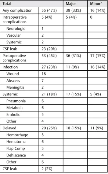
The cohort’s complication profile is presented in►Table 1. Major intraoperative complications occurred infive (4%) cases and included two systemic complications (one case of intra-operative asystole and one case of profound hypotension). One vascular complication occurred when excessive intraoperative hemorrhage required vascular and interventional radiology. Two neurological complications occurred causing transient neurological injury (one case in which durotomies were created by a separate surgical team during a cranioplasty and one case in which hemineglect was discovered postoper-atively). No predictors of intraoperative complications or CSF leaks were found; however, smoking approached significance with intraoperative complications (8 vs. 0%;p¼0.06).
Postoperative complications occurred in 53 (45%) cases, of which 36 (31%) were major complications. Infection was the most common postoperative complication and occurred after 27 (23%) cases and included 18 superficial wound infections (1 resulting in permanent neurological damage), 7 postoperative abscesses (1 resulting in transient neurological damage), and 2 cases of meningitis. One case of postoperative meningitis resulted in permanent neurological injury and subsequently death of a patient already in hospice care. Postoperative delayed wound complications occurred in 29 (25%) cases with hemorrhage (8) and hematoma (6) the most common wound complications. Two cases of postoperative delayed complications resulted in transient neurological deficits. Post-operative systemic complications occurred in 21 (18%) cases with pneumonia (6) and metabolic disturbances (6) as the most common complications. No deaths resulted from post-operative systemic complications. Two CSF leaks occurred postoperatively (one highflow and one lowflow).
Complications stratified by surgical approach are pre-sented in ►Table 2. Intraoperative, all-type postoperative and postoperative infectious, systemic, and delayed compli-cation were all found to have significantly different rates between approach types (p<0.005). Differences in all type postoperative complications between approach types ap-proached significance (p¼0.053). More postoperative com-plications occurred after infratemporal fossa (48%) and CFR (54%) approaches compared with orbitozygomatic (20%) and transfrontal (27%). In all classes of complications, “other” approaches had higher rates of complication. A separate linear regression model explored differences in complica-tion rates between subtypes of ITF approach noted
in►Table 2. No significant differences were observed.
Ma-lignancy, fat and free grafting, age, and obesity were not associated with open-approach complications (p> 0.05)
(►Table 3). The need forflap reconstruction was signifi
cant-ly associated with all-type postoperative complications (55 vs. 34%; odds ratio [OR]¼2.27; 95% confidence interval [CI]: 1.03–5.04) and postoperative systemic complications (26 vs. 8%; OR¼3.98; 95% CI: 1.34–11.86). Flap reconstruction also resulted in higher rates of delayed postoperative complica-tions (32 vs. 16%; OR¼2.08; 95% CI: 0.73–5.93). Smoking trended toward higher intraoperative complication rates (8 vs. 0%;p¼0.06) and postoperative systemic complications (23 vs. 11%; OR¼2.4; 95% CI: 0.86–6.74). Prior radiation
Table 1 Open surgeries resulting in complications
Total Major Minora
Any complication 55 (47%) 39 (33%) 16 (14%)
Intraoperative complications
5 (4%) 5 (4%) 0
Neurologic 1
Vascular 2
Systemic 2
CSF leak 23 (20%)
Postoperative complications
53 (45%) 36 (31%) 17 (15%)
Infection 27 (23%) 11 (9%) 16 (14%)
Wound 18
Abscess 7
Meningitis 2
Systemic 21 (18%) 17 (15%) 5 (4%)
Pneumonia 6
Metabolic 6
Embolic 5
Other 4
Delayed 29 (25%) 18 (15%) 11 (9%)
Hemorrhage 8
Hematoma 6
Flap Comp 5
Dehiscence 4
Other 6
CSF leak 2 (2%)
Abbreviation: CSF, cerebrospinalfluid; Flap Comp,flap related complications. aSurgeries resulting in any major complication were only counted as such
regardless of concurrent minor complications.
Table 2 Open and endoscopic complication rates
Open Endo
Intraoperative complications 5 (4%) 6 (2%)
CSF leak 23 (20%) 133 (40%)
Postoperative complications 53 (45%) 41 (12%)
Infection 27 (23%) 9 (3%)
Systemic 21 (18%) 20 (6%)
Delayed 29 (25%) 12 (4%)
Abbreviation: CSF, cerebro spinalfluid.
Table 3 A ss o ci at io n s o f o p e ra ti ve var ia b le s an d o p e n sk u ll b as e co m p lica ti o n s In tr a o p e ra ti ve Po st o p er at iv e A n y O R (95 % C I) A n y O R (9 5 % C I) In fe ct io n O R (9 5 % C I) Sys te mi c O R (9 5 % C I) D e laye d O R (95 % C I) A g e (a vg) 53 1.03 (0.74 – 1.45) 0.99 (0.90 – 1.10) 45 0.91 (0.80 – 1.03) 53 1.04 (0.90 – 1.20) 50 0.99 (0.88 – 1.12) BM I (av g) 25.5 0 .84 (0.51 – 1.41) 27. 5 0 .90 (0.75 – 1.09) 26.3 0 .95 (0.83 – 1.09) 28.4 1 .00 (0.90 – 1.11) 26.3 0 .82 (0.61 – 1.12) Tu m o r Be n ig n 1 (2%) 1 .75 (0.22 – 14.1) 19 (39%) 1 .75 (0.79 – 3.88) 10 (20%) 1 .41 (0.54 – 3.68) 7 (14%) 1 .69 (0.63 – 4.55) 11 (22%) 1 .48 (0.89 – 2.48) M a ligna n t 4 (6%) 34 (51%) 1 7 (25%) 1 4 (21%) 1 8 (27%) Sm o ker No 0 b p ¼ 0.06 c 22 (42%) 1 .28 (0.61 – 2.67) 13 (25%) 0 .76 (0.31 – 1.82) 6 (11%) b 2.4 (0.86 – 6.74) 12 (23%) 1 .01 (0.41 – 2.51) Ye s 5 (8 % ) b 30 (48%) 1 4 (23%) 1 4 (23%) b 17 (27%) Pr io r ra d ia ti o n N o 3 (4%) 1 .13 (0.18 – 7.33) 32 (41%) 2 .02 (0.87 – 4.75) 14 (18%) b 2.65 (0.89 – 7.88) 13 (17%) 1 .44 (0.56 – 3.71) 18 (23%) 1 .40 (0.74 – 2.66) Y e s 2 (5%) 21 (57%) 1 3 (35%) b 8 (22%) 1 1 (30%) Fa t g ra ft N o 4 (4%) 3 .18 (0.30 – 34.22) 49 (45%) 1 .33 (0.35 – 5.14) 25 (23%) 1 .22 (0.25 – 5.81) 19 (17%) 1 .59 (0.28 – 8.98) 28 (26%) 2 .76 (0.21 – 35.5) Y e s 1 (13%) 4 (50%) 2 (25%) 2 (25%) 1 (13%) Fr e e g ra ft N o 4 (4%) 2 .21 (0.21 – 23.0) 48 (45%) 1 .00 (0.29 – 3.40) 25 (24%) 0 .76 (0.14 – 4.13) 20 (19%) 0 .42 (0.05 – 3.55) 27 (26%) 0 .77 (0.31 – 1.91) Y e s 1 (9%) 5 (46%) 2 (18%) 1 (9 %) 2 (18%) Fl
ap No
1 (2%) 2 .22 (0.42 – 11.7) 17 (34%) a 2.27 (1.03 – 5.04) 8 (16%) 2 .01 (0.65 – 6.20) 4 (8%) a 3.98 (1.34 – 11.86) 8 (16%) 2 .08 (0.73 – 5.93) Y e s 4 (6%) 36 (55%) a 19 (29%) 1 7 (26%) a 21 (32%) Ab br ev ia ti on s: Avg , ave ra g e; B M I, b od y m as s in d e x (k g/ m 2); C I, con fi de nc e in te rva l; OR , o dd s ra tio . ap < 0. 05 . bp < 0. 1. cFi sh er e xa ct tes t.
trended toward higher postoperative infections (35 vs. 18%; OR¼2.65; 95% CI: 0.89–7.88).
Discussion
The open approach to the skull base has been significantly refined since itsfirst description by Ketcham et al in 1963.7 Advances in imaging, surgical tools, techniques, and recon-struction initially increased the scope and safety of the open approach. Ketcham et al8reported a 54% overall complication rate in the author’sfirst 89 patients. Nine years later, Kraus et al10and Janecka et al19described 39 and 33% overall compli-cation rates in 85 and 183 patients, respectively. In 2000, Solero et al20described a 30% major complication rate in 168 patients who underwent combined craniofacial resection.
Although the aforementioned authors have noted a steady trend toward lower complication rates in open skull base surgery in the past,20,21our study demonstrated an overall major complication rate (33%) comparable to that of series prior to the year 2000. In our study, we observed a 9% major and 14% minor postoperative infection rate and 15% major and 9% minor delayed wound complication rate. Prior to the rapid growth of endoscopic skull base surgery between 1985 and 2000, the reported infectious complications of open surgery for benign and malignant skull base lesions ranged from 4 to 34%.8,13,20,22The International Collaborative Study Group23found a 20% major wound complication rate in 1193 patients undergoing open skull base surgery from 1970 to 2000 when including infections. The author’s data suggest that current infection and delayed wound complication rates are comparable to those prior to the endoscopic era.
Systemic complications occurred after 21 surgeries (18%) in our study. A significant portion of our systemic complica-tions were classified as major (81%) and were largely driven by postoperative pneumonia, metabolic disturbances, and embolic events (►Table 1). Metabolic disturbances included five cases of renal insufficiency and one case of diabetes insipidus. Embolic events included two deep-vein thrombi, one upper extremity venous thrombosis, and one case of postoperative pulmonary embolus. Additional causes of sys-temic complication included two cases of recurrent gastro-stomy tube malfunction requiring hospitalization, peripheral nerve neuropathy from intraoperative positioning, and one episode of asystole during hospitalization. Contrastingly, reported systemic complication rates for the open resection of benign and malignant skull base lesions from 1985 to 2000 ranged from 6 to 12%, with a mean of 8%.10,13,19Additionally, the International Collaborative Study Group23found only a 5% systemic complication rate, suggesting an increase in the systemic complication rate of our cohort compared with those reported on prior to the year 2000.
The International Collaborative Study Group created the largest open skull base cohort to date which comprised surgeries performed prior to the advent of the endoscopic era.11,23While the cohort contained only malignant tumors, it comprised individual series from prior studies that investi-gated both benign (9–26%) and malignant (65–91%) disease processes with similar complication profiles to those of the
collaborative cohort.8,13,20,22,24,25 Similar to our study, the most common histologies were squamous cell carcinoma (32%) and adenocarcinoma (15%). The collaborative cohort comprised 47% stage IV and 10% stage III tumors,11while the malignant tumors in our cohort were 92% stage IV and 3% stage III disease. Additionally, 42% of malignancies in our study required preoperative head and neck radiation, while only 28% of surgeries were performed after head and neck radiation in the International Collaborative Study.11
Larger resections have long been known to predispose to higher complications as early studies found that involvement of both the anterior fossa and temporal regions is the largest predictor of postoperative complication and that surgical involvement of more than one skull base site is significantly associated with postoperative complications.24,26Zada et al27 recently postulated that the few remaining indications for pursuing an open approach to the skull base include lesions necessitating larger resections of more morbid disease, with greater intracranial extension, morefirm tumor consistency, and increased proximity or encasement of vital structures.
Additionally, prolonged operative time from the resec-tion of more extensive and complicated disease has been associated with increased systemic complications second-ary to operative physiologic stress.28,29 Patients with higher-stage disease and more extensive resection also frequently require more advanced reconstruction and in-herently have more comorbidities and prolonged hospital stays, which may have contributed to our relatively in-creased systemic complications. Our patients underwent flap reconstruction in 66 (56%) cases, and our analysis did reveal a significant association with flap reconstruction and postoperative systemic complications. Many of our systemic complications such as pneumonia, deep venous thrombosis, and pulmonary embolism are often associated with prolonged hospital stays,30 which are frequently necessitated after flaps or other complex reconstruc-tions.31 Subsequently, our study likely found relatively increased systemic complications, unchanged rates of in-fection, and delayed complications because the open ap-proach was reserved for the largest, most complex lesions, which are inherently associated with increased morbidity. This increased morbidity emphasizes the current selection bias when comparing open and endoscopic series. We com-pared the complication profiles of this provider’s endoscopic and open skull base practices, and agreeing with previous studies, the open cohort experienced higher rates of all-type postoperative complications (45 vs. 12%). Endoscopic surgery comprised 74% of this provider’s skull base practice. Malig-nancies comprised only 24% of endoscopic cases and 86% of these tumors were clinically staged as T4. Open approaches had higher rates of postoperative infection (23 vs. 3%), systemic complications (18 vs. 6%), and delayed wound complications (25 vs. 4%). Open and endoscopic approaches resulted in similar rates of intraoperative complications (4 vs. 2%, respectively). Rates of endoscopic and open complications are compared in ►Table 4. Although, as previously men-tioned, significant selection bias is present in such compar-isons, continued comparisons remain justified as the territory
of endoscopic skull base surgery expands and surgeons face more frequent decisions between endoscopic or open approaches.
Some authors advocate initial attempts at endoscopic approaches even in the setting of historically relative contra-indications.27Subsequently, the endoscopic era has added a new paradigm to preoperative decision-making and counsel-ing, highlighting the importance of exploring predictors of open complications. As discussed, our analysis didfind an association betweenflap reconstruction and postoperative complications as freeflap reconstruction is commonly used for only the largest soft tissue defects.23We also explored age, malignancy, obesity, smoking status, and prior radiation because these factors are readily known preoperatively, thus could aid in risk stratification before recommending open surgery to patients. Our data suggest that smokers may have a higher risk of intraoperative and postoperative com-plications after open skull base surgery. Patients with prior head and neck radiation may also have a higher risk of postoperative wound infections.
Given the significant evident increase in complication rates of undergoing an open approach to the anterior skull base, it is important to characterize complication profiles of popular open approaches. As noted in►Table 2, we didfind signifi -cantly different complication profiles between approach types. Orbitozygomatic and transfrontal approaches had less all-type postoperative, postoperative infectious, systemic, and delayed complications than ITF and CFR approaches. This was despite the fact that ITF and CFR approaches were utilized approxi-mately twice as often.“Other”approaches had significantly higher complication rates. This may be secondary to lesser frequency of use and subsequent lower experience with these respective approaches, including transoral, transplanar, cervi-cal, and temporal bone resections. Given the frequency at which the ITF approach was used, we felt it useful to explore any differences in complication rates between ITF approach subtypes (transparotid, transmaxillary, and mandibulectomy/ mandibulotomy), and no significant differences in postopera-tive complications were observed between these subtypes (p¼0.477).
Although our study is retrospective, we performed a continual and consecutive series with the only inclusion criteria being skull base surgery. There is an inherent lack of uniformity when comparing our complications with those of prior studies, which limits our assessments. However, by classifying our complications by the criteria of Kassam et al18 and continuing to do so in future studies, we can improve the standardization of skull base outcomes research and allow for improved inter- and intra- study comparisons. Additionally, surgeries were performed by one surgeon, which may limit the applicability of our results to other institutions. However, our goal was to examine this specific provider’s skull base practice as it is uniquely comprehensive (open and endoscop-ic) and portrays open complications in such a practice.
Based on our experience, we believe that the complication risk of open skull base surgery will initially rise as surgeons increasingly adopt endoscopic techniques. Our practice has attempted to be early adopters of minimally invasive skull base surgery as the landscape has evolved with 74% of skull base surgeries performed endoscopically over 4 years of practice. Subsequently, our data may forecast the complica-tion profiles of other skull base practices that have not yet adopted a similar endoscopic volume. As the feasibility and safety of endoscopic skull base surgery is further established and the approach is adopted for increasing types of lesions that would historically be reserved for open surgery, surgeons will face new decisions between open and endoscopic ap-proaches. Considering the complication profile found in this study, the decision to pursue lesions with an open approach must not be taken lightly and patients should be counseled appropriately. Consequently, future studies are needed to delineate indications for the open approach in the endoscopic era.
Conclusion
The open approach to the skull base was found to have higher rates of postoperative infections, delayed complications, and systemic complications than the endoscopic approach. Flap reconstruction was significantly associated with increased
Table 4 Total open complications by approach
Intra-opa Any POb PO infectiona PO systemica PO delayeda
ITF 4 (10%) 19 (48%) 10 (25%) 9 (23%) 12 (30%)
Transparotid 2 (18%) 4 (36%) 4 (36%) 3 (27%) 3 (27%)
Mandibular 1 (13%) 6 (75%) 3 (38%) 2 (25%) 4 (50%)
Transmaxillary 1 (13%) 3 (38%) 1 (13%) 2 (25%) 2 (25%)
CFR 0 (0%) 19 (54%) 9 (26%) 6 (17%) 10 (29%)
Orbitozygomatic 1 (7%) 3 (20%) 2 (13%) 0 (0%) 1 (7%)
Transfrontal 0 (0%) 4 (27%) 2 (13%) 1 (7%) 2 (13%)
Other 0 (0%) 8 (67%) 4 (33%) 5 (42%) 4 (33%)
Abbreviations: PO, postoperative; ITF, infratemporal fossa; CFR, craniofacial resection. Note: Logistic regression models explored complications between the ITF approaches. ap<0.05.
bp<0.06.
complications after open skull base surgery, while smoking and prior head and neck radiation trended toward increased complications after open surgery. Our study found higher rates of complications in our open skull base cohort than those reported in studies prior to the advent of the endo-scopic era. Our patients appeared to have, on average, higher-stage disease than patients in prior open-approach studies, suggesting that the selection bias from selecting only the most complicated cases for open surgery may modestly increase the complication profile of the open approach in the endoscopic era. Finally, we recommend that patients’and physicians’expectations be adjusted accordingly when pur-suing an open approach to the skull base.
Conflict of Interest
None.
Note
This study was performed at the University of North Carolina Hospitals, Chapel Hill, NC.
Acknowledgments
We would like to acknowledge the University of North Carolina Translational and Clinical Sciences Institute for their help with study design and statistics.
References
1 Kassam A, Snyderman CH, Mintz A, Gardner P, Carrau RL. Expand-ed endonasal approach: the rostrocaudal axis. Part II. Posterior clinoids to the foramen magnum. Neurosurg Focus 2005;19(1):E4 2 Kassam A, Snyderman CH, Mintz A, Gardner P, Carrau RL. Expand-ed endonasal approach: the rostrocaudal axis. Part I. Crista galli to the sella turcica. Neurosurg Focus 2005;19(1):E3
3 Kassam AB, Gardner P, Snyderman C, Mintz A, Carrau R. Expanded endonasal approach: fully endoscopic, completely transnasal ap-proach to the middle third of the clivus, petrous bone, middle cranial fossa, and infratemporal fossa. Neurosurg Focus 2005;19(1):E6 4 Kassam AB, Snyderman C, Gardner P, Carrau R, Spiro R. The
expanded endonasal approach: a fully endoscopic transnasal approach and resection of the odontoid process: technical case report. Neurosurgery 2005;57(1, Suppl):E213, discussion E213 5 Kassam AB, Thomas AJ, Zimmer LA, et al. Expanded endonasal
approach: a fully endoscopic completely transnasal resection of a skull base arteriovenous malformation. Childs Nerv Syst 2007; 23(5):491–498
6 Kassam AB, Vescan AD, Carrau RL, et al. Expanded endonasal approach: vidian canal as a landmark to the petrous internal carotid artery. J Neurosurg 2008;108(1):177–183
7 Ketcham AS, Wilkins RH, Vanburen JM, Smith RR. A combined intracranial facial approach to the paranasal sinuses. Am J Surg 1963;106:698–703
8 Ketcham AS, Van Buren JM. Tumors of the paranasal sinuses: a therapeutic challenge. Am J Surg 1985;150(4):406–413 9 Richtsmeier WJ, Briggs RJ, Koch WM, et al. Complications and early
outcome of anterior craniofacial resection. Arch Otolaryngol Head Neck Surg 1992;118(9):913–917
10 Kraus DH, Shah JP, Arbit E, Galicich JH, Strong EW. Complications of craniofacial resection for tumors involving the anterior skull base. Head Neck 1994;16(4):307–312
11 Patel SG, Singh B, Polluri A, et al. Craniofacial surgery for malignant skull base tumors: report of an international collaborative study. Cancer 2003;98(6):1179–1187
12 Eloy JA, Vivero RJ, Hoang K, et al. Comparison of transnasal endoscopic and open craniofacial resection for malignant tumors of the anterior skull base. Laryngoscope 2009;119(5):834–840 13 Dias FL, Sá GM, Kligerman J, et al. Complications of anterior
craniofacial resection. Head Neck 1999;21(1):12–20
14 Germani RM, Vivero R, Herzallah IR, Casiano RR. Endoscopic reconstruction of large anterior skull base defects using acellular dermal allograft. Am J Rhinol 2007;21(5):615–618
15 Patel MR, Taylor RJ, Hackman TG, et al. Beyond the nasoseptalflap: outcomes and pearls with secondaryflaps in endoscopic endo-nasal skull base reconstruction. Laryngoscope 2014;124(4): 846–852
16 Greene FL, Page DL, Fleming ID, et al. AJCC Cancer Staging Manual. 6th ed. New York: Springer; 2002
17 Edge SB, Compton CC. The American Joint Committee on Cancer: the 7th edition of the AJCC cancer staging manual and the future of TNM. Ann Surg Oncol 2010;17(6):1471–1474
18 Kassam AB, Prevedello DM, Carrau RL, et al. Endoscopic endonasal skull base surgery: analysis of complications in the authors’initial 800 patients. J Neurosurg 2011;114(6):1544–1568
19 Janecka IP, Sen C, Sekhar LN, et al. Cranial base surgery: results in 183 patients (Reprinted from Otolaryngology-Head Neck-Sur-gery). J Neurooncol 1994;20(3):281–289
20 Solero CL, DiMeco F, Sampath P, et al. Combined anterior craniofa-cial resection for tumors involving the cribriform plate: early postoperative complications and technical considerations. Neuro-surgery 2000;47(6):1296–1304, discussion 1304–1305
21 O’Malley BW Jr, Janecka IP. Evolution of outcomes in cranial base surgery. Semin Surg Oncol 1995;11(3):221–227
22 Donald PJ. Complications in skull base surgery for malignancy. Laryngoscope 1999;109(12):1959–1966
23 Ganly I, Patel SG, Singh B, et al. Complications of craniofacial resection for malignant tumors of the skull base: report of an International Collaborative Study. Head Neck 2005;27(6): 445–451
24 Irish JC, Gullane PJ, Gentili F, et al. Tumors of the skull base: outcome and survival analysis of 77 cases. Head Neck 1994;16(1):3–10 25 Janecka IP, Sen C, Sekhar LN, et al. Cranial base surgery: results in
183 patients. Otolaryngol Head Neck Surg 1994;110(6): 539–546
26 Wornom IL III, Neifeld JP, Mehrhof AI Jr, Young HF, Chinchilli VM. Closure of craniofacial defects after cancer resection. Am J Surg 1991;162(4):408–411
27 Zada G, Du R, Laws ER Jr. Defining the“edge of the envelope”: patient selection in treating complex sellar-based neoplasms via transsphenoidal versus open craniotomy. J Neurosurg 2011; 114(2):286–300
28 Girod DA, McCulloch TM, Tsue TT, Weymuller EA Jr. Risk factors for complications in clean-contaminated head and neck surgical procedures. Head Neck 1995;17(1):7–13
29 Ryan MW, Hochman M. Length of stay after freeflap reconstruc-tion of the head and neck. Laryngoscope 2000;110(2, Pt 1):210–216
30 Heyland DK, Cook DJ, Griffith L, Keenan SP, Brun-Buisson C; The Canadian Critical Trials Group. The attributable morbidity and mortality of ventilator-associated pneumonia in the critically ill patient. Am J Respir Crit Care Med 1999;159(4, Pt 1):1249–1256 31 Kroll SS, Evans GR, Goldberg D, et al. A comparison of resource costs for head and neck reconstruction with free and pectoralis majorflaps. Plast Reconstr Surg 1997;99(5):1282–1286