PATHOGENESIS AND MECHANISMS OF ETHANOL-INDUCED LIMB DEFECTS
Corey S. Johnson
A dissertation submitted to the faculty of the University of North Carolina at
Chapel Hill in partial fulfillment of the requirements for the degree of Doctor of Philosophy in the Department of Cell and Developmental Biology
Chapel Hill 2006
Approved by:
ABSTRACT
Corey S. Johnson: Pathogenesis and mechanisms of ethanol-induced limb defects (Under the direction of Kathleen K. Sulik, Ph.D.)
Fetal alcohol spectrum disorders encompass a variety of structural and behavioral
abnormalities attributable to maternal alcohol consumption. This study was designed to investigate the mechanistic and pathogenic role of retinoic acid (RA) in the genesis of ethanol-induced forelimb defects. The mouse limb bud has proven to be an ideal model with which to study effects of ethanol because its development is more thoroughly understood than other regions or organ systems. The hypothesis that RA-deficient mouse embryos/fetuses manifest similar limb defects and distal limb cell death patterns to those of ethanol treated dams was tested. A RA receptor (RAR) antagonist and an aldehyde dehydrogenase (ALDH) inhibitor produced limb malformations consistent with those following ethanol exposure. Similarly, cell death was observed in the same region of the limb following exposure to each chemical. Secondly, the hypothesis that exogenous RA can prevent ethanol-induced cell death in the limb was tested. A subteratogenic dose of RA was co-administered with ethanol. Limb buds exposed to ethanol and RA exhibited low levels of distal limb cell death, comparable to control limbs, demonstrating that RA acts antagonistically to ethanol in the limb and suggesting that ethanol interferes with RA-mediated development. Also, in support of this premise, the results of in situ hybridization analysis reveal that ethanol represses RA-dependant gene
expression in the limb at 8 and 18 hours, post treatment. Importantly, however, microarray
iii
ACKNOWLEDGEMENTS
I extend my deepest gratitude to my dissertation advisor, Dr. Kathleen K. Sulik for her persistence, patience, and commitment to proper training. She was particularly determined to see improvement in my writing skills. If not for her dedication to my development in this regard, the body of work presented herein would be unreadable. She has shown great generosity in allowing me to pursue my teaching aspirations during my tenure under her direction, underscoring her commitment to training well-rounded scientists. Lastly, I would like to express my gratefulness for her model of
professionalism. Dr. Sulik has always conducted herself in the most appropriate manner, whether presenting research, teaching, writing, or managing temperamental students. Her attitude and thoughtfulness towards the execution of her trade is worthy of emulation.
I would like to thank Dr. E. Sydney Hunter III for his mentorship. Under Sid’s direction, I was able to conduct much of my research at the EPA and benefit from the great collection of minds such as Dr. Mitch Rosen, Dr. John Rogers, Judy Schmid, and Carmen Wood. Most of all, I am grateful for Sid’s intelligence and willingness to communicate his ideas. Though we often exchanged ideas, it was always enjoyable to sit back and listen to Sid discuss his latest schemes. Since I spent most of my time under Sid’s watchful eye, it was through him that I learned the most about conducting research, designing experiments, and evaluating results. I’ve learned so much under Sid’s direction in the past 7 years about conducting research that I bet I could do it over in 3 years!
v
I also wish to thank two members of the Department of Biology, Dr. William Kier and Dr. Albert Harris. Bill Kier has given me employment, and for that I am grateful. He also was influential in my desire to teach. While auditing his comparative physiology course I learned an appreciation for the craft of teaching, and the importance of being prepared and thoughtful in designing lectures. Albert Harris has been an absolute inspiration to me. The fervor that he has for his subject and for science, in general, has influenced and driven me to continue my studies and remain passionate about my research and my teaching. I have greatly enjoyed my conversations with him and thank him for his friendship and mentorship. Both of these outstanding scientists have greatly contributed, however indirectly, to the completion of this work.
TABLE OF CONTENTS
List of tables………..………...………...……….………....…... viii
List of figures………...……….……….………... ix
List of abbreviations………....x
CHAPTER I. BACKGROUND 1.1 A brief history of Fetal Alcohol Syndrome and Fetal Alcohol Spectrum Disorders………..………...…………...1
1.2 Mouse models of FASD: ethanol’s dysmorphogenesis…..…….….………..…...3
1.3 Cellular effects of embryonic ethanol exposure……..………...…….…...5
1.3.1 Ethanol’s mechanism of action………..……...…………....7
1.4 Early development of the limb..……..……….………….……….………...11
1.5 The components of Retinoic acid synthesis, degradation, and signaling, with particular reference to the limb …….………..…….……..…..……...14
1.6 Hypothesis: embryonic RA-mediated development is impacted by ethanol………..…………..…..………..…....18
1.7 Specific aims………...…….………..……...…...19
II. PERTURBATION OF RETINOIC ACID (RA)-MEDIATED LIMB DEVELOPMENT SUGGESTS A ROLE FOR DIMINISHED RA SIGNALING IN ETHANOL’S TERATOGENESIS. 2.1 Abstract………..…….……...…………..………....21
2.2 Introduction………...….…………..………....………....22
2.3 Materials and Methods………..……..………....26
vii
III. A MICROARRAY ANALYSIS EXPLORING ETHANOL’S TERATOGENESIS IN THE MOUSE FORELIMB
3.1 Abstract ………...…….……..…...………..………...39
3.2 Introduction………..……….…...……..…………...………....40
3.3 Materials and Methods…...………..…...……...……….………....44
3.4 Results………...………....………...………...47
3.5 Discussion………..…...………..………....52
IV. CONCLUSIONS.………..………..…………..…..…….………....62
LIST OF TABLES
Table
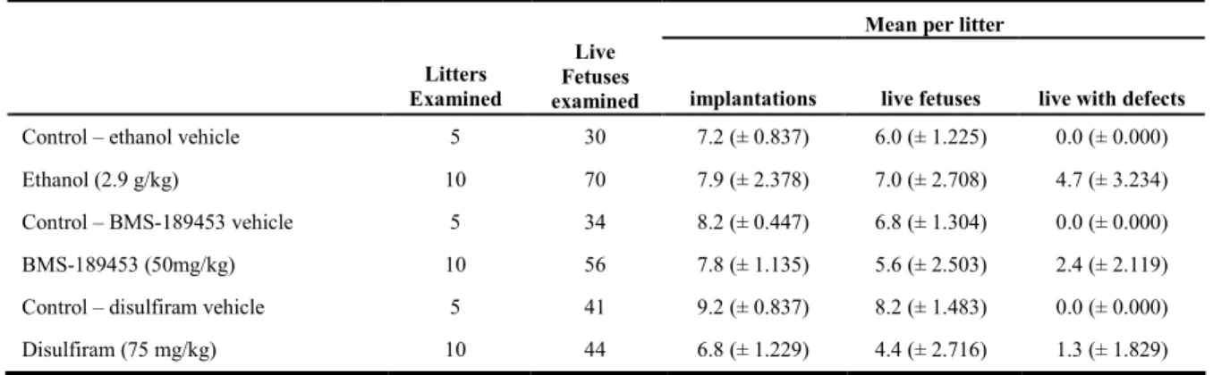
2.1 Summary of the effects of ethanol, BMS-189453, and disulfiram
on implantation, and forelimb development…….………...…41 3.1 Network analysis of significant transcripts found in ethanol-
exposed forelimb buds……….………70 3.2 Functional analysis of significant transcripts found in ethanol-
exposed forelimb buds……….………....……71 3.3 Gene ontology analysis of significant transcripts in forelimb
buds after 4 hours of ethanol exposure….……….………..……72 3.4 Canonical pathways affected in the forelimb by 2, 4, or 6 hours
following ethanol exposure……….…73
3.5 Correlation of significant transcripts in BMS-189453 and
ethanol-exposed forelimb buds ……….………..…74 3.6 Genes involved in (a) redox balance or (b) NF-IB activity
ix
LIST OF FIGURES
Figure
1.1 Diagram of developmental regulators of forelimb initiation,
outgrowth, and patterning……….……..…...…..…………21 1.2 The synthesis and signaling of retinoic acid……….………....………..……22 2.1 Skeletal dysmorphology resulting from maternal ethanol,
BMS-189453, and disulfiram treatment……….…….….……….…...…...42 2.2 NBS staining of ethanol, BMS-189453, and disulfiram exposed embryos…...…..…43 2.3 LysoTracker Red staining of embryos demonstrating the prevention
of ethanol's effects with exogenous retinoic acid……….…..…...…..44 2.4 In situ hybridization demonstrating ethanol-induced changes
in developmentally significant genes……….……...…..45 2.5 Quantitative real-time PCR analysis of transcriptional changes in
forelimb buds of ethanol and BMS-189453 exposed embryos…………...……46 3.1 Hierarchial clustering analysis of significant transcripts in forelimb
buds exposed to ethanol for 2, 4, and 6 hours….………..….….……76 3.2 Network analysis results demonstrating the connections between
ethanol affected genes in the largest network……….….…..…….77 3.3 Tukey post-hoc analysis. Pair-wise comparisons of PBS
control and ethanol treatment groups……….………….….…..….78 3.4 Hierarchial clustering analysis of significant transcripts in forelimb
buds exposed to ethanol for 2, 4, and 6 hours or BMS-189453….……….…...…….79 3.5 Tukey post-hoc analysis. Pair-wise comparisons of ethanol
LIST OF ABBREVIATIONS
ADH Alcohol dehydrogenase AER Apical ectodermal ridge ALDH Aldehyde dehydrogenase
AP Anterio-posterior
IGF Insulin-like growth factor FAS Fetal Alcohol Syndrome
FASD Fetal Alcohol Spectrum Disorders
GD Gestational day
GO Gene Ontology
IPA Ingenuity Pathway Analysis
IPKB Ingenuity Pathway Knowledge Base
PG Prostaglandins
LPM Lateral plate mesoderm
LTR LysoTracker Red
NBS Nile Blue Sulfate
qPCR Quantitative, real-time PCR
PD Proximo-distal
CHAPTER I Background
Section 1.1 A brief history of Fetal Alcohol Syndrome and Fetal Alcohol Spectrum Disorders
The first written record cautioning against maternal alcohol consumption comes from the Bible (Judges 13:7), wherein the pregnant mother of Samson was commanded to abstain from “wine and strong drink.” Aristotle, perhaps the first embryologist, suggested a connection between maternal alcohol consumption and the “morose and languid” nature of their children in the 4thcentury BC
(reviewed by Warner and Rosett 1975). During the so-called gin epidemic of the mid-1700s in England, when the consumption of gin rose to approximately 1 gallon per capita per year, concerns rose over the possible detriment of alcohol to the developing fetus (Mitchell and Deane, 1962; Abel, 2001). Not until the late 1800s would the anecdotal observations of physicians be subject to scientific inquiry.
In 1848, Samuel Howe presented the first epidemiological evidence that ethanol consumption may be a detriment to embryological development (Warner and Rosett 1975). Later, Sullivan (1899) published a report indicating that alcoholic women incarcerated during pregnancy had lower rates of miscarriage, stillbirths, and children with epilepsy than alcoholic women who were not incarcerated. Although these studies indicated the negative effects of ethanol consumption during pregnancy, the public remained largely ignorant of their importance.
Concurrently, experimental evidence began to contribute to the knowledge of the embryo-toxic effects of ethanol. Fere (1893) exposed chicken embryos to ethanol in ovo, noting its
result of alcohol teratogenesis were perhaps overstated, it brought to the public an awareness of the potential health risk of ethanol consumption during pregnancy. Pearl, like Fere before him, found that ethanol had deleterious effects on chick embryos (Pearl 1917). Pearl, and later Stockard considered alcohol a beneficial “selective agent” for the human race.
Not all evidence supported the hypothesis that ethanol was dysmorphogenic. The widely influential epidemiological study of Elderton and Pearson (1910) found no correlation between maternal alcohol consumption and birth defects. This study later gained support from F.B. Hanson, whose meticulous and exhaustive studies on the effects of alcohol on rats found no detrimental effects of ethanol whatsoever (reviewed in Pauly 1996), leaving the question of ethanol’s teratogenicity unanswered.
Finally, after a long period of relatively little research on ethanol’s effects on reproduction and development, Lemoine et al. (1968), having described over 100 individuals, took notice of the common features of children known to have been exposed to ethanol in utero. Although the work of Lemoine et al., was at first met with hesitation, the scientific and medical communities were soon impressed with similar observations by Jones and Smith, leaving unanimity in the affirmation of alcohol’s teratogenicity (Jones and Smith 1973; Jones et al., 1973; 1974; reviewed in Armstrong et al., 1998). By 1980, The Research Society on Alcoholism recommended clinical guidelines for diagnosis of Fetal Alcohol Syndrome (FAS; Rosett 1980), and in 1981 the US Surgeon General issued an advisory warning of the dangers of maternal alcohol consumption. The guidelines for FAS identification were recently updated by the Centers for Disease Control and Prevention (CDC, 2005). FAS consists of craniofacial abnormalities, central nervous system deficiencies, and prenatal and/or postnatal growth retardation (Rosett 1980).
3
related nerurodevelopmental disorders (ARND). Neural structures commonly affected by ethanol include the basal ganglia, corpus callosum, cerebellum, and hippocampus (reviewed in Mattson et al., 2001). Cognitive deficits involving attention, learning, memory, language, motor, and visuo-spatial abilities are also present. Such cognitive disorders likely have a structural basis, yet definitive evidence is currently unavailable (reviewed in Mattson and Riley 1998). All known
ethanol-associated birth defects, structural and behavioral, have been given the umbrella classification, Fetal Alcohol Spectrum Disorders (FASD). Sampson et al. (1997) report that the incidence of FAS in the United States is 2-5 per 1000 births. FASD is suggested to be about 3 times as prevalent as FAS according to the Centers for Disease Control and Prevention.
Embryonic ethanol exposure is known to result in a variety of defects of the heart, ears, eyes, joints, and limbs (Abel 1984). Alcohol-induced limb defects are of particular interest for the current work. Limb defects have often been reported in conjunction with FAS (Spiegel et al., 1979; Jaffer et al., 1981; Cremin and Jaffer 1981; Viljoen et al., 2005). Froster and Baird (1992) indicated that the incidence of limb defects attributable to “severe maternal alcohol problems” is 1.6 per 10,000 live births in a study in British Columbia. These limb defects preferentially affect the right arm and hand. Herrmann and colleagues (1980) have described a number of patients with alcohol-induced limb defects including ectrodactyly, shortened or missing metacarpals and metatarsals, digit hypoplasia, unusually large gaps between digits, camptodactyly, clinocamptodactyly, soft tissue syndactyly, branched digits, and club foot. Others have described amelia (van Rensburg, 1981; Aro, 1983; Pauli and Feldman, 1986). Less dramatic defects of the limb, such as abnormal palmar creases and radioulnar synostosis, are often found in conjunction with FAS (Jones et al., 1973; Speigel 1979).
Section 1.2 Mouse models of FASD: ethanol’s dysmorphogenesis
dams were fed an ethanol-containing diet throughout their pregnancy. Offspring of these mice exhibited craniofacial and heart abnormalities bearing much similarity to ethanol-induced defects seen in humans. Acute ethanol exposure of mouse embryos in utero has also been used with particular success to achieve developmental stage-specific defects (Draft et al., 1996; Sulik 1985; Sulik et al., 1981; Kronick 1976). For the study of ethanol’s action on the brain, the ‘Pup in a Cup’ model has been useful. This model allows the study of ethanol exposure to postnatal rodents during a period of time analogous to the third trimester in humans. The use of these various models allows an in depth evaluation of the factors leading to dysmorphogenesis throughout the course of development; variables including dose, length of exposure, diet and nutrition, genetic background, and concomitant drug exposure (West et al., 1994; Abel Hannigan, 1995).
Of the abundant information provided by studies of embryonic ethanol exposure of mice, two pieces of information emerge as especially important to our understanding of the impact of prenatal ethanol exposure in humans. First, genetic background influences the incidence and severity of ethanol-induced malformations. Chernoff (1977) demonstrated that two strains of mice exhibited variable responses to ethanol. One strain had more severe and higher percentages of malformations than another strain. A recent study has shown that genetic variability does, indeed, influence the incidence of FAS in humans (Viljoen et al., 2001), as was predicted by animal studies. Secondly, doses of ethanol administered at different windows in development result in different types of defects, demonstrating specific windows of exposure in which distinct body regions are sensitive to ethanol (West et al., 1994; Coles, 1994; Becker et al., 1996). This observation has allowed investigators to determine the sensitive windows for malformations in humans. For example, exposure to ethanol during gastrulation (day 7 of C57BL/6J mouse development, 3rd week of human development) results
in typical FAS craniofacial characteristics (Webster et al., 1980; Sulik et al., 1981; Sulik and Johnston, 1983). Later, on day 8.5 of mouse development (4th week of human development)
5
(4th – 5th week in humans), limb and urogenital malformations are prevalent following ethanol
exposure (Kronick 1976; Webster et al., 1980; 1983; Gage and Sulik, 1991; Kotch et al., 1992). Exposure of mouse embryos to ethanol at later stages of development primarily affects the developing and maturing CNS, causing cell death in the hippocampus and cerebellum (Barnes and Walker, 1981; Goodlet et al., 1990; Bonthius and West, 1990; Maier et al., 1999). The array of malformations that may occur throughout development cautions alcohol use at any stage of pregnancy.
Regarding models for ethanol-induced limb defects, C57BL/6J mice have been particularly useful. Defects caused by prenatal ethanol exposure in these mice include polydactyly (extra digits), syndactyly (fusion of skeletal and/or soft tissues of the digits), and ectrodactyly (missing digits). High incidences of right-sided, forelimb postaxial ectrodactyly are produced by an acute exposure to ethanol on the 9thday of gestation (Kronick 1976; Webster et al., 1983; Kotch et al., 1992). In such
malformations, severity ranges from the loss of digit 5, to the loss of digits 2-5 and the ulna. Studies by Kotch et al. (1992) have provided information regarding the underlying
pathogenesis of ethanol-induced limb deficiencies. Examination of gestation day (GD) 12 embryos that were exposed to ethanol on the 9th day of gestation illustrated that some forelimbs were missing a
sizable mass of tissue on the posterior aspect of the limb bud, from which the digits are formed. Examination at an earlier time-point demonstrated the presence of excessive amounts of cell death along the apical ectodermal ridge (AER), an embryonic structure involved in limb outgrowth. Kotch et al. (1992) proposed that excessive cell death in the AER accounts for the variety and severity of limb defects seen in the mouse, following embryonic ethanol exposure.
Section 1.3 Cellular effects of embryonic ethanol exposure
One might surmise that the loss of cells over the course of development, such as occurs following cell death, premature differentiation, or cessation of proliferation, contributes substantially to ethanol-induced dysmorphogenesis. The consequences of cell loss may include alterations in inductive influences, patterning, and the processes of growth and migration.
Bannigan and colleagues (1982, 1984) noted that cell death is commonly observed in specific regions of the mouse embryo following ethanol exposure. Sulik et al. (1988) have since observed that the regions particularly sensitive to ethanol-induced cell death, are those that, as a normal part of development, undergo a limited amount of programmed cell death. Craniofacial (Sulik et al., 1988), urinary tract (Gage and Sulik, 1991), and neural (Kotch and Sulik 1992) defects are characterized by the presence of excessive cell death in their primordia following ethanol exposure. It remains to be determined why cell death occurs in these regions in untreated embryos, and it is equally unclear whether the consequences of excessive cell death are strictly morphological.
7
Section 1.3.1 Ethanol’s mechanism of actionThere are a number of existing hypotheses regarding ethanol’s mechanisms of teratogenesis. They can be reduced to two broad categories: maternal effects of ethanol and direct effects of ethanol on the conceptus.
The primary hypotheses concerning the maternal contribution to FASD implicate dysfunction of the placenta or yolk sac. That ethanol has the affect of constricting umbilical vessels in rats, primates, and humans (Jones et al., 1981; Mukberjee and Hodgen 1982; Altura et al., 1983; Savoy-Moore et al., 1989) implicates reduced blood flow as a major contributor. The most obvious consequence of reduced placental blood flow is hypoxia. Though once a popular hypothesis, experimental evidence linking hypoxia to FASD is lacking. However, research indicates that placental transport of amino acids, glucose, vitamins (including folate, thaiamine, and vitamin B6),
and zinc are diminished following ethanol exposure (reviewed in Schenker et al., 1990). Many of these occurances may be a secondary consequence of reduced placental blood flow following ethanol exposure.
There is some discussion in the literature that suggests that ethanol, per se, does not mediate FASD. Rather, ethanol’s metabolite, acetaldehyde, has been suggested as the proximate teratogen (Veghelyi et al., 1978). The major site of the conversion of ethanol to acetaldehyde occurs in the maternal liver; the embryo has relatively little capability to metabolize ethanol. Investigators have shown that acetaldehyde does, in fact, account for some of the defects associated with ethanol’s teratogenesis, although at a much lower incidence than ethanol exposure during the same window of development (Webster et al., 1983). In vitro evidence supports the hypothesis that acetaldehyde is, in part, responsible for ethanol’s teratogenicicty (Campbell and Fantel, 1983).
Beginning with Stockard (1910), researchers have known that ethanol affects
contribution to the effects of ethanol is removed (Brown et al., 1979; Priscott, 1982; Kotch et al., 1996).
Prostaglandins (PG), which are known to be essential in normal development (Persaud 1978; Challis and Patrick 1980), may be elevated following maternal alcohol consumption in several tissue types (Anggard 1983) to the detriment of the embryo. In fact, ethanol-induced growth retardation in mice can be prevented with the application of PG synthesis inhibitors (Pennington et al., 1985), indicating that PG may contribute significantly to the hypoplastic effects of ethanol. In the developing limb, however, PG synthesis inhibition acts synergistically with ethanol to produce malformations in mice (Padmanabhan and Pallot 1995), suggesting that ethanol’s effects on PG synthesis may be dependant upon the cellular context or other contributing factors.
Several investigators have implicated altered protein synthesis as a mediator of ethanol’s effects. RNA transport and aminoacyl transfer RNA synthases in the embryo are negatively impacted by ethanol (Henderson and Schenker 1977). Additionally, ethanol depresses ribosomal function (Horbach et al., 1989). Some have suggested that ethanol’s affect on protein synthesis is primarily a function of the availability of precursors, protein degradation, or hypothermia (reviewed in Schenker et al., 1990; Henderson et al., 1980).
The observation that Ca2+ levels rise very quickly after exposure of preimplantation embryos
to ethanol (Stachecki and Armant, 1996) has given rise to the hypothesis that ethanol’s effects on other developing systems are mediated by intracellular Ca2+ signaling (Debelak-Kragtorp et al., 2003).
Calcium-mediated stimulation of phospholipase-C (PLC) signaling has been shown to account for ethanol-induced cell death in chick neural crest (Debelak-Kragtorp et al., 2003). Because of the large number of signaling pathways that utilize PLC, the potential targets of ethanol’s action are
innumerable.
9
number of effects, including ethanol’s induction of intracellular Ca2+, altered membrane protein
conformation, decreased cell adhesion, and attenuated migration (reviewed in Schenker et al., 1990). Due to the advancements in the fields of molecular and developmental biology, it has become possible to examine ethanol-induced gene changes in the context of a known developmental system. Importantly, investigators using mouse and chick models have noted that expression of Shh, a major developmental patterning gene is downregulated in response to ethanol exposure (Chrisman et al., 2004; Ahlgren et al., 2002). However, it is not clear whether the decreased expression is the result of ethanol exposure per se, or the secondary result of tissue loss that follows ethanol exposure as described by Kotch et al. (1992). That the effect on Shh expression is primary is suggested by the phenotype of the Shh null mutant mouse, which bears limb and craniofacial characteristics of acute ethanol exposure.
Other genes, including those important to gastrulation, are likewise affected by ethanol exposure. In the frog, Xenopus, expression of Chordin,gsc, and Otx2 is upregulated in response to ethanol, and gastrulation is delayed (Yelin et al., 2005). Murine homologs to these genes are present and important for gastrulation in mouse embryos, although no study has described the response of these genes to ethanol.
A microarray analysis (Dunty 2002) has shown that acute ethanol exposure perturbs a number of significant pathways in the developing brain, including RA, and Wnt signaling pathways. Several other genes involved in the cell cycle-related processes of proliferation, and differentiation and cell death are perturbed following embryonic ethanol exposure (Dunty 2002; Leach et al., 1999; Gu et al., 2001). Though not patterning genes, ethanol-induced perturbation of these cell cycle genes may play an equally important role in dysmorphogenesis, as it is these processes that execute the instructions of developmental patterning. That ethanol affects these essential regulatory pathways and cellular processes is not surprising, given the significant dysmorphogenic effects of ethanol. Gene knock out studies involving components of these pathways have demonstrated their pivotal roles in
pathways, and the potential for significant changes in cellular constituency, experiments designed to examine the targets of ethanol and other teratogens must consider the biological consequences of exposure as close to the time of exposure as is possible.
Two particularly important genes involved in cell cycle processes are epidermal growth factor (EGF) and insulin-like growth factor (IGF). The perturbation of these highly significant, developmentally regulated growth factors has a considerable effect on growth and development (Thesleff et al., 1995; Rother and Accili 2000). Ethanol targets EGF-dependant cell division by depressing EGF-R phosphorylation (Henderson et al., 1989). Impaired IGF signaling has also been demonstrated in response to ethanol (Resnicoff et al., 1993; 1996; Seiler et al., 2000). These results may be interpreted to support the hypothesis that ethanol-induced changes in cell cycle processes are a result of its perturbation of growth factor signaling.
One of the more compelling hypotheses is that oxidative stress accounts for the teratogenicity of ethanol. Molecules that induce oxidative stress are free radicals (superoxide, hydroxyl ion, and nitrogen dioxide radicals) and reactive oxygen species (ROS; hydrogen peroxide, nitric oxide, and peroxynitrate ion). Ethanol metabolism is suspected of inducing oxidative stress through the accumulation of NADH and mitochondrial generation of hydroxyethyl or superoxide radicals (Goodlet et al., 2005). Ethanol also reduces antioxidant levels, which provide cellular defenses against oxidative stresses such as vitamins C and E, glutathione, superoxide dismutase, and retinol (Reyes et al., 1993; Montoliu et al., 1995; Henderson et al., 1995; Addolorato et al., 1997). The unopposed accumulation of oxidative stressors has two effects. First, it results in the oxidation of macromolecules, which may lead to cellular demise. Secondly, ROS act as signaling molecules in certain cellular contexts, mediating growth factor signaling, Ca2+ channel regulation, and gene
11
That oxidative stress may mediate ethanol’s effects is evidenced by the fact that ROS appear after ethanol exposure in embryonic tissue (Chen and Sulik 1996; Johnson et al., 2004; Goodlet et al., 2005), followed by cell death. Furthermore, the incidence of ethanol induced malformations is diminished by the application of antioxidants (Kotch et al., 1996; Chen et al., 2004; the author’s unpublished observations).
Lastly, and of particular relevance to this thesis, is the hypothesis that ethanol-induced malformations result from the inhibition of retinoic acid (RA) synthesis by ethanol. This hypothesis was advanced by Duester (1991) and Pullarkat (1991), who noticed two important pieces of
information. First, ethanol and RA utilize the same enzymes for their metabolism. These investigators proposed that ethanol would competitively inhibit the enzyme(s) necessary for the production of RA. Han et al. (1998) have since identified three alcohol dehydrogenase enzymes responsible for synthesizing RA, and have shown that RA production is diminished by ethanol’s inhibition of these enzymes. Secondly, Duester and Pullarkat also observed that offspring of laboratory animals exposed to ethanol share morphological similarity with animals whos dams were vitamin A deficient during pregnancy (summarized in Zachman and Grummer 1998).
Deltour and colleagues (1996) have since demonstrated that ethanol lowers RA
concentrations in embryos. Because of ethanol’s affect on RA concentrations, and RA’s integral role in development (described below), reduced RA levels would be predicted to result in developmental abnormalities, a premise that warrants examination as a possible factor in ethanol’s teratogenesis.
Section 1.4 Early development of the limb (figure 1.1)
Wnt-2b. It is expressed in the LPM and intermediate mesoderm at the anterio-posterior level of the forelimb field (Kawakami et al., 2001). Another gene, Tbx5, is necessary for forelimb initiation (Ahn et al., 2002) and is dependant on RA for its expression (Mic et al., 2004). Together, Tbx5 and Wnt-2b impart identity and induce forelimb bud initiation by stimulating expression of Fgf10 in the LPM of the limb field (Ng et al., 2002). Fgf10 then induces the overlying ectoderm to express Fgf8
(Ohuchi et al., 1997) by way of Wnt-3a in the chick (Ng et al., 2002), and an unidentified signal in the mouse. Together Fgf8 in the ectoderm and Fgf10 in the mesoderm stimulate each other in a positive feedback loop, promoting the proliferation of limb mesenchyme (Xu et al., 1998; figure 1.1). As proliferation begins in the LPM the first signs of the limb bud appear.
While the above process of initiation ensues, the limb bud divides into two transcriptionally distinct regions. The first is characterized by the proximal expression of Meis genes; Meis1 and Meis2 (Ceccini et al., 1997; Capdevila et al., 1999). This region will develop into the stylopod, that region of the forelimb containing the humerus. RA, synthesized in the presumptive forelimb mesoderm, intermediate mesoderm, and somitic mesoderm prior to limb outgrowth (Swindell et al., 1999; Mic et al., 2002), is the signal that regulates where Meis genes will be expressed (Mercader et al., 2000). The second, meis-negative, region permits expression of characteristically distal gene expression and gives rise to more distal limb structures (Mercader et al.,1999; 2000).
A signal from the distal ectoderm prevents Meis expression and is responsible for the distal, Meis-negative, expression domain. This distalizing signal is Fgf8, which arises from a specialized epithelial structure known as the apical ectodermal ridge (AER) and also its primordium, the distal ectoderm. As described above, Fgf8 is first expressed in the ectoderm of the limb field at the
13
While the proximo-distal (PD) axis is being established, the anterior-posterior (AP) axis begins to emerge. The distal limb is patterned by an organizer found in the posterior mesenchyme (Saunders and Gasseling 1968) known as the zone of polarizing activity (ZPA). The ZPA produces the protein, Shh, which is responsible for the patterning effect of the ZPA (Chiang et al., 2001; Kraus et al., 2001). Shh expression, however, is stimulated by an earlier signal. A proximal source of RA, previously integral to Tbx5 mediated initiation and Meis mediated outgrowth, is next used to stimulate two genes in the posterior half of the limb bud, dHand and Hoxb8 (Lu et al., 1997; Charite et al., 2000 Fernandez-Teran et al., 2000; Mic et al., 2004). Together, dHand and Hoxb8 stimulate Shh expression in the ZPA. The restriction of dHand to the posterior mesenchyme is believed to result from the reciprocal repression between dHand and the Gli3 repressor (Gli3-R). Each of these gene products prevents the expression of the other in their respective posterior and anterior
compartments (Welscher et al., 2002). When Shhexpression increases, it replaces dHand and interacts with Gli3-R in a similar manner. Together, Shh and Gli3-R specify the number of digits produced by the limb as well as the AP identity of those digits.
With the onset of Shh expression and the formation of the SRM on GD 9, the regulation of the AP and PD axes becomes coupled as the AER sustains both regions through positive feedback loops. The AER and ZPA are responsible for maintaining their respective axes, and ultimately coordinating the translation of genetic signals into morphological structures. Each of the two organizers maintains one another. Shh, in the ZPA, and Fgf4 and Fgf8, in the posterior AER, form a positive feedback loop (Niswander et al., 1994). So, while RA initiates the establishment of both axes in separate pathways, the two axes become co-dependant. The dorso-ventral axis similarly relates to the AER and the ZPA through signals from the dorsal and ventral ectoderm (reviewed in Capdevila and Izpisúa Belmonte 2001).
characteristic of a morphogen is that it acts by producing a concentration gradient. In favor of this hypothesis is the spatial opposition of regions of RA synthesis and degradation. The expression of RALDH2, a major RA synthetic enzyme, is restricted to the trunk and proximal region of the early limb bud (Yashiro et al., 2004; Mic and Duester 2003), forming a “source” of RA. The expression domain of an enzyme responsible for degrading RA, CYP26A1, is opposed to the source at the distal ectoderm of the limb bud forming a “sink” for RA (Fujii et al., 1997). While there is no evidence that the limb responds to a gradient of RA, it is clear that proximal regions depend on RA for normal development, and that distal regions operate without RA or with very low concentrations.
Section 1.5 The components of Retinoic acid synthesis, degradation, and signaling, with particular
reference to the limb
All-trans retinoic acid (RA) is synthesized from retinol in a two-step process (figure 1.2). First, retinol is converted to an aldehyde, retinal, in a rate-limiting reaction catalyzed by a class of enzymes known as alcohol dehydrogenases (ADHs). Aldehyde dehydrogenases (ALDHs) then convert retinal to RA (Kim et al., 1992; Blaner et al., 1994). The three alcohol dehydrongenases known to convert retinol to retinal in the mouse embryo are ADH1, ADH3, and ADH4. ADH 3 is expressed ubiquitously from GD 6.5 day to GD 9.5 (Ang et al, 1996). ADH1 is expressed on the 9th
day of gestation in the trunk and mesonephros, while ADH4 is expressed from GD 8.5 through 9.5 in craniofacial regions (Ang et al, 1996; Haselbeck and Duester 1998). Other ADH enzymes are present, however, they are not known to contribute significantly to the production of RA.
There are three ALDHs known to synthesize RA from retinal in the mouse embryo:
retinaldehyde dehydrogenase (RALDH) 1, RALDH2, and RALDH3 (Haselbeck et al., 1999; Mic et al., 2000). They are present at different but overlapping windows in development. RALDH1 is present in the cranial regions, including the eye and ear rudiments of the embryo, beginning around GD 9, while RALDH2 appears on the 7th day of gestation. The latter is localized to the LPM and
15
and outgrowth (Mic et al., 2002). RALDH3 is exclusively expressed in the ear, eye, and nasal rudiments (Mic et al., 2000; Li et al., 2000). Under normal circumstances, RA is likely produced by the combined activities of RALDH2 and ADH3 enzymes in the limb bud and proximal mesoderm.
Of note is the presence of a third class of enzymes that may participate in the production of RA. These are the short-chain dehydrogenases/reductases (SDR) which include RDH1, RDH5, CRAD1, CRAD2, CRAD3 (RDH9), and retSDR1 (reviewed in Duester et al., 2003). There is very little information regarding these enzymes, though it is clear that some of them have retinol
dehydrogenase (ADH) activity while others likely participate in steroid metabolism. RDH5, Crad1, and Crad2 are present in the embryo during limb development (Ulven et al., 2000). Unfortunately, there is no data indicating whether they are localized to the limb.
There are three cytochrome P450 enzymes responsible for the degradation of RA to the inactive forms, 18-OH-RA and 4-OH-RA. CYP26A1 and CYP26B1 are found in the limb bud. However, there expression is slightly different, in that the A1 type is localized to the distal ectoderm, and the B1 type is expressed in the distal mesoderm (Abu-Abed et al., 2002). The third member, CYP26C1, is not found in the developing limb, being localized to the craniofacial and hindbrain regions of the embryo (Tahayato et al., 2003).
revealed significant functional differences among isoforms of the same RAR type (reviewed in Lohnes et al., 1995). Dimerization occurs between receptors as they bind RA and translocate to the nucleus of the cell. There, the RA-RAR dimer complex binds a specific enhancer region of DNA known as a RA response element (RARE; Umesono et al., 1988) that participates in the regulationof the transcription of RA-responsive genes.
A comprehensive study of the expression patterns of RAR was undertaken by Dolle et al. (1989), beginning with GD 10 embryos. These investigators found that RARTand RARVexpression was localized to the mesenchyme of the limb bud. RARUwas found in the most proximal region of the limb and the adjacent flank. By GD 12.5 cartilage has begun to form, and RARVis localized to the centers of precartilaginous nodules and undifferentiated mesenchyme in the distal limb bud. At this same time, RARTand RARUmaintain their earlier expression domains throughout the limb mesenchyme and in the proximal limb bud, respectively.
Another group of retinoid-binding receptors include the retinoid-X-receptors (RXR; Mangelsdorf et al., 1990) that bind metabolites of RA, primarily 9-cis RA. (Heyman et al., 1992; Levin et al., 1992; Allenby et al., 1993). Although they do not share homology with RAR, they are capable of forming heterodimers with RAR and bind RARE. While heterodimerization between RAR and RXR occurs in vitro, it is unknown whether all combinations of RAR and RXR heterodimerize in vivo. RXR also form homodimers with themselves and heterodimers with
peroxisome proliferator-activated receptors (PPAR), thyroid receptors, and vitamin D receptor (Yu et al., 1991; Berrodin et al., 1992; Bugge et al., 1992; Kliewer et al., 1992a; Kliewer et al., 1992b; Leid et al., 1992; Zhang et al., 1992). The expression of RXRTand Uis nearly ubiquitous, while RXRVis limited to cells of a myogenic lineage during embryogenesis (Mangelsdorf et al., 1992; Dolle et al., 1994). RXRTappears to be necessary for the teratogenic effects of excessive RA in vivo (Sucov et al., 1995).
17
development. The retinoid orphan receptors (ROR) are a group of three (RORT, -U, -V) receptors belonging to the steroid-hormone receptor superfamily (Mangelsdorf DJ, et al., 1995). Like RAR they function by binding a known response element (RORE; Medvedev et al., 1996). RORTand -V are known to be important to cerebellum and lymph node development, respectively (Dussault et al., 1998, Kurebayashi et al., 2000; reviewed in Jetten et al., 2004), but little else is known of their role in development. Expression of the ROR in the developing limb is unknown.
In addition to those components that synthesize, degrade, and transduce RA, there are binding proteins that regulate the availability and distribution of RA or retinol, intra- and inter-cellularly. Retinol binding proteins (RBP) are typically found in the blood plasma, as they distribute retinol to the cells of the body. Maternal RBP does not cross the placenta, but sufficient retinoids may pass to the embryo as retinyl esters bound to lipoproteins (Quadro et al., 2004).
Intracellularly, types I and II cellular retinol binding proteins (CRBP), and cellular types I and II RA binding proteins (CRABP) control the availability of the retinol and RA, respectively (reviewed in Blomhoff et al., 1990 and Wolf et al., 1991). In the adult, CRBP-I binds retinol and promotes esterification to retinyl esters, which are stored in the liver (Yost et al., 1988). The exact role of CRBPs and CRABPs in embryonic development, however, is the subject of much speculation. The consensus view is that they influence or take part in cytoprotection, cellular uptake of retinol, and modulation of intracellular retinol metabolism (reviewed in Li and Norris 1996, and Siegenthaler 1996). Investigators have shown that overexpression of CRABP-I prevents activation of RA-induced genes, suggesting CRABP I functions to limit the availability of RA (Boylan and Gudas, 1991). Fiorella and Napoli (1991) provide evidence that CRABP-I has a role in catabolism of RA.
region and very low in the proximal limb bud. CRABP-II, like CRABP-I, is expressed in limb mesenchyme to the exclusion of the ectoderm (Ruberte, et al., 1992). CRBP-II is present in embryos from GD 6.5-9.5 (Ulven et al., 2000), however there is no information regarding its expression in the limb bud. The reasons for this distribution are unknown and may present a significant obstacle to understanding the functioning of RA.
Section 1.6 Rationale
The teratogenicity of ethanol is well established, however, the means by which it interacts with the developing embryo to produce malformations is unclear. The premise that ethanol causes perturbation of RA-mediated development is supported in many ways. First, RA is necessary for normal development. As has been demonstrated using the hypovitaminosis A model, RAR
antagonists, and gene knockouts of RAR and RALDH2, RA is integral to the normal development of a variety of embryonic structures including the limb. Researchers have demonstrated that,
particularly in the limb, RA plays a role in signaling to regulators of pattern formation (section 1.4). Secondly, ethanol exposure lowers the concentrations of RA in adult and developing animals (Sato and Lieber, 1982; Deltour et al., 1996). The means by which ethanol has been proposed to accomplish the reduction of RA concentration, is through the competitive inhibition of the enzyme(s) necessary for RA synthesis (Duester 1991; Pullarkat 1991). As further evidence of the biological relevance of ethanol’s impact on RA concentrations, ethanol is known to alter a number of RA signaling components including CRABP-I (Bi et al., 2001), RART, -U, and -V(summarized in Zachman and Grummer, 1998). These changes can be seen as an indication of a change in RA concentrations, as many of the components of RA signaling are directly or indirectly regulated by RA.
19
However, it is a requisite piece of evidence. The RAR antagonist AGN 193109 produces craniofacial defects similar to those caused by ethanol (Sulik et al., 1981; Kochhar et al., 1998). In addition to malformations induced by acute ethanol exposure and antagonist treatment, malformations resulting from chronic ethanol exposure and chronic maternal hypovitaminosis A bear similarity to one another (reviewed in Zachman and Grummer, 1998). That malformations resulting from both acute and chronic ethanol exposure recapitulate those of acute and chronic RA signal abrogation, suggests a similarity in their mechanism or pathogenesis.
Section 1.7 Objective and specific aims
The objective of this study was to understand the contribution of RA signal abrogation to the genesis of ethanol-induced limb malformations. The specific aims and hypotheses that this study addressed were as follows:
1: The hypothesis that Retinoic Acid (RA)-deficient mouse embryos/fetuses manifest comparable limb defects and cell death patterns to those embryos/fetuses from ethanol treated dams was tested. To this end, C57BL/6J mouse embryos were exposed to a teratogenic dose of ethanol on gestational day (GD) 9 and 6 hours (9:6), followed by a second dose 4 hours later at GD 9:10. Skeletal
abnormalities of the forelimbs were assessed on GD 18 following Alcian Blue/Alizarin Red staining. To ascertain whether the abrogation of the RA signal results in similar morphological abnormalities to those induced by maternal ethanol treatment, disulfiram or BMS-189453 was administered to mice on the 9th day of gestation. GD 18 fetal skeletons were examined and comparisons between each of the treatment and control groups made, based on the type and incidence of defects involving the forelimbs. Because cell death is a strong indicator of ethanol-induced limb dysmorphology (Kotch et al., 1992), Nile Blue Sulfate (NBS) staining of GD 9:14 embryos was used to document and compare the patterns of cell death among the three treatment groups. These in vivo studies demonstrate that RA-deficiency-mediated and ethanol-induced limb malformations are pathogenically and
2: The hypothesis that maternal RA supplementation can ameliorate ethanol-induced cell death and dysmorphogenesis was tested. Based on the knowledge that ethanol reduces RA concentrations, this study was designed to determine whether exogenous RA could rescue ethanol-induced limb defects and cell death. Pregnant mice were administered 25 mg/kg maternal body weight of RA following ethanol administration. Skeletal examinations were be made on GD 18 to determine whether
maternal RA administration reduced the incidence of limb defects. Using NBS as an indicator of cell death, spatial and qualitative assessments were made regarding the effectiveness of RA in
diminishing cell death in the presumptive apical ectodermal ridge (AER) of GD 9:14 ethanol-exposed embryos. LysoTracker Red staining and laser scanning confocal microscopy were utilized to confirm these data, and three-dimensional reconstructions were made illustrating the relative distribution and intensity of cell death.
21
Figure 1.1. Illustration of the successive, early stages of limb development involving retinoic acid. (a,b,d,e,g,h): The cranial direction is towards the top of the page and the caudal towards the bottom. The right forelimb is shown developing to the right, and somites and lateral plate mesoderm are to the left of each illustration. (a,b): Known players in limb bud initiation: The domain of Fgf10 expression (light blue) is initiated or specified by Wnt-2b and Tbx5. The ectoderm expresses Fgf8 (orange), which maintains Fgf10 in the mesenchyme, causes proliferation, and results in the formation of the limb bud. (d,e): Initial patterning of the proxiomodistal axis: RA-mediated Meis genes expression defines the proximal limb, which is antagonized by Fgf8 in the nascent AER. (g,h): Patterning of the anterioposterior axis: RA-mediated dHand expression is limited to the posterior limb mesenchyme by Gli3-R. Shh, and the ZPA that it defines is positioned by dHand and Fgf8 in the posterior border of the developing AER. Shh will later determine the identity and number of posterior digits. (c,f,i): Illustration of a transverse plane through the developing limb corresponding to the developmental stage immediately to it’s left.
a
b
d
e
g
h
c
f
Figure 1.2. The metabolism and biological activity of retinoic acid. Retinoic acid (RA) is produced from retinol by ADH and RALDH enzymes. Catabolism of RA is accomplished by a CYP26A1enzyme. RA binds to cytoplasmic receptors, RAR homodimers or RAR/RXR heterodimers. RA binding allows nuclear
CHAPTER II
Perturbation of Retinoic Acid (RA)-mediated limb development suggests a role for diminished RA signaling in ethanol’s teratogenesis.
Section 2.1 Abstract
Background: Several lines of evidence link ethanol’s teratogenicity to hypovitaminosis A. Many regions affected by ethanol exposure bear similarity to those affected in retinoic acid (RA)-deficient embryos. In the present study, the dysmorphogenic effects of acute embryonic exposure to disulfiram (an aldehyde dehydrogenase inhibitor) or BMS-189453 (a pan-retinoic acid (RA) receptor (RAR) antagonist) on forelimb morphogenesis were examined to determine whether a temporary abrogation of the RA signal would result in limb defects comparable to those caused by acute ethanol exposure. Excessive cell death in the apical ectodermal ridge (AER), a component of ethanol’s pathogenesis, was assessed in embryos exposed to BMS-189453 and disulfiram to determine whether they exhibit common effects. Also, the ability of exogenous RA to prevent the effects of ethanol in vivo was investigated to further test the hypothesis that ethanol interferes with RA-mediated development. Finally, expression of key developmental regulatory genes for limb development was examined to determine whether those that are RA-dependent were affected by embryonic ethanol exposure.
Methods: Ethanol, disulfiram, or BMS-189453 was administered to C57BL/6J mice on the 9th day of
3-dimensional representation of cell death in limb buds. In situ hybridization and real-time PCR was used to examine gene expression in the ethanol-exposed limb buds.
Results: Treatment with ethanol, disulfiram, or BMS-189453 resulted in postaxial ectrodactyly. Although less frequently observed, intermediate ectrodactyly and other digital defects also occurred in the three treatment groups. Relative to control embryos, excessive NBS staining was evident in the presumptive apical ectodermal ridge (AER) following ethanol, disulfiram, and BMS-189453
exposure. Ethanol-induced NBS staining was prevented by RA supplementation in vivo. Within 6 hours of ethanol exposure dHand and Shh expression levels were lower than control; within 18 hours Tbx5 was decreased relative to control.
Conclusions: Ethanol-induced forelimb defects and excessive cell death in the AER are recapitulated by abrogation of the RA-signal, strongly implicating a common pathogenesis. The prevention of ethanol-induced cell death by RA indicates that ethanol may compromise the RA signal necessary for AER maintenance. In addition, ethanol-mediated reduction of RA-dependent gene expression is consistent with perturbation of RA-mediated development. Transcriptional repression of the ZPA and AER within only hours of exposure suggests a direct insult to these developmental centers integral to normal limb morphogenesis.
Key words: retinoic acid, ethanol, limb development, fetal alcohol spectrum disorders
Section 2.2 Introduction
25
Hypoplastic nails and abnormal palmar creases are also common defects of the limb (Jones et al., 1973; Tillner and Majewski 1978; Viljoen 2005).
Limb defects have also been reported in an FASD mouse model (Kronick 1976; Kotch et. al 1992). They include postaxial ectrodactyly, intermediate digit ectrodactyly, syndactyly, and
abnormally large digital spacing. As reported by Kotch et al. (1992), the pathogenesis of ethanol-induced limb malformations involves localized cell death in the developing apical ectodermal ridge (AER), a region of specialized epithelium that promotes proximo-distal limb outgrowth and participates in the maintenance of the anterio-posterior signaling center, the zone of polarizing activity (ZPA).
Since the dysmorphogenic effects of ethanol upon the embryo were first described (Lemoine 1968; Jones and Smith 1973), many experiments have probed the biochemical, physiological, and developmental processes impacted by ethanol exposure. Investigators have observed that
morphological similarities are present between humans with FAS and experimental animals exposed to a vitamin A (retinol) deficient diet (VAD). Pullarkat (1991) and Duester (1991) noted that RA synthesis depends on oxidation of retinol and retinal by enzymes that also metabolize ethanol: alcohol dehydrogenase (ADH) and aldehyde dehydrogenase (ALDH). Pullarkat and Duester hypothesized that ethanol’s morphological effects result from diminished concentrations of RA, as a result of the competitive inhibition of RA synthetic enzymes by ethanol. Indeed, it has been
It is well established that RA is essential for normal limb development. Of relevance to the present study, is the role of RA in the formation of the ZPA and the AER. RA was initially described as the signal from the ZPA that induced ectopic digits when grafted to the anterior margin of a host limb. The proximate signal was later identified as Shh, a RA-inducible factor. Shh is stimulated by and dependent upon RA (Helms et. al 1994; Stratford et. al 1996). There is some evidence that the RA dependency of Shh may be mediated by dHand, a posteriorly restricted mesenchymal
transcription factor. Localization of Shh depends on the RA-dependant dHand localized to the posterior mesenchyme, and Fgf8 secreted from the overlying posterior AER (Lewandoski et al., 2000; Moon and Capecchi 2000). After the ZPA is established, it functions to sustain the posterior AER and a positive feedback loop is established.
RA’s function in AER formation is less clearly understood. During the initiation phase of limb bud development, the ectoderm expresses Fgf8. The mesenchyme proliferates in response to Fgf8, and produces Fgf10 which in turn maintains the ectoderm. As limb initiation concludes, the domain of Fgf8 expressing ectoderm narrows as a result of dorsal and ventral ectoderm signals, forming a discrete domain of specialized ectoderm at the ventro-distal margin of the limb bud. With the outgrowth of the limb bud, the influence of Fgf8 is restricted to a distal, mitotically active
subpopulation of mesenchyme beneath the developing AER, the subridge mesenchyme (SRM). Fgf8 continues to maintain expression of Fgf10 in the mesoderm (Mahmood et al., 1995; Vogel et al., 1996; Ohuchi et al., 1997; Moon and Capecchi, 2000) necessary for SRM proliferation and AER maintenance. The SRM appears to require the RA-dependent factor, Tbx5 to maintain Fgf10 expression; failure of Tbx5 expression is associated with failed AER maintenance and formation. Like the ZPA, the SRM forms a positive feedback loop with the AER, mediated by Fgf8 and Fgf10. As many investigations have shown, RA is integral to the formation of two important signaling centers, the AER and ZPA.
27
mouse embryos correlated with the occurrence of limb malformations. Subsequent failure of posterior mesenchyme formation was characteristic of many limb buds examined on GD 12. Chrisman et al, (2004) demonstrated that perturbations in the AER and ZPA are accompanied by the loss of their respective molecular markers, Fgf8 and Shh. Other studies suggest that Shh may be important to ethanol-induced malformations. Shh is important in the development of numerous structures, such as the limb, CNS, and craniofacial region (Wilson et al., 2005, Motoyama 2006). Ahlgren et al. (2002) showed that Shh is decreased in craniofacial regions following exposure to ethanol and rescue of the ethanol-induced phenotype was possible with application of Shh protein. Given the similarity between the limbs of Shh null mutants and those exposed to ethanol, and the ability of Shh to rescue ethanol’s effects in other embryonic structures, a Shh-mediated pathogenesis of ethanol-induced limb malformations seems likely. However, as Chrisman et al. (2004) suggest, perturbation of the AER’s ability to sustain Shh expression may be a more direct cause of Shh downregulation.
As an upstream regulator of Shh, perturbation of RA seems a likely mechanism of ethanol-induced limb malformations. However, because of the dissimilarities between limbs exposed to ethanol and those permanently lacking normal RA signaling, ethanol’s possible perturbation of RA-mediated limb development hasn’t been previously considered. Limb reduction defects related to RA signaling failure were described in RAR- and RAR- double knockout mice (Lohnes et al., 1994), and included malformations of the scapulae, radius, carpals, and digits. Mice lacking the major RA synthetic enzyme in the limb, RALDH2, that have been given limited dietary supplementation or gavage administration of RA exhibit a similar range of limb defects (Niederreither et al., 2002). We sought to determine whether temporary abrogation of the RA signal results in limb defects
BMS-189453 (Schulze et. al 2001), were examined. Comparisons of the results of the three treatments sought to establish whether temporary abrogation of the RA signal results in a more limited, “ethanol-like” dysmorphology than other studies have shown. Because excessive cell death in the AER is detectable within hours of ethanol exposure, the presence of abnormal cell death in the limbs of ethanol, disulfiram, or BMS-189453 exposed embryos was compared in order to ascertain whether a common pathogenesis exists. Subsequently, the hypothesis was tested that RA could restore normal limb development to embryos exposed to ethanol. To establish a link between ethanol exposure and abrogation of the RA signal, the RA-dependent genes Tbx5, dHand, and Shh were analyzed using in situ hybridization and real-time PCR. Transcripts were examined within hours of ethanol exposure in order to minimize ambiguity regarding the cellular targets of ethanol. These experiments provide evidence that RA-mediated limb development is impacted by ethanol exposure.
Section 2.3 Materials and methods
Animals: C57BL/6J mice were purchased from The Jackson Laboratory, Bar Harbor, Maine and housed in a temperature and humidity-controlled vivarium on a 14 hr light, 10 hr dark cycle. Mice were mated at the beginning of the light cycle at 8 am and inspected for a vaginal plug at 10 am. The presence of a plug was designated as gestational day (GD) 0:0.
In vivo exposure to ethanol: At day 9, hour 6 (9:6) and 9:10, pregnant mice were given intraperitoneal (ip) injections of 25% (v/v) ethanol in phosphate buffered saline (PBS). The ethanol solution was administered at 0.015 ml/g of maternal body weight, resulting in a teratogenic dose of 2.9 g/kg (Sulik et. al 1981). Control animals were treated with two injections of PBS at 0.015 ml/g on GD 9:6 and 9:10. At selected times the embryos or fetuses were removed from the uteri and placed in PBS for subsequent analyses.
29
et al., 1999) that is effective in embryonic limb bud cells (Ali-Khan and Hales, 2005). Mice were given a single ip injection of 50 mg/kg BMS-189453 in DMSO; ip injection (0.005 ml/g of maternal body weight ) was performed on day 9:10, based on pilot studies of time-dependent effects (data not shown). Control animals were treated with DMSO at 0.005 ml/g at day 9:10. At the time selected for observation, the embryos or fetuses were removed from the uteri and placed in PBS.
In vivoexposure to disulfiram: Mice were given a single 75 mg/kg ip injection of disulfiram in DMSO on day 9:6 of pregnancy. Injection volumes were 0.005 ml/g of maternal body weight. This dose was determined based on pilot studies of dose-dependent effects (data not shown). Control animals were treated with DMSO at 0.005 ml/g at day 9:6. At the time selected for observation, the embryos or fetuses were removed from the uteri and placed in PBS.
Skeletal morphology: GD 18 fetuses dissected from extraembryonic membranes were transferred to, and maintained in 95% (v/v) ethanol for at least 3 days. When ready to stain, the head, skin, and viscera were removed from the trunk. The trunks were transferred to a 0.015% Alcian blue solution in 75% (v/v) ethanol and 20% (v/v) glacial acetic acid for 3-4 days of staining. After clearing in a 1% (w/v) KOH solution for 1-2 days, they were stained for 1 day in 0.025% (w/v) Alizerin Red in 1% KOH. Finally, stained and cleared skeletons were rinsed once in water and transferred to a 1:1 solution of glycerin and 70% ethanol (v/v) for photography and storage. Six litters for each of the PBS and DMSO control groups were examined; ten litters were examined for each experimental group. The numbers of resorptions, live fetuses, and defects were noted for each litter, and the type and severity of limb malformation(s) were noted for each fetus. Percentages of live fetuses at GD 18 and live fetuses with defects were calculated; the percentages of postaxial ectrodactyly, intermediate ectrodactyly, and other defects were determined.
incubated for 30 minutes at 37oC with agitation every 10 minutes. Following staining, embryos were
rinsed in cold Ringers solution and the right forelimbs photographed using a Nikon D70 camera mounted on a Leica DMRB microscope. At least three embryos from each of three litters were stained and photographed for visual comparison.
Whole mount TUNEL staining: Following treatments, embryos at GD 9:14 were dissected from extraembryonic membranes and transferred to 4% paraformaldehyde overnight at 4oC.
Embryos were washed 3x in PBS and then washed 3x 30 min in PBX (PBS + 1% Triton X). Embryos were pre-incubated 30 min at 37oC in TdT labeling buffer. The labeling reaction was
conducted using the reaction mixture provided by Trevigen (Gaithersburg, MD) for 3 hours at 37 oC.
After a series of PBX washes, a 1:500 dilution of Streptavidin-Fluorescein (Jackson ImmunoResearch Laboratories, Inc., West Grove PA ) solution was used to label the TdT enzyme. Embryos were washed and visualized on a TE300 Nikon fluorescence microscope and photographed with a RT Slider Spot Camera. Three embryos from each of three litters were stained and photographed for visual comparison.
LysoTracker Red staining: To examine the ability of RA administration to prevent ethanol-induced cell death, pregnant mice were treated on day 9:6 with ethanol alone (as described above) or with ethanol plus 2.5 mg/ml RA in corn oil. RA was administered by gavage 30 minutes following the first ethanol injection. Control groups included a vehicle control (corn oil and PBS) or a dose of 25 mg/kg RA and 0.015 ml/g PBS. Three embryos from three litters of each control or experimental group were stained with LysoTracker Red (LTR). For visualization of cell death patterns, a
31
568 nm laser line; the emission fluorescence was observed between 580-630 nm. Specimens were approximately 1.2 mm thick and were analyzed at 20 Wm intervals. Using Leica software, data were prepared for presentation as a maximum projection.
Probe preparation and whole-mount in situ hybridization: At GD 9:12 the embryos were removed from the uteri and placed in cold PBS. Three stage-matched embryos from each of three litters were utilized to provide a reliable sample of embryos. Antisense RNA probes were hybridized to embryos as described by Correia and Conlon (2001). The probes for dHand, Shh, and Tbx5 were kindly provided by E. Olsen, E. Michaud, and V. Papaioannou,respectively. Three embryos from each of three litters were stained and photographed for visual comparison.
RNA extraction, reverse transcription and real-time PCR: Right forelimb buds from a single litter were obtained for quantitative real-time PCR (qPCR). They were put into a microtube, and immediately placed on dry ice before transferring to -80 degree storage. Five samples (1 litter per sample) were used for each treatment or control group. Total RNA was extracted from each sample using the RNeasy Mini Kit (Qiagen) according to the provided protocol. Traces of genomic DNA were removed using the RNase-free DNase Set (Qiagen). RNA was eluted in 40 ul of RNase-free water. For reverse transcription, 20 ul of reaction mixture from the High Capacity cDNA Archive Kit (Applied Biosystems) was mixed with 400 ng of sample RNA in 20 ul of water. Reverse
transcription reactions were run according to the suggested protocol.
TaqMan Universal PCR MasterMix and probes (Applied Biosystems, Foster City, CA, USA) were used for the PCR step: Cyp26a1 - Mm00514486_m1; dHand - Mm00439247_m1; Fgf8 - Mm00438921_m1; Shh - Mm00436527_m1; Tbx5 - Mm00803521_m1. Amplification and detection were performed using the ABI PRISM 7700 Sequence Detection System (Applied Biosystems) with the following profile: 1 cycle at 94 degrees for 10 min, and 40 cycles each at 95 degrees for 15 sec and 60 degrees for 1 min.
obtained by subtracting the average Ct value of GAPDH mRNA from the Ct value of each mRNA: the YCt. YCt calculations were based on mean GAPDH Ct values for each treatment or control group. YYCt values were obtained by subtracting the YCt value of a 0 hour group from that of each treatment or control group. Data are represented as fold change: 2-(YYCt). T-test comparisons were
conducted on YCt values. Because a fraction of embryos develop limb malformations, P values < 0.10 were considered to have biological significance.
Section 2.4 Results
Skeletal morphology
As evident in skeletons stained with Alcian blue and Alizarin red (figure 2.1), each
33
Cell death
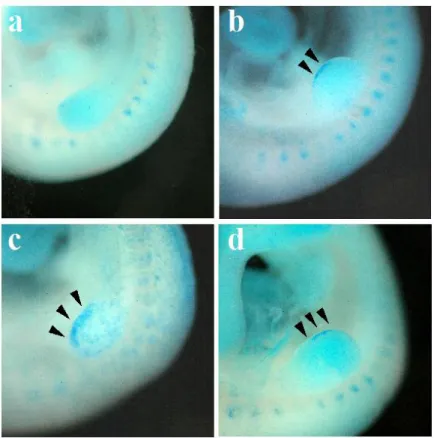
Limbs of control embryos exhibited very little NBS staining in the developing AER at GD 9:14. However, excessive NBS staining was obvious in GD 9:14 embryos exposed to ethanol. At 18 hours after the onset of ethanol exposure (GD 10:0) a high level of staining was evident relative to control groups (figure 2.2b). Staining was found at the distal apex of the AER near the center of the preaxial-postaxial axis. A minority of specimens had a slight postaxial shift of this staining pattern. BMS-189453-treated embryos likewise exhibited a similar localization of NBS to the AER at GD 9:14 and GD 10:0 (figure 2.2c). While ethanol and BMS-189453 both produced similar NBS localization, ethanol-treated embryos were more intensely stained than those treated with BMS-189453. Disulfiram treatment resulted in a cell death pattern similar to BMS-189453 and ethanol treatment by GD 10:0 (figure 2.2d), but were not distinguishable from control limb buds at GD 9:14.
Prevention of ethanol-induced cell death by Retinoic Acid
As with NBS, LTR staining was evident within 8 hours of exposure to ethanol (figure 2.3c). Staining of the AER was intense in ethanol-exposed embryos, contrary to the staining in vehicle and RA-treated embryos (figures 2.3a,b). LTR staining revealed mesenchymal staining within the limb buds of ethanol-treated embryos, a phenomenon not observed using whole mount TUNEL or NBS staining techniques. Control or RA-treated limb buds had very little LTR staining in the AER or mesenchyme. Embryos exposed to both ethanol and RA exhibited a greatly reduced LTR-positive domain, compared to those embryos exposed to ethanol, alone. However, the mesenchymal staining observed in ethanol-exposed embryos was not prevented with RA co-treatment. Whole mount TUNEL and NBS staining techniques provided identical findings with respect to the AER of control and treatment group limbs.
In the forelimb buds of GD 9:12 embryos, dHand was expressed in the postaxial
mesenchyme. It was also present in the lateral plate mesoderm proximal and caudal to the limb bud. In most GD 9:12 embryos exposed to ethanol for 6 hours, the dHand expression domain was
noticeably smaller and less intense, relative to embryos exposed to PBS (figure 2.4 a,b). Quantitative Real-time PCR (qPCR) indicated that dHand is decreased by ethanol relative to controls at GD 9:12 and GD 10:0 (P = 0.355 and P = 0.084, respectively; figure 2.5a). BMS-189453 did not have an affect on dHand transcription at either time.
Shh expression was undetectable in the limb buds of GD 9:12 embryos. However, by GD 10:0 a strong signal was present on the postaxial margin of the limb bud in control embryos (figure 2.4g). Embryos exposed to ethanol exhibited little or no observable Shh expression at GD 10:0. In the few instances in which Shh staining was observed in ethanol-exposed embryos a weaker expression domain was present than in the limbs of comparably-staged control embryos. qPCR demonstrated that expression of Shh is extremely low at GD 9:6, the time of initial ethanol exposure (Ct = 32 cycles). In control embryos, six hours later (GD 9:12), expression increased 5.0 fold before climbing an additional 12.8 fold by 14 hours (GD 10). Ethanol decreased Shhexpression
substantially at 6 hours and 18 hours of exposure (P = 0.006 and 0.062, respectively; figure 2.5b). BMS-189453 had no affect on Shhexpression after 2 hours of exposure (GD 9:12), but by GD 10:0, expression was decreased 75% (P = 0.009).
Tbx5 expression was present throughout the limb buds of GD 9:12 control embryos and those exposed to ethanol. Ethanol had no discernable effect on the level or distribution of Tbx5 expression in the limb at this time (figure 2.4c,d). However, by GD 10:0 Tbx5 expression was substantially higher in the limb buds of control embryos, compared to the expression of Tbx5 in the limbs of ethanol-exposed embryos (figure 2.4e,f). qPCR confirmed these findings; no substantial change was evident at GD 9:12 following ethanol exposure, but by GD 10:0 ethanol had decreased Tbx5