Review
Mechanisms of Post-Replication DNA Repair
Yanzhe Gao1,*, Elizabeth Mutter-Rottmayer1,2, Anastasia Zlatanou1, Cyrus Vaziri1and Yang Yang1
1 Department of Pathology and Laboratory Medicine, University of North Carolina at Chapel Hill,
Chapel Hill, NC 27599, USA; [email protected] (E.M.-R.); [email protected] (A.Z.); [email protected] (C.V.); [email protected] (Y.Y.)
2 Curriculum in Toxicology, University of North Carolina at Chapel Hill, Chapel Hill, NC 27599, USA
* Correspondence: [email protected]
Academic Editor: Eishi Noguchi
Received: 5 December 2016; Accepted: 3 February 2017; Published: 8 February 2017
Abstract: Accurate DNA replication is crucial for cell survival and the maintenance of genome stability. Cells have developed mechanisms to cope with the frequent genotoxic injuries that arise from both endogenous and environmental sources. Lesions encountered during DNA replication are often tolerated by post-replication repair mechanisms that prevent replication fork collapse and avert the formation of DNA double strand breaks. There are two predominant post-replication repair pathways, trans-lesion synthesis (TLS) and template switching (TS). TLS is a DNA damage-tolerant and low-fidelity mode of DNA synthesis that utilizes specialized ‘Y-family’ DNA polymerases to replicate damaged templates. TS, however, is an error-free ‘DNA damage avoidance’ mode of DNA synthesis that uses a newly synthesized sister chromatid as a template in lieu of the damaged parent strand. Both TLS and TS pathways are tightly controlled signaling cascades that integrate DNA synthesis with the overall DNA damage response and are thus crucial for genome stability. This review will cover the current knowledge of the primary mediators of post-replication repair and how they are regulated in the cell.
Keywords: DNA damage tolerance; post replication repair; DNA damage response; trans-lesion synthesis; template switching
1. Introduction
Accurate and efficient DNA replication is crucial for the health and survival of all living organisms. Under optimal conditions, the replicative DNA polymerasesε,δ, andαcan work in concert to ensure that the genome is replicated efficiently with high accuracy in every cell cycle [1]. However, DNA is constantly challenged by exogenous and endogenous genotoxic threats, including solar ultraviolet (UV) radiation and reactive oxygen species (ROS) generated as a byproduct of cellular metabolism. Damaged DNA can act as a steric block to replicative polymerases, thereby leading to incomplete DNA replication or the formation of secondary DNA strand breaks at the sites of replication stalling. Incomplete DNA synthesis and DNA strand breaks are both potential sources of genomic instability [2]. As discussed elsewhere in this special issue, an arsenal of DNA repair mechanisms exists to repair various forms of damaged DNA and minimize genomic instability. Most DNA repair mechanisms require an intact DNA strand as template to fix the damaged strand. In this review, we will discuss the mechanisms behind Post-Replication Repair (PRR) that specifically help cells tolerate damage on the single stranded DNA template.
Genes2017,8, 64 2 of 16
2. DNA Damage Repair and Complications at the Replication Fork
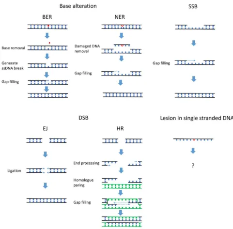
DNA damage can be categorized by structural changes in the DNA such as base alteration, single stranded break (SSB), and double stranded break (DSB), each repaired via a distinct mechanism [3]. As summarized in Figure1, a broad spectrum of DNA repair mechanisms has evolved to remove lesions that occur on double stranded DNA. Most DNA repair mechanisms rely on information from an undamaged DNA strand, either the complementary strand of the double helix (nucleotide excision repair (NER), base excision repair (BER) and SSB repair) or the sister chromatid and homologous allele (homologous recombination). Utilizing an undamaged template prevents aberrant alteration of the genetic coding on the damaged DNA strand. A major limitation to template-based repair mechanisms is that sometimes an undamaged DNA template strand is unavailable. This problem is frequently encountered during DNA replication, in the synthesis (S) phase of the cell cycle.
Genes 2017, 8, 64 2 of 15
2. DNA Damage Repair and Complications at the Replication Fork
DNA damage can be categorized by structural changes in the DNA such as base alteration, single stranded break (SSB), and double stranded break (DSB), each repaired via a distinct mechanism [3]. As summarized in Figure 1, a broad spectrum of DNA repair mechanisms has evolved to remove lesions that occur on double stranded DNA. Most DNA repair mechanisms rely on information from an undamaged DNA strand, either the complementary strand of the double helix (nucleotide excision repair (NER), base excision repair (BER) and SSB repair) or the sister chromatid and homologous allele (homologous recombination). Utilizing an undamaged template prevents aberrant alteration of the genetic coding on the damaged DNA strand. A major limitation to template-based repair mechanisms is that sometimes an undamaged DNA template strand is unavailable. This problem is frequently encountered during DNA replication, in the synthesis (S) phase of the cell cycle.
Figure 1. Many mechanisms efficiently repair DNA damage on the DNA double helix. Lesions in the double stranded DNA can be efficiently repaired by mechanisms corresponding to the specific type of DNA damage. Base-specific damage can be directly reversed by particular enzymes such as photolyases and O6-methylguanine DNA methyltransferase (MGMT) (reviewed in [4]). The majority of base-specific damage is repaired by base excision repair (BER) and nucleotide excision repair (NER). In BER and NER, the damaged base or surrounding DNA is excised from the double stranded DNA. The gap left behind is then filled by a DNA polymerase. Single stranded breaks (SSBs) are recognized by poly(ADP-ribose) polymerase 1 (PARP1), which activates downstream signaling that leads to gap-filling by DNA polymerases. Double stranded breaks (DSBs) are repaired by end joining (EJ) or by homologous recombination (HR). EJ directly ligates the exposed DSB with DNA ligase, while, during HR, break sites are replicated using undamaged homologous sequences of sister
Genes2017,8, 64 3 of 16
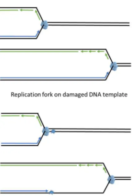
DNA replication is a multistep process with two key events; (1) unwinding of the annealed double helix to expose ssDNA and (2) using this ssDNA as template to synthesize daughter strands. During an unperturbed S phase, DNA unwinding, carried out by the replicative helicase (the CDC45-MCM2-7-GINS or “CMG” complex), is strictly coupled with polymerase activity at replication forks (reviewed in [5]). Three replicative polymerases, polεon the leading strand and polδand pol αon the lagging strand, copy the template DNA with an error rate less than 10−4[1]. The compact catalytic sites of replicative DNA polymerases confer high fidelity but preclude DNA damage-tolerant synthesis when using templates harboring bulky DNA lesions. As a result, replicative polymerases stall when a lesion is encountered (Figure2). Fork-stalling DNA lesions are very prevalent in cells. In the human body there are approximately 30,000 lesions in every cell at any given time due to aerobic metabolism and endogenous depurination and deamination events [6]. It is inevitable that replication forks will be challenged by fork stalling lesions during DNA synthesis. Lesions encountered at replicating DNA are unique because the DNA in the vicinity of a replication fork is not double-helical. Excising the lesion from the ssDNA, as seen in BER and NER, will generate DNA strand breaks and result in fork collapse.
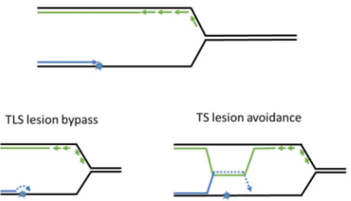
To survive fork-stalling DNA lesions, cells have developed post replication DNA repair mechanisms (PRR), which allow replication forks to progress through the lesions on damaged templates. The main role of PRR is to “patch” ssDNA gaps in the daughter strand and restore DNA to its double-stranded state for subsequent DNA repair via other mechanisms (covered in Figures2and3
and later sections of the paper). There are two mechanisms of PRR; trans-lesion synthesis (TLS), which employs TLS polymerases to directly replicate across the DNA lesion [7], and template switching (TS), which “borrows” the genetic information from the newly synthesized sister chromatid as a replication template [8] and thus avoids the lesion (Figure3). As described in detail below, the TLS and TS pathways are coordinated to facilitate ongoing DNA synthesis on damaged genomes.
chromatid templates. In contrast, DNA lesions in single stranded DNA (ssDNA) cannot be repaired by BER, NER, HR, or EJ and must be remediated using alternative mechanisms (as suggested by the question mark in the figure). Post replication repair is a mechanism specialized in tolerating lesions in single stranded template.
DNA replication is a multistep process with two key events; (1) unwinding of the annealed double helix to expose ssDNA and (2) using this ssDNA as template to synthesize daughter strands. During an unperturbed S phase, DNA unwinding, carried out by the replicative helicase (the CDC45-MCM2-7-GINS or “CMG” complex), is strictly coupled with polymerase activity at replication forks (reviewed in [5]). Three replicative polymerases, pol ε on the leading strand and pol δ and pol α on the lagging strand, copy the template DNA with an error rate less than 10−4[1]. The
compact catalytic sites of replicative DNA polymerases confer high fidelity but preclude DNA damage-tolerant synthesis when using templates harboring bulky DNA lesions. As a result, replicative polymerases stall when a lesion is encountered (Figure 2). Fork-stalling DNA lesions are very prevalent in cells. In the human body there are approximately 30,000 lesions in every cell at any given time due to aerobic metabolism and endogenous depurination and deamination events [6]. It is inevitable that replication forks will be challenged by fork stalling lesions during DNA synthesis. Lesions encountered at replicating DNA are unique because the DNA in the vicinity of a replication fork is not double-helical. Excising the lesion from the ssDNA, as seen in BER and NER, will generate DNA strand breaks and result in fork collapse.
To survive fork-stalling DNA lesions, cells have developed post replication DNA repair mechanisms (PRR), which allow replication forks to progress through the lesions on damaged templates. The main role of PRR is to “patch” ssDNA gaps in the daughter strand and restore DNA to its double-stranded state for subsequent DNA repair via other mechanisms (covered in Figures 2 and 3 and later sections of the paper). There are two mechanisms of PRR; trans-lesion synthesis (TLS), which employs TLS polymerases to directly replicate across the DNA lesion [7], and template switching (TS), which “borrows” the genetic information from the newly synthesized sister chromatid as a replication template [8] and thus avoids the lesion (Figure 3). As described in detail below, the TLS and TS pathways are coordinated to facilitate ongoing DNA synthesis on damaged genomes.
Figure 2. DNA lesions in single stranded DNA (ssDNA) are detrimental to the replication fork. DNA lesions on an ssDNA template act as road blocks for replicative polymerases but not for the replicative DNA helicase. An uncoupling of replicative DNA polymerase and DNA helicase activities generates single stranded DNA tracts. Persistent ssDNA is fragile and prone to breakage, generating lethal DSB.
Genes2017,8, 64 4 of 16
Genes 2017, 8, 64 4 of 15
Figure 3. Post replication repair efficiently returns lesions in ssDNA to double helix. PRR utilizes trans-lesion synthesis (TLS) or template switching (TS) to bypass or avoid DNA lesions and prevent accumulation of ssDNA gaps. After being restored to its double-stranded state, damaged DNA may be repaired via the mechanisms described in Figure 1.
3. Activation of Post Replication Repair
Proliferating cell nuclear antigen (PCNA) is a ring shaped homo-trimeric protein complex that surrounds the DNA and is a central player in PRR. During the initiation of DNA replication, PCNA is loaded onto the chromatin by the Replication Factor C (RFC) clamp loader [9]. Upon completion of DNA replication, ATAD5 (Elg1 in yeast) unloads PCNA from chromatin [10,11]. Chromatin-bound PCNA slides along the DNA strand and serves as a processivity factor for DNA polymerases. In addition to tethering polymerases to template DNA, PCNA is a platform for a wide variety of proteins that participate in DNA replication and damage repair [12]. The interactions between PCNA and its binding partners are typically mediated by the PCNA inter-domain connecting loop (IDCL) and the PCNA interacting peptide (PIP) motif on its binding partner [13]. PCNA can also be modified by ubiquitin and SUMO (Small Ubiquitin Modifier) to create additional interfaces for binding partners during the S-phase or when the replication fork is under stress [12,14,15]. These post-translational modifications of PCNA are crucial events in PRR.
When the replication fork encounters a bulky DNA lesion, replicative polymerases stall but the MCM helicases continue unwinding the double helix ahead of the polymerase. The uncoupling of DNA polymerase and helicase at the stalled replication fork generates long stretches of ssDNA covered by replication protein A (RPA) that activates the DNA replication checkpoint [16]. RPA-coated ssDNA generated by fork-stalling recruits Rad18 (a PCNA-directed E3 ubiquitin ligase) to the vicinity of the DNA lesion [17–19]. Chromatin-bound Rad18 and its associated E2 ubiquitin-conjugating enzyme (Rad6) mono-ubiquitinate PCNA at the conserved residue, K164 [14,15]. Mono-ubiquitinated PCNA initiates PRR by recruiting TLS polymerases to replace the activity of replicative polymerase at the stalled replication fork [20–22]. Although RPA-coated ssDNA is necessary for Rad18 chromatin-binding, multiple regulators have been shown to modulate the recruitment of Rad18 to PCNA. For example, TLS Pol η can facilitate the Rad18-PCNA interaction by binding to both proteins with its C-terminus domain and enhancing PCNA ubiquitination [23]. NBS1 (mutated in Nijmegen Breakage Syndrome) interacts with Rad18 at the Rad6-interacting domain to help recruit Rad18 to damaged DNA [24]. Additionally, BRCA1 facilitates efficient recruitment of RPA and Rad18 to damaged DNA and promotes PRR [25]. SIVA1 physically bridges chromatin-bound Rad18 and its substrate PCNA and promotes PCNA ubiquitination. However, SIVA1 is not required for Rad18 recruitment to DNA damage sites [26]. Spartan/DVC1 interacts with Rad18 and PCNA and is necessary for UV-tolerance, although the molecular mechanism by which Spartan/DVC1 regulates PRR is unclear [27–31]. Of note, Rad18 might not be the only enzyme that mono-ubiquitinates PCNA. PCNA mono-ubiquitination has been observed in both Rad18−/− DT40 cells and in Rad18 KO mice, suggesting other E3 ubiquitin ligases
may use PCNA as a substrate [32–34].
Figure 3. Post replication repair efficiently returns lesions in ssDNA to double helix. PRR utilizes trans-lesion synthesis (TLS) or template switching (TS) to bypass or avoid DNA lesions and prevent accumulation of ssDNA gaps. After being restored to its double-stranded state, damaged DNA may be repaired via the mechanisms described in Figure1.
3. Activation of Post Replication Repair
Proliferating cell nuclear antigen (PCNA) is a ring shaped homo-trimeric protein complex that surrounds the DNA and is a central player in PRR. During the initiation of DNA replication, PCNA is loaded onto the chromatin by the Replication Factor C (RFC) clamp loader [9]. Upon completion of DNA replication, ATAD5 (Elg1 in yeast) unloads PCNA from chromatin [10,11]. Chromatin-bound PCNA slides along the DNA strand and serves as a processivity factor for DNA polymerases. In addition to tethering polymerases to template DNA, PCNA is a platform for a wide variety of proteins that participate in DNA replication and damage repair [12]. The interactions between PCNA and its binding partners are typically mediated by the PCNA inter-domain connecting loop (IDCL) and the PCNA interacting peptide (PIP) motif on its binding partner [13]. PCNA can also be modified by ubiquitin and SUMO (Small Ubiquitin Modifier) to create additional interfaces for binding partners during the S-phase or when the replication fork is under stress [12,14,15]. These post-translational modifications of PCNA are crucial events in PRR.
PCNA mono-ubiquitination has been observed in both Rad18−/−DT40 cells and in Rad18 KO mice, suggesting other E3 ubiquitin ligases may use PCNA as a substrate [32–34].
PCNA K164 mono-ubiquitination can be further extended to K63-linked poly-ubiquitin chains by another E3 ubiquitin-ligase, which in yeast is Rad5 [14]. The interaction between Rad18 and Rad5 brings Ubc13/Mms2-Rad5 to the vicinity of stalled replication forks [35]. Ubc13/Mms2 and Rad5-mediated poly-ubiquitination of PCNA directs lesion avoidance using the TS pathway [36]. InXenopus laevis, PCNA poly-ubiquitination is induced by DNA damage, although it is unclear why PCNA is also modified when replicating undamaged DNA in this system [37]. In humans, the two human Rad5 orthologues, SNF2 histone-linker PHD-finger RING-finger helicase (SHPRH) [38,39] and helicase-like transcription factor (HLTF) [40,41], mediate PCNA poly-ubiquitination. Although PCNA poly-ubiquitination is a less abundant modification than PCNA mono-ubiquitination [42], it is clear that the human RAD5 homologues do contribute to DNA damage tolerance [43]. There are also some studies suggesting that Rad5 has an Mms2-Ubc13-independent role in the TLS pathway [44–46].
SUMOylation of PCNA, catalyzed by UBC9 and Siz1/2, has also been observed on lysines K127 and/or K164 during normal DNA replication or following sub-lethal DNA-damaging treatments [14,15]. SUMOylated PCNA interacts with Srs2, a helicase that displaces the Rad51 recombinase from ssDNA. Since Rad51 is essential for DNA repair via homologous recombination, Rad51 displacement prevents recombinational repair [47,48]. Consequently, the inhibition of homologous recombination by Srs2 at the replication fork further limits the pathway choices to PRR when a DNA lesion is encountered on the single stranded template [49]. Interestingly, PCNA SUMOylation also has been shown to facilitate Rad18 E3 ligase activity towards PCNA by physically linking Rad18 and PCNA in yeast [50]. Although this is not an evolutionally conserved mechanism for Rad18 activation, it still exemplifies the cross talk between different PCNA modifications.
4. Trans-Lesion Synthesis
The Trans-Lesion Synthesis branch of PRR employs specialized DNA polymerases to perform replicative bypass of DNA lesions. In a process termed “polymerase switching” the TLS polymerases are recruited to stalled replication forks where they transiently replace the replicative polymerases. There are three TLS polymerases RAD30 (η), Rev1, andζ, in budding yeast and two additional TLS polymerases,κandι, in vertebrates. Of these TLS polymerases,η,κ,ι, and Rev1 belong to the Y-family, while Polζbelongs to the B family [51]. The unique structure of TLS polymerases allows them to synthesize across lesions that block the conventional replicative polymerases. Compared to replicative polymerases, TLS polymerases have larger catalytic sites that are able to make loose contact with the template DNA and incoming nucleotide. This structure makes TLS polymerases more promiscuous in their selection of template DNA and allows them to accommodate templates with bulky adducts and abasic sites [52]. Furthermore, TLS polymerases lack the proofreading exonuclease domain that is present in the replicative ones and which is critical for accurate DNA synthesis. Therefore, utilizing TLS polymerases to replicate damaged templates can confer damage-tolerant DNA synthesis at the cost of reduced replication accuracy [7].
Genes2017,8, 64 6 of 16
Due to the intrinsic ability of TLS polymerases to accommodate a wide variety of lesions, sometimes lesion bypass can be carried out by a “non-ideal” error-prone polymerase, especially when the “correct” polymerase is not available. This phenomenon of compensatory error-prone lesion bypass by inappropriate DNA polymerases is exemplified by xeroderma pigmentosum variant (XP-V) patients, in which Polηis mutated [57]. XP-V patients experience extreme sunlight sensitivity and have an increased incidence of skin cancer. In XP-V patients, UV-induced DNA damage is bypassed by other Y-family DNA polymerases such as Polι[58] and Polκ[59], resulting in high mutation rates. These studies suggest that, despite the presence of intact nucleotide excision repair, selecting the correct TLS polymerase to accurately bypass the DNA lesion is crucial for the prevention of elevated mutagenesis. As mentioned previously, the recruitment of TLS polymerases to stalled replication forks is facilitated by Rad18-mediated PCNA mono-ubiquitination. TLS polymerases possess a higher affinity towards PCNA in its mono-ubiquinated state [20,21] and may displace the processive DNA polymerases to replicate through damaged DNA [60]. Interestingly, lysine 164 of PCNA is not located at the IDCL, the protein-protein interacting domain on PCNA that mediates the interaction with the PIP motif of target proteins [13]. Instead, the K164-linked ubiquitin is attached to the back face of PCNA, creating a distinct interacting motif for TLS polymerases [22] (Figure4). In addition to a PIP-motif, all Y family polymerases contain at least one Ubiquitin-Binding Zinc finger (UBZ) or Ubiquitin-Binding Motif (UBM) at the C-terminus of the protein [61]. The ubiquitin-binding domain, together with the PIP motif on TLS polymerases, mediates the preferential interaction with mono-ubiquitinated PCNA. In this structure, the binding of Polηto PCNA does not interfere with the binding of Polδ. Instead, Pol ηis resting at the back face of PCNA, while Polδis contacting the front surface of PCNA [22] (Figure4). It is important to note that PCNA is a trimeric ring, which in theory could interact with three DNA polymerases at the same time. In fact, the structural study by Freudenthal et al. favors the notion that the PCNA ring acts as a molecular ‘tool belt’, carrying both TLS and replicative polymerases to cope with damage on ssDNA, similar to theβsliding clamp in E.coli [62]. Unlike the Y-family TLS polymerases (η,κ,ι, REV1), Polζ(a B-family DNA polymerase) does not contain a UBZ domain. However, Polζrecruitment to stalled replication forks is mediated by the Y family polymerases, such as REV1 [63], and therefore might have some dependency on PCNA mono-ubiquitination.
Genes 2017, 8, 64 6 of 15
variant (XP-V) patients, in which Pol η is mutated [57]. XP-V patients experience extreme sunlight sensitivity and have an increased incidence of skin cancer. In XP-V patients, UV-induced DNA damage is bypassed by other Y-family DNA polymerases such as Pol ι [58] and Pol κ [59], resulting in high mutation rates. These studies suggest that, despite the presence of intact nucleotide excision repair, selecting the correct TLS polymerase to accurately bypass the DNA lesion is crucial for the prevention of elevated mutagenesis.
As mentioned previously, the recruitment of TLS polymerases to stalled replication forks is facilitated by Rad18-mediated PCNA mono-ubiquitination. TLS polymerases possess a higher affinity towards PCNA in its mono-ubiquinated state [20,21] and may displace the processive DNA polymerases to replicate through damaged DNA [60]. Interestingly, lysine 164 of PCNA is not located at the IDCL, the protein-protein interacting domain on PCNA that mediates the interaction with the PIP motif of target proteins [13]. Instead, the K164-linked ubiquitin is attached to the back face of PCNA, creating a distinct interacting motif for TLS polymerases [22] (Figure 4). In addition to a PIP-motif, all Y family polymerases contain at least one Ubiquitin-Binding Zinc finger (UBZ) or Ubiquitin-Binding Motif (UBM) at the C-terminus of the protein [61]. The ubiquitin-binding domain, together with the PIP motif on TLS polymerases, mediates the preferential interaction with mono-ubiquitinated PCNA. In this structure, the binding of Pol η to PCNA does not interfere with the binding of Pol δ. Instead, Pol η is resting at the back face of PCNA, while Pol δ is contacting the front surface of PCNA [22] (Figure 4). It is important to note that PCNA is a trimeric ring, which in theory could interact with three DNA polymerases at the same time. In fact, the structural study by Freudenthal et al. favors the notion that the PCNA ring acts as a molecular ‘tool belt’, carrying both TLS and replicative polymerases to cope with damage on ssDNA, similar to the β sliding clamp in E.coli [62]. Unlike the Y-family TLS polymerases (η, κ, ι, REV1), Pol ζ (a B-family DNA polymerase) does not contain a UBZ domain. However, Pol ζ recruitment to stalled replication forks is mediated by the Y family polymerases, such as REV1 [63], and therefore might have some dependency on PCNA mono-ubiquitination.
Figure 4. Structure of a monoubiquitinated PCNA ring (picture from reference [22]). (A) Back view of monoubiquitinated PCNA ring showing the two domains of a single PCNA subunit and the inter-domain connecting loop (IDCL). Ubiquitin is shown in red. Three individual PCNA molecules (shown in blue, green, and yellow) constitute the ring shape; (B) Side view of the monoubiquitinated PCNA where the back surface is to the left and the front surface is to the right of the figure. Notice that the ubiquitin is located on the back surface of the PCNA ring while IDCL is to the front side of the PCNA ring.
Despite the extensive studies suggesting that PCNA and its ubiquitination facilitate TLS activation, the absolute requirement of PCNA mono-ubiquitination is still being debated. Several
Despite the extensive studies suggesting that PCNA and its ubiquitination facilitate TLS activation, the absolute requirement of PCNA mono-ubiquitination is still being debated. Several lines of evidence suggest that TLS can, in some instances, proceed without the need for PCNA mono-ubiquitination. Gueranger and collegues showed that the PolηPIP-box mutant could completely restore the UV resistance in a pol η-deficient cell line [64]. Acharya and colleagues additionally found that the ubiquitin-binding domain of polηis dispensable for its TLS function [65,66]. Embryonic fibroblasts from a genetically-engineered PCNA K164R “knock-in” mouse show some attenuation of TLS activity, yet retain lesion bypass activity [67]. In a recent study, polηwas shown to interact with unmodified and mono-ubiquitinated PCNA with equivalent affinities in vitro. Furthermore, mono-ubiquitinated PCNA did not enhance the lesion bypass activity of polη[68]. Together, these studies suggest that PCNA ubiquitination is important for high-capacity TLS and efficient recruitment of TLS polymerases in normal cells. However, TLS may also occur in the absence of PCNA mono-ubiquitination, most likely due to residual UBZ-independent interactions between PCNA and the PIP motifs.
Even with our mechanistic understanding of trans-lesion synthesis, it is still not known how cells recruit appropriate and specific TLS polymerases to their cognate DNA lesions. It is formally possible that unknown factors “read” the structure of distorted DNA and then signal for the recruitment of the specific polymerases. However, as clearly documented in XP-V cells, CPD lesions can be bypassed by non-cognate TLS polymerases when Polηis absent. Therefore, it is possible that mono-ubiquitinated PCNA does not discriminate between different TLS polymerases and serves as a recruitment platform that interacts with all TLS polymerases equally. Perhaps all the TLS polymerases are recruited to the damage site randomly and attempt to replicate through the lesion. In this “trial-and-error” mechanism, the “correct” polymerase bypasses a cognate lesion with the lowest energy expenditure. Non-ideal TLS polymerases would only perform a bypass when the “correct” polymerase is unavailable. Therefore, it might be the nature of the lesion itself that determines which polymerase engages and replicates across a specific type of DNA damage.
5. Template Switching
The Template Switching branch of PRR enables the stalled replication fork to use the newly synthesized daughter strand as template to avoid damaged DNA (Figure 3). Similar to TLS, TS is also mediated by PCNA post-translational modifications, specifically poly-ubiquitination and SUMOylation. The extension of Rad18-induced K164 mono-ubiquitination to poly-ubiquitination by Ubc13-Mms2 and Rad5 redirects the PRR mode to TS.
Most of our understanding of TS was generated from a series of elegant studies in yeast, which provided the basis for the existence of an error-free form of PRR [69,70]. This error-free mechanism requires Rad5 [71,72], Ubc13/Mms2 [73,74], DNA Polδ[75], a subset of the RAD52 epistasis group [76] and involves recombination between partially replicated sister strands [77]. A groundbreaking study by Branzei and colleagues combining 2D gel electrophoresis of DNA replication intermediates and yeast genetics identified TS intermediates and defined their relationship with the previously mentioned TS factors [78].
Genes2017,8, 64 8 of 16
Rad5, Rad51, and Sgs1, there is a growing body of evidence that other DNA replication and damage repair factors are also involved in TS. For example, the 9-1-1 complex and Exo1 nuclease are essential for the initiation of TS [81], and Ctf4 was found to establish the connection between Polα/primase and the MCM helicase to protect the replication fork structure that favors TS [82].
Although Ubc13-Mms2 and Rad5-mediated poly-ubiquitination of PCNA at K164 is a crucial event in TS, the structure and function of the PCNA poly-ubiquitin chain formed during TS remains elusive. In contrast, the significance of PCNA SUMOylation has been studied in more detail and is better understood. PCNA SUMOylation occurs both during normal, unperturbed DNA replication and in response to DNA damage [14,15]. SUMOylated PCNA provides an interaction platform for the recruitment of the Srs2 helicase [47,48]. Similar to other PCNA-interacting proteins, Srs2 contains a non-canonical PIP box motif that mediates PCNA binding. However, the interaction between the Srs2 PIP box and PCNA is fairly weak until a second interaction is established between SUMOylated PCNA and a SUMO-interacting motif at the C terminus of Srs2 [83]. PCNA-bound Srs2 helicase functions as a safeguard that limits unscheduled recombination at the replication fork by disrupting Rad51 filament formation on ssDNA during normal replication [84,85]. A small controversy still exists regarding why PCNA SUMOylation is required for the TS pathway; Srs2 actively removes Rad51 from the replication fork while template switching requires Rad51 activity. For this reason, it is generally believed that SUMOylation antagonizes the effect of PCNA ubiquitination and inhibits the TS pathway [14,15].
A recent study may help resolve this paradox; a SUMO-like domain protein, Esc2, was found to be recruited to stalled replication forks and displace Srs2, thereby creating a microenvironment that is permissive for Rad51 chromatin-binding [86]. Therefore, PCNA SUMOylation facilitates the usage of PRR on a challenged replication fork by suppressing homologous recombination. When TS is initiated by PCNA poly-ubiquitination, replication fork binding factors such as Esc2 alleviate the inhibition of recombination by PCNA SUMOylation and allow DNA damage avoidance [47].
Error-free DNA damage avoidance is a conserved PRR mechanism in metazoans [43,87]. Although the identity of the human Srs2 orthologue is still being debated, human PCNA SUMOylation has also been shown to suppress unscheduled DNA recombination via PARI (PCNA-associated recombination inhibitor), suggesting a conserved mechanism of regulating HR at the replication fork [88–90]. PCNA is also poly-ubiquitinated in human cells in response to DNA damage. Blocking K63 linked poly-ubiquitination chain formation sensitizes cells to DNA damage, increases UV-induced mutagenesis, and increases the reliance of cells on TLS for DNA damage tolerance [43]. Rad5 has evolved into two orthologues, SHPRH [38,39] and HLTF, in higher organisms [40,41]. Both SHPRH and HLTF can poly-ubiquitinate PCNA in vitro but via distinct mechanisms. SHPRH extends Rad18 mediated PCNA mono-ubiquitination, while HLTF transfers the pre-assembled poly-ubiquitin chain to Rad6-Rad18 and eventually onto unmodified PCNA [38,39,91]. Depletion of SHPRH and HLTF sensitizes the cell to DNA damaging agents and reduces PCNA poly-ubiquitination; however, SHPRH−/−HLTF−/− double knockout mouse embryonic fibroblasts are still able to poly-ubiquitinate PCNA, suggesting that other Rad5 orthologues might exist in higher organisms [92]. In addition to poly-ubiquitination of PCNA, HLTF has acquired additional functions in DNA damage tolerance. In response to UV damage, HLTF is able to mono-ubiquitinate PCNA and promote Polη recruitment [93]. Furthermore, HLTF can also facilitate DNA strand invasion and D-loop formation in a Rad51-independent manner [94].
In addition to TS, there are other recombination-based mechanisms, such as complementary strand transfer repair (CSTR) [95] and replication fork reversal [96–99], that have also been shown to contribute to DNA damage avoidance.
6. Timing of Post Replication Repair
developed sophisticated mechanisms to control the timing of DNA post replication repair by limiting the availability of crucial PRR factors [100–102]. Interestingly, two studies using temporally controlled expression of Rad18 or Polηfound that it is possible to delay the onset of PRR without significantly affecting cell viability. Moreover, limiting PRR in the G2/M phase of the cell cycle does not significantly delay the progression of the S-phase [103,104]. These studies suggest that it is possible to detach the PRR with bulk DNA synthesis in the S-phase without compromising its function.
Nevertheless, the delayed onset of PRR during S-phase could potentially lead to the accumulation of dangerously long and fragile ssDNA stretches, especially on the leading strand. Exposed ssDNA in cells is frequently observed when the replicative polymerase is blocked. However, these ssDNA gaps are usually small in size and are located inside a single replicon, regardless of whether they are on the leading or the lagging strand. However extremely long ssDNA gaps (>3 kb) are rarely observed. This suggests that the leading strand is also synthesized discontinuously when replicating a damaged DNA template, similar to the discontinuous synthesis of the lagging strand [105].
Restart of replication requires a de-novo re-priming mechanism downstream (30) of the stalled leading strand DNA polymerase. This repriming activity is carried out by DnaG inE. coli[106], and by a specialized polymerase PrimPol in higher organisms [107–110]. This repriming mechanism of PRR explains why UV-induced lesions only cause a slight reduction in fork speed even when Polη is mutated in human cells [111]. The ability of PRR to function distal (50) to a newly-primed leading strand may provide ample time to select the optimal DNA damage tolerance mechanism. It is also well established that TLS is functional outside the S phase of the cell cycle and can patch ssDNA arising in the G0 and G1 phases [112–114].
7. Conclusions and Outlook
Although neither TLS nor TS directly repair DNA damage, both PRR mechanisms enable an immediate response to polymerase stalling DNA lesions. PRR during S phase prevents gross chromosomal rearrangements and ensures that replication is completed in a timely manner.
A deficiency in PRR could lead to replication fork collapse and the accumulation of DNA DSBs. In the absence of PRR, DSB repair mechanisms could allow for tolerance of replication-associated DNA damage. However, DSB repair pathways have limitations; DNA end-joining frequently results in mutations, while HR serves as the salvage pathway and creates complex and unstable repair intermediates through the use of a homologous strand from another DNA molecule. (For more insight into salvage and other homologous recombination-mediated DNA damage tolerance, we invite readers to read a recent review on this topic [115]) Therefore, PRR is perhaps the least genome-destabilizing option for the tolerance of DNA lesions arising in S-phase.
Both branches of PRR are important for cells to tolerate and survive DNA damage. In terms of maintaining genome stability, TS has a great advantage over TLS because it does not induce base mutations. Because of its intrinsic error-propensity, TLS has been linked to both increased mutation rates and might, therefore, fuel carcinogenesis [116–118]. In established cancers, TLS is also suggested to be responsible for a high mutation frequency and elevated treatment resistance [116,119,120]. However, it is not known why untransformed cells would utilize the error-prone TLS pathway when error-free TS is available.
Genes2017,8, 64 10 of 16
Acknowledgments:This manuscript was supported by grant R01 ES009558 from the National Institutes of Health to Cyrus Vaziri, a Tier 1 Pilot Award from the University of North Carolina Lineberger Comprehensive Cancer Center to Yanzhe Gao, the Leon and Bertha Golberg Postdoctoral Fellowship to Yanzhe Gao, a PhRMA Foundation Pre-Doctoral Award in Pharmacology/Toxicology to Elizabeth Mutter-Rottmayer, and a NC TraCS Institute CTSA grant UL1TR001111 to Yang Yang.
Conflicts of Interest:The authors declare no conflict of interest.
References
1. Burgers, P.M.J. Polymerase Dynamics at the Eukaryotic DNA Replication Fork. J. Biol. Chem. 2009,284, 4041–4045. [CrossRef] [PubMed]
2. Lehmann, A.R. Replication of UV-damaged DNA: New insights into links between DNA polymerases, mutagenesis and human disease.Gene2000,253, 1–12. [CrossRef]
3. Ciccia, A.; Elledge, S.J. The DNA Damage Response: Making It Safe to Play with Knives.Mol. Cell2010,40, 179–204. [CrossRef] [PubMed]
4. Yi, C.; He, C. DNA Repair by Reversal of DNA Damage.Cold Spring Harb. Perspect. Biol.2013. [CrossRef] [PubMed]
5. Masai, H.; Matsumoto, S.; You, Z.; Yoshizawa-Sugata, N.; Oda, M. Eukaryotic Chromosome DNA Replication: Where, When, and How?Ann. Rev. Biochem.2010,79, 89–130. [CrossRef] [PubMed]
6. Nakamura, J.; Mutlu, E.; Sharma, V.; Collins, L.; Bodnar, W.; Yu, R.; Lai, Y.; Moeller, B.; Lu, K.; Swenberg, J. The Endogenous Exposome.DNA Repair2014,19, 3–13. [CrossRef] [PubMed]
7. Sale, J.E.; Lehmann, A.R.; Woodgate, R. Y-family DNA polymerases and their role in tolerance of cellular DNA damage.Nat. Rev. Mol. Cell Biol.2012,13, 141–152. [CrossRef] [PubMed]
8. Branzei, D. Ubiquitin family modifications and template switching. FEBS Lett. 2011, 585, 2810–2817. [CrossRef] [PubMed]
9. Majka, J.; Burgers, P.M.J. The PCNA–RFC Families of DNA Clamps and Clamp Loaders.Prog. Nucleic Acid
Res. Mol. Biol.2004,78, 227–260. [PubMed]
10. Lee, K.Y.; Fu, H.; Aladjem, M.I.; Myung, K. ATAD5 regulates the lifespan of DNA replication factories by modulating PCNA level on the chromatin.J. Cell Biol.2013,200, 31–44. [CrossRef] [PubMed]
11. Kubota, T.; Nishimura, K.; Kanemaki, M.T.; Donaldson, A.D. The Elg1 replication factor C-like complex functions in PCNA unloading during DNA replication.Mol. Cell2013,50, 273–280. [CrossRef] [PubMed] 12. Moldovan, G.-L.; Pfander, B.; Jentsch, S. PCNA, the Maestro of the Replication Fork.Cell2007,129, 665–679.
[CrossRef] [PubMed]
13. Gulbis, J.M.; Kelman, Z.; Hurwitz, J.; O'Donnell, M.; Kuriyan, J. Structure of the C-terminal region of p21(WAF1/CIP1) complexed with human PCNA.Cell1996,87, 297–306. [CrossRef]
14. Hoege, C.; Pfander, B.; Moldovan, G.L.; Pyrowolakis, G.; Jentsch, S. RAD6-dependent DNA repair is linked to modification of PCNA by ubiquitin and SUMO.Nature2002,419, 135–141. [CrossRef] [PubMed] 15. Stelter, P.; Ulrich, H.D. Control of spontaneous and damage-induced mutagenesis by SUMO and ubiquitin
conjugation.Nature2003,425, 188–191. [CrossRef] [PubMed]
16. Byun, T.S.; Pacek, M.; Yee, M.-c.; Walter, J.C.; Cimprich, K.A. Functional uncoupling of MCM helicase and DNA polymerase activities activates the ATR-dependent checkpoint.Genes Dev.2005,19, 1040–1052. [CrossRef] [PubMed]
17. Davies, A.A.; Huttner, D.; Daigaku, Y.; Chen, S.; Ulrich, H.D. Activation of ubiquitin-dependent DNA damage bypass is mediated by replication protein a.Mol. Cell2008,29, 625–636. [CrossRef] [PubMed] 18. Huttner, D.; Ulrich, H.D. Cooperation of replication protein A with the ubiquitin ligase Rad18 in DNA
damage bypass.Cell Cycle2008,7, 3629–3633. [CrossRef]
19. Tsuji, Y.; Watanabe, K.; Araki, K.; Shinohara, M.; Yamagata, Y.; Tsurimoto, T.; Hanaoka, F.; Yamamura, K.; Yamaizumi, M.; Tateishi, S. Recognition of forked and single-stranded DNA structures by human RAD18 complexed with RAD6B protein triggers its recruitment to stalled replication forks.Genes Cells Devot. Mol.
Cell. Mech.2008,13, 343–354. [CrossRef] [PubMed]
21. Kannouche, P.L.; Wing, J.; Lehmann, A.R. Interaction of Human DNA Polymeraseηwith Monoubiquitinated PCNA: A Possible Mechanism for the Polymerase Switch in Response to DNA Damage.Mol. Cell2004,14, 491–500. [CrossRef]
22. Freudenthal, B.D.; Gakhar, L.; Ramaswamy, S.; Washington, M.T. Structure of monoubiquitinated PCNA and implications for translesion synthesis and DNA polymerase exchange.Nat. Struct. Mol. Biol.2010,17, 479–484. [CrossRef] [PubMed]
23. Durando, M.; Tateishi, S.; Vaziri, C. A non-catalytic role of DNA polymerase eta in recruiting Rad18 and promoting PCNA monoubiquitination at stalled replication forks.Nucleic Acids Res.2013,41, 3079–3093. [CrossRef] [PubMed]
24. Yanagihara, H.; Kobayashi, J.; Tateishi, S.; Kato, A.; Matsuura, S.; Tauchi, H.; Yamada, K.; Takezawa, J.; Sugasawa, K.; Masutani, C.; et al. NBS1 recruits RAD18 via a RAD6-like domain and regulates Pol eta-dependent translesion DNA synthesis.Mol. Cell2011,43, 788–797. [CrossRef] [PubMed]
25. Tian, F.; Sharma, S.; Zou, J.; Lin, S.Y.; Wang, B.; Rezvani, K.; Wang, H.; Parvin, J.D.; Ludwig, T.; Canman, C.E.; et al. BRCA1 promotes the ubiquitination of PCNA and recruitment of translesion polymerases in response to replication blockade.Proc. Natl. Acad. Sci. USA2013,110, 13558–13563. [CrossRef] [PubMed]
26. Han, J.; Liu, T.; Huen, M.S.; Hu, L.; Chen, Z.; Huang, J. SIVA1 directs the E3 ubiquitin ligase RAD18 for PCNA monoubiquitination.J. Cell Biol.2014,205, 811–827. [CrossRef] [PubMed]
27. Centore, R.C.; Yazinski, S.A.; Tse, A.; Zou, L. Spartan/C1orf124, a Reader of PCNA Ubiquitylation and a Regulator of UV-Induced DNA Damage Response.Mol. Cell2012,46, 625–635. [CrossRef] [PubMed] 28. Mosbech, A.; Gibbs-Seymour, I.; Kagias, K.; Thorslund, T.; Beli, P.; Povlsen, L.; Nielsen, S.V.; Smedegaard, S.;
Sedgwick, G.; Lukas, C.; et al. DVC1 (C1orf124) is a DNA damage-targeting p97 adaptor that promotes ubiquitin-dependent responses to replication blocks.Nat. Struct. Mol. Biol.2012,19, 1084–1092. [CrossRef] [PubMed]
29. Juhasz, S.; Balogh, D.; Hajdu, I.; Burkovics, P.; Villamil, M.A.; Zhuang, Z.; Haracska, L. Characterization of human Spartan/C1orf124, an ubiquitin-PCNA interacting regulator of DNA damage tolerance.Nucleic
Acids Res.2012,40, 10795–10808. [CrossRef] [PubMed]
30. Davis, E.J.; Lachaud, C.; Appleton, P.; Macartney, T.J.; Nathke, I.; Rouse, J. DVC1 (C1orf124) recruits the p97 protein segregase to sites of DNA damage.Nat. Struct. Mol. Biol.2012,19, 1093–1100. [CrossRef] [PubMed] 31. Machida, Y.; Kim, M.S.; Machida, Y.J. Spartan/C1orf124 is important to prevent UV-induced mutagenesis.
Cell Cycle2012,11, 3395–3402. [CrossRef] [PubMed]
32. Arakawa, H.; Moldovan, G.-L.; Saribasak, H.; Saribasak, N.N.; Jentsch, S.; Buerstedde, J.-M. A Role for PCNA Ubiquitination in Immunoglobulin Hypermutation.PLoS Biol.2006,4, e366. [CrossRef] [PubMed] 33. Simpson, L.J.; Ross, A.L.; Szuts, D.; Alviani, C.A.; Oestergaard, V.H.; Patel, K.J.; Sale, J.E. RAD18-independent
ubiquitination of proliferating-cell nuclear antigen in the avian cell line DT40.EMBO Rep.2006,7, 927–932. [CrossRef] [PubMed]
34. Shimizu, T.; Tateishi, S.; Tanoue, Y.; Azuma, T.; Ohmori, H. Somatic hypermutation of immunoglobulin genes in Rad18 knockout mice.DNA Repair (Amst)2017,50, 54–60. [CrossRef] [PubMed]
35. Ulrich, H.D.; Jentsch, S. Two RING finger proteins mediate cooperation between ubiquitin-conjugating enzymes in DNA repair.Embo. J.2000,19, 3388–3397. [CrossRef]
36. Haracska, L.; Torres-Ramos, C.A.; Johnson, R.E.; Prakash, S.; Prakash, L. Opposing effects of ubiquitin conjugation and SUMO modification of PCNA on replicational bypass of DNA lesions in Saccharomyces cerevisiae.Mol. Cell Biol.2004,24, 4267–4274. [CrossRef] [PubMed]
37. Leach, C.A.; Michael, W.M. Ubiquitin/SUMO modification of PCNA promotes replication fork progression in Xenopus laevis egg extracts.J. Cell Biol.2005,171, 947–954. [CrossRef] [PubMed]
38. Motegi, A.; Sood, R.; Moinova, H.; Markowitz, S.D.; Liu, P.P.; Myung, K. Human SHPRH suppresses genomic instability through proliferating cell nuclear antigen polyubiquitination. J. Cell Biol. 2006,175, 703–708. [CrossRef] [PubMed]
39. Unk, I.; Hajdu, I.; Fatyol, K.; Szakal, B.; Blastyak, A.; Bermudez, V.; Hurwitz, J.; Prakash, L.; Prakash, S.; Haracska, L. Human SHPRH is a ubiquitin ligase for Mms2-Ubc13-dependent polyubiquitylation of proliferating cell nuclear antigen.Proc. Natl. Acad. Sci. USA2006,103, 18107–18112. [CrossRef] [PubMed] 40. Unk, I.; Hajdu, I.; Fatyol, K.; Hurwitz, J.; Yoon, J.H.; Prakash, L.; Prakash, S.; Haracska, L. Human HLTF
functions as a ubiquitin ligase for proliferating cell nuclear antigen polyubiquitination.Proc. Natl. Acad.
Genes2017,8, 64 12 of 16
41. Motegi, A.; Liaw, H.J.; Lee, K.Y.; Roest, H.P.; Maas, A.; Wu, X.; Moinova, H.; Markowitz, S.D.; Ding, H.; Hoeijmakers, J.H.; et al. Polyubiquitination of proliferating cell nuclear antigen by HLTF and SHPRH prevents genomic instability from stalled replication forks.Proc. Natl. Acad. Sci. USA2008,105, 12411–12416. [CrossRef] [PubMed]
42. Kannouche, P.L.; Lehmann, A.R. Ubiquitination of PCNA and the polymerase switch in human cells.
Cell Cycle2004,3, 1011–1013. [CrossRef] [PubMed]
43. Chiu, R.K.; Brun, J.; Ramaekers, C.; Theys, J.; Weng, L.; Lambin, P.; Gray, D.A.; Wouters, B.G. Lysine 63-polyubiquitination guards against translesion synthesis-induced mutations.PLoS Genet.2006,2, e116. [CrossRef]
44. Gangavarapu, V.; Haracska, L.; Unk, I.; Johnson, R.E.; Prakash, S.; Prakash, L. Mms2-Ubc13-dependent and -independent roles of Rad5 ubiquitin ligase in postreplication repair and translesion DNA synthesis in Saccharomyces cerevisiae.Mol. Cell. Biol.2006,26, 7783–7790. [CrossRef]
45. Pages, V.; Bresson, A.; Acharya, N.; Prakash, S.; Fuchs, R.P.; Prakash, L. Requirement of Rad5 for DNA polymerase zeta-dependent translesion synthesis in Saccharomyces cerevisiae.Genetics2008,180, 73–82. [CrossRef] [PubMed]
46. Xu, X.; Lin, A.; Zhou, C.; Blackwell, S.R.; Zhang, Y.; Wang, Z.; Feng, Q.; Guan, R.; Hanna, M.D.; Chen, Z.; et al. Involvement of budding yeast Rad5 in translesion DNA synthesis through physical interaction with Rev1.
Nucleic Acids Res.2016,44, 5231–5245. [CrossRef] [PubMed]
47. Pfander, B.; Moldovan, G.L.; Sacher, M.; Hoege, C.; Jentsch, S. SUMO-modified PCNA recruits Srs2 to prevent recombination during S phase.Nature2005,436, 428–433. [CrossRef] [PubMed]
48. Papouli, E.; Chen, S.; Davies, A.A.; Huttner, D.; Krejci, L.; Sung, P.; Ulrich, H.D. Crosstalk between SUMO and ubiquitin on PCNA is mediated by recruitment of the helicase Srs2p. Mol. Cell2005,19, 123–133. [CrossRef] [PubMed]
49. Schiestl, R.H.; Prakash, S.; Prakash, L. The SRS2 suppressor of rad6 mutations of Saccharomyces cerevisiae acts by channeling DNA lesions into the RAD52 DNA repair pathway.Genetics1990,124, 817–831. [PubMed] 50. Parker, J.L.; Ulrich, H.D. A SUMO-interacting motif activates budding yeast ubiquitin ligase Rad18 towards
SUMO-modified PCNA.Nucleic Acids Res.2012,40, 11380–11388. [CrossRef] [PubMed]
51. Sale, J.E. Translesion DNA synthesis and mutagenesis in eukaryotes.Cold Spring Harb. Perspect. Biol.2013,
5, a012708. [CrossRef] [PubMed]
52. Prakash, S.; Johnson, R.E.; Prakash, L. Eukaryotic translesion synthesis DNA polymerases: Specificity of structure and function.Annu. Rev. Biochem.2005,74, 317–353. [CrossRef] [PubMed]
53. Johnson, R.E.; Prakash, S.; Prakash, L. Efficient Bypass of a Thymine-Thymine Dimer by Yeast DNA Polymerase, Polη.Science1999,283, 1001–1004. [CrossRef] [PubMed]
54. Zhang, Y.; Yuan, F.; Wu, X.; Wang, M.; Rechkoblit, O.; Taylor, J.-S.; Geacintov, N.E.; Wang, Z. Error-free and error-prone lesion bypass by human DNA polymeraseκin vitro. Nucleic Acids Res. 2000,28, 4138–4146. [CrossRef] [PubMed]
55. Bi, X.; Slater, D.M.; Ohmori, H.; Vaziri, C. DNA polymerase kappa is specifically required for recovery from the benzo[a]pyrene-dihydrodiol epoxide (BPDE)-induced S-phase checkpoint.J. Biol. Chem.2005,280, 22343–22355. [CrossRef] [PubMed]
56. Zhang, Y.; Yuan, F.; Wu, X.; Taylor, J.-S.; Wang, Z. Response of human DNA polymeraseιto DNA lesions.
Nucleic Acids Res.2001,29, 928–935. [CrossRef] [PubMed]
57. Masutani, C.; Kusumoto, R.; Yamada, A.; Dohmae, N.; Yokoi, M.; Yuasa, M.; Araki, M.; Iwai, S.; Takio, K.; Hanaoka, F. The XPV (xeroderma pigmentosum variant) gene encodes human DNA polymerase [eta].Nature
1999,399, 700–704. [PubMed]
58. Wang, Y.; Woodgate, R.; McManus, T.P.; Mead, S.; McCormick, J.J.; Maher, V.M. Evidence that in Xeroderma Pigmentosum Variant Cells, which Lack DNA Polymeraseη, DNA PolymeraseιCauses the Very High Frequency and Unique Spectrum of UV-Induced Mutations.Cancer Res. 2007,67, 3018–3026. [CrossRef] [PubMed]
59. Ziv, O.; Geacintov, N.; Nakajima, S.; Yasui, A.; Livneh, Z. DNA polymerase zeta cooperates with polymerases kappa and iota in translesion DNA synthesis across pyrimidine photodimers in cells from XPV patients.
60. Zhuang, Z.; Johnson, R.E.; Haracska, L.; Prakash, L.; Prakash, S.; Benkovic, S.J. Regulation of polymerase exchange between Polηand Polδby monoubiquitination of PCNA and the movement of DNA polymerase holoenzyme.Proc. Natl. Acad. Sci. USA2008,105, 5361–5366. [CrossRef] [PubMed]
61. Bienko, M.; Green, C.M.; Crosetto, N.; Rudolf, F.; Zapart, G.; Coull, B.; Kannouche, P.; Wider, G.; Peter, M.; Lehmann, A.R.; et al. Ubiquitin-binding domains in Y-family polymerases regulate translesion synthesis.
Science2005,310, 1821–1824. [CrossRef] [PubMed]
62. Indiani, C.; McInerney, P.; Georgescu, R.; Goodman, M.F.; O'Donnell, M. A sliding-clamp toolbelt binds high-and low-fidelity DNA polymerases simultaneously.Mol. Cell2005,19, 805–815. [CrossRef] [PubMed] 63. Kikuchi, S.; Hara, K.; Shimizu, T.; Sato, M.; Hashimoto, H. Structural basis of recruitment of DNA polymerase
zeta by interaction between REV1 and REV7 proteins. J. Biol. Chem. 2012,287, 33847–33852. [CrossRef] [PubMed]
64. Gueranger, Q.; Stary, A.; Aoufouchi, S.; Faili, A.; Sarasin, A.; Reynaud, C.-A.; Weill, J.-C. Role of DNA polymerasesη,ιandζin UV resistance and UV-induced mutagenesis in a human cell line.DNA Repair2008,
7, 1551–1562. [CrossRef] [PubMed]
65. Acharya, N.; Yoon, J.H.; Gali, H.; Unk, I.; Haracska, L.; Johnson, R.E.; Hurwitz, J.; Prakash, L.; Prakash, S. Roles of PCNA-binding and ubiquitin-binding domains in human DNA polymerase eta in translesion DNA synthesis.Proc. Natl. Acad. Sci. USA2008,105, 17724–17729. [CrossRef] [PubMed]
66. Acharya, N.; Yoon, J.H.; Hurwitz, J.; Prakash, L.; Prakash, S. DNA polymerase eta lacking the ubiquitin-binding domain promotes replicative lesion bypass in humans cells.Proc. Natl. Acad. Sci. USA
2010,107, 10401–10405. [CrossRef] [PubMed]
67. Hendel, A.; Krijger, P.H.L.; Diamant, N.; Goren, Z.; Langerak, P.; Kim, J.; Reißner, T.; Lee, K.-Y.; Geacintov, N.E.; Carell, T.; et al. PCNA Ubiquitination Is Important, But Not Essential for Translesion DNA Synthesis in Mammalian Cells.PLoS Genet.2011,7, e1002262. [CrossRef] [PubMed]
68. Hedglin, M.; Pandey, B.; Benkovic, S.J. Characterization of human translesion DNA synthesis across a UV-induced DNA lesion.Elife2016. [CrossRef] [PubMed]
69. Prakash, L. Characterization of postreplication repair in Saccharomyces cerevisiae and effects of rad6, rad18, rev3 and rad52 mutations.Mol. Gen. Genet. MGG1981,184, 471–478. [CrossRef] [PubMed]
70. Prakash, S.; Sung, P.; Prakash, L. DNA repair genes and proteins of Saccharomyces cerevisiae.
Annu. Rev. Genet.1993,27, 33–70. [CrossRef] [PubMed]
71. Xiao, W.; Chow, B.L.; Broomfield, S.; Hanna, M. The Saccharomyces cerevisiae RAD6 group is composed of an error-prone and two error-free postreplication repair pathways.Genetics2000,155, 1633–1641. [PubMed] 72. Minca, E.C.; Kowalski, D. Multiple Rad5 Activities Mediate Sister Chromatid Recombination to Bypass DNA
Damage at Stalled Replication Forks.Mol. Cell2010,38, 649–661. [CrossRef] [PubMed]
73. Hofmann, R.M.; Pickart, C.M. NoncanonicalMMS2-Encoded Ubiquitin-Conjugating Enzyme Functions in Assembly of Novel Polyubiquitin Chains for DNA Repair.Cell1999,96, 645–653. [CrossRef]
74. Broomfield, S.; Chow, B.L.; Xiao, W. MMS2, encoding a ubiquitin-conjugating-enzyme-like protein, is a member of the yeast error-free postreplication repair pathway.Proc. Natl. Acad. Sci. USA1998,95, 5678–5683. [CrossRef] [PubMed]
75. Torres-Ramos, C.A.; Prakash, S.; Prakash, L. Requirement of Yeast DNA Polymeraseδin Post-replicational Repair of UV-damaged DNA.J. Biol. Chem.1997,272, 25445–25448. [CrossRef] [PubMed]
76. Gangavarapu, V.; Prakash, S.; Prakash, L. Requirement of RAD52 group genes for postreplication repair of UV-damaged DNA in Saccharomyces cerevisiae.Mol. Cell Biol.2007,27, 7758–7764. [CrossRef] [PubMed] 77. Zhang, H.; Lawrence, C.W. The error-free component of the RAD6/RAD18 DNA damage tolerance pathway
of budding yeast employs sister-strand recombination.Proc. Natl. Acad. Sci. USA2005,102, 15954–15959. [CrossRef] [PubMed]
78. Branzei, D.; Vanoli, F.; Foiani, M. SUMOylation regulates Rad18-mediated template switch.Nature2008,456, 915–920. [CrossRef] [PubMed]
79. Wu, L.; Hickson, I.D. The Bloom’s syndrome helicase suppresses crossing over during homologous recombination.Nature2003,426, 870–874. [CrossRef] [PubMed]
Genes2017,8, 64 14 of 16
81. Karras, G.I.; Fumasoni, M.; Sienski, G.; Vanoli, F.; Branzei, D.; Jentsch, S. Noncanonical Role of the 9-1-1 Clamp in the Error-Free DNA Damage Tolerance Pathway.Mol. Cell2013,49, 536–546. [CrossRef] [PubMed] 82. Fumasoni, M.; Zwicky, K.; Vanoli, F.; Lopes, M.; Branzei, D. Error-Free DNA Damage Tolerance and Sister Chromatid Proximity during DNA Replication Rely on the Polα/Primase/Ctf4 Complex.Mol. Cell2015,57, 812–823. [CrossRef] [PubMed]
83. Kim, S.O.; Yoon, H.; Park, S.O.; Lee, M.; Shin, J.-S.; Ryu, K.-S.; Lee, J.-O.; Seo, Y.-S.; Jung, H.S.; Choi, B.-S. Srs2 possesses a non-canonical PIP box in front of its SBM for precise recognition of SUMOylated PCNA.J. Mol.
Cell Biol.2012,4, 258–261. [CrossRef] [PubMed]
84. Krejci, L.; Van Komen, S.; Li, Y.; Villemain, J.; Reddy, M.S.; Klein, H.; Ellenberger, T.; Sung, P. DNA helicase Srs2 disrupts the Rad51 presynaptic filament.Nature2003,423, 305–309. [CrossRef] [PubMed]
85. Veaute, X.; Jeusset, J.; Soustelle, C.; Kowalczykowski, S.C.; Le Cam, E.; Fabre, F. The Srs2 helicase prevents recombination by disrupting Rad51 nucleoprotein filaments.Nature2003,423, 309–312. [CrossRef] [PubMed] 86. Urulangodi, M.; Sebesta, M.; Menolfi, D.; Szakal, B.; Sollier, J.; Sisakova, A.; Krejci, L.; Branzei, D. Local regulation of the Srs2 helicase by the SUMO-like domain protein Esc2 promotes recombination at sites of stalled replication.Genes Dev.2015,29, 2067–2080. [CrossRef] [PubMed]
87. Izhar, L.; Ziv, O.; Cohen, I.S.; Geacintov, N.E.; Livneh, Z. Genomic assay reveals tolerance of DNA damage by both translesion DNA synthesis and homology-dependent repair in mammalian cells.Proc. Natl. Acad.
Sci. USA2013,110, E1462–E1469. [CrossRef] [PubMed]
88. Moldovan, G.-L.; Dejsuphong, D.; Petalcorin, M.I.R.; Hofmann, K.; Takeda, S.; Boulton, S.J.; D’Andrea, A.D. Inhibition of Homologous Recombination by the PCNA-Interacting Protein PARI.Mol. Cell2012,45, 75–86. [CrossRef] [PubMed]
89. Gali, H.; Juhasz, S.; Morocz, M.; Hajdu, I.; Fatyol, K.; Szukacsov, V.; Burkovics, P.; Haracska, L. Role of SUMO modification of human PCNA at stalled replication fork.Nucleic Acids Res.2012,40, 6049–6059. [CrossRef] [PubMed]
90. Burkovics, P.; Dome, L.; Juhasz, S.; Altmannova, V.; Sebesta, M.; Pacesa, M.; Fugger, K.; Sorensen, C.S.; Lee, M.Y.W.T.; Haracska, L.; et al. The PCNA-associated protein PARI negatively regulates homologous recombination via the inhibition of DNA repair synthesis.Nucleic Acids Res.2016,44, 3176–3189. [CrossRef] [PubMed]
91. Masuda, Y.; Suzuki, M.; Kawai, H.; Hishiki, A.; Hashimoto, H.; Masutani, C.; Hishida, T.; Suzuki, F.; Kamiya, K. En bloc transfer of polyubiquitin chains to PCNA in vitro is mediated by two different human E2-E3 pairs.Nucleic Acids Res.2012,40, 10394–10407. [CrossRef] [PubMed]
92. Krijger, P.H.; Lee, K.Y.; Wit, N.; van den Berk, P.C.; Wu, X.; Roest, H.P.; Maas, A.; Ding, H.; Hoeijmakers, J.H.; Myung, K.; et al. HLTF and SHPRH are not essential for PCNA polyubiquitination, survival and somatic hypermutation: Existence of an alternative E3 ligase. DNA Repair (Amst)2011,10, 438–444. [CrossRef] [PubMed]
93. Lin, J.-R.; Zeman, M.K.; Chen, J.-Y.; Yee, M.-C.; Cimprich, K.A. SHPRH and HLTF Act in a Damage-Specific Manner to Coordinate Different Forms of Postreplication Repair and Prevent Mutagenesis.Mol. Cell2011,
42, 237–249. [CrossRef] [PubMed]
94. Burkovics, P.; Sebesta, M.; Balogh, D.; Haracska, L.; Krejci, L. Strand invasion by HLTF as a mechanism for template switch in fork rescue.Nucleic Acids Res.2014,42, 1711–1720. [CrossRef] [PubMed]
95. Adar, S.; Izhar, L.; Hendel, A.; Geacintov, N.; Livneh, Z. Repair of gaps opposite lesions by homologous recombination in mammalian cells.Nucleic Acids Res.2009,37, 5737–5748. [CrossRef] [PubMed]
96. Ciccia, A.; Nimonkar, A.V.; Hu, Y.; Hajdu, I.; Achar, Y.J.; Izhar, L.; Petit, S.A.; Adamson, B.; Yoon, J.C.; Kowalczykowski, S.C.; et al. Polyubiquitinated PCNA Recruits the ZRANB3 Translocase to Maintain Genomic Integrity after Replication Stress.Mol. Cell2012,47, 396–409. [CrossRef] [PubMed]
97. Weston, R.; Peeters, H.; Ahel, D. ZRANB3 is a structure-specific ATP-dependent endonuclease involved in replication stress response.Genes Dev.2012,26, 1558–1572. [CrossRef] [PubMed]
98. Kile, A.C.; Chavez, D.A.; Bacal, J.; Eldirany, S.; Korzhnev, D.M.; Bezsonova, I.; Eichman, B.F.; Cimprich, K.A. HLTF’s Ancient HIRAN Domain Binds 30DNA Ends to Drive Replication Fork Reversal.Mol. Cell2015,58, 1090–1100. [CrossRef] [PubMed]
100. Waters, L.S.; Walker, G.C. The critical mutagenic translesion DNA polymerase Rev1 is highly expressed during G(2)/M phase rather than S phase. Proc. Natl. Acad. Sci. USA2006,103, 8971–8976. [CrossRef] [PubMed]
101. Edmunds, C.E.; Simpson, L.J.; Sale, J.E. PCNA Ubiquitination and REV1 Define Temporally Distinct Mechanisms for Controlling Translesion Synthesis in the Avian Cell Line DT40.Mol. Cell2008,30, 519–529. [CrossRef] [PubMed]
102. Ortiz-Bazán, M.Á.; Gallo-Fernández, M.; Saugar, I.; Jiménez-Martín, A.; Vázquez, M.V.; Tercero, J.A. Rad5 Plays a Major Role in the Cellular Response to DNA Damage during Chromosome Replication.Cell Rep.
2014,9, 460–468. [CrossRef] [PubMed]
103. Daigaku, Y.; Davies, A.A.; Ulrich, H.D. Ubiquitin-dependent DNA damage bypass is separable from genome replication.Nature2010,465, 951–955. [CrossRef] [PubMed]
104. Karras, G.I.; Jentsch, S. The RAD6 DNA damage tolerance pathway operates uncoupled from the replication fork and is functional beyond S phase.Cell2010,141, 255–267. [CrossRef] [PubMed]
105. Lopes, M.; Foiani, M.; Sogo, J.M. Multiple Mechanisms Control Chromosome Integrity after Replication Fork Uncoupling and Restart at Irreparable UV Lesions.Mol. Cell2006,21, 15–27. [CrossRef] [PubMed]
106. Yeeles, J.T.P.; Marians, K.J. The Escherichia coli replisome is inherently DNA damage tolerant.Science2011,
334, 235–238. [CrossRef] [PubMed]
107. Wan, L.; Lou, J.; Xia, Y.; Su, B.; Liu, T.; Cui, J.; Sun, Y.; Lou, H.; Huang, J. hPrimpol1/CCDC111 is a human DNA primase-polymerase required for the maintenance of genome integrity.EMBO Rep.2013,14, 1104–1112. [CrossRef] [PubMed]
108. García-Gómez, S.; Reyes, A.; Martínez-Jiménez, M.I.; Chocrón, E.S.; Mourón, S.; Terrados, G.; Powell, C.; Salido, E.; Méndez, J.; Holt, I.J.; et al. PrimPol, an Archaic Primase/Polymerase Operating in Human Cells.
Mol. Cell2013,52, 541–553.
109. Bianchi, J.; Rudd, S.G.; Jozwiakowski, S.K.; Bailey, L.J.; Soura, V.; Taylor, E.; Stevanovic, I.; Green, A.J.; Stracker, T.H.; Lindsay, H.D.; et al. PrimPol bypasses UV photoproducts during eukaryotic chromosomal DNA replication.Mol. Cell2013,52, 566–573. [CrossRef] [PubMed]
110. Mourón, S.; Rodriguez-Acebes, S.; Martínez-Jiménez, M.I.; García-Gómez, S.; Chocrón, S.; Blanco, L.; Méndez, J. Repriming of DNA synthesis at stalled replication forks by human PrimPol.Nat. Struct. Mol. Biol.
2013,20, 1383–1389. [CrossRef] [PubMed]
111. Elvers, I.; Johansson, F.; Groth, P.; Erixon, K.; Helleday, T. UV stalled replication forks restart by re-priming in human fibroblasts.Nucleic Acids Res.2011,39, 7049–7057. [CrossRef] [PubMed]
112. Zlatanou, A.; Despras, E.; Braz-Petta, T.; Boubakour-Azzouz, I.; Pouvelle, C.; Stewart, G.S.; Nakajima, S.; Yasui, A.; Ishchenko, A.A.; Kannouche, P.L. The hMsh2-hMsh6 Complex Acts in Concert with Monoubiquitinated PCNA and Polηin Response to Oxidative DNA Damage in Human Cells.Mol. Cell
2011,43, 649–662. [CrossRef] [PubMed]
113. Yang, Y.; Durando, M.; Smith-Roe, S.L.; Sproul, C.; Greenwalt, A.M.; Kaufmann, W.; Oh, S.; Hendrickson, E.A.; Vaziri, C. Cell cycle stage-specific roles of Rad18 in tolerance and repair of oxidative DNA damage.Nucleic
Acids Res.2013,41, 2296–2312. [CrossRef] [PubMed]
114. Nakajima, S.; Lan, L.; Kanno, S.-i.; Usami, N.; Kobayashi, K.; Mori, M.; Shiomi, T.; Yasui, A. Replication-dependent and -independent Responses of RAD18 to DNA Damage in Human Cells.J. Biol. Chem.
2006,281, 34687–34695. [CrossRef] [PubMed]
115. Lambert, S.; Carr, A.M. Replication stress and genome rearrangements: Lessons from yeast models.
Curr. Opin. Genet. Dev.2013,23, 132–139. [CrossRef] [PubMed]
116. Xie, K.; Doles, J.; Hemann, M.T.; Walker, G.C. Error-prone translesion synthesis mediates acquired chemoresistance.Proc. Natl. Acad. Sci. USA2010,107, 20792–20797. [CrossRef] [PubMed]
117. Bavoux, C.; Leopoldino, A.M.; Bergoglio, V.; O-Wang, J.; Ogi, T.; Bieth, A.; Judde, J.-G.; Pena, S.D.J.; Poupon, M.-F.; Helleday, T.; et al. Up-Regulation of the Error-Prone DNA PolymeraseκPromotes Pleiotropic Genetic Alterations and Tumorigenesis.Cancer Res.2005,65, 325–330. [PubMed]
Genes2017,8, 64 16 of 16
119. Ceppi, P.; Novello, S.; Cambieri, A.; Longo, M.; Monica, V.; Lo Iacono, M.; Giaj-Levra, M.; Saviozzi, S.; Volante, M.; Papotti, M.; et al. Polymerase eta mRNA expression predicts survival of non-small cell lung cancer patients treated with platinum-based chemotherapy.Clin. Cancer Res.2009,15, 1039–1045. [CrossRef] [PubMed]
120. Doles, J.; Oliver, T.G.; Cameron, E.R.; Hsu, G.; Jacks, T.; Walker, G.C.; Hemann, M.T. Suppression of Rev3, the catalytic subunit of Polζ, sensitizes drug-resistant lung tumors to chemotherapy.Proc. Natl. Acad. Sci. USA
2010,107, 20786–20791. [CrossRef] [PubMed]
121. Gao, Y.; Mutter-Rottmayer, E.; Greenwalt, A.M.; Goldfarb, D.; Yan, F.; Yang, Y.; Martinez-Chacin, R.C.; Pearce, K.H.; Tateishi, S.; Major, M.B.; et al. A neomorphic cancer cell-specific role of MAGE-A4 in trans-lesion synthesis.Nat. Commun.2016. [CrossRef] [PubMed]
![Figure 4. Structure of a monoubiquitinated PCNA ring (picture from reference [22]). (A) Back view of monoubiquitinated PCNA ring showing the two domains of a single PCNA subunit and the inter-domain connecting loop (IDCL)](https://thumb-us.123doks.com/thumbv2/123dok_us/8306496.2199991/6.892.149.746.722.995/figure-structure-monoubiquitinated-reference-monoubiquitinated-domains-subunit-connecting.webp)
