QUADRICEPS MUSCLE QUALITY FOLLOWING ANTERIOR CRUCIATE LIGAMENT RECONSTRUCTION: A POTENTIAL MECHANISM FOR QUADRICEPS
DYSFUNCTION AND POST-TRAUMATIC KNEE OSTEOARTHRITIS
Christopher David Johnston
A dissertation submitted to the faculty of The University of North Carolina at Chapel Hill in partial fulfillment of the requirements for the degree of Doctor of Philosophy in the
Curriculum of Human Movement Science in the School of Medicine.
Chapel Hill 2020
Approved by: Troy Blackburn Brian Pietrosimone
ii © 2020
iii
ABSTRACT
Christopher David Johnston: Quadriceps Muscle Quality Following Anterior Cruciate Ligament Reconstruction: A Potential Mechanism for Quadriceps Dysfunction and
Post-Traumatic Knee Osteoarthritis
(Under the direction of Dr. Troy Blackburn)
Context: Individuals with ACLR are at an increased risk of PTOA. This increased risk is driven by quadriceps dysfunction that contributes to aberrant gait biomechanics and poorer self-reported outcomes. Rehabilitation following ACLR focuses on improving quadriceps function, however, dysfunction persists for years after surgery. Neurological deficits and atrophy may be accompanied by morphological alterations in quadriceps composition leading to poorer quadriceps muscle quality (QMQ). Identifying mechanisms contributing to quadriceps dysfunction could lead to improved therapies that combat long term joint degradation. Objective: Evaluate QMQ following ACLR and identify
iv
and 3 months compared to baseline and to the contralateral limb. Greater VL EI associates with smaller peak knee flexion angles during gait. Greater asymmetries in RF EI are
associated to larger interlimb differences in KEM. Larger VL EI asymmetries are associated to larger interlimb differences in peak knee flexion angle and knee flexion excursion.
v
ACKNOWLEDGEMENTS
To my advisor Dr. Troy Blackburn, thank you for inspiring and leading me over these last 4 years. Your guidance, patience, and mentorship has helped mold me into an
independent researcher, better scholar, and a mentor for undergraduate and graduate students. Thank you for continuing to challenge me to “get in the lab and screw things up” and
allowing me to “figure it out,” whether it be the hard way or the easy way. Your support means the world to me and I appreciate everything you have done for me. I look forward to moving on from here as colleagues and will forever be grateful for all you have taught me.
To committee member Dr. Brian Pietrosimone, thank you for taking me on 9 years ago as a master’s student at the University of Toledo. At the time, I was raw with little to no research interest or experience, but your passion and drive for your research took hold and inspired me to learn more. Thank you for your constant support over these last 9 years, and mentorship through my time as a doctoral student and for continuing to inspire me to be better each day.
To committee member Dr. Darin Padua, thank you for your help and guidance throughout my time, not only as a doctoral student, but as an undergraduate athletic training student. You have been a great role model over the past 13 years, and I appreciate
everything you have done to assist along the way.
vi
previous work peaked my interest in how I could utilize some of those techniques with our injured cohorts. Thank you for allowing me to pick your brain and helping mentor me throughout this dissertation process.
To committee member Dr. Jeffrey Spang, thank you for giving me access to your patients and allowing me to perform assessments in your orthopedic clinic. This project does not get off the ground without your help and willingness to participate.
To my research assistants, you guys are the heroes behind the scenes. Our labs could not run without you. Thank you for the countless hours processing ultrasound data and biomechanics and for being in the lab to lend a hand when needed.
To all the other doctoral students, thank you for all the support over these last 4 years. Thank you for all the good times, but more important, thank you for being there when things were tough. We started out as lab and classmates that over time have turned into lifelong friends and colleagues.
vii
TABLE OF CONTENTS
LIST OF FIGURES ... xi
LIST OF TABLES ... xii
CHAPTER I: INTRODUCTION ...1
Specific Aim 1. ...7
Specific Aim 2 ...8
Specific Aim 3 ...8
CHAPTER II: REVIEW OF THE LITERATURE ...10
Knee Osteoarthritis ...10
Anterior Cruciate Ligament Injury ...11
ACL Injury Risk ...13
Neuromuscular Consequences of ACLR ...15
Alterations in Gait Biomechanics Following ACLR ...22
Morphological changes to the quadriceps following ACLR. ...28
Ultrasound to Measure Quadriceps Muscle Quality ...32
US and Obtaining an Image ...34
Patient and Probe Position ...36
viii
Summary ...37
CHAPTER III: EXPERIMENTAL DESIGN AND METHODS ...39
Subjects ...39
Aims 1-3 ...39
Experimental Design ...39
Assessments ...40
Ultrasonography of the Quadriceps ...40
Ultrasonographic Image Processing ...42
Walking Gait Biomechanical Assessment ...43
Biomechanics Processing ...44
Quadriceps Function ...46
Hopping Assessment ...46
Patient reported outcomes ...47
Data Analysis ...47
CHAPTER IV: SUMMARY RESULTS ...50
CHAPTER V: MANUSCRIPT 1...53
Introduction ...53
Methods ...55
Experimental Design ...55
ix
Assessments ...56
Statistical Analyses ...57
Results ...58
Quadriceps Echo Intensity ...58
Quadriceps Cross-sectional Area ...60
Discussion ...62
Conclusions ...67
CHAPTER VI: MANUSCRIPT 2 ...75
Introduction ...75
Methods ...77
Experimental Design ...77
Subjects ...77
Assessments ...78
Ultrasonography of the Quadriceps ...78
Walking Gait Biomechanical Assessment ...80
Statistical Analyses ...81
Results ...82
Discussion ...83
Conclusions ...86
x
Introduction ...93
Methods ...95
Experimental Design ...95
Assessments ...96
Ultrasonography of the Quadriceps ...96
Patient Reported Outcomes ...98
Results ...99
Correlations between EI and Patient Report Outcomes ...99
Correlations between CSA and Patient Report Outcomes ...100
Discussion ...100
Conclusions ...104
xi
LIST OF FIGURES
Figure 1: Schematic of the Gamma Loop Pathway ...18
Figure 2: Schematic of Arthrogenic Muscle Inhibition (Rice and McNair 2010) ...20
Figure 3. US Assessment of the Quadriceps ...41
Figure 4. Selection of Region of Interest for EI and CSA ...42
Figure 5. Gait Biomechanics Assessment; A) Static Trial; B) Level Walking ...44
Figure 6. Quadriceps Function Setup...46
Figure 7.Top: Vastus lateralis; Bottom: Rectus femoris. . ...68
Figure 8 . Rectus Femoris Echointensity ...71
xii
LIST OF TABLES
Table 1. Subject Demographics (mean ± sd) ...69
Table 2 Average Echointensity at Each Time point ...70
Table 3. Average Cross-Sectional Area at Each Time point ...73
Table 4. Average EI and CSA Limb Symmetry at Each Time point ...74
Table 5. Gait Biomechanics ...88
Table 6. Average EI, CSA, % Change, and LSI ...89
Table 7. Partial Correlations Between EI and Biomechanical Variables. ...90
Table 8. Partial Correlations Between CSA and Biomechanical Variables. ...91
Table 9. Partial Correlations Between US Measures and Gait ...92
Table 10. Echointensity, CSA, and % Change at each time point. ...105
Table 11. Average Patient Reported Outcome Scores ...106
Table 12. Correlation Coefficients for Measures of Echointensity and PRO's ...107
1
CHAPTER I: INTRODUCTION
Anterior cruciate ligament injury and surgical reconstruction (ACLR) are common in young, physically active individuals, affecting as many as 250,000 individuals in the United States annually.1,2 ACLR using patellar tendon or hamstring tendon autografts is the
standard of care for replacing the native ligament and restoring knee stability.3,4 Despite restoration of mechanical stability with surgical intervention,5,6 those with ACLR are at a dramatically increased risk of long-term joint health complications in the form post-traumatic knee osteoarthritis (PTOA).7–10 As many as 30-80% of ACLR patients develop PTOA within the first two decades,7,8,11,12 with evidence of joint degradation occurring within two
years.13,14 Because those with ACLR typically develop PTOA at a younger age than those who develop idiopathic OA, the financial costs represent a substantial financial burden with an annual lifetime cost in the United States of $7.6 billion.2 Currently, there is no cure for PTOA and many individuals progress to total knee arthroplasty (TKA).15 In an active military population, 74.3% of individuals who required TKA had prior ligament, meniscal, and/or chondral injuries with ACL injury being most common, leading to a mean time to TKA of 23.1 years post-injury.16 Identifying targetable mechanisms contributing to PTOA development following ACLR is a crucial step in enhancing long-term quality of life and reducing the associated economic burden.
2
cascade of mechanical and metabolic changes within the knee that leads to the breakdown of joint tissues. Despite restored joint stability and return to physical activity, alterations in the loading environment within the knee persist many years following ACLR.19–21 It is well documented that those with ACLR display an altered gait pattern that changes the
mechanical loading of the knee.22–24 Multiple studies suggest that smaller sagittal plane knee moments and angles are primary contributors to PTOA development. A meta-analysis shows that the surgical limb displays lesser peak knee flexion angles and external sagittal plane moments compared to healthy controls and the contralateral limb.22 Additionally, patients who develop PTOA within 5 years post-ACLR demonstrate lower peak knee flexion angles and lesser external peak knee flexion moments.25 These smaller sagittal plane angles and moments are also seen in those who already have knee osteoarthritis compared to heathy controls,26,27 and are linked to changes in cartilage morphology following ACLR.28 These changes in sagittal angles and moments lead to asymmetrical loading, potentially altering the metabolism of articular cartilage and initiating tissue breakdown.23,29,30
Animal models suggest that greater rates of loading lead to a stiffened loading
3
Quadriceps dysfunction in the form of weakness and activation failure is a common, lingering complication following ACLR.36–40 Individuals with ACLR consistently
demonstrate lesser sagittal plane knee moments and angles, both of which have been
attributed to quadriceps weakness.22,25,41–43 The quadriceps muscles are the primary dynamic stabilizers of the knee joint and eccentrically control knee flexion during walking.
Individuals with ACLR and OA with weaker quadriceps display a gait pattern with smaller sagittal plane knee angles and moments compared to healthy controls.42,44 Poorer isometric quadriceps rate of torque development has also been linked to greater loading rates in those with ACLR.43 This evidence suggests that quadriceps dysfunction potentially influences PTOA risk following ACLR by contributing to aberrant gait biomechanics.
Quadriceps weakness and atrophy following ACLR are due in part to altered neural factors, both peripherally and centrally, that reduce the capacity for individuals to voluntarily activate the muscle.37,38,45–50 These deficits in voluntary activation are in caused in part by partial deafferentation due to loss of the native ligament leading to compromised afferent signaling to the central nervous system.38,46,51 The altered afferent signaling from the
4
In addition to the neural contributors to quadriceps dysfunction, concomitant changes in muscle morphology have been observed that likely impair force producing capacity following ACLR. Muscle biopsies demonstrate that muscle fiber cross-sectional area (CSA) decreases, the concentration of type II muscle fibers declines, and the extracellular matrix between fibers increases in size.57 These adaptations have been reported an average of 6 months post-ACLR, thus they persist throughout the rehabilitation period and the resumption of physical activity. Additionally, our preliminary data suggest that the surgical limb
displays poorer quadriceps muscle quality (QMQ) measured via ultrasound (US) compared to the contralateral limb an average of 49 months post-ACLR. Poorer QMQ is caused by an increase in non-contractile (fatty and fibrous tissue) elements within the muscle.58 The increase in extracellular matrix leads to lesser area represented by active contractile components within the muscle. Decreases in activity, muscle activation deficits, mobility limitations, and injury are some of the leading causes of increased intramuscular fat.59 Higher concentrations of fat within a muscle limit its ability to produce force.59,60 In turn, reduced muscle function incurs greater infiltration of fatty tissue in a detrimental cycle of cause and effect.59 Following ACL injury and ACLR, patients exhibit lower physical
5
Techniques for evaluating muscle quality can be expensive, time consuming, and cause patient pain. The gold standard for evaluating muscle composition is via biopsy.57 This process allows direct assessment of muscle characteristics but can be painful to the patient, requires substantial time and processing costs, and requires training to derive and interpret the findings that is inconsistent with clinical expertise. As such, this method is not feasible for longitudinal clinical assessments. MRI does not cause pain, but is expensive, requires substantial space and time, and requires additional certifications to operate. Ultrasound is a cost-effective tool that can be used to obtain a surrogate measure of muscle quality in a variety of settings by a wide range of clinical providers with no known risks to patients.65 Ultrasound echo-intensity (EI) is highly correlated with intramuscular fat content derived from MRI.66 EI refers to the brightness of a region of interest in an ultrasound image, with higher EI representing greater noncontractile tissue (i.e. fat, connective tissue, fibrous tissue) within a muscle and poorer muscle quality.58,66,67 When compared with CT and MRI, ultrasound is a reliable alternative and more clinically applicable modality for assessing quadriceps muscle characteristics.
6
because muscles with higher amounts of adipose tissue will appear brighter, indicating a less capable muscle. Because greater intramuscular fat is linked to quadriceps dysfunction, it is reasonable to assume that those with ACLR are at risk of compositional changes within the muscle that may hinder quadriceps function and exacerbate faulty gait mechanics,
contributing to PTOA development. Our preliminary data in individuals with ACLR supports this notion, demonstrating associations between poorer QMQ and limitations in strength and functional ability. Additionally, our preliminary data indicate that QMQ is poorer in the ACLR limb compared to the contralateral limb an average of 4 years post-ACLR. However, the time course of changes in QMQ following ACLR are unknown. Furthermore, the implications of poor QMQ for gait biomechanics linked to PTOA development are unclear.
Individuals with ACLR exhibit prolonged deficits in quadriceps function leading to altered gait biomechanics and increasing the risk of PTOA. Therapies for improving
quadriceps function are often minimally effective, likely due to an incomplete understanding of the underlying causes and may be partially attributed to changes in muscle composition as observed in our preliminary data approximately 4 years removed from surgery. Use of US would allow clinicians to observe changes in QMQ that have an impact on quadriceps function. However, it is unclear how composition of the quadriceps muscle changes early following ACL injury and reconstruction. Patients are restricted from performing tasks that are indicative of quadriceps function such as maximal knee extension effort or dynamic tasks such as hopping for several months following ACLR in an effort to protect the graft.
7
correct, clinicians will be able to track changes in composition following surgery and
alter/adjust rehabilitation paradigms based on the effectiveness of the rehab to mitigate these changes. Following 6 months of physical therapy, most patients no longer receive formal rehabilitation and care. Maximizing the efficacy of therapies during the first 6 months post ACLR to address all the underlying factors related to quadriceps function may improve long term knee joint health. Additionally, the aforementioned changes in quadriceps structure have been reported using MRI and muscle biopsy methods which are not feasible for routine clinical use. The effects of changes in muscle composition on walking gait biomechanics following ACLR, both in the short term and long term, are also poorly understood. Linking compositional changes to aberrant mechanics provides a mechanism clinicians can
potentially target immediately following ACLR that may improve gait characteristics further removed from surgery. Therefore, the purpose of this study is to longitudinally evaluate QMQ following ACLR and the determine concomitant effects in quadriceps function and gait biomechanics. We will address our overall objectives of this dissertation with the following specific aims:
Specific Aim 1: Identify the time course of changes in QMQ measured via ultrasound
over the first 6 months following ACLR.
We hypothesize that QMQ will decline over the first 6 months following ACLR. At 1, 3, and 6
months, the ACLR limb will display poorer QMQ compared to baseline (pre surgery) and the
contralateral limb. At 6 months QMQ will also be worse in the ACLR limb compared to 1 and 3
months post-ACLR. At 3 months, we expect QMQ to be poorer compared to 1 month and baseline in
the ACLR limb. QMQ will also be worse at all timepoints post-ACLR compared to a healthy
8
Specific Aim 2: Determine the associations between QMQ and walking gait
biomechanics at 3 and 6 months following ACLR.
We hypothesize that individuals with poorer QMQ and larger declines in QMQ over the first 6
months post-ACLR will display a gait profile associated with PTOA development during level
treadmill walking as evidenced by lesser internal knee extension moments, knee flexion excursion,
and peak vGRF, and greater vGRF loading rates.
Specific Aim 3: Determine the associations between QMQ at 1, 3, and 6 months
post-ACLR and clinical outcomes (quadriceps function, single leg hop distance, and patients
self-report outcomes) at 6 months post-ACLR.
We hypothesize that those with poorer QMQ and greater declines in QMQ 1, 3, and 6 months
following ACLR will display poorer quadriceps function at 6 months evidenced by lower peak torque
and rate of torque development during maximal voluntary isometric contraction, as well as shorter
single leg hop distances. We also hypothesize that poorer QMQ will result in greater asymmetries in
quadriceps function and hopping. We also hypothesize that poorer QMQ and greater declines in
QMQ will be associated with poorer self-report outcomes for all subscales of the knee injury and
osteoarthritis outcome score (KOOS) at 6 months post-ACLR.
The proposed study will advance understanding of the significance of changes in quadriceps composition following ACLR. Understanding the mechanisms behind quadriceps dysfunction will inform development of superior therapies for improving function and
9
10
CHAPTER II: REVIEW OF THE LITERATURE
Knee Osteoarthritis
Knee osteoarthritis (KOA) is a common disability that hinders many people across the world.72 In fact, symptomatic KOA affects up to 12% of the entire population of the United States, making it one of the most common chronic medical conditions.73,74 Affecting over one third of people over the age of 65, OA is one of the leading causes of disability and costs Americans on average $51 billion annually.75–77 OA can be characterized as a gradual reduction of articular cartilage within a joint and presents with cartilage loss, bony changes, inflammation within the joint, and degradation of the menisci.78 The most common approach to clinical diagnosis includes a physical examination and analysis of patient symptoms. Radiographs are also commonly used as a metric to track the severity and progression of OA by examining joint space narrowing and identifying osteophytes. Grading of radiographs is performed using the Kelgren Lawrence Scale, and patients receive a score from 1 (minimal narrowing) to 4 (large osteophytes and major narrowing).79 As a result of the inflammatory and structural changes within the joint, individuals with knee OA experience functional disability.80,81
11
function.86 Over half of those with OA do not meet the recommended daily guidelines for physical activity and spend more time being sedentary, exacerbating the aforementioned comorbidities. 87,88 Currently, there is no cure for OA and treatment strategies are extremely poor. The only effective treatment for end stage OA is total joint replacement.16,72 Because current diagnosis relies on symptoms and radiographic changes, irreversible damage to the articular surfaces has already occurred at the time of diagnosis, yielding most treatment strategies ineffective.
The majority of OA cases develop idiopathically, meaning there is no known
definitive cause. The next leading category of OA cases is those that result from sustaining a significant joint injury.89,90 This is classified as post-traumatic osteoarthritis (PTOA). A joint injury, and especially ACL injury, is an inciting event that initiates the development of PTOA. Up to 80% of those with ACL injury will go on to develop PTOA within the first decade following ACLR.8,12 This makes individuals with ACLR a great model for studying contributors to PTOA development. Those with idiopathic OA typically are diagnosed around the age of 55,91 but those with PTOA develop the disease at a much younger age. This leads to living with the disease for a longer period of time and a greater financial burden, with a lifetime annual cost of up to $7.6 billion following ACLR.2
Anterior Cruciate Ligament Injury
12
with approximately 1 ACL injury occurring every 1500 hours of participation.1,92–94 These injuries result in instability of the knee joint and decreased lower extremity function in most patients.92 Pain is also a major source of dysfunction following ACL injury.92 In order to mitigate symptoms, ACLR is performed as the standard of care using autografts or allografts to replace the native ligament and regain stability of the knee3,4 Patellar tendon and
hamstring tendon autografts are the two most common choices for graft type4, though the use of the quadriceps tendon for the ACL graft is beginning to increase.95 Short term outcomes are generally positive following ACLR with approximately 82% of patients resuming physical activity at some level.61
Despite the restoration of mechanical stability with surgical intervention,5,6 those with ACLR are at a dramatically increased risk of long-term joint health complications in the form post-traumatic knee osteoarthritis (PTOA).7–10 This is accompanied by lower extremity dysfunction that persists for years even with return to physical activity.41,55,62,96 As many as 30-80% of ACLR patients go on to develop PTOA within the first two decades from
13
within the United States of $7.6 billion.2 Currently, there is no cure for OA and many individuals eventually require total knee replacement surgery.15 In the active military population, 74.3% of individuals who required TKA had prior ligament, meniscal, and/or chondral injuries with ACL injury being most common, leading to a mean time to TKA of 23.1 years post-injury.16
ACL Injury Risk
ACL injuries are primarily caused by non-contact mechanisms, accounting for up to 80% of all ACL ruptures. These injuries are multifactorial with no single definitive cause identified in the literature.1,94,100 Common identifiable risk factors fall into anatomical, biomechanical and/or physiological categories.100
Multiple anatomical factors may predispose an individual to greater ACL injury risk. Greater knee joint laxity has been identified as a risk factor for ACL injury,101–104 as this factor allows for the ligament to reach kinematic extremes that introduce greater stress and lead to injury. The width of the intercondylar notch has also been noted as an anatomical risk factor for injury,105,106 with notch width, identified using radiographs, being smaller in the ACL-injured limb compared to the contralateral uninjured limb.105 Greater BMI has also been linked to increased risk for ACL injury and this risk is greater in those who are taller.107,108 Taller individuals are potentially at a disadvantage due to longer limbs and increased moment arms leading to torques about the knee joint.
14
linked to collagen synthesis of the ligament, potentially altering the strength of the
ACL.112,113 Females are at a much greater risk of sustaining an ACL injury, and fluctuation in hormones may play a key factor in the high prevalence of ACL injuries in females compared to their male counterparts.94,114,115
15
combination of greater GRFs, lesser knee flexion moment and angle, and greater rotational and valgus forces are likely the biomechanical factors that lead to ACL injury risk.
The greatest risk factor for ACL injury is having sustained a previous ACL injury.107,108,129 The risk for sustaining a second ACL injury is not only greater in the
surgical limb, but also in the contralateral limb.130 This may be due impart to fallacies within current rehabilitation programs for those with ACLR.131 Currently, these paradigms are often not effective and restoring proper neuromuscular and biomechanical functions. Following ACLR, patients demonstrate similar biomechanical patterns as mentioned previously that may have caused the initial injury.42,46,132 These mechanics include a jump landing pattern with smaller knee flexion angles, greater knee rotation, and vGRF.17,42
Neuromuscular Consequences of ACLR
16
Following ACLR, deficits in activation are extremely common and, in some cases, approach a 50% reduction in CAR.47 Even with strict rehabilitation and strengthening, deficits in activation are present years after injury and surgical reconstruction. Deficits in activation can have a deleterious effect on strength as shown by Palmieri-Smith et al.135 Unfortunately, decreases in activation are in both the ACLR and contralateral limbs, with some studies reporting up to a 24% decrease in activation in the uninjured limb.54,132 These deficits in activation in both the injured and contralateral limbs suggests that AMI is caused by both peripheral mechanisms around the joint and central mechanisms that influence the initiation of movement and muscle function.45
The sensory receptors within the knee joint are important to understand with regards to changes in afferent signaling following injury and surgical reconstruction. Not only do the ACL and surrounding structures contribute to the passive stability of the knee, they also contain essential mechanoreceptors that send sensory information about joint position, motion, and loading forces across the joint to the central nervous system.45,46,136 The sensory receptors within the knee are categorized into two major classifications.45,137 These two classes are group II afferents, classified as large, myelinated, afferent fibers, and the others are group III/IV afferents that are smaller and less myelinated.137 The group II afferents are sensitive to different types of mechanical stimuli such as stress and pressure and include Ruffini endings, Pacini-form corpuscles and Golgi-like endings that sense tension across the ACL.136–139 The majority of the nerves within the knee are group III/IV afferents.137,138,140,141 These receptors are free nerve endings that are sensitive to mechanical, thermal, and
17
are essential for sending information regarding pain, swelling, and inflammation to the central nervous system.
Initial swelling following ACL injury is a primary contributor to AMI. Frobell et al.142 demonstrated that swelling can last up to 3 months following injury and up to 12 months following ACLR. Even in the absence of pain and inflammation, swelling alone has been revealed to induce inhibition by altering the firing frequency of group II afferents.143,144 Multiple studies demonstrate that artificially induced joint effusion leads to inhibition and concomitant weakness of the quadriceps in the absence of pain.145,146 Knee joint effusion has been shown to reduce quadriceps EMG activity, spinal reflexive responses, and force
output.147,148 Greater swelling is hypothesized to increase intra articular pressure causing an increase in group II afferent firing, which elicits a strong inhibitory response to the
18
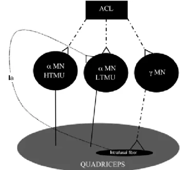
Changes in afferent discharge following ACL injury leads to AMI partially through the spinal reflexive pathways within the spinal cord. The alterations in afferent signaling inhibit excitation of the α-motoneuron pool of the quadriceps causing incomplete activation
of the quadriceps during voluntary tasks.45,46,150,151 Three different spinal pathways that influence excitability of the α-motoneuron pool of the quadriceps are believed to contribute to AMI. These are the
nonreciprocal (Ib) inhibitory pathway, the flexion reflex, and the Gamma (γ) loop.45,46 It is believed that swelling affects discharge of group II afferents and may facilitate the Ib inhibitory interneuron, thus inhibiting the quadriceps α-motoneuron
pool.46,152 The flexion reflex is believed to contribute to AMI by
facilitation of the knee flexors while inhibiting the quadriceps. Animal models show that increased inflammation within a joint causes an increase in flexion reflex excitability and reduced quadriceps activation.153 In order to achieve maximal activation, it is essential to have normal function of the γ-loop spinal reflex circuit. This circuit innervates muscle
spindles that transmit excitatory signals to the α-motoneuron pool. Any disruption of this
pathway contributes to AMI.154 Following ACLR, decreases in Ia input from the muscle spindles to the α-motoneuron pool are observed for up to 20 months.155,156 This is due to a
19
depletion of available neurotransmitter and a heightened Iα threshold suggesting γ-loop dysfunction from the injury and surgery is caused by increasing presynaptic inhibition thus reducing the effectiveness of the Ia afferent to signal the α-motoneuron pool and the ability to
activate the quadriceps maximally (Figure 1).133,157 It is hypothesized that damage to the ACL during injury alters the feedback from receptors within the ACL and disrupts the signaling to the γ-motoneurons and the supraspinal areas, diminishing α-motoneuron
activation during maximal activation.45,154
AMI is not entirely caused by alterations within spinal reflexive pathways. Joint afferent pathways have many connections to the supraspinal centers in conjunction with the spinal pathways.158–160 Following unilateral ACL injury, motor cortex activation threshold is reduced, as measured via transcranial magnetic stimulation, compared to healthy controls and to the contralateral limb.160,161 Lepley et al.160 reported that while spinal reflexive pathways were inhibited early on following ACLR in the acute stages of recovery, there were no
20
and the flexion reflex. Injury to the knee joint enhances descending output from portions of the brainstem increasing the aforementioned flexion reflex, thus increasing AMI.164,165 AMI is multifactorial and disruptions from spinal reflexive or corticospinal pathways likely contribute to this dysfunction (Figure 2).
The most common consequence of AMI following ACLR is quadriceps weakness, which has been well established to persist for years following injury and surgical
reconstruction. Reductions in quadriceps strength influence a variety of outcomes following ACLR and likely play a vital role in the development of PTOA.44,50,56 Sixty-one percent of the variance in self-reported disability following ACLR can be predicted from quadriceps strength.50,166 Tourville et al.56 demonstrated that patients with reduced quadriceps strength at approximately 4 years post-ACLR also presented with significant reductions in
tibiofemoral joint space width, a hallmark indicator of cartilage health. The quadriceps’ role during ambulation is to attenuate shock and evenly distribute loads across the knee joint.
21
Quadriceps dysfunction alters the ability of the muscle to eccentrically control the limb during walking, altering the
dissipation of forces across the joint. Reductions in quadriceps strength influence lower extremity biomechanical patterns with weak individuals displaying a reduction in knee flexion angle and internal knee extension moments
compared to health control
participants.42 A reduction in knee flexion angles during the stance phase of gait is related to greater vGRF magnitude and loading rate, which may increase degenerative changes to the articular cartilage.167 Animal models suggest that greater magnitude and rate of loading influence cartilage breakdown and has been display during walking in females following ACLR.31–33 Individuals who can produce higher rate of torque development (RTD) during isometric contractions display lower magnitude and loading rate of the vGRF and reduced magnitude of the heel strike transient.43 Throughout the stance phase of gait, the quadriceps perform an eccentric action that allows for smooth motion through controlled knee flexion to attenuate force across the knee joint.42 Reductions in strength, leading to lesser knee flexion, contribute to lesser internal knee extension moment (KEM). KEM is a surrogate for the net quadriceps contribution during loading, with lower KEM potentially indicating less energy absorption by the quadriceps, and a greater amount of force being applied to the knee joint.133,168 This indicates that those with better quadriceps function following ACLR walk
22
with a more normal gait pattern, potentially reducing the risk of PTOA development. Andriacchi et al.17,169 have demonstrated that quadriceps dysfunction from AMI directly influences gait biomechanics, and these mechanics change both the mechanical and biological environment within the knee joint (Figure 3).
Alterations in Gait Biomechanics Following ACLR
Quadriceps weakness and dysfunction following ACLR is a primary contributor to altered walking gait biomechanics following ACLR.41,42,133 As mentioned previously, the quadriceps attenuate forces during gait and evenly disperse load across the joint. Any alteration in quadriceps function alters how loads are distributed and changes both the magnitude and the rate of loading, which is disadvantageous to cartilage health.31,51,170 Articular cartilage is viscoelastic in nature and is therefore highly influenced by the rate at which it is loaded.171,172 Animal models show that repetitive loading of articular cartilage at higher rates of loading causes a stiffening response followed by fissuring and breakdown, mimicking rapid degeneration.31,32,172 The rapid degeneration of cartilage seen as early as one year following ACLR is likely attributed to changes in loading rate and magnitude during gait.13 Blackburn et al.173 demonstrated that poorer quadriceps function was
23
symptomatic and greater than 24 months removed from surgery display greater vGRF.174 This suggest that symptomatic individuals may offload the joint early post-ACLR but over time develop a gait strategy that overloads the joint.
Healthy cartilage is able to adapt to loading during cyclical tasks such as walking, biking, and jogging by increasing cartilage thickness.175 Loading in healthy cartilage leads to greater proteoglycan production and an increase in chondrocytes, thus causing a thickening response.176 The thickest region of femoral articular cartilage in healthy individuals is highly related to knee flexion angle at heel strike, and typically the thickest tibial and femoral cartilage regions align near full extension.177 On the contrary, immobilization of a joint causes cartilage thinning, indicating that a certain amount of loading is necessary to maintain cartilage health.178 Alterations in kinematics are believed to contribute to cartilage
degeneration by shifting the region of loading from areas of thicker cartilage to thinner areas that are not accustomed to repetitive loading.49,169 A change in the loading pattern to areas of unconditioned, thinner cartilage leads to greater fibrillation and breakdown of collagen, speeding up the progression of PTOA.169
A change in the overall tibiofemoral contact force may also contribute to cartilage degeneration. The quadriceps and hamstring musculature are important contributors to force applied on the joint. Any alteration in muscle activation, may drastically alter force
24
protective mechanism. Saxby et al.179 demonstrate that those with ACLR exhibit lower tibiofemoral contact force during walking, running, and side stepping. These lower contact forces, are believed to be attributed to lower sagittal plane angles and moments.179 While the contact forces were lower, the ACLR knees underwent less total range of motion, therefore reducing the total contact area, leading to focused load in a finite area of the cartilage. On the contrary, Gardinier et al.182 demonstrate smaller contact forces in both the medial and lateral compartments of the ACLR limb compared to the contralateral limb, without a change in contact area. Changes in tibiofemoral contact force are persistent and some individuals continue to display alterations following the resumption of sport like activity.20 These alterations in contact force may be a critical variable in the development of PTOA as those with ACLR who go on to develop PTOA within 5 years of surgery demonstrate greater interlimb differences in contact forces at only 6 months post-surgery compared to those who do not go on to develop PTOA.24 Caution must be taken with the interpretation of the literature regarding tibiofemoral contact forces. Most of these studies utilize EMG driven models to estimate contact force and area and may not truly represent the true contact forces presented. EMG driven models introduce the potential for a multitude of errors that need to be considered when interpreting the data. These models assume that EMG during maximal contractions at a given angle are representative of the forces at different joint angles during dynamic tasks and this may not be completely accurate. In a pathological population, inhibition and atrophy can lead to altered EMG activity that may not be a true representation of the status of the muscles of interest, thus invalidating the EMG driven model.
25
contractions is related to greater antagonist activation of the hamstring musculature.183 Individuals with ACLR also demonstrate greater quadriceps-hamstring co-activation during walking gait at heel strike and during the preparatory phases of gait compared to the
contralateral limb and to a healthy control cohort.184 Greater co-activation in these same individuals was related to lesser knee flexion displacement and smaller internal KEM.184 Greater co-activation during gait may have a deleterious impact on cartilage loading by making the limb more stiff, thus increasing the compressive forces between the femoral and tibial articulating surfaces.185 Individuals with knee OA are also reported to walk with greater co-activation.186 Those who display a longer duration of co-activation are also more likely to develop knee OA at a quicker rate than those who display lesser co-activation throughout the gait cycle.187
Changes in gait biomechanics theorized to contribute to the risk of PTOA
development are consistently linked to alterations in the sagittal plane, and this is driven by quadriceps dysfunction.42 Decreased quadriceps strength, commonly seen in individuals with ACLR, is related to reduced sagittal plane moments and deficits in knee flexion angles.42 Dynamic stability of the knee joint is highly dependent on the quadriceps, and as noted previously, is essential for mediating loading rates and dissipating forces during gait.44,188,189 The quadriceps produces an internal knee extension moment in response to large external flexion moments. These sagittal plane kinetics and kinematics are consistently reported in the literature as being altered following ACLR and contributing to the development of PTOA by altering contact area and forces within the knee joint.22,25,28,181,190–193 At 6 months
26
resolved with rehabilitation. Other evidence suggests smaller knee flexion angles are seen in those between 1- and 3-years post ACLR compared to the contralateral limb.196 Compared to the contralateral knee, data consistently supports the notion that the ACLR displays lesser knee flexion angles throughout walking gait.19,21,197 Di Stasi et al.19 demonstrate that both those who pass and fail return to sport criteria display lesser peak knee flexion angles and external flexion moments compared to the uninvolved limb. Those who failed return to sport testing presented with lesser knee flexion angles and smaller sagittal moments during
walking compared those who passed testing, further suggesting that quadriceps function plays an essential role gait biomechanics following ACLR.19 Scanlan et al.28 reported that individuals following ACLR who walk with smaller sagittal plane moments display greater changes in cartilage morphology, therefore linking a potential mechanism between sagittal plane alteration and PTOA development. Khandha et al.25 support this notion by showing that those 5 years removed from ACLR who develop PTOA present with a gait pattern characterized by smaller knee flexion angles and sagittal moments. Wellsandt et al.24 also showed that those who display smaller sagittal moments at 2 years post ACLR were more likely to develop OA by 5 years post-surgery.
Changes in sagittal plane angles and moments are believed to be the primary
27
tibiofemoral contact area and force, it is likely that deficits in quadriceps activation and strength lead to the inability to support eccentric control of the limb during loading phases of gait. This leads to a reduction in knee flexion angle and potentially greater co-activation of the quadriceps and hamstrings, thus increasing tibiofemoral contact forces over a smaller contact area and altering changes in loading of articular cartilage that may lead to cartilage degradation.
Gait biomechanics are consistently reported as a primary contributor to PTOA development following ACLR, however, discrepancies in the literature with regards of timing following surgery and directionality of certain variables make it difficult to draw succinct conclusions about the long-term effects of gait biomechanical deviations on joint health. A systematic review and meta-analysis by Hart et al.192 indicate that individuals within 6-12 months following ACLR demonstrate smaller sagittal angles and moments during gait and this persists up to 3 years following injury. However, after 3 years, there is conflicting evidence that may suggest sagittal anomalies resolve. Conversely, Goetschius et al.191 report lower sagittal plane moments early and long term following ACLR compared to both control and contralateral limbs. The discrepancies in gait biomechanics may be
28
they require a greater contribution from the quadriceps as evident from greater EMG activity of the quadriceps compared to level walking.199 In those with a total knee replacement, joint loading is greater when walking downhill compared to healthy controls, but not during level walking.200 As grade increases, the demand for greater quadriceps activation continues to increase.201 During stair walking, both up and down, those with ACLR display lower sagittal moments in the ACLR compared to the contralateral limb and healthy controls, with the contralateral limb displaying greater sagittal moment than healthy controls.202 This suggests that following ACLR, during daily ambulation involving stairs, the quadriceps in the ACLR limb contribute much less than the contralateral limb, therefore causing the contralateral limb to work harder to complete the task as a compensatory mechanism. More complex tasks, such as walking up or down stairs/hills, in those with greater quadriceps dysfunction may display greater abnormalities due to the increased level of difficulty. Those with knee OA display lesser sagittal moments during level, upstairs, and down stairs walking.26 Liikavainio et al.203 demonstrate that impulsive loading in the lower extremity is more prevalent during stair descent compared to level walking in those with OA and is related to activation of the quadriceps musculature activation during this specific task. Based on the aforementioned literature, we believe that those with greater quadriceps dysfunction following ACLR will present with greater interlimb differences when presented with the more challenging task of walking inclined and declined. Those with poorer QMQ will also present with lesser sagittal moments and angles, but greater vGRF and loading rates in the ACLR limb.
Morphological changes to the quadriceps following ACLR.
Following ACLR, atrophy of the is one of the most common measurable
29
30
lateralis, and vastus medialis volume post-surgery. This study must be interpreted with caution as only 4 individuals were included in the analysis.
31
to the changes in medicine, surgical approach, and rehabilitation changes over the last 30 years. Lindstrom et al.213 show that over the course of 1-year, the quadriceps undergoes a morphological change both in size and quality. Using CT scans at 1 week pre-op and 1 year post op, the quadriceps CSA was smaller at 1 year compared to pre-op measures and
compared to the uninjured limb. Interestingly, the quadriceps muscle at 1 year demonstrated lower attenuation compared to pre-op and to the uninjured limb.213 Attenuation from CT was measured using Hounsfield measurements, with lower numbers indicating greater water or fat content.
Intramuscular fat can have a negative effect on the function of a muscle leading to a multitude of deleterious outcomes.59 Kumar at al.214 report that individuals with knee OA who had greater intramuscular fat were likely to be more symptomatic and present with a greater severity of joint degradation measured via MRI. Quadriceps strength was also lower in OA individuals with great intramuscular fat, even with no difference in CSA of the quadriceps musculature.214 Greater increases in intramuscular fat over time are also related to greater cartilage degradation over a 3 year period;215 with each % increase in fatty infiltration of the quadriceps, there was an 83% increase in the risk of cartilage degradation as measured via MRI. This indicates that greater fat content within the quadriceps affects its ability to function properly, potentially altering how the muscle disperses forces, and leading to altered forces across the joint and greater cartilage breakdown. Lower attenuation of the quadriceps musculature, previously mentioned, potentially mimics the effect seen in those with OA and may be a precursor to cartilage degradation and PTOA.213
Increased intramuscular fat (IMAT) occurs due to a combination of different
32
to IMAT. As BMI increases, generally, the fat content within a muscle will also increase.59 Inactivity, deficits in muscle activation, immobilization, and injury are also primary
contributors to fatty infiltration within a muscle.59 In turn, greater IMAT leads to further deficits in strength, activation, and mobility limitations, providing a catalyst for more IMAT in a deleterious cycle of cause and effect.59 Rahemi et al.60 report the deleterious effects of greater IMAT by showing that greater fat within a muscle fiber dramatically reduced the ability to generate force. Greater IMAT increases stiffness within muscle fiber, reducing the ability to shorten, and therefore reducing the overall force production capacity of the
muscle.60 Greater IMAT also contributes to reduced capacity to voluntarily activate the quadriceps muscle.64 MRI derived measures of IMAT are strongly negatively associated with CAR of the quadriceps, indicating that greater amounts of IMAT are related to reduced voluntary activation capacity.64 Baum et al.216 reported a strong negative relationship
between isometric knee extension strength and MRI-derived IMAT with weaker relationships related to quadriceps size. Following ACLR, the literature suggests there is a significant decrease in physical activity immediately following injury, as well as persistent inhibition of the quadriceps, thus providing a mechanism for fat to infiltrate the quadriceps. Even in the absence of size differences, an alteration in the fat content with the quadriceps following ACLR may drastically impact quadriceps function, thus contributing to the risk of PTOA development.
Ultrasound to Measure Quadriceps Muscle Quality
33
Ultrasound is an increasingly used tool to analyze muscle size and compositional characteristics due to its ease of use, time saving capabilities, cheaper costs, and clinical feasibility compared to MRI and CT scans. Palmer et el.65 establish panoramic US as a valid and reliable tool for investigating muscle composition via the evaluation of EI. Young et al.66 demonstrate strong associations between EI obtained via US and intramuscular fat derived from MRI, indicating that EI is a practical and reproducible method for evaluating intramuscular fat. Pillen et al.58 in a study looking a fibrous tissue in golden retrievers, validated EI measured via US to muscle biopsies. In the elderly68,69,217 and in those with OA70 poorer muscle quality is related to deleterious effects on strength and function that affect activities of daily living. Yoshida et al.64 even found that the presence of greater intramuscular fat in the elderly was linked to deficits in central activation ratio. In stroke survivors, those with higher echo intensities within the lower extremity muscles also display mobility limitations during simple tasks such as walking.218 Our preliminary data show the ACLR limb in individuals an average of 48 months post-ACLR has higher EI compared to the contralateral limb. Greater EI in this ACLR cohort was also strongly related to poor isometric strength and deficits during a single limb hopping task. This suggests that
34
US and Obtaining an Image
US is an imagining technique that uses sound frequencies, undetectable to the human ear, to penetrate tissues and pixelate images based on reflected sound. Piezoelectric crystals within a transducer vibrate rapidly to create sound waves that move through tissues at known speeds. The sound waves reflect off anatomical structures, and the returning sound creates an image. The depth of the image is determined by the time it takes for the sound wave to return to the transducer. Many factors play a role in how a sound beam is reflected to a transducer. When a transducer pulsates a sound beam into a human tissue, 4 things happen with the signal:
1. Absorption: Some of the signal is absorbed into the tissues and not returned to the transducer. Absorption of a sound beam is directly related to the frequency. Higher frequencies = more absorption of the signal. Higher frequencies may be beneficial for better image quality of superficial tissues but not for deeper tissues as more signal is absorbed before reaching the deeper underlying structures. This can also be
described as attenuation of the US signal.
2. Refraction: occurs when the angle of incidence of the transducer to the tissue of interest is not perfectly 90 degrees. The sound beam reflects off a tissue but does not return back at a 90 degree angle to the transducer. Some of the signal is lost and in extreme circumstances, the location of the image is distorted relative to the probe position. This also occurs when imaging curved structures. In human anatomy, rarely are structures perfectly flat. The sound beam hitting a curved structure causes refraction of the signal away from the transducer. This can be an issue when
35
the sound hits the surface and reflects back the signal at a 90 degree angle so the images is clearer, but as you approach the medial and lateral condyles, the sound beams hit bone at an acute angle causing less clarity in the image.
3. Scattering: Scattering occurs when the sound beam is disbursed in multiple directions but not returning to the transducer.
4. Reflection: Reflection is how we obtain an image. Sound beams that reflect off tissue and back to the transducer are processed to derive an image. Reflections occur whenever the sound beam passes from one type of tissue into another. In skeletal muscle for instance, the sound beam travels through gel, into the skin, through subcutaneous fat, fascia, muscle, fat and fibrous tissue in the muscle, and eventually hitting bone. Each type of tissue has a specific acoustic impedance that affects the speed at which the sound beam travels. When two tissues of difference acoustic impedances are next to each other, a reflection will occur. The greater the difference in impedance, the greater the reflectivity. The amount of reflectivity is derived from the equation: % reflected = ((Z2-Z1)/(Z2+Z1))^2 *100, where Z1 and Z2 are acoustic impedances of different tissues. The greater the change in acoustic impedance
between tissues, the greater the reflectivity.
36
sufficient for capturing the entirety of the quadriceps, this depth was insufficient for some patients. Gain is also important but is machine dependent. Therefore, it is important to keep the gain consistent throughout a study as it will greatly affect EI.
Patient and Probe Position
For the vastus lateralis and rectus femoris, the literature suggests obtaining US images at 50% of the femoral thigh segment length as measured from the most lateral aspect of the greater trochanter to the lateral joint space of the knee.65,67 Palmer et al.65 suggest that panoramic US is a useful and reliable tool for measuring thigh CSA and EI. The knee should be placed in 50 degrees of flexion and supported with a bolster.219 Probe placement should be flush with the skin at an angle of 90 degrees for optimal angle of incidence. A pad can be used to maintain alignment as a cross sectional cut across the thigh is obtained, generally moving slowly from laterally to medially. Ample gel is needed as a medium, as any air between the surface of the skin and the transducer can distort the image. Keeping the probe positioned consistently through the cross sectional, panoramic assessment is essential to prevent overlap of the pixels. For panoramic images, one image can be used to obtain a cross section of both the vastus lateralis and rectus femoris concomitantly, or separate images for each muscle. Obtaining separate images is recommended to reduce motion artifact and error.
Image analysis
37
outside of the muscle, excluding the fascial borders. Using built in algorithms based on the set scale, the CSA of the muscle of interest is obtained. EI is also calculated from the region of interest. EI is the average brightness within the muscle. This arbitrary measure is based on a gray scale scored from 0-255. The higher the number, the higher the EI. EI is inversely related to muscle quality. Greater amounts of fat within the muscle result in greater
reflection of the sound wave, thus resulting in a brighter image and a higher EI. An image that of exclusively muscle tissue would be almost completely black (low EI; high muscle quality). Therefore, higher EI equates to poorer muscle quality. Subcutaneous fat thickness must also be accounted for as described by Young et al.66 and further validated by Ryan.67 When soundwaves pass through greater amounts of subcutaneous fat, more of the signal is lost and cause the underlying muscle to appear darker. Darker muscle represents higher quality, therefore, not accounting for subcutaneous fat may lead to an interpretation that a muscle may be of better quality than it actually is. For each muscle, three measures of subcutaneous fat thickness via the straight-line function should be obtained an averaged. One measure from near the middle of the image, one from the lateral aspect of the muscle, and a third from the medial aspect of the muscle are average and input to an equation from Young et al.66 along with the raw EI, to derive a corrected EI value. Thicker SF can distort the image underneath the layer of fat, and therefore must be accounted for.
Summary
Following ACLR, the literature suggests that quadriceps dysfunction is common and persists for many years. The most commonly attributed mechanism to quadriceps
38
Traditional therapies for addressing quadriceps dysfunction following ACLR are often inadequate. Poor quadriceps function manifests in altered walking gait biomechanics, specifically changes in the sagittal plane moment and angles. These changes in the sagittal plane potentially alter the magnitude and rate of loading of cartilage within the knee joint. Changes in sagittal plane moment and angle also alter the loading contact area and contact force of tibiofemoral joint. Overall, changes in biomechanics due to quadriceps dysfunction are considered primary contributors to the increased risk of PTOA development following ACLR. Therapies fail at decreasing the risk of PTOA as they may not target all mechanisms for quadriceps dysfunction. Based on our preliminary data and previous studies evaluating compositional changes in the quadriceps following ACLR, there is potential for increased fatty infiltration in the quadriceps that may not be targeted with current rehabilitation approaches. Greater intramuscular fat has deleterious affects on quadriceps activation and strength, and may facilitate aberrant gait biomechanics in individuals with ACLR.
39
CHAPTER III: EXPERIMENTAL DESIGN AND METHODS
Subjects
Aims 1-3
Thirty individuals who are between the ages of 12 and 35 years with a unilateral ACL injury and plan for surgical reconstruction will be recruited to participate in this study. Individuals with a history of other lower extremity surgery, previous ACLR to either limb, a lower extremity injury other than primary ACL injury within the 6 months prior to
participation, concussion within the 6 months prior to participation, or neurological disorder will be excluded. Subjects will be recruited from local orthopedic surgeons and
rehabilitation clinics that treat high volumes of ACLR patients each year. The surgeons contributing this study performed a total of 351 ACL reconstructions during the year of 2018 with approximately 241 falling within our age range for participation. Thirty healthy, uninjured controls will also be recruited for aim 1 for a comparison of changes in QMQ over the same time periods
Experimental Design
Aims 1-3
40
office or on campus in the MOTION Science institute at 1-month post ACLR. The third fourth sessions will take place in the MOTION Science Institute at the University of North Carolina at Chapel Hill at 3 and 6 months post-ACLR. Visit 1 and 2 will consist of
completion of informed consent, an US assessment of the quadriceps, and completion of patient reported outcome surveys (PROs). Subjects will complete the same US assessment and PROs at the 3rd and 4th visits. Sessions 3 and 4 will also involve an analysis of walking gait biomechanics during which subjects will walk for 2-minute intervals on an instrumented treadmill (Figure 3). Quadriceps function and hopping tasks will also be assessed during the 4th session. The healthy control cohort will only complete the ultrasound assessment and all data collection will be in the MOTION Science Institute at the University of North Carolina at Chapel Hill
Assessments
Ultrasonography of the Quadriceps
41
measurements will be obtained with the subjects lying supine with the legs fully relaxed and bolstered to 50° as depicted in Figure 1. Transmission gel will be liberally applied to the skin to enhance acoustic coupling and minimize image artifact. Panoramic US images of the VL and RF will be
obtained to determine echo intensity (EI) with the following settings: gain (56 dB), depth (6 cm), and frequency (10 MHz). A high-density foam pad will be
positioned perpendicular to the longitudinal axis of the thigh segment to help
stabilize the probe during the panoramic scan. This will allow the investigator to move the US probe perpendicular to the skin (from lateral to medial) during the imaging assessments (Figure 1). For each assessment, the probe will be moved with consistent speed and minimal pressure to minimize motion artifact and avoid compression of the underlying structures. From each image, we will also obtain anatomical cross-sectional area (CSA) and
subcutaneous fat thickness (SFT). SFT will be used to correct EI during image analysis.66,67 While CSA is not part of our primary aims, it will be collected for a secondary analysis investigating concurrent changes in muscle size relative to changes in muscle quality.
42
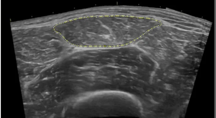
Ultrasonographic Image Processing
A single investigator who will not be involved with obtaining the ultrasound images and blinded to the limb condition (surgical or non-surgical) and time (Baseline, 3 months, and 6 months) will use ImageJ software (National Institutes of Health, Bethesda, MD, USA) for all US imaging analyses. Using the
straight-line function, each image will be scaled individually from pixels to centimeters. To determine EI, the polygon function will be used to select the region of interest (ROI), including as much of the muscle as possible
and avoiding the surrounding fascia as depicted in Figure 2. EI will be assessed to determine QMQ using computer-aided gray-scale ranging between 0 (black) and 255 (white) arbitrary units (AU) assigned to each pixel from which the mean across the ROI is calculated. CSA will be calculated as the area (cm2) of the same region of interest. Values of EI will then be corrected for subcutaneous fat using the following equation: Corrected EI = Uncorrected EI + (SFT × 40.5278) to account for the influence of fat on the brightness of the image of the underlying muscle.66 US signals are attenuated as they travel through more tissues due to a variety of factors including reflection.221 Greater subcutaneous fat results in more reflective superficially such that the signal is weaker when it reaches the underlying muscle, resulting in a darker image at greater depths.67 SFT will be determined by measuring the distance from deepest skin border to the superficial fascia surface in three different locations (lateral, middle, medial) anterior to the muscle of interest and the mean of the three distances will be used for correction.
43
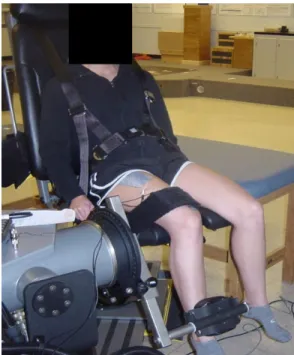
Walking Gait Biomechanical Assessment
A modified retroreflective marker bony landmark setup will be used for data collection. Markers will be affixed with double sided tape bilaterally on the anterior thigh, anterior tibia, base of the 1st and 5th metatarsals, calcaneus, the anterior superior iliac spine, greater trochanter, medial/lateral femoral epicondyle, medial/lateral malleoli, and acromion processes. A cluster of markers will then be placed over the sacrum with the addition of a marker over L4/L5 vertebra and the manubrium of the sternum. A static trial will be recorded at the beginning of each session for marker identification and used to create knee and ankle joint centers (Figure 3C). Medial epicondyle and medal malleoli markers will then be removed during dynamic trials. Marker trajectories will be collected using an 8 camera 3D motion capture system with a sampling frequency of 200Hz with Qualisys Track
Manager Software (Qualisys Motion Capture Systems, Göteborg Sweden) and lowpass filtered at 10Hz.222 The cameras will be interfaced with a split belt instrumented treadmill collecting ground reaction force data at 2000 Hz and lowpass filtered at 10 Hz.33,223,224 Five over ground walking trials will be performed at a self-selected, comfortable speed between infrared timing gates to determine the preferred walking speed. For aim 2, we will
44
using the first minute to familiarize the subject with walking on the split belt treadmill. (Figure 5).
Biomechanics Processing
All walking trials will be initially assessed in QTM software to identify and label marker trajectories. Once labeled, the marker trajectory data will be synced with the ground reaction force data and exported from QTM to Visual 3D software as a C3D file for further analysis. Visual 3D uses the labeled trajectories to construct a 3D model for each participant. The lower extremity and trunk segments for each participant will be modeled as rigid bodies using the individual markers with at least 3 non-collinear markers representing each segment as listed below:
• Foot: calcaneus, base of the 1st metatarsal, base of the 5th metatarsal Figure 5. Gait Biomechanics Assessment; A) Static Trial; B) Level Walking
45
• Shank: tibia, right lateral malleolus, right medial malleolus, right lateral epicondyle, right medial epicondyle
• Thigh: thigh, right lateral epicondyle, right medial epicondyle, right greater trochanter
• Pelvis: Sacral cluster, right anterior superior iliac spine, left anterior superior iliac spine
• Trunk: right acromion process, left acromion process, sternal notch, L4-L5
Joint centers will be calculated from the rigid body segments of the static trials. Ankle joint centers will be created from the midpoint between the lateral and medial malleolus markers. Knee joint centers will be defined as the midpoint between the lateral and medial epicondyle markers. We will use the Bell method to determine the hip joint centers.225 After joint centers are created, the local coordinate system for each segment will be aligned to the global axis system. The medial/lateral axis of each local system will be aligned to the world Y-axis, anterior/posterior to the world X-axis, and the inferior/superior to the world Z-axis similar to the ISB recommendations for joint coordinate systems.226
Analysis will be restricted to the first 50% of the stance phase via custom software (LabVIEW, National Instruments Corp, Austin, TX). The stance phase will be defined as the
interval from heel strike (vertical ground reaction force [vGRF] ≥ 20N) to toe off (vGRF ≤
20 N). Kinetic outcomes will include the peak vGRF, time derivative) and peak internal extension moment EXT. Moments will be calculated by combining kinetics, kinematics, and anthropometric data through a standard inverse dynamic procedure to yield net internal joint moments. Forces will be normalized to body weight (xBW) while moments will be


![Table 2 Average Echointensity at Each Time point [Mean (SD)]](https://thumb-us.123doks.com/thumbv2/123dok_us/7946852.2112048/82.918.135.712.326.636/table-average-echointensity-time-point-mean-sd.webp)
![Table 3. Average Cross-Sectional Area at Each Time point [Mean (SD)]](https://thumb-us.123doks.com/thumbv2/123dok_us/7946852.2112048/85.918.133.790.235.522/table-average-cross-sectional-area-time-point-mean.webp)
