Original Research Article
Correlation of fine needle aspiration cytology with histopathological
diagnosis in assessing breast lumps at a tertiary care hospital
Greeshma Ann George
1, Philips Antony
2*
INTRODUCTION
Breast cancer is one of the most common malignancies in women worldwide (22%) and India ranks the second after cervical cancer.1 The most common symptoms reported
are pain, a palpable mass or nipple discharge. There is an increasing awareness with associated anxiety and stress among women, who perceive every symptom in breast as cancer, compelling them to seek medical advice. It is sometimes very difficult to determine simply from
clinical assessment whether a suspicious lump is benign or malignant.2
As it is the one of the leading causes of cancer death in women, the major concern of the surgeon and the responsibility of the surgical pathologist lies in the ability to differentiate a benign lesion from a malignant one.3-4
Women should be educated and should be aware of the symptoms of Breast cancer. Screening can be done stringently to detect the cancer at the earliest. So, we are
1Department ofPathology, Kanachur Institute of Medical Sciences, Mangalore, Karnataka, India
2Department of Respiratory Medicine, Kanachur Institute of Medical Sciences, Mangalore, Karnataka, India
Received: 09 September 2018
Accepted: 06 October 2018
*Correspondence:
Dr. Philips Antony,
E-mail: phil.antony@gmail.com
Copyright: © the author(s), publisher and licensee Medip Academy. This is an open-access article distributed under the terms of the Creative Commons Attribution Non-Commercial License, which permits unrestricted non-commercial use, distribution, and reproduction in any medium, provided the original work is properly cited.
ABSTRACT
Background: Breast cancer is one of the most common malignancies in women worldwide (22%) and India ranks the second after cervical cancer. The diagnostic accuracy of FNAC increases to 99% when it is combined with clinical and radiological examination. In this study, authors plan to correlate the cytological findings with histopathological examinations for breast lesions and determine the accuracy of fine needle aspiration cytology in the diagnosis of breast lesions.
Methods: It is a retrospective study carried out in the Department of Pathology at a Tertiary Care Hospital. All the FNAC results of breast lesions during the one year period were collected. Gauge needle maximum of 3 passes were made and the slides were fixed in 70-80% alcohol and stained with routine haematoxylin and eosin stain.
Results: Among 200 patients, 197 were females and 3 were males. Benign breast lesions were found in 158 cases; among which fibroadenoma was the commonest lesion. Malignancy was observed in 25 cases. Two cases of phyllodes tumour were incorrectly reported as fibroadenoma on cytology. Of 12 cases which were diagnosed to have atypical lesions, 4 cases were papillary neoplasm, and 8 cases were atypical ductal hyperplasia.
Conclusions: This study concludes that breast FNAC is a reliable, easy, cheap and effective procedure for the diagnosis. It reduces the need of core needle biopsies and very well correlated with histopathological examination. FNAC differentiates non neoplastic from the neoplastic by which it reduces the patient’s anxiety and helps the surgeons in planning the mode of treatment.
Keywords: Breast, Cytological, FNAC, Histopathological
in a main way to detect breast lesions at the earliest and to reduce the disease burden.
Self-breast examination along with triple test which includes clinical examination, mammogram and Fine Needle Aspiration Cytology (FNAC) has reduced the false negative rate to less than 1%.5 The diagnostic
accuracy of FNAC increases to 99% when it is combined with clinical and radiological examination and accuracy further increases to 100% when it is combined with cell block preparation.6-7
Although open surgical biopsy is the ‘gold standard’ for diagnosis of palpable breast lesions, two types of minimally invasive breast biopsy techniques, core needle biopsy (CNB) and fine needle aspiration cytology (FNAC), have become established for the diagnostic evaluation of palpable breast lesions in the recent years.8
Core biopsy is also an effective mean to diagnose breast lumps, but it is time consuming and expensive, and can be associated with complications like haematoma and rarely pneumothorax.9-10
Adnan K et al, described some of the difficulties and pitfalls in FNAC such as inadequate samples, suspicious malignancies and overlapping features of different lesions.11 In settings like ours where resources are poor,
FNAC comes readily useful for its obvious advantages. It is a cheap, fast, and reliable diagnostic method. It also reduces the need and the frequencies for taking up the patient for open biopsies.12 It is also possible that the
smears may be acellular (no cells are harvested) making cytological analysis impossible. These are described as inadequate aspirates and cases have to be converted to CNB which can mostly solve the problem.13-14
In this study, we plan to correlate the cytological findings with histopathological examinations for breast lesions and determine the accuracy of fine needle aspiration cytology in the diagnosis of breast lesions.
METHODS
A cross sectional and retrospective study was undertaken to study the clinical profile and correlation between FNAC and Histopathology reports of breast lumps. This study was conducted in Department of Pathology, at a tertiary care hospital for one-year period (March 2017-March 2018). A total of 258 patients attending the outpatient department were chosen as study sample. Out of these cases 58 of them did not have histological correlation available and were excluded from the study. All the patients aged more than 15 years presenting with breast lumps were included in the study. Patients with breast pain of any cause were excluded from the study.
The patients thus selected were subjected for detailed history including general physical examination, systemic and local examination. The patients were also subjected
investigations. The FNAC report and histopathological report were correlated.
Procedure for FNAC
FNAC procedure was explained and an informed consent was taken from the patients. FNA was done under strict aseptic precaution utilizing 5ml or 10ml disposable syringe for each prick and for each patient. Maximum of 3 passes were made, no local anaesthetic was utilized. The needle was inserted into the palpable lesions, either once or twice depending upon the size of the nodule. Cellular material was aspirated into a syringe and expelled onto slides. Three to four slides were prepared for each patient. A minute or medium-sized drop of aspirate was put near the frosted cessation of a slide that was placed on a table. A second slide was acclimated to spread the aspirated material in the same manner used to prepare a peripheral blood smear. One of the smears was wet fine-tuned in 95% methanol and stained with Hematoxylin and Eosin (H and E). The other onez were air dried smear and were stained with Giemsa stain. The procedure was repeated if the sample was inadequate. The procedure was done within one hour, and the reports were signed out within 1-2days. Smears were screened by two pathologists and the FNAC results were reported. The cytology reports were classified as benign, atypical, suspicious, malignancy, and unsatisfactory.
Procedure for histopathology
The biopsy specimens were fine-tuned in 10% formalin for 24 hours. Then gross examination was done in the Department of Pathology by consultant histopathologists. The gross and cut section findings were noted. Several bits were taken from opportune sites for processing and paraffin embedding. From each block, sections were cut at 4-5 microns thickness and stained with H and E.
The data thus obtained was entered in a predesigned proforma. The data was analysed using Statistical Package for social services (SPSS vs 18). The categorical variables were analysed using frequencies and percentages.
RESULTS
The cytology reports were classified as benign, atypical,
suspicious, malignancy, and unsatisfactory (Table 1). Of the benign lesions, fibroadenoma to be followed by fibrocystic disease was the most common cytological diagnosis. Fibroadenoma was diagnosed in 98 patients with histology correlating with cytology in 96 cases. Two cases of phyllodes tumour were incorrectly reported as fibroadenoma on cytology. Of 12 cases who were diagnosed to atypical lesions in cytology (4 cases were papillary neoplasm, and 8 cases were atypical ductal hyperplasia) while the final histopathology showed 4 cases of papillary carcinoma, 2 cases of papilloma, 3 had atypical ductal hyperplasia, 1 case had ductal carcinoma in situ, and 2 cases had DCIS with a foci of invasive
carcinoma. Cytology was useful in diagnosing various malignancies like neuroendocrine cancer, papillary neoplasms, and metastatic neoplasm from stomach.
Table 1: Cytology impression of the patients who underwent FNAC of breast lumps.
Report No. of cases
Total number of patients 200
Benign 158
Atypical 12
Suspicious 02
Malignancy 25
Unsatisfactory 03
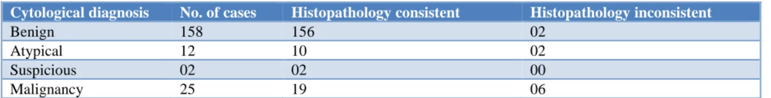
Table 2: Comparison of cytology and histopathology diagnosis.
Cytological diagnosis No. of cases Histopathology consistent Histopathology inconsistent
Benign 158 156 02
Atypical 12 10 02
Suspicious 02 02 00
Malignancy 25 19 06
Twenty patients underwent ultrasound guided FNAC for deeply seated small lesions, of which 8 had invasive cancer, one was a case of papillary lesion later confirmed as papilloma, 4 were inflammatory lesions, and 7 patients had fibrocystic disease. Three patients with unsatisfactory reports who were less than 30 years of age were repeated again and in histology as well as cytology, the lump appeared to be benign. Eight cases were incorrectly diagnosed by FNAC (2 cases of phyllodes reported as fibroadenoma and 6 cases of carcinoma diagnosed as atypical hyperplasia) (Table 2). Two cases were reported as suspicious for malignancy in cytology.
DISCUSSION
The earliest large-scale use of Fine Needle aspiration Cytology FNAC as a diagnostic tool in the management of palpable masses was recorded in Memorial Hospital, New York, United States in the 1930s but it did not gain much during the early years. The technique had a come back in Scandinavia during the 1950s and 1960s, where it flourished and kept spreading to other parts of the world.15
True FNAC for breast aspirations were first introduced by Franzen and Zajicek at the Karolinska Hospital in Stockholm.16 Despite their success, it was not until 1980s
that FNAC became widely used. The reasons included lack of confidence in the sensitivity and specificity of the procedure and there is fear of tumour implantation in the needle track.17
Fibroadenoma was the commonest benign lesion and Invasive ductal carcinoma was the commonest malignant lesion which was similar to a study done by Bhaskar et al.18 Two cases which were cytologically diagnosed as
lesions “suspicious for malignancy” were confirmed as malignant lesions on doing histopathological studies and this was similar in the study done by Panjvani SI et al.19
FNAC is a simple rapid technique performed as an outpatient procedure with less chance of complications unlike core biopsy which has risk of bleeding and occasionally rare complications like pneumothorax. It does not require any anaesthesia or hospitalization and is very cost effective. Experience and expertise in sampling and interpretation of specimen decide the effectiveness of FNAC. The other limitation of FNAC is that it cannot differentiate few lesions like fibroadenoma from phyllodes, papilloma from papillary carcinoma, phyllodes from metaplastic carcinoma breast and atypical ductal hyperplasia from ductal carcinoma insitu.
It is highly useful as an initial method of pathological assessment for palpable breast lumps. If the initial FNAC is inadequate, CNB can be a useful second line method of pathological diagnosis. Excision biopsy should be the last option to obtain a pathological diagnosis.20 In cases
Though a triple assessment is advocated for all palpable breast lumps, it is not feasible in developing countries like India, where availability and affordability are an issue at all centres and clinical decision takes a major role. The most common reason for a false negative result is failure to localize the lesion exactly. This can be minimised to an extent by performing the test under image guidance.
Hence, we would like to bring in light that FNAC is still an acceptable mode of diagnosis in experienced hands especially where there is limitation of resources. It is our conclusion therefore that the surgeons and pathologists should continue to deploy the procedure towards the early detection of breast cancer. This will also significantly reduce patient’s waiting time for incision/excision biopsy.
CONCLUSION
This study concludes that breast FNAC is a reliable, easy, cheap and effective procedure for the diagnosis of breast lumps. It reduces the need of core needle biopsies and very well correlated with histopathological examination. FNAC differentiates non neoplastic from the neoplastic by which it reduces the patient’s anxiety and helps the surgeons in planning the mode of treatment. It is also helpful in the administration of neoadjuvant therapy in the cases of malignancy. FNAC can be also be combined with cell block to increase the diagnostic accuracy. Immunohistochemistry and molecular techniques like PCR, FISH and genomic imprinting can be carried out in the cytology itself to define the cancer biology. Authors conclude from this study that diagnosis of breast lump based on FNAC should be a practice as there is high correlation with histopathological finding. FNAC should be used as a routine diagnostic procedure due to its cost effectiveness, thus maximizing the availability of health care to patients with breast lesions.
ACKNOWLEDGEMENTS
Authors would like to thank all the staffs and students from Pathology Department for their sincere help and support.
Funding: No funding sources Conflict of interest: None declared
Ethical approval: The study was approved by the Institutional Ethics Committee
REFERENCES
1. Ramachandra K, Kamaleshwar S, Sand TS. A study on risk factors of breast cancer among patients attending the tertiary care hospital in Udupi district. Indian J Community Med. 2013;38(2);95-99. 2. Yong WS, Chia KH, Poh WT and Wong CY. A
comparison of trucut biopsy with fine needle
aspiration cytology in the diagnosis of breast cancer. Singapore Med J. 1999;40(9):587-9.
3. Rahman MZ, Islam S. Fine needle aspiration cytology of palpable breast lump: a study of 1778 cases. Surgery. S12:001.
4. Obaseki DE, Olu-Edo AN, Ogunbiyi JO. Diagnostic accuracy of fine needle aspiration cytology of palpable breast masses in Benin City, Nigeria. West Afr J Med. 2010;29(4):259-62.
5. Kaufman Z, Shpitz B, Shapiro M, Rona R, Lew S, Dinbar A. Triple approach in the diagnosis of dominant breast masses: combined physical examination, mammography, and fine‐needle aspiration. J Surg Oncol. 1994 Aug;56(4):254-7. 6. Daramola AO, Odubanjo MO, Obiajulu FJ, Ikeri
NZ, Banjo AA. Correlation between fine-needle aspiration cytology and histology for palpable breast masses in a Nigerian Tertiary Health Institution. International Journal of breast Cancer. 2015;2015. 7. Ukah CO, Oluwasola OO. The clinical effectiveness
of fine needle aspiration biopsy in patients with palpable breast lesions seen at the University College Hospital, Ibadan, Nigeria: A 10-year retrospective study. J Cytol. 2011;28(3):111-3. 8. Basnet S, Talwar OP. Role of cell block preparation
in neoplastic lesions. J Pathology Nepal. 2012 Jan 1;2(4):272-6.
9. Dowlatshahi K, Jokich PM, Schmidt R, Bibbo M, Dawson PJ. Cytologic diagnosis of occult breast lesions using stereotaxic needle aspiration: a preliminary report. Archives Surg. 1987 Nov 1;122(11):1343-6.
10. Evans WP, Cade SH. Needle localization and fine-needle aspiration biopsy of nonpalpable breast lesions with use of standard and stereotactic equipment. Radiology. 1989 Oct;173(1):53-6. 11. Adnan K, Razza J, Muneeb J, Maria T. Correlation
of fine needle aspiration cytology and histopathology diagnosis in the evaluation of breast lumps. Inter J Medical Students. 2014;2(2):40-3. 12. Jindal U, Singh K, Kochhar A. Fine needle
aspiration cytology of breast lumps with histopathological correlation: a four year and eight month study from rural India. Internet J Pathol. 2012;13(3).
13. Walker SR. A randomized controlled trial comparing a 21 G needle with a 23 G needle for Fine needle aspiration of breast lumps. R Coll Surg Edinb. 1998;43:322-3.
14. Abdel-Hadi M, Abdel-Hamid GF, Abdel-Razek N, Fawzy RK. Should fine-needle aspiration cytology be the first choice diagnostic modality for the assessment of all nonpalpable breast lesions? The experience of a breast cancer screening center in Alexandria, Egypt. Diag Cytopathol. 2010;38(12):880-9.
16. Franzen S, Zajicek J. Aspiration biopsy in the
diagnosis of palpable lesions of the breast: Critical review of 3479 consecutive biopsies. Acta Radiol Ther Phys Biol. 1968;7:241-62.
17. Berner A, Torill Sauer T. Fine-needle Aspiration Cytology of the Breast. Ultra Pathol. 2011;35(4):162-7.
18. Thakkar B, Parekh M, Trivedi NJ, Agnihotri AS, Mangar U. Role of fine needle aspiration cytology in palpable breast lesions and its correlation with histopathological diagnosis. National J Med Res. 2014;4(4):283-8.
19. Panjvani SI, Parikh BJ, Parikh SB, Chaudhari BR, Patel KK, Gupta GS, et al. Utility of fine needle
aspiration cytology in the evaluation of breast lesions. JCDR. 2013 Dec;7(12):2777-79.
20. Pruthi S. Detection and evaluation of a palpable breast masses. Mayo Clin Pro. 2001;76(6):641-8.
Cite this article as: George GA, Antony P. Correlation of fine needle aspiration cytology with histopathological diagnosis in assessing breast lumps at a tertiary care hospital. Int J Res Med Sci