CELLULAR & MOLECULAR BIOLOGY LETTERS http://www.cmbl.org.pl
Received: 25 April 2011 Vollume 16 (2011) pp 493-514 Final form accepted: 11 July 2011 DOI:10.2478/s11658-011-0019-7 Published online: 18 July 2011 © 2011 by the University of Wrocław, Poland
* Author for correspondence. e-mail: mwitkowska@wum.edu.pl, phone: +48 22 628 63 34, fax: +48 22 628 78 46
Abbreviations used: AFCs – amniotic fluid-derived mesenchymal stromal cells; AM-MSCs – amniotic membrane mesenchymal stromal cells; BM-MSCs – bone marrow mesenchymal stem cells; C-MSCs – chorionic mesenchymal stem/stromal cells; EGF – epidermal growth factor; ESCs – embryonic stem cells; FGF – fibroblast growth factor; HLA – human leukocyte antigen; HSCs – hematopoietic stem cells; IGF – insulin growth factor; Il – interleukin; MHC – major histocompatibility complex; MSCs – mesenchymal stem cells; PMSCs – placenta mesenchymal stem/stromal cells; TGF-β – transforming growth factor-β; UC-MSCs – umbilical cord mesenchymal stromal/stem cells; VEGF – vascular endothelial growth factor
Review
PERINATAL SOURCES OF MESENCHYMAL STEM CELLS: WHARTON’S JELLY, AMNION AND CHORION
MALGORZATA WITKOWSKA-ZIMNY* and EDYTA WROBEL Department of Biophysics and Human Physiology, Medical University of
Warsaw, Chalubinskiego 5, 02-004 Warsaw, Poland
Abstract: Recently, stem cell biology has become an interesting topic,
especially in the context of treating diseases and injuries using transplantation therapy. Several varieties of human stem cells have been isolated and identified
in vivo and in vitro. Ideally, stem cells for regenerative medical application
should be found in abundant quantities, harvestable in a minimally invasive procedure, then safely and effectively transplanted to either an autologous or allogenic host. The two main groups of stem cells, embryonic stem cells and adult stem cells, have been expanded to include perinatal stem cells. Mesenchymal stem cells from perinatal tissue may be particularly useful in the clinic for autologous transplantation for fetuses and newborns, and after banking in later stages of life, as well as for in utero transplantation in case of genetic disorders.
This review highlights the characteristics and therapeutic potential of three human mesenchymal stem cell types obtained from perinatal sources: Wharton's jelly, the amnion, and the chorion.
Key words: Perinatal stem cells, Wharton's jelly, Amnion, Chorion, Placenta,
INTRODUCTION
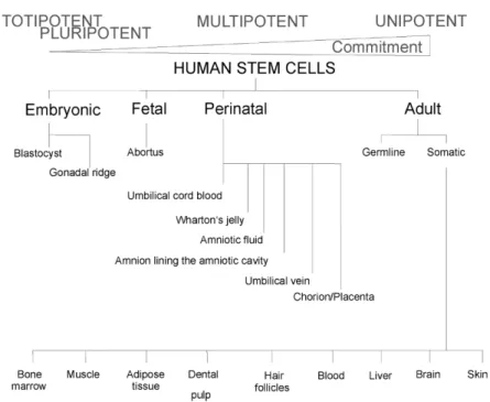
The term ‘stem’, originally derived from old botanical monographs, was used in reference to the apical root meristems responsible for the regenerative capacity of plants [1]. Nowadays, the term ‘stem cells’ may refer to various types of cells which (i) are unspecialized and can generate one or more cell lineage types of the germ layer, (ii) have the ability to replenish their own number (self-renewal). The ability of stem cells to differentiate into various mature cell types is variable for different cells. Stem cell division is organized hierarchically from the most primitive to the targeted cells into mature tissue, and on the basis of this criterion the following stem cell classification has been proposed: totipotent, pluripotent, multipotent and unipotent stem cells.
The most primitive stem cell, with the greatest opportunities for differentiation, is the totipotent cell of the zygote or first blastomere, which dates from the first division of the zygote. These cells can divide to give rise to both the embryo and the placenta. At the 32-cell stage of the embryo, known as the morula, the cells have already lost their totipotency and are pluripotent. They contribute to all three germ layers in the developing embryo. The stem cells with lower potency are multipotent stem cells that can differentiate into a number of cells, but only in the same germ layer. Multipotent stem cells give rise to unipotent stem cells that are committed to particular organs/tissues [2].
Stem cells are isolated from embryos, fetuses and adults. Based on their origin, they can be divided into four main types: (i) embryonic stem cells, (ii) fetal stem cells, (iii) perinatal stem cells, and (iv) adult/somatic stem cells (Fig. 1).
Embryonic stem cells (ESCs) are a prototype of progenitor cells which can give rise to the whole organism and yield all tissue types. However, there are still many ethical dilemmas concerning the use of these stem cells. Another potential limitation which involves the use of ESCs in regenerative medicine is their unlimited ability to proliferate – which can potentially increase the risk of malignancy in the medical use of these stem cells [3]. Tumorigenicity and ethical considerations have impeded the widespread use of embryonic stem cells in clinical applications.
Fetal tissue as a source for human stem cells also raises important ethical considerations. Moreover, it is clear that the use of human fetal tissue involves allogenic transplantation [4].
Thus, adult stem cells have aroused much greater interest. The best known example of an adult stem cell is the bone marrow stem cell, hematopoietic and non-hematopoietic, that is able to specialize into blood cells and other mesenchymal lineages. Adult stem cells are isolated from several tissue sources, including the skeletal muscle, bone, pancreas, and adipose tissue. Most of these cells preferentially generate mature cells of the same germ line as their tissue of origin: hematopoietic stem cells generate blood cells; bone marrow mesenchymal stem cells generate mesodermal cell types. However, several recent transplant studies indicate that at least a fraction of stem cells in these populations can generate cells of a different embryonic lineage in vitro and in vivo, for example: adipose derived stromal/stem cells have the ability to form bone and muscle of a mesodermal lineage; hepatocytes and pancreatic islets of an endodermal lineage; and neurons and oligodendrocytes of an ectodermal lineage [5]. Unfortunately, adult stem cells are present in miniscule quantities, their self-renewal is not as successful as ESCs, and they do not proliferate to the same degree. Moreover, because adult stem cells are not as young as ESCs, they may contain more DNA abnormalities acquired with age.
Regenerative medicine and tissue engineering are searching for a novel stem cell-based therapeutic strategy. Because of many limitations in the use of embryonic and adult stem cells, a perinatal source of stem cells seems to be an important potential source of stem cells. The term ‘perinatal’ refers to around the time of birth, but technically covers the period from 20 weeks of pregnancy through the first 28 days of the neonatal period. Normally discarded as medical waste, the umbilical cord and perinatal tissue proved to be a rich source of stem cells. The most popular perinatal stem cell source is the umbilical cord blood. Umbilical cord blood is collected at the time of birth, and provides a pool of undifferentiated cells which has been shown to be therapeutically useful for rescuing patients with bone marrow-related deficits and inborn metabolic defects [6]. In addition, cord blood may be banked, and thus is available for autologous and allogeneic cell replacement.
efficiently to either an autologous or allogenic host. Hence, many new studies are dedicated to finding new sources of stem cells.
Due to the easy access, low immunogenicity, extensive proliferation potential, multipotency, cryopreservation, and minimal ethical concerns associated with stem cells from umbilical cord blood, intensive studies on the presence of stem cells in other perinatal tissues have been started. Multipotent cells have been found in the chorion, placenta, perivascular areas, amnion, amniotic fluid, and in the tissue surrounding the umbilical cord vessels, i.e., Wharton's jelly [7-9]. In this review paper we focus on the biology and therapeutic potential of mesenchymal stem cells derived from the umbilical cord, Wharton's jelly and two fetal membranes: the amnion and the chorion.
WHARTON’S JELLY AS A MESENCHYMAL STEM CELL SOURCE
Human umbilical cord is an extra-embryonic tissue linking the mother and fetus. In the umbilical cord two arteries and one vein are surrounded by a mucoid connective tissue, called Wharton's jelly. Wharton's jelly is composed of myofibroblast-like stromal cells, collagen fibers, and proteoglycans [10]. In 2003, the existence of primitive cell types within the gelatinous connective tissue from the umbilical cord was reported for the first time [11]. Cells isolated from Wharton's jelly, referred to as umbilical cord mesenchymal stromal/stem cells (UC-MSCs), adhere to plastic. There are a few methods for the isolation of mesenchymal stem cells from the umbilical cord, e.g. density gradient centrifugation and two-step enzymatic digestion. The most efficient method is via enzymatic degradation of the extracellular matrix to release the cells from the Wharton's jelly. These cells can be isolated in large numbers, approximately ~1.5x10⁶ cells per centimeter of the umbilical cord [12]. Proliferation analysis
revealed that human UC-MSCs have a faster population doubling time than BM-MSCs. Wharton's jelly cells may be isolated and cultured for more than 80 population doublings with no indication of senescence or changes in morphology [11]. It was also shown that isolated cells are characteristic of other types of stem cells and are capable of multipotential differentiation. Cells isolated from Wharton's jelly of the umbilical cord (UC-MSCs) meet the basic criteria for mesenchymal stem cells established by the Mesenchymal and Tissue Stem Cell Committee of the International Society for Cellular Therapy in 2006. They (i) are plastic-adherent when maintained under standard culture conditions, (ii) have the capacity for osteogenic, adipogenic, and chondrogenic differentiation, and (iii) express CD73, CD90, and CD105, but do not express the hematopoietic lineage markers CD14, CD11b, CD34, CD45, CD19, and CD79 [13].
immunomodulatory activities [14, 15]. The immune properties of UC-MSCs were studied in detail, and the conclusion of Troyer and Weiss was that there is no evidence for a frank immune rejection of undifferentiated UC-MSCs in vivo, and they can be tolerated well in allogenic transplantation [16]. The transcriptome analysis of human UC-MSCs showed increased expression of genes associated with the immune system, including IL6, VEGF (important in the immunosuppressive properties of mesenchymal cells), and CD200 (prevents fetal rejection in pregnancies; a mouse model demonstrated a role of CD200 in immune tolerance of allogeneic skin and cardiac transplantation) [17-19]. Additionally, studies have demonstrated high expression of human leukocyte antigen G (HLA-G) belonging to HLA class I, which plays a role in immune tolerance during implantation and in pregnancy [20, 21]. Chen et al. for the first time demonstrated that the immunosuppressive effects of UC-MSCs depend on the prostaglandin E2 (PGE2) mechanism (blocking PGE2 biosynthesis completely
abolished the immunosuppression activities of human UC-MSCs), which probably inhibits T-cell proliferation [22]. Published in 2011, the first report of detailed transcriptome profiling of stem cells isolated from Wharton's jelly revealed and provided answers concerning certain unique properties of human UC-MSCs compared to human embryonic and other mesenchymal stem cells. Human UC-MSCs express a low level of the molecular markers typical for the pluripotent embryonic stem cell phenotype – OCT4, Nanog, Sox2 and Lin28 – thus explaining why they do not produce teratomas in vivo, in immunodeficient mice and rats [12, 23]. It was demonstrated that human UC-MSCs attenuate the growth of human breast carcinoma cells in vitro, and also in vivo, in a mouse xenograft study [24]. The anticancer effects of UC-MSCs may include upregulated expression of some genes associated with the induction of apoptosis and a tumor suppressor (IL12). It was shown that un-engineered UC-MSCs attenuated the Akt and MAPK phosphorylation in cancer cells [18, 24]. The mechanism of tumor regression is not clear, but there are suggestions that it may be via cell-to-cell contact or via factors secreted by the UC-MSCs. It suggests that UC-MSCs are a potential cytotherapeutic tool for cancer therapy.
The identification and characterization of the potential anticancer factors of UC-MSCs would be very important and of tremendous value for clinical application. No risk of malignant transformation and anticancer effects – these features strongly distinguish UC-MSCs from embryonic stem cells.
Importantly, Wharton's jelly cells reveal telomerase activity, which is found in human embryonic stem cells [25]. High telomerase activity is correlated with an early stage of differentiation and a high level of division activity in the cells. Hence, UC-MSCs may have the capability to undergo a high number of cellular divisions.
endothelial cells, cardiomyocytes, hepatocyte-like and pancreas beta cells [26-28]. In this context, UC-MSCs are an example of multipotent or even pluripotent stem cells.
Neuronal differentiation
UC-MSCs express a marker of neural precursors, nestin, without exposure to differentiation signals [11]. Under neurogenic conditions (medium supplemented with DMSO, butylated hydroxyanisole, valproic acid, forskolin, hydrocortisone and insulin), Wharton's jelly cells undergo changes in morphology, and express at a higher level several proteins consistent with the neuronal, astrocyte and oligodendrocyte phenotype: neuron-specific enolase (NSE), glial fibrillary acidic protein (GFAP), and 2',3'-cyclic nucleotide-3'-phospodiesterase (CNPase), respectively. The potential for exploring the capacity of UC-MSCs to differentiate into mature neural cells holds much promise for treating a number of devastating neuronal diseases [11, 12]. Hence, Wharton's jelly of the umbilical cord may represent an alternative source of stem cells for central nervous system cell transplantations.
Osteogenic differentiation
Various studies have demonstrated that UC-MSCs possess osteogenic potential and may be an alternative cell source for bone repair in regenerative medicine [29-31]. For bone tissue engineering, it is important to analyze the in vitro
behavior of cell-biomaterial hybrids concerning their integration in vivo into the injured tissue. UC-MSCs have expression and synthesis of many proteins which is different than BM-MSCs: upregulation of extracellular proteins, i.e. metalloproteinases MMP-1 and MMP-2; and downregulation of osteogenic markers, i.e. osteopontin and alkaline phosphatase on the collagenous matrix under osteogenic conditions [30, 32]. However, the observed differences did not affect the final result of osteogenic differentiation of these perinatal umbilical mesenchymal stem cells. The studies of Schneider et al. suggest a mechanism of UC-MSCs for bone formation which is different compared to bone marrow mesenchymal stem cells. The osteogenic differentiation properties of UC-MSCs are enhanced by their contact with collagen, whereas contact between BM-MSCs stimulates the synthesis of extracellular matrix proteins, including collagen type I, III and IV [32]. Also of interest is the impact of various factors associated with the set of properties of UC-MSCs. Their osteogenic potential does not depend on the baby's gender and mode of delivery. Interestingly, low birth weight and a shortened duration of pregnancy have a significant impact on the highest degree of osteogenic differentiation [33].
Endothelial differentiation
the umbilical cord is also a rich source of vascular endothelial cells (hUVECs), which may be isolated from the umbilical cord vein [35]. UC-MSCs may also facilitate the engraftment of hematopoietic stem cells.
Recent studies have provided encouraging results regarding the use of human UC-MSCs in tissue regeneration and repair in several models of disease, such as treating rat liver fibrosis, mice diabetes mellitus, rescuing the photoreceptors and the visual function in a rodent model of retinal disease, and ameliorating apomorphine-induced rotations in a rodent model of Parkinson's disease [12, 36-38]. UC-MSCs may be transfected with either DNA or mRNA, so they may be an efficient tool for cell-based gene therapy [39]. Wharton's jelly from the umbilical cord is a rich reservoir of mesenchymal stem cells, and has a high concentration of many growth factors (EGF – epidermal growth factor, FGF – fibroblast growth factor, IGF-I – insulin-like growth factor I), which may stimulate division and differentiation of these cells [40].
In many findings it is suggested that UC-MSCs offer advantages over stem cells as a source of therapeutic cells. Firstly, they are derived from an uncontroversial and inexhaustible source, and may be harvested noninvasively at low cost. Secondly, UC-MSCs did not induce teratomas – they have anticancer effects. Therefore, UC-MSCs are attractive cells for medical therapy.
AMNION AS A SOURCE OF STEM CELLS
The amnion is a membrane forming the amniotic sac that surrounds and protects the developing fetus. This fetal tissue is composed of three major layers: a single epithelial layer, a thick basement membrane, and an avascular mesenchyme [41]. The interior of the amniotic sac is filled with amniotic fluid, which creates the environment of the intrauterine life. Not only does it allow the fetus to move freely in the womb, but it also constitutes a kind of shock absorber protecting the fetus from mechanical injury. Besides protecting the fetus from drying out and temperature fluctuations, as well as isolating the child from strong stimuli from the outside world, it also participates in the metabolism of the fetus and the exchange to allow the transport of nutrients. Both the amniotic membrane and the amniotic fluid contain cells that may be of therapeutic interest.
Human AECs are unique cells exhibiting the characteristics of stem cells, i.e. in
vitro the ability to produce cells of three germ layers [43].
Amniotic membrane – human mesenchymal stem/stromal cells (AM-MSCs)
The transplantation of human amniotic membrane has been performed in ophthalmology since the 1950s in the treatment of ocular disorders. Amniotic membrane was used as a substitute, but also as a scaffold upon which cells may migrate and regenerate, forming healthy tissue. The existence of a high number of mesenchymal stem cells with osteogenic and adipogenic potential within the amniotic membrane was reported for the first time in 2004 by In 't Anker and colleagues [44]. AM-MSCs are easily isolated through mechanical and sequential trypsin and collagenase digestion, in a significant number, approximately 2x10⁶ per gram of the amnion [45]. They exhibit a fibroblast-like
morphology and have a capability to differentiate towards not only adipogenic and osteogenic, but also chondrogenic, skeletal myogenic and endothelial lineages [46]. AM-MSCs have the capacity to support the hematopoiesis of CD34+ cells in co-cultures, even in the absence of exogenous cytokines. There is
a possibility that the co-transplantation of the umbilical cord blood-derived hematopoietic stem cells with AM-MSCs might induce earlier and more complete recovery of hematopoiesis. This was observed in a mouse model [47] and if this observation is also confirmed in humans, this method would provide new insights for cord blood stem cell transplants. Of note, the amniotic membrane is avascular tissue, which does not contain endothelial cells; nevertheless, AM-MSCs have angiogenic abilities. Alviano et al. showed that AM-MSCs spontaneously form capillary-like structures when they are cultured in semisolid medium. Supplementing the culture medium with vascular endothelial growth factor (VEGF) strengthens the AM-MSCs angiogenic behavior [46]. AM-MSCs are capable of secreting several growth factors that support angiogenesis and tissue remodeling, and decrease inflammation, such as: transforming growth factor-β (TGF-β), basic fibroblast growth factor (FGF), epidermal growth factor (EGF), keratinocyte growth factor, and hepatocyte growth factor (HGF) [48]. The AM-MSCs retained a stable morphology for more than 20 passages. The phenotype of AM-MSCs (CD29, CD73, CD44 positive and CD14, CD34, CD45 negative) is similar to MSCs derived from adult bone marrow and umbilical cord blood [8]. The genetic profiles of the AM-MSCs were compared with the umbilical cord blood and bone marrow-derived mesenchymal stem cells (BM-MSCs). The AM-MSCs express octamer-binding transcription factors (Oct-4) – molecular markers typical for embryonic stem cells – at higher levels than BM-MSCs, which may suggest a higher position of these cells within the stem cell hierarchy compared with bone marrow derived-mesenchymal stem cells. The primitiveness of AM-MSCs also confirmed their anti-inflammatory and low immunogenic characteristics. It has been shown that AM-MSCs have the highest proliferation rates in vitro
Amniotic fluid-derived mesenchymal stromal cells (AF-MSCs)
It is known that amniotic fluid contains many cell types that come from a developing fetus. Amniotic fluid is easily obtained in a low-risk procedure of genetic diagnostics – amniocentesis – at any time point, from week 14 until the end of the pregnancy. The composition and origin of cells in the amniotic fluid change as the pregnancy progresses [50]. Due to their varied origins, these perinatal stem cells are a heterogeneous population. Among the cells in the amniotic fluid are found the epithelioid E-type cells, amniotic fluid-specific AF-type cells and fibroblast F-type cells [51].
Numerous recent publications devoted to methods of cell isolation from the stem cell niche reflect a great interest in the cells present in the amniotic fluid. [52-55]. Unfortunately, so far a considerable number of tests have been carried out on a mixed population of adherent cells isolated from the amniotic fluid without division into E-type, AF-type and F-type cells. The class of adherent cells obtained from the amniotic fluid (AF-type) appears to fulfill the criteria for MSCs, and based on their morphology, phenotypes, and differentiation potential
in vitro they may be considered as mesenchymal stem/stromal cells. Amniotic
fluid mesenchymal stromal cells (AF-MSCs) are negative for hematopoietic markers CD45 and CD34, and positive for mesenchymal characteristics surface markers CD29, CD73, CD90 and CD105 [53]. The surface markers’ profile of AF-MSCs and the expression of Oct4 suggest that they represent an early stage of cell differentiation: multipotency or even pluripotency [56]. They have high
in vitro proliferation potential of over 250 population doublings without
doubling time changes. Only a few studies concerned mesenchymal cells isolated from the amniotic fluid or single-cell derived AF-MSC clones [53, 57, 58]. Approximately 1% of cells in a culture obtained from human amniocentesis comprises the population of mesenchymal stem cells. They may be isolated as c-kit-positive (CD117) cells [53]. The multilineage differentiation capacity of these cells is confirmed by in vitro differentiation assays while cultured under specific conditions. AF-MSCs are able to differentiate into neurogenic lineages and thus contribute to the ectodermal layer; into hepatocytic cells as part of the endodermal lineages; or into osteoblasts, fibroblasts, chondrocytes, and adipocytes, as part of the mesodermal lineages [59]. The in vivo applications of AF-MSCs are at an early stage. It should be mentioned that sometimes cells at an early stage of differentiation fail in vivo to reproduce the same results obtained in vitro. The studies of Chiavegato et al. demonstrated that human AF-MSCs are responsible for the spontaneous development of chondrogenic masses in the heart of immuno-deficient rat, despite the cardiomyocyte-like phenotype obtained in vitro [60].
on cells isolated from the amniotic fluid, this source of stem cells is particularly interesting for pediatric tissue engineering. Since many malformations are detectable already during pregnancy, and should be corrected directly or shortly after birth, the ideal pediatric tissue engineering paradigm would comprise a prenatal cell harvest to provide time for the in vitro fabrication of an autologous implant that is ready to use at birth. With a minimally invasive procedure for the collection of amniotic fluid, amnion fluid-derived stem cells are particularly attractive for innovative therapies used in pediatrics and neonatology. Besides, in the future, the possibility of banking these cells may provide sources both for autologous cell replacement in later life and for allogenic transplants.
CHORION AS A SOURCE OF STEM CELLS
The chorion is the outermost membrane that is formed between the developing fetus and the mother's organism during pregnancy. In the first trimester of pregnancy the chorion undergoes rapid proliferation and forms numerous chorionic villi, which develop (vascular fingers) and give rise to the placenta. The placenta is a highly vascularised hematopoietic tissue, and contains immature hematopoietic progenitors and hematopoietic stem cells [61].
Potential mesenchymal stromal cells have been isolated from chorionic mesenchymal and trophoblastic regions of the chorion. Therefore, two classes of primitive cells may be distinguished: chorionic mesenchymal stem/stromal cells (C-MSCs) and chorionic villous/placenta mesenchymal stem/stromal cells (PMSCs). The morphological features of these cells isolated from the chorion are similar to those described for BM-MSCs and other types of mesenchymal stem cells, and include plastic adherence and fibroblast-like growth. The effectiveness of the procedures of isolation of mesenchymal cells from chorionic tissues was confirmed by the presence of surface markers characteristic for BM-MSCs (CD13, CD29, CD44, CD54, CD73, CD105, CD166), and the lack of hematopoietic (CD3, CD14, CD34, CD45) and endothelial (CD31) markers [62]. Mesenchymal cells derived from the chorion have a significantly better chondrogenic, osteogenic, myogenic, and neurogenic differentiation potential than amnion-derived cells [63]. This may be related to the different origins of these two membranes: the chorion is derived from the trophoblast, the amnion from the embryoblast – the inner layer of the blastocyst.
Unfortunately, there are a few reports stating that to date mesenchymal chorion-derived cells could not be cultured in vitro beyond five passages [63-65]. Despite this, the chorion and the placenta, as fetomaternal organs discarded following birth, represent important, valuable and promising sources of mesenchymal and hematopoietic stem cells.
Chorionic mesenchymal stem/stromal cells (C-MSCs)
After enzymatic digestion of the entire chorionic membranes, the isolation protocol typically yields 21x106 plastic-adherent and fibroblast-like chorionic
immediately after the isolation and during the culture at passages showed marker expression similar to that of AM-MSCs and BM-MSCs [64]. However, after 15 passages, discrete morphological changes were observed in the C-MSCs population. Many reports have shown that placenta-derived cells, including C-MSCs, are able to persist in culture for a maximum of 10 passages [62, 66]. Perhaps these cells have specific, unknown culture requirements. When the C-MSCs were cultured in differentiation media, they demonstrated broad morphogenetic plasticity and differentiated into osteoblasts, osteocytes, chondrocytes, myocytes, adipocytes, and neural cells [63, 64]. They can also differentiate into hepatocytes, endothelial cells, and cardiomyocytes [46, 67, 68]. Despite the high plasticity of these cells, there is little experience with chorion-derived stem cells, which may be related to their poor survival in advanced passages.
Placenta/chorionic villous mesenchymal stem/stromal cells
In the first trimester chorionic villi are usually obtained from aborted fetuses or by a transcervical biopsy for a cytogenetic diagnosis [63, 65]. Very little definite information has been published on chorionic villous mesenchymal stem/stromal cells to date. An immunohistochemical and telomere length analysis of mesenchymal cells of the first trimester and of a term placenta showed no significant differences between early and late pregnancy-isolated cells, indicating that mesenchymal cells do not undergo a noticeable change after the first trimester [65, 69]. These observations and the more easily acquired material from mature placenta caused that the vast majority of research is carried out on mature placenta mesenchymal stem/stromal cells (PMSCs) instead of the mesenchymal stem/stromal cells collected from chorionic villi. The advantages of PMSCs are the lack of ethical controversies and the easily accessible source – without invasive procedures – of these cells for future experimental and clinical applications. The number of potential donors of tissue for isolating PMSCs is also high. PMSCs are readily isolated from placenta using a variety of methods. The most popular one involves mechanical mincing, hemolysis for removing red blood cells, and trypsin digestion. Cells may also be isolated during an ongoing pregnancy using relatively minimally invasive techniques such as chorionic villus sampling (CVS).
induction of tolerance to allografts. Li and colleagues demonstrated that PMSCs do not induce allogenic lymphocyte proliferation and may inhibit a mixed lymphocyte reaction (MLR) [73]. However, the mechanisms underlying these processes remain unknown. Additionally, PMSCs also have the ability to support the proliferation of hematopoietic stem cells as feeder cells [62]. The fact that the placenta is one of the most important perinatal sources of stem cells is confirmed by the growing number of preclinical and clinical studies of their use (Tab. 2). In order to strengthen the links among researchers who use placenta-derived stem cells, in September 2009 the International Placenta Stem Cell Society (IPLASS) was established.
PERSPECTIVES
MSCs from perinatal tissue demonstrate greater proliferation capacity as compared to adult stem cells, and they are capable of differentiation into lineages of three germ layers, which confirms their pluripotentiality. Selected reports of the potential for differentiating mesenchymal stem cells derived from Wharton’s jelly, the amnion and the chorion are summarized in Tab. 1.
Tab. 1. Reported differentiation potential of mesenchymal stem cells from Wharton's jelly (UC-MSCs), amniotic membrane (AM-MSCs), amniotic fluid (AF-MSCs), chorion membrane (C-MSCs) and placenta/chorionic villi (PMSCs).
Differentiation potential Mesenchymal cell type
UC-MSCs AM-MSCs AF-MSCs C-MSCs PMSCs
Adipogenic differentiation [26] [44, 46, 63, 64] [59] [63, 64] [70] Chondrogenic differentiation [26] [44, 46, 63, 64] [59, 74] [63, 64] [70, 71] Osteogenic differentiation [29-31] [44, 46, 63, 64] [59] [63, 64] [70, 75]
Myogenic differentiation [26] [46, 76] [59, 77-79] [63] [89] Neuronal differentiation [11] [63, 80] [81-83] [63, 64, 82, 84] [7, 84, 90] Endothelial differentiation [67] [46] [59] [46] [7, 84, 90] Hepatocyte-like cells [27, 86] [87] [88] [67] [67, 90]
cell destiny. Stem cell technology provides unprecedented opportunities for identifying new molecular targets, discovering and developing new drugs, and for testing them for safety. The practical applications of these human stem cells may be in toxicological screening of candidate drug molecules, especially in embryotoxicological approaches. Practically, the possibilities of their differentiation into diverse cell types have made them interesting candidates in transplantation studies for tissue reconstruction. The population of perinatal stem cells, secreting several growth factors at a high level, could also be used to deliver stimulatory factors to tissues and endogenous stem cells.
The biological features and the absence of ethical issues concerning the application of fetal MSCs suggest that these cells might be promising candidates for tissue engineering and stem cell therapy. Although the understanding of the mechanisms and molecular processes underlying stem cell differentiation and cell fate determination is not complete, many clinical applications using the above cells have been described and applied.
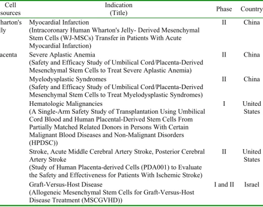
Tab. 2. Current status of Wharton's jelly and placenta mesenchymal stem cells in clinical trials (based on www.clinicaltrials.gov).
Cell sources
Indication
(Title) Phase Country
Wharton's
jelly Myocardial Infarction (Intracoronary Human Wharton's Jelly- Derived Mesenchymal Stem Cells (WJ-MSCs) Transfer in Patients With Acute Myocardial Infarction)
II China
Placenta Severe Aplastic Anemia
(Safety and Efficacy Study of Umbilical Cord/Placenta-Derived Mesenchymal Stem Cells to Treat Severe Aplastic Anemia)
II China
Myelodysplastic Syndromes
(Safety and Efficacy Study of Umbilical Cord/Placenta-Derived Mesenchymal Stem Cells to Treat Myelodysplastic Syndromes)
II China
Hematologic Malignancies
(A Single-Arm Safety Study of Transplantation Using Umbilical Cord Blood and Human Placental-Derived Stem Cells From Partially Matched Related Donors in Persons With Certain Malignant Blood Diseases and Non-Malignant Disorders (HPDSC))
I United States
Stroke, Acute Middle Cerebral Artery Stroke, Posterior Cerebral Artery Stroke
(Study of Human Placenta-derived Cells (PDA001) to Evaluate the Safety and Effectiveness for Patients With Ischemic Stroke)
II United States
Graft-Versus-Host Disease
(Allogeneic Mesenchymal Stem Cells for Graft-Versus-Host Disease Treatment (MSCGVHD))
I and II Israel
the results of clinical investigations using stem cells from perinatal tissues is limited. No studies were found using stem cells from the amnion. The ongoing human clinical trials using mesenchymal stem cells isolated from Wharton’s jelly and placenta are summarized in Tab. 2.
There are also some safety issues inherent in stem cell therapy. Like any other new technology, it is completely unknown what the long-term effects of such interference with nature could be. Once placed into a patient's body, stem cells intended to treat or cure a disease could end up wreaking havoc simply because they are no longer under the control of the clinician. Another disadvantage of perinatal stem cells is the poorly understood biology of these stem cells and molecular mechanisms of differentiation. Many issues must be explored before the safe application of these cells in the clinical setting. However, with appropriate validation of cell types and optimal performance, and further characterization, perinatal stem cells should yield a demonstrable benefit in cell therapy.
SUMMARY
Human perinatal tissue – Wharton's jelly, amnion and chorion/placenta – provides an abundant source of developmentally young somatic cells that can be stored, differentiated and/or even reprogrammed for regenerative medicine. Since the umbilical cord, amnion and placenta are normally discarded at birth, they provide an easily accessible alternative source of stem cells. These cells have a high expansion potential and genetic stability. In addition, they have strong immunosuppressive properties that can be exploited for successful autologous as well as heterologous transplantation.
The stem cells described in the present article may emerge as a remarkable tool for the cell therapy of multiple diseased tissues. Advances in the purification, isolation and identification of stem cells and their success in clinical application would give a solid foundation for further development of regenerative medicine based on cell therapy. Stem cells may be used both in the regeneration of damaged tissue and to create ex vivo organ fragments in the future. Further studies are required to understand better the precise nature of perinatal stem cells and to explore their potential clinical applications.
Acknowledgments. This work was supported by project no. N N302157037
from the Polish funds for scientific research in 2009-2012.
REFERENCES
1. Kiessling, A.A. and Anderson, S.C. Human embryonic stem cells. (Jonas and Bartlett), Boston (2003).
3. Solter, D. From teratocarcinomas to embryonic stem cells and beyond: a history of embryonic stem cell research. Nat. Rev. Genet. 7 (2006) 319-327. 4. Campagnoli, C., Roberts, I. A., Kumar, S., Bennett, P.R., Bellantuono, I. and Fisk, N.M. Identification of mesenchymal stem/progenitor cells in human first-trimester fetal blood, liver, and bone marrow. Blood 98 (2001) 2396-2402. 5. Witkowska-Zimny, M. and Walenko, K. Stem cells from adipose tissue.
Cell Mol. Biol. Lett. 16 (2011) 236-257.
6. van de Ven, C., Collins, D., Bradley, M.B., Morris, E. and Cairo, M.S. The potential of umbilical cord blood multipotent stem cells for nonhematopoietic tissue and cell regeneration. Exp. Hematol. 35 (2007) 1753-1765.
7. Miao, Z., Jin, J., Chen, L., Zhu, J., Huang, W., Zhao, J., Qian, H. and Zhang, X. Isolation of mesenchymal stem cells from human placenta: comparison with human bone marrow mesenchymal stem cells. Cell Biol. Int. 30 (2006) 681-687. 8. Mizokami, T., Hisha, H., Okazaki, S., Takaki, T., Wang, X.L., Song, C.Y.,
Li, Q., Kato, J., Hosaka, N., Inaba, M., Kanzaki, H. and Ikehara, S. Preferential expansion of human umbilical cord blood-derived CD34-positive cells on major histocompatibility complex-matched amnion-derived mesenchymal stem cells. Haematologica 94 (2009) 618-628.
9. Romanov, Y.A., Svintsitskaya, V.A. and Smirnov, V.N. Searching for alternative sources of postnatal human mesenchymal stem cells: candidate MSC-like cells from umbilical cord. Stem Cells 21 (2003) 105-110.
10.Kobayashi, K., Kubota, T. and Aso, T. Study on myofibroblast differentiation in the stromal cells of Wharton's jelly: expression and localization of alpha-smooth muscle actin. Early Hum Dev. 51 (1998) 223-233. 11.Mitchell, K.E., Weiss, M.L., Mitchell, B.M., Martin, P., Davis, D., Morales, L., Helwig, B., Beerenstrauch, M., Abou-Easa, K., Hildreth, T., Troyer, D. and Medicetty, S. Matrix cells from Wharton's jelly form neurons and glia. Stem
Cells 21 (2003) 50-60.
12.Weiss, M.L., Medicetty, S., Bledsoe, A.R., Rachakatla, R.S., Choi, M., Merchav, S., Luo, Y., Rao, M.S., Velagaleti, G. and Troyer, D. Human umbilical cord matrix stem cells: preliminary characterization and effect of transplantation in a rodent model of Parkinson's disease. Stem Cells 24 (2006) 781-792.
13.Dominici, M., Le Blanc, K., Mueller, I., Slaper-Cortenbach, I., Marini, F., Krause, D., Deans, R., Keating, A., Prockop, D. and Horwitz, E. Minimal criteria for defining multipotent mesenchymal stromal cells. The International Society for Cellular Therapy position statement. Cytotherapy 8 (2006) 315-317.
14.La Rocca, G., Anzalone, R. and Farina, F. The expression of CD68 in human umbilical cord mesenchymal stem cells: new evidences of presence in non-myeloid cell types. Scand. J. Immunol. 70 (2009) 161-162.
human umbilical cord Wharton's jelly-derived cells. Stem Cells 26 (2008) 2865-2874.
16.Troyer, D.L. and Weiss, M.L. Wharton's jelly-derived cells are a primitive stromal cell population. Stem Cells 26 (2008) 591-599.
17.Fong, C.Y., Chak, L.L., Biswas, A., Tan, J.H., Gauthaman, K., Chan, W.K. and Bongso, A. Human Wharton's jelly stem cells have unique transcriptome profiles compared to human embryonic stem cells and other mesenchymal stem cells. Stem Cell. Rev. 7 (2011) 1-16.
18.Fong, C.Y., Chak, L.L., Subramanian, A., Tan, J.H., Biswas, A., Gauthaman, K., Choolani, M., Chan, W.K. and Bongso, A. A three dimensional anchorage independent in vitro system for the prolonged growth of embryoid bodies to study cancer cell behaviour and anticancer agents. Stem Cell Rev. 5 (2009) 410-419.
19.Djouad, F., Charbonnier, L.M., Bouffi, C., Louis-Plence, P., Bony, C., Apparailly, F., Cantos, C., Jorgensen, C. and Noel, D. Mesenchymal stem cells inhibit the differentiation of dendritic cells through an interleukin-6-dependent mechanism. Stem Cells 25 (2007) 2025-2032.
20.La Rocca, G., Anzalone, R., Corrao, S., Magno, F., Loria, T., Lo Iacono, M., Di Stefano, A., Giannuzzi, P., Marasa, L., Cappello, F., Zummo, G. and Farina, F. Isolation and characterization of Oct-4+/HLA-G+ mesenchymal stem cells from human umbilical cord matrix: differentiation potential and detection of new markers. Histochem. Cell Biol. 131 (2009) 267-282. 21.Le Blanc, K. Immunomodulatory effects of fetal and adult mesenchymal
stem cells. Cytotherapy 5 (2003) 485-489.
22.Chen, K., Wang, D., Du, W.T., Han, Z.B., Ren, H., Chi, Y., Yang, S.G., Zhu, D., Bayard, F. and Han, Z.C. Human umbilical cord mesenchymal stem cells hUC-MSCs exert immunosuppressive activities through a PGE2-dependent mechanism. Clin. Immunol. 135 (2010) 448-458.
23.Fong, C.Y., Richards, M., Manasi, N., Biswas, A. and Bongso, A. Comparative growth behaviour and characterization of stem cells from human Wharton's jelly. Reprod. Biomed. Online 15 (2007) 708-718.
24.Ayuzawa, R., Doi, C., Rachakatla, R.S., Pyle, M.M., Maurya, D.K., Troyer, D. and Tamura, M. Naive human umbilical cord matrix derived stem cells significantly attenuate growth of human breast cancer cells in vitro and in vivo. Cancer Lett. 280 (2009) 31-37.
25.Angelucci, S., Marchisio, M., Di Giuseppe, F., Pierdomenico, L., Sulpizio, M., Eleuterio, E., Lanuti, P., Sabatino, G., Miscia, S. and Di Ilio, C. Proteome analysis of human Wharton's jelly cells during in vitro expansion. Proteome Sci. 8 (2010) 18.
26.Wang, H.S., Hung, S.C., Peng, S.T., Huang, C.C., Wei, H.M., Guo, Y.J., Fu, Y.S., Lai, M.C. and Chen, C.C. Mesenchymal stem cells in the Wharton's jelly of the human umbilical cord. Stem Cells 22 (2004) 1330-1337.
human Wharton’s jelly mesenchymal stem cells: immunological features and hepatocyte-like differentiative capacity. Stem Cells Dev. 19 (2010) 423-438. 28.Anzalone, R., Lo Iacono, M., Loria, T., Di Stefano, A., Giannuzzi, P.,
Farina, F. and La Rocca, G., Wharton’s Jelly mesenchymal stem cells as candidates for beta cells regeneration: extending the differentiative and immunomodulatory benefits of adult mesenchymal stem cells for the treatment of type 1 diabetes. Stem Cell Rev. 7 (2011) 342-363, DOI: 10.1007/s12015-010-9196-4.
29.Baksh, D., Yao, R. and Tuan, R.S. Comparison of proliferative and multilineage differentiation potential of human mesenchymal stem cells derived from umbilical cord and bone marrow. Stem Cells 25 (2007) 1384-1392. 30.Hou, T., Xu, J., Wu, X., Xie, Z., Luo, F., Zhang, Z. and Zeng, L. Umbilical
cord Wharton's Jelly: a new potential cell source of mesenchymal stromal cells for bone tissue engineering. Tissue Eng. Part A 15 (2009) 2325-2334. 31.Hildebrandt, C., Buth, H. and Thielecke, H. Influence of cell culture media
conditions on the osteogenic differentiation of cord blood-derived mesenchymal stem cells. Ann. Anat. 191 (2009) 23-32.
32.Schneider, R.K., Puellen, A., Kramann, R., Raupach, K., Bornemann, J., Knuechel, R., Perez-Bouza, A. and Neuss, S. The osteogenic differentiation of adult bone marrow and perinatal umbilical mesenchymal stem cells and matrix remodelling in three-dimensional collagen scaffolds. Biomaterials 31 (2010) 467-480.
33.Penolazzi, L., Vecchiatini, R., Bignardi, S., Lambertini, E., Torreggiani, E., Canella, A., Franceschetti, T., Calura, G., Vesce, F. and Piva, R. Influence of obstetric factors on osteogenic potential of umbilical cord-derived mesenchymal stem cells. Reprod. Biol. Endocrinol. 7 (2009) DOI10.1186/1477-7827-7-106.
34.Chen, M.Y., Lie, P.C., Li, Z.L. and Wei, X. Endothelial differentiation of Wharton's jelly-derived mesenchymal stem cells in comparison with bone marrow-derived mesenchymal stem cells. Exp. Hematol. 37 (2009) 629-640. 35.Kadam, S.S., Tiwari, S. and Bhonde, R.R. Simultaneous isolation of
vascular endothelial cells and mesenchymal stem cells from the human umbilical cord. In Vitro Cell Dev. Biol. Anim. 45 (2009) 23-27.
36.Tsai, P.C., Fu, T.W., Chen, Y.M., Ko, T.L., Chen, T.H., Shih, Y.H., Hung, S.C. and Fu, Y. The therapeutic potential of human umbilical mesenchymal stem cells from Wharton's jelly in the treatment of rat liver fibrosis. Liver
Transpl. 15 (2009) 484-495.
37.Wang, H.S., Shyu, J.F., Shen, W.S., Hsu, H.C., Chi, T.C., Chen, C.P., Huang, S.W., Shyr, Y.M., Tang, K.T. and Chen, T.H. Transplantation of insulin producing cells derived from umbilical cord stromal mesenchymal stem cells to treat NOD mice. Cell Transplant. (2010) DOI: 10.3727/096368910X522270.
Mistry, S.K. Cells isolated from umbilical cord tissue rescue photoreceptors and visual functions in a rodent model of retinal disease. Stem Cells 25 (2007) 602-611.
39.Friedman, R., Betancur, M., Boissel, L., Tuncer, H., Cetrulo, C. and Klingemann, H. Umbilical cord mesenchymal stem cells: adjuvants for human cell transplantation. Biol. Blood Marrow Transplant. 13 (2007) 1477-1486.
40.Malkowski, A., Sobolewski, K., Jaworski, S. and Bankowski, E. FGF binding by extracellular matrix components of Wharton's jelly. Acta
Biochim. Pol. 54 (2007) 357-363.
41.Benirschke, K.K.P. Pathology of the human placenta. Springer-Verlag, New York (1995).
42.Horwitz, E.M., Le Blanc, K., Dominici, M., Mueller, I., Slaper-Cortenbach, I., Marini, F.C., Deans, R.J., Krause, D.S. and Keating, A. Clarification of the nomenclature for MSC: The International Society for Cellular Therapy position statement. Cytotherapy 7 (2005) 393-395.
43.Miki, T., Lehmann, T., Cai, H., Stolz, D.B. and Strom, S.C. Stem cell characteristics of amniotic epithelial cells. Stem Cells 23 (2005) 1549-1559. 44.In 't Anker, P.S., Scherjon, S.A., Kleijburg-van der Keur, C., de Groot-Swings, G.M., Claas, F.H., Fibbe, W.E. and Kanhai, H.H. Isolation of mesenchymal stem cells of fetal or maternal origin from human placenta.
Stem Cells 22 (2004) 1338-1345.
45.Bilic, G., Zeisberger, S.M., Mallik, A.S., Zimmermann, R. and Zisch, A.H. Comparative characterization of cultured human term amnion epithelial and mesenchymal stromal cells for application in cell therapy. Cell Transplant. 17 (2008) 955-968.
46.Alviano, F., Fossati, V., Marchionni, C., Arpinati, M., Bonsi, L., Franchina, M., Lanzoni, G., Cantoni, S., Cavallini, C., Bianchi, F., Tazzari, P.L., Pasquinelli, G., Foroni, L., Ventura, C., Grossi, A. and Bagnara, G.P. Term Amniotic membrane is a high throughput source for multipotent Mesenchymal Stem Cells with the ability to differentiate into endothelial cells in vitro. BMC Dev. Biol. 7 (2007) DOI:10.1186/1471-213X-7-11. 47.Zhang, Y., Adachi, Y., Suzuki, Y., Minamino, K., Iwasaki, M., Hisha, H.,
Song, C.Y., Kusafuka, K., Nakano, K., Koike, Y., Wang, J., Koh, E., Cui, Y., Li, C. and Ikehara, S. Simultaneous injection of bone marrow cells and stromal cells into bone marrow accelerates hematopoiesis in vivo. Stem
Cells 22 (2004) 1256-1262.
48.Koizumi, N.J., Inatomi, T.J., Sotozono, C.J., Fullwood, N.J., Quantock, A.J. and Kinoshita, S. Growth factor mRNA and protein in preserved human amniotic membrane. Curr. Eye. Res. 20 (2000) 173-177.
49.Fauza, D. Amniotic fluid and placental stem cells. Best Pract. Res. Clin.
Obstet. Gynaecol. 18 (2004) 877-891.
51.Prusa, A.R. and Hengstschlager, M. Amniotic fluid cells and human stem cell research: a new connection. Med. Sci. Monit. 8 (2002) 253-257.
52.Tsai, M.S., Lee, J.L., Chang, Y.J. and Hwang, S.M. Isolation of human multipotent mesenchymal stem cells from second-trimester amniotic fluid using a novel two-stage culture protocol. Hum. Reprod. 19 (2004) 1450-1456. 53.De Coppi, P., Bartsch, G., Jr., Siddiqui, M.M., Xu, T., Santos, C.C., Perin, L.,
Mostoslavsky, G., Serre, A.C., Snyder, E.Y., Yoo, J.J., Furth, M.E., Soker, S. and Atala, A. Isolation of amniotic stem cell lines with potential for therapy.
Nat. Biotechnol. 25 (2007) 100-106.
54.Antonucci, I., Iezzi, I., Morizio, E., Mastrangelo, F., Pantalone, A., Mattioli-Belmonte, M., Gigante, A., Salini, V., Calabrese, G., Tete, S., Palka, G. and Stuppia, L. Isolation of osteogenic progenitors from human amniotic fluid using a single step culture protocol. BMC Biotechnol. 9 (2009) DOI:10.1186/1472-6750-9-9.
55.Phermthai, T., Odglun, Y., Julavijitphong, S., Titapant, V., Chuenwattana, P., Vantanasiri, C. and Pattanapanyasat, K. A novel method to derive amniotic fluid stem cells for therapeutic purposes. BMC Cell Biol. 11 (2010) DOI: 10.1186/1471-2121-11-79.
56.Prusa, A.R., Marton, E., Rosner, M., Bernaschek, G. and Hengstschlager, M. Oct-4-expressing cells in human amniotic fluid: a new source for stem cell research? Hum. Reprod. 18 (2003) 1489-1493.
57.Schmidt, D., Achermann, J., Odermatt, B., Breymann, C., Mol, A., Genoni, M., Zund, G. and Hoerstrup, S.P. Prenatally fabricated autologous human living heart valves based on amniotic fluid derived progenitor cells as single cell source. Circulation 116 (2007) 64-70.
58.Tsai, M.S., Hwang, S.M., Tsai, Y.L., Cheng, F.C., Lee, J.L. and Chang, Y.J. Clonal amniotic fluid-derived stem cells express characteristics of both mesenchymal and neural stem cells. Biol. Reprod. 74 (2006) 545-551. 59.Klemmt, P.A., Vafaizadeh, V. and Groner, B. Murine amniotic fluid stem
cells contribute mesenchymal but not epithelial components to reconstituted mammary ducts. Stem Cell Res. Ther. 1 (2010) DOI: 10.1186/scrt20. 60.Chiavegato, A., Bollini, S., Pozzobon, M., Callegari, A., Gasparotto, L.,
Taiani, J., Piccoli, M., Lenzini, E., Gerosa, G., Vendramin, I., Cozzi, E., Angelini, A., Iop, L., Zanon, G.F., Atala, A., De Coppi, P. and Sartore, S. Human amniotic fluid-derived stem cells are rejected after transplantation in the myocardium of normal, ischemic, suppressed or immuno-deficient rat. J. Mol. Cell Cardiol. 42 (2007)746-759.
61.Dzierzak, E. and Robin, C. Placenta as a source of hematopoietic stem cells.
Trends Mol. Med. 16 (2010) 361-367.
62.Zhang, Y., Li, C., Jiang, X., Zhang, S., Wu, Y., Liu, B., Tang, P. and Mao, N. Human placenta-derived mesenchymal progenitor cells support culture expansion of long-term culture-initiating cells from cord blood CD34+ cells.
63.Portmann-Lanz, C.B., Schoeberlein, A., Huber, A., Sager, R., Malek, A., Holzgreve, W. and Surbek, D.V. Placental mesenchymal stem cells as potential autologous graft for pre- and perinatal neuroregeneration. Am. J.
Obstet. Gynecol. 194 (2006) 664-673.
64.Soncini, M., Vertua, E., Gibelli, L., Zorzi, F., Denegri, M., Albertini, A., Wengler, G.S. and Parolini, O. Isolation and characterization of mesenchymal cells from human fetal membranes. J. Tissue Eng. Regen. Med. 1 (2007) 296-305.
65.Castrechini, N.M., Murthi, P., Gude, N.M., Erwich, J.J., Gronthos, S., Zannettino, A., Brennecke, S.P. and Kalionis, B. Mesenchymal stem cells in human placental chorionic villi reside in a vascular Niche. Placenta 31 (2010) 203-212.
66.Fukuchi, Y., Nakajima, H., Sugiyama, D., Hirose, I., Kitamura, T. and Tsuji, K. Human placenta-derived cells have mesenchymal stem/progenitor cell potential. Stem Cells 22 (2004) 649-658.
67.Chien, C.C., Yen, B.L., Lee, F.K., Lai, T.H., Chen, Y.C., Chan, S.H. and Huang, H. I. In vitro differentiation of human placenta-derived multipotent cells into hepatocyte-like cells. Stem Cells 24 (2006) 1759-1768.
68.Zhao, Y., Wang, H. and Mazzone, T. Identification of stem cells from human umbilical cord blood with embryonic and hematopoietic characteristics. Exp. Cell Res. 312 (2006) 2454-2464.
69.Poloni, A., Maurizi, G., Babini, L., Serrani, F., Berardinelli, E., Mancini, S., Costantini, B., Discepoli, G. and Leoni, P. Human mesenchymal stem cells from chorionic villi and amniotic fluid are not susceptible to transformation after extensive in vitro expansion. Cell Transplant. (2010) DOI: 10.3727/096368910X536518.
70.Igura, K., Zhang, X., Takahashi, K., Mitsuru, A., Yamaguchi, S. and Takashi, T.A. Isolation and characterization of mesenchymal progenitor cells from chorionic villi of human placenta. Cytotherapy 6 (2004) 543-553. 71.Zhang, X., Mitsuru, A., Igura, K., Takahashi, K., Ichinose, S., Yamaguchi, S.
and Takahashi, T.A. Mesenchymal progenitor cells derived from chorionic villi of human placenta for cartilage tissue engineering. Biochem. Biophys.
Res. Commun. 340 (2006) 944-952.
72.Cargnoni, A., Gibelli, L., Tosini, A., Signoroni, P. B., Nassuato, C., Arienti, D., Lombardi, G., Albertini, A., Wengler, G.S. and Parolini, O. Transplantation of allogeneic and xenogeneic placenta-derived cells reduces bleomycin-induced lung fibrosis. Cell Transplant. 18 (2009) 405-422.
73.Li, C., Zhang, W., Jiang, X. and Mao, N. Human-placenta-derived mesenchymal stem cells inhibit proliferation and function of allogeneic immune cells. Cell Tissue Res. 330 (2007) 437-446.
74.Kolambkar, Y.M., Peister, A., Soker, S., Atala, A. and Guldberg, R.E. Chondrogenic differentiation of amniotic fluid-derived stem cells. J. Mol.
75.Brooke, G., Rossetti, T., Pelekanos, R., Ilic, N., Murray, P., Hancock, S., Antonenas, V., Huang, G., Gottlieb, D., Bradstock, K. and Atkinson, K. Manufacturing of human placenta-derived mesenchymal stem cells for clinical trials. Br. J. Haematol. 144 (2009) 571-579.
76.Ilancheran, S., Michalska, A., Peh, G., Wallace, E.M., Pera, M. and Manuelpillai, U. Stem cells derived from human fetal membranes display multilineage differentiation potential. Biol. Reprod. 77 (2007) 577-588. 77.Zhao, P., Ise, H., Hongo, M., Ota, M., Konishi, I. and Nikaido, T. Human
amniotic mesenchymal cells have some characteristics of cardiomyocytes.
Transplantation 79 (2005) 528-535.
78.Yeh, Y.C., Wei, H.J., Lee, W.Y., Yu, C.L., Chang, Y., Hsu, L.W., Chung, M.F., Tsai, M.S., Hwang, S.M. and Sung, H.W. Cellular cardiomyoplasty with human amniotic fluid stem cells: in vitro and in vivo studies. Tissue Eng.
Part A 16 (2010) 1925-1936.
79.Yeh, Y.C., Lee, W.Y., Yu, C.L., Hwang, S.M., Chung, M.F., Hsu, L.W., Chang, Y., Lin, W.W., Tsai, M.S., Wei, H.J. and Sung, H.W. Cardiac repair with injectable cell sheet fragments of human amniotic fluid stem cells in an immune-suppressed rat model. Biomaterials 31 (2010) 6444-6453.
80.Sakuragawa, N., Kakinuma, K., Kikuchi, A., Okano, H., Uchida, S., Kamo, I., Kobayashi, M. and Yokoyama, Y. Human amnion mesenchyme cells express phenotypes of neuroglial progenitor cells. J. Neurosci. Res. 78 (2004) 208-214.
81.Portmann-Lanz, C.B., Schoeberlein, A., Portmann, R., Mohr, S., Rollini, P., Sager, R. and Surbek, D.V. Turning placenta into brain: placental mesenchymal stem cells differentiate into neurons and oligodendrocytes.
Am. J. Obstet. Gynecol. 202 (2010) 294e1-e11.
82.Portmann-Lanz, C.B., Baumann, M.U., Mueller, M., Wagner, A.M., Weiss, S., Haller, O., Sager, R., Reinhart, U. and Surbek, D.V. Neurogenic characteristics of placental stem cells in preeclampsia. Am. J. Obstet.
Gynecol. 203 (2010) 391-397.
83.Prusa, A.R., Marton, E., Rosner, M., Bettelheim, D., Lubec, G., Pollack, A., Bernaschek, G. and Hengstschlager, M. Neurogenic cells in human amniotic fluid. Am. J. Obstet. Gynecol. 191 (2004) 309-314.
84.Yen, B.L., Chien, C. C., Chen, Y.C., Chen, J.T., Huang, J.S., Lee, F.K. and Huang, H.I. Placenta-derived multipotent cells differentiate into neuronal and glial cells in vitro. Tissue Eng. Part A 14 (2008) 9-17.
85.Wu, C.C., Chao, Y.C., Chen, C.N., Chien, S., Chen, Y.C., Chien, C.C., Chiu, J.J. and Linju Yen, B. Synergism of biochemical and mechanical stimuli in the differentiation of human placenta-derived multipotent cells into endothelial cells. J. Biomech. 41 (2008) 813-821.
87.Tamagawa, T., Oi, S., Ishiwata, I., Ishikawa, H. and Nakamura, Y. Differentiation of mesenchymal cells derived from human amniotic membranes into hepatocyte-like cells in vitro. Hum. Cell 20 (2007) 77-84. 88.Saulnier, N., Lattanzi, W., Puglisi, M.A., Pani, G., Barba, M., Piscaglia, A. C.,
Giachelia, M., Alfieri, S., Neri, G., Gasbarrini, G. and Gasbarrini, A. Mesenchymal stromal cells multipotency and plasticity: induction toward the hepatic lineage. Eur. Rev. Med. Pharmacol. Sci. 13 (2009) 71-78. 89.Park, T.S., Gavina, M., Chen, C.W., Sun, B., Teng, P.N., Huard, J., Deasy, B.M.,
Zimmerlin, L. and Peault, B. Placental perivascular cells for human muscle regeneration. Stem Cells Dev. 20 (2011) 451-463.
90.Strom, S. and Miki, T. Placental derived stem cells and uses thereof, United States Patent Application Publications, (2003) US2003/0235563.