http://www.cmbl.org.pl
DOI: 10.2478/s11658-007-0014-1 Received: 04 August 2006
Revised form accepted: 26 January 2007
Published online: 15 March 2007 © 2007 by the University of Wrocław, Poland
* Author for correspondence; e-mail: [email protected], tel.: +48 22 3277 177, fax: +48 22 3277 200
Abbreviations used: CFTR – cystic fibrosis transmembrane conductance regulator; GFP – green fluorescence protein; HA –hemagglutinin antigen; NBF2 – nucleotide-binding fold 2
Short communication
THE CFTR-DERIVED PEPTIDES AS A MODEL OF SEQUENCE-SPECIFIC PROTEIN AGGREGATION
DANIEL BĄK1,2*, GARRY R. CUTTING3 and MICHAŁ MILEWSKI1 1Laboratory of Cell Biology, Department of Medical Genetics, Institute
of Mother and Child, Kasprzaka 17A, 01-211 Warsaw, Poland, 2Postgraduate
School of Molecular Medicine, Żwirki i Wigury 61, 02-091 Warsaw, Poland,
3Institute of Genetic Medicine, Johns Hopkins University School of Medicine,
733 N Broadway, Baltimore, MD 21287-3914, USA
Thus, our results indicate that even subtle alterations within the aggregating peptide can affect many different aspects of the aggregation process.
Key Words:Protein aggregation, Conformational diseases, CFTR, Site-directed mutagenesis
INTRODUCTION
The ability of a protein to perform a physiological function depends to a significant extent on its tertiary structure forming correctly during the process of protein folding. Although misfolded proteins should be degraded by the ubiquitin/proteosome system, some stable intermediates are able to avoid the degradation pathway, and can go on to form intra- or extracellular protein aggregates. Such protein aggregation is a characteristic feature of many diseases affecting humans (e.g. Alzheimer’s disease and Huntington’s disease) and other animals (e.g. bovine spongiform encephalopathy) [1]. These conformational diseases constitute a great challenge to biomedical science, as their molecular pathogenesis is not well understood and no effective cures are available.
Numerous studies have indicated that different polypeptides use common mechanisms to form aggregates. As a consequence, many unrelated proteins can accumulate in similar aggregation-related structures, with extracellular amyloid fibrils and intracellular aggresomes being the best known examples [2, 3]. On the other hand, certain features of the aggregation process seem to be specific to particular proteins or even to particular amino acid sequences. For instance, elongated polyglutamine stretches are not only responsible for the disease-related aggregation of several human proteins, but are apparently also involved in the intracellular sequestration of polyglutamine-containing transcription factors and other crucial cellular proteins, which may additionally contribute to disease-specific clinical symptoms [4-9]. It has also been demonstrated that sequences other than the polyglutamine repeats determine the specific nuclear or cytoplasmic localization of aggregates formed by mutated huntingtin, a protein critical for the pathogenesis of Huntington’s disease [10-12].
As the number of similar examples increases, protein aggregation is becoming perceived as a more sequence-specific phenomenon. However, the exact amino acids contributing to this specificity are known in only a few cases [13-16]. This exposes the need for new protein models that would facilitate the exploration of the sequence-specific mechanisms governing the aggregation process.
the contribution of different amino acids to the aggregation process. Therefore, we used this model system to investigate to what extent the background of the ag region within the CFTR C-terminal sequence can influence the peptide’s ability to form intracellular aggregates.
MATERIALS AND METHODS
Construction of expression plasmids
The construction of the eukaryotic expression plasmids encoding the CFTR C-terminal fragment (a.a. 1370-1480 of the full-length protein), fused to the green fluorescence protein (GFP) or tagged with the hemagglutinin (HA) epitope, was described elsewhere [23]. The introduction of the deletion encompassing amino acids 1395-1403 (Δag) of CFTR was also previously described [23].
The Transformer site-directed mutagenesis system (Becton Dickinson) was applied to introduce the ag region or the HR motif into different positions within the CFTR C-terminal sequence. The sequences of all the oligonucleotide primers used to create these insertions are available upon request. The nucleotide sequence of the newly created constructs was verified by DNA sequencing.
Cell culture and transient transfections
The IB3-1 cells (immortalized bronchial epithelial cells derived from a patient with cystic fibrosis [24]) were cultured in LHC-8 medium (BioSource) containing 5% FBS (Gibco). These cells lack functional CFTR [24] and do not express detectable amounts of mutant CFTR, as measured by immunoprecipitation followed by radioactive in vitro phosphorylation [25 and our unpublished results]. For routine culture, the cells were grown in polystyrene flasks (Sarstedt) coated with collagen (Cohesion), in 5% CO2-balanced air at
37ºC. For protein aggregation analysis, the cells were grown on collagen-coated glass coverslips in 6-well culture plates (Sarstedt). After reaching 60-80% confluency, the cells were transiently transfected with Lipofectin (Invitrogen), in accordance with the manufacturer’s instructions. Twenty-two hours after transfection, the cells were fixed with 4% PFA (Sigma) for 20 minutes. If immunostaining was not necessary (as for the GFP-tagged constructs), the cells were mounted in SlowFade (Molecular Probes) containing 0.05 mg/ml of DAPI (Sigma). For Western blot analysis, the cells were grown in 6-well plates and harvested 22 hours after transfection.
Western blot analysis
Immunofluorescent staining
The HA-tagged peptides were detected using monoclonal (12CA5, Roche) or polyclonal (SG77, Zymed Laboratories Inc.) anti-HA antibodies. The mitochondrial marker Grp75 was detected with the anti-Grp75 antibody (SPA-825, Stressgen). The transfected cells were fixed in 4% PFA for 20 minutes, washed with PBS, and permeabilized with 0.1% Triton X-100 (Sigma) for 3 minutes, and then immunostained. Non-specific binding was blocked by incubation with 2.5% goat serum (Sigma) in PBS for 30 minutes. Immunostaining was carried out in two steps separated by a short PBS wash. The cells were first stained with the primary antibody for 1.5 hours and then with the proper secondary antibody, conjugated to Cy3 or FITC (Sigma), for 30 minutes. The coverslips were then washed with PBS and mounted in SlowFade with DAPI.
Fluorescence microscopy and statistical analysis
Cells were counted and images were taken using the Eclipse E400 (Nikon) fluorescence microscope. The CytoVision 2.75 software (Applied Imagining Corp.) was used to process the images.
To estimate the percentage of cells with aggregates, at least 150 transfected cells were analyzed in multiple random visual fields on each slide in quadruplicate. To estimate the frequency of occurrence of the different morphological forms of aggregates, at least 50 transfected cells were counted in multiple random visual fields on each slide in quadruplicate. For analysis of aggregates’ morphology, the protein depositions in cells expressing the GFP-tagged peptides were classified as aggregates of either compact or loose structure. The aggregates in cells expressing the HA-tagged peptides with the ag region in a different position were classified as being: (1) very small and numerous (> 20 per cell); (2) medium-sized and less numerous (5-20 per cell); or (3) very large but scarce (< 5 per cell). Since some cells showed the presence of both small and large protein depositions, they were counted only once as having large aggregates. Such a classification of aggregates was not performed for the Δag peptides, which rarely aggregated. Additionally, their infrequent aggregates showed no distinct morphology or localization pattern. The aggregates formed by the HA-tagged peptides modified by the introduction of the HR motif were classified as: (1) very small and dispersed throughout the cytoplasm, (2) very small and concentrated in one region within the cytoplasm, (3) medium-sized cytoplasmic, (4) large cytoplasmic, or (5) nuclear. The investigators worked blind, not knowing the identity of constructs used for transfection.
The standard chi2 test was used to estimate the statistical significance of the
mutagenesis. Only three positions, all generated by mutagenesis, were compared in the case of the HR motif. Statistical significance was assumed at P < 0.05.
RESULTS AND DISCUSSION
The position of the ag region affects the peptide’s ability to aggregate
To examine first the intrinsic potential of the CFTR-derived ag region to promote protein aggregation, we tested whether this short 9-amino acid sequence alone is able to cause aggregation when attached to a soluble non-CFTR protein. The ag sequence (C-T-V-I-L-C-E-H-R; a.a. 1395-1403 of CFTR) was fused directly to the C-terminus of GFP, as is commonly practiced when fusing this green fluorescence reporter protein to other polypeptides. By contrast to the
non-aggregating GFP protein, the GFP-ag fusion formed large cytoplasmic
aggregates of irregular shape in 13.4 ± 2.1% of transiently transfected IB3-1 cells (P < 0.05). This indicated that the ag region is indeed able to induce protein aggregation when present in the non-native amino acid context.
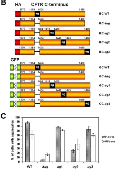
To further explore the ability of the ag region to induce protein aggregation, we tested whether the position of this region within the C-terminus of CFTR would affect the aggregation rate of the CFTR-derived peptides. A series of GFP- or HA-tagged constructs was created in which the ag region was deleted from the native position and introduced into one of the three new locations within the CFTR C-terminal sequence (Fig. 1A and 1B). The first of these locations (ag1) fell within the predicted C-terminal part of the second nucleotide-binding fold (NBF2) [26], a domain also encompassing the native ag region and disrupted by the creation of the CFTR C-terminal peptides. In the second position (ag2), the ag region was placed within the unstructured region between the NBF2 domain and the C-terminus. Finally, when present in the third position (ag3), the ag region extended the carboxyl terminus of CFTR, thus masking the native C-terminal PDZ-binding motif (D-T-R-L>) [27].
aggregation level of the ag2 peptides of both series was significantly lower than in the case of other constructs containing the ag region. This indicated that the amino acid background may substantially influence the ability of this region to induce protein aggregation.
The overall association between the four different positions of the ag region and the peptide’s ability to aggregate was evident for both series of constructs (P < 0.001). However, this association was markedly stronger for the HA-tagged (V = 0.52) than for the GFP-tagged (V = 0.23) peptides. Numerous studies have reported that GFP may influence the aggregation process when fused to other proteins [9, 15, 22, 28]. Thus, it seems likely that the presence of GFP enhanced the ag-independent mechanism of protein aggregation, which in turn weakened the observed relationship between the position of the ag region and the peptide’s aggregation rate.
Aggregates formed by modified peptides show different morphologies
Previous studies have shown that fusing a reporter protein to an aggregating peptide may affect the subcellular distribution and morphology of aggregates [22]. Therefore, we tested whether changing the position of the ag region within the CFTR-derived sequence would lead to similar alterations in the aggregation pattern.
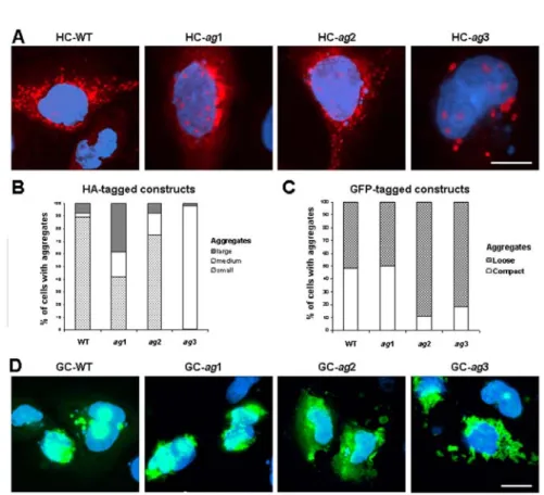
The analysis of the morphology of intracellular aggregates in cells expressing the modified HA-tagged peptides revealed that only the HC-ag2 peptide predominantly accumulated in small and numerous (> 20 per cell) aggregates resembling those formed by the wild type construct (HC-WT) (Fig. 2A). This similarity included not only the size and the overall distribution of the aggregates within the cell, but also their previously reported association with the mitochondria (data not shown) [22]. On the other hand, two other modified peptides, HC-ag1 and HC-ag3, showed significantly different aggregation patterns. The HC-ag1 peptide often accumulated in large perinuclear aggregates that were much less frequent in cells expressing the wild-type peptide (38.2 ± 3.3 and 7.5 ± 1.0%, respectively, p < 0.001). An even more unique aggregation pattern was associated with the HC-ag3 construct, accumulating almost exclusively (97.5 ± 2.5% of all cells with aggregates) in aggregates of medium size and uniform spherical shape (Fig. 2A). These cytoplasmic aggregates were usually less numerous (10-20 per cell) than the previously described small aggregates, and unlike them, showed no association with the mitochondria (data not shown). Both the above-described predominant forms of aggregates formed by the HC-ag1 and HC-ag3 peptides were frequently accompanied by small nuclear deposits (Fig. 2A).
Fig. 2. The morphology and localization of aggregates formed by modified CFTR-derived peptides. A – The subcellular distribution of peptides tagged with the HA epitope (red) in transiently transfected IB3-1 cells. The cells were counterstained with DAPI (blue). Bars, 10 μm. B and C – Frequencies of different types of aggregates in cells transfected with HA-tagged (B) or GFP-tagged (C) constructs. D – The subcellular distribution of peptides fused to GFP (green) in transiently transfected IB3-1 cells. The cells were counterstained with DAPI (blue). Bars, 10 μm.
dispersed structure (Fig. 2D), suggesting that despite the predominant impact of the GFP protein, the position of the ag region within the CFTR-derived sequence may also contribute to the morphology of the aggregates.
Summarizing the above observations, the morphology of aggregates formed by the ag1-3 peptides of both series showed significant associations with the position of the ag region within the CFTR-derived sequence (P < 0.001). This overall relationship was stronger for the HA (V = 0.64) than for the GFP series (V = 0.35), suggesting again that the GFP fusion may additionally influence the aggregation process.
Insertion of the HR motif affects the aggregation pattern
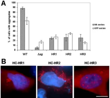
The histidine-arginine (HR) motif within the ag region was previously shown to be critical for the aggregation of the CFTR C-terminus [22]. To investigate whether this dipeptide motif alone could increase the aggregation rate when present in a different amino acid background, we altered its position within the CFTR C-terminus. Similarly to the strategy used for the whole ag region, the HR motif was inserted into three different positions, replacing amino acids 1419 and 1420 (HR1), 1442 and 1443 (HR2) or 1479 and 1480 (HR3) of the HA- or GFP-tagged peptide devoid of the ag region.
The effect of the HR insertions on the peptides’ ability to aggregate in transfected IB3-1 cells was noticeably different from the outcome produced by the ag insertions. The overall aggregation level of the HR1, HR2 and HR3 peptides was never as high as in the case of the original peptide with the intact ag region (Fig. 3A). However, with the one exception of the GFP-tagged HR3 construct, the aggregation level was significantly higher than in the case of the peptide modified by the deletion of the ag region only. Together, these results indicated that the HR motif alone, when extracted from the context of the ag region, shows only limited ability to induce protein aggregation following its insertion into a non-native amino acid background.
Although having only a moderate influence on the peptide’s overall ability to aggregate, the insertion of the HR motif was still able to significantly alter the subcellular distribution of protein aggregates, at least in the case of peptides that were not fused to GFP (Fig. 3B). For example, in most (53.3 ± 2.3%) aggregate-containing cells expressing the HC-HR1 construct, the modified peptide accumulated in small aggregates concentrated in one region within the cytoplasm (Fig. 3B). By contrast, the HC-HR3 peptides aggregated predominantly (61.3 ± 6.1%) within the nucleus, where they formed small discrete accumulations (Fig. 3B). Only the HC-HR2 construct was able to form the small mitochondria-associated aggregates typical for the wild type peptide (data not shown). These examples clearly indicate that even relatively subtle changes within the amino acid sequence of the aggregating peptide may substantially influence the subcellular localization of the aggregates.
Concluding remarks
Our results demonstrate that the ag region shows remarkable potential to induce protein aggregation, as can be judged from its ability to increase the aggregation rate following insertion into a new amino acid context. However, the position of this region within the aggregating peptide may influence the aggregation process, suggesting that the ability of the ag region to induce aggregation has certain natural limits. This is supported by the fact that when present in the native position within the NBF2 domain of CFTR, the ag region does not cause protein aggregation as long as the integrity of the whole domain is preserved. In our studies, the position associated with a decreased aggregation rate (ag2, between a.a. 1442 and 1443) corresponds to the presumably unstructured fragment of CFTR, suggesting that the ag region causes stronger protein aggregation when present within this relatively highly structured non-native background. Insertion of the ag region could introduce a structural element that disrupts a local fold and exposes neighboring aggregation-prone sequences that are normally buried inside the molecule.
proteins have indicated that the localization of protein aggregates is critical for their toxicity [29-33]. However, little is known about the sequences in disease-related proteins that modulate the localization of protein aggregates. Identifying these sequences and the mechanisms responsible for targeting protein aggregates to different subcellular compartments should facilitate designing new therapies aimed at directing the aggregating protein to locations associated with lower toxicity.
Acknowledgments. This study was supported by a grant from State Committee for Scientific Research (PBZ/KBN/042/P05/06 and 2 P05A 157 29), Poland.
REFERENCES
1. Carrell, R.W. and Lomas, D.A. Conformational disease. Lancet 350 (1997) 134-138.
2. Kisilevsky, R. and Fraser, P.E. A beta amyloidogenesis: unique, or variation on a systemic theme? Crit. Rev. Biochem. Mol. Biol. 32 (1997) 361-404. 3. Kopito, R.R. Aggresomes, inclusion bodies and protein aggregation. Trends
Cell Biol. 10 (2000) 524-530.
4. Donaldson, K.M., Li, W., Ching, K.A., Batalov, S., Tsai, C.C. and Joazeiro C.A. Ubiquitin-mediated sequestration of normal cellular proteins into polyglutamine aggregates. Proc. Natl. Acad. Sci. USA 100 (2003) 8892-8897.
5. Kazantsev, A., Preisinger, E., Dranovsky, A., Goldgaber, D. and Housman, D. Insoluble detergent-resistant aggregates form between pathological and nonpathological lengths of polyglutamine in mammalian cells. Proc. Natl. Acad. Sci. USA 96 (1999) 11404-11409.
6. Preisinger, E., Jordan, B.M., Kazantsev, A. and Housman, D. Evidence for a recruitment and sequestration mechanism in Huntington's disease. Philos. Trans. R. Soc. Lond B Biol. Sci. 354 (1999) 1029-1034.
7. Schaffar, G., Breuer, P., Boteva, R., Behrends, C., Tzvetkov, N., Strippel, N., Sakahira, H., Siegers, K., Hayer-Hartl, M. and Hartl, F.U. Cellular toxicity of polyglutamine expansion proteins: mechanism of transcription factor deactivation. Mol. Cell. 15 (2004) 95-105.
8. Suhr, S.T., Senut, M.C., Whitelegge, J.P., Faull, K.F., Cuizon, D.B. and Gage, F.H. Identities of sequestered proteins in aggregates from cells with induced polyglutamine expression. J. Cell Biol. 153 (2001) 283-294.
10.Chai, Y., Wu, L., Griffin, J.D. and Paulson, H.L. The role of protein composition in specifying nuclear inclusion formation in polyglutamine disease. J. Biol. Chem. 276 (2001) 44889-44897.
11.Nozaki, K., Onodera, O., Takano, H. and Tsuji, S. Amino acid sequences flanking polyglutamine stretches influence their potential for aggregate formation. Neuroreport 12 (2001) 3357-3364.
12.DiFiglia, M. Huntingtin fragments that aggregate go their separate ways.
Mol. Cell. 10 (2002) 224-225.
13.Ziegler, J., Viehrig, C., Geimer, S., Rosch, P. and Schwarzinger, S. Putative aggregation initiation sites in prion protein. FEBS Lett. 580 (2006) 2033-2040.
14.Gautreau, A., Fievet, B.T., Brault, E., Antony, C., Houdusse, A., Louvard, D. and Arpin, M. Isolation and characterization of an aggresome determinant in the NF2 tumor suppressor. J. Biol. Chem. 278 (2003) 6235-6242.
15.Link, C.D., Fonte, V., Hiester, B., Yerg, J., Ferguson, J., Csontos, S., Silverman, M.A. and Stein, G.H. Conversion of green fluorescent protein into a toxic, aggregation-prone protein by C-terminal addition of a short peptide. J. Biol. Chem. 281 (2006) 1808-1816.
16.Giasson, B.I., Murray, I.V., Trojanowski, J.Q. and Lee, V.M. A hydrophobic stretch of 12 amino acid residues in the middle of alpha-synuclein is essential for filament assembly. J. Biol. Chem. 276 (2001) 2380-2386. 17.Johnston, J.A., Ward, C.L. and Kopito, R.R. Aggresomes: a cellular
response to misfolded proteins. J. Cell Biol. 143 (1998) 1883-1898.
18.Bence, N.F., Sampat, R.M. and Kopito, R.R. Impairment of the ubiquitin-proteasome system by protein aggregation. Science 292 (2001) 1552-1555. 19.Rajan, R.S., Illing, M.E., Bence, N.F. and Kopito, R.R. Specificity in
intracellular protein aggregation and inclusion body formation. Proc. Natl. Acad. Sci. USA 98 (2001) 13060-13065.
20.Corboy, M.J., Thomas, P.J. and Wigley, W.C. CFTR degradation and
aggregation. Methods Mol. Med. 70 (2002) 277-294.
21.Mukai, H., Isagawa, T., Goyama, E., Tanaka, S., Bence, N.F., Tamura, A., Ono, Y. and Kopito, R.R. Formation of morphologically similar globular aggregates from diverse aggregation-prone proteins in mammalian cells.
Proc. Natl. Acad. Sci. USA 102 (2005) 10887-10892.
22.Milewski, M.I., Mickle, J.E., Forrest, J.K., Stanton, B.A. and Cutting, G.R. Aggregation of misfolded proteins can be a selective process dependent upon peptide composition. J. Biol. Chem. 277 (2002) 34462-34470.
23.Milewski, M.I., Mickle, J.E., Forrest, J.K., Stafford, D.M., Moyer, B.D., Cheng, J., Guggino, W.B., Stanton, B.A. and Cutting, G.R. A PDZ-binding motif is essential but not sufficient to localize the C terminus of CFTR to the apical membrane. J. Cell Sci. 114 (2001) 719-726.
immortalization by adeno-12-SV40 infection. Am. J. Respir. Cell Mol. Biol. 4 (1991) 313-319.
25.Jiang, X., Hill, W.G., Pilewski, J.M. and Weisz, O.A. Glycosylation differences between a cystic fibrosis and rescued airway cell line are not CFTR dependent. Am. J. Physiol. 273 (1997) L913-L920.
26.Eudes, R., Lehn, P., Ferec, C., Mornon, J.P. and Callebaut, I. Nucleotide binding domains of human CFTR: a structural classification of critical residues and disease-causing mutations. Cell Mol. Life Sci. 62 (2005) 2112-2123.
27.Moyer, B.D., Duhaime, M., Shaw, C., Denton, J., Reynolds, D., Karlson, K.H., Pfeiffer, J., Wang, S., Mickle, J.E., Milewski, M., Cutting, G.R., Guggino, W.B., Li, M. and Stanton, B.A. The PDZ-interacting domain of cystic fibrosis transmembrane conductance regulator is required for functional expression in the apical plasma membrane. J. Biol. Chem. 275 (2000) 27069-27074.
28.Thomas, C.L. and Maule, A.J. Limitations on the use of fused green fluorescent protein to investigate structure-function relationships for the cauliflower mosaic virus movement protein. J. Gen. Virol. 81 (2000) 1851-1855.
29.Peters, M.F., Nucifora, F.C., Jr., Kushi, J., Seaman, H.C., Cooper, J.K., Herring, W.J., Dawson, V.L., Dawson, T.M. and Ross, C.A. Nuclear targeting of mutant Huntingtin increases toxicity. Mol. Cell Neurosci. 14 (1999) 121-128.
30.Gutekunst, C.A., Li, S.H., Yi, H., Mulroy, J.S., Kuemmerle, S., Jones, R., Rye, D., Ferrante, R.J., Hersch, S.M. and Li, X.J. Nuclear and neuropil aggregates in Huntington's disease: relationship to neuropathology. J. Neurosci. 19 (1999) 2522-2534.
31.Yang, W., Dunlap, J.R., Andrews, R.B. and Wetzel, R. Aggregated
polyglutamine peptides delivered to nuclei are toxic to mammalian cells.
Hum. Mol. Genet. 11 (2002) 2905-2917.
32.Schilling, G., Savonenko, A.V., Klevytska, A., Morton, J.L., Tucker, S.M., Poirier, M., Gale, A., Chan, N., Gonzales, V., Slunt, H.H., Coonfield, M.L., Jenkins, N.A., Copeland, N.G., Ross, C.A. and Borchelt, D.R. Nuclear-targeting of mutant huntingtin fragments produces Huntington's disease-like phenotypes in transgenic mice. Hum. Mol. Genet. 13 (2004) 1599-1610. 33.Duennwald, M.L., Jagadish, S., Muchowski, P.J. and Lindquist, S. Flanking