Open Access
R E S E A R C H
BioMed
Central
© 2010 Miravitlles et al; licensee BioMed Central Ltd. This is an Open Access article distributed under the terms of the Creative CommonsAttribution License (http://creativecommons.org/licenses/by/2.0), which permits unrestricted use, distribution, and reproduction in any medium, provided the original work is properly cited.Research
Colour of sputum is a marker for bacterial
colonisation in chronic obstructive pulmonary
disease
Marc Miravitlles*
1, Alicia Marín
2, Eduard Monsó
3, Sara Vilà
1, Cristian de la Roza
4, Ramona Hervás
3, Cristina Esquinas
1,
Marian García
3, Laura Millares
3, Josep Morera
3and Antoni Torres
5Abstract
Background: Bacterial colonisation in chronic obstructive pulmonary disease (COPD) contributes to airway inflammation and modulates exacerbations. We assessed risk factors for bacterial colonisation in COPD.
Methods: Patients with stable COPD consecutively recruited over 1 year gave consent to provide a sputum sample for microbiologic analysis. Bronchial colonisation by potentially pathogenic microorganisms (PPMs) was defined as the isolation of PPMs at concentrations of ≥102 colony-forming units (CFU)/mL on quantitative bacterial culture. Colonised
patients were divided into high (>105 CFU/mL) or low (<105 CFU/mL) bacterial load.
Results: A total of 119 patients (92.5% men, mean age 68 years, mean forced expiratory volume in one second [FEV1] [% predicted] 46.4%) were evaluated. Bacterial colonisation was demonstrated in 58 (48.7%) patients. Patients with and without bacterial colonisation showed significant differences in smoking history, cough, dyspnoea, COPD
exacerbations and hospitalisations in the previous year, and sputum colour. Thirty-six patients (62% of those colonised) had a high bacterial load. More than 80% of the sputum samples with a dark yellow or greenish colour yielded PPMs in culture. In contrast, only 5.9% of white and 44.7% of light yellow sputum samples were positive (P < 0.001). Multivariate analysis showed an increased degree of dyspnoea (odds ratio [OR] = 2.63, 95% confidence interval [CI] 1.53-5.09, P = 0.004) and a darker sputum colour (OR = 4.11, 95% CI 2.30-7.29, P < 0.001) as factors associated with the presence of PPMs in sputum.
Conclusions: Almost half of our population of ambulatory moderate to very severe COPD patients were colonised with PPMs. Patients colonised present more severe dyspnoea, and a darker colour of sputum allows identification of individuals more likely to be colonised.
Background
Exacerbations are the main cost driver in chronic obstructive pulmonary disease (COPD), have a negative impact on the clinical course of the patients and are asso-ciated with increased mortality [1-3]. Around 70% of exacerbations are infectious in nature, either bacterial, viral or mixed [4-7]. It has been shown that airway bacte-rial load in the stable state contributes to airway inflam-mation and modulates the character and frequency of exacerbations [8,9]. There is also evidence that bronchial
colonisation influences the decline in lung function over time [10]. Different studies in which respiratory samples were obtained by the protected specimen brush (PSB) technique have shown a high prevalence of bronchial col-onisation in COPD patients [5,11,12]. However, the prac-tice of bronchoscopy to assess bronchial colonisation in routine clinical practice is not feasible and data that sup-port the use of sputum samples to identify patients colo-nised by potentially pathogenic microorganisms (PPMs) are required.
Consequently, a cross-sectional study was designed to assess the frequency of bronchial bacterial colonisation using sputum samples and to identify risk factors for col-onisation in stable ambulatory patients with COPD. The
* Correspondence: marcm@separ.es
clinical characteristics of patients colonised and non-col-onised with PPMs were compared as were those of patients with low and high bacterial loads in sputum sam-ples.
Methods
A cross-sectional study was carried out to assess clinical characteristics associated with bronchial colonisation in stable ambulatory COPD patients. These patients were visited at the outpatient respiratory clinics of two acute-care tertiary hospitals in Barcelona, Spain and were con-secutively recruited over one year. After completing the collection of data for this study, patients with bronchial colonisation were included in a randomised trial of anti-biotic treatment the results of which have been reported elsewhere [13]. The protocol was approved by the institu-tional review board and all patients gave written informed consent.
Study population
Eligible patients were adults over 40 years of age, smokers or ex-smokers of at least 10 pack-years, with stable COPD, defined as a post-bronchodilator forced expira-tory volume in one second (FEV1)/forced vital capacity (FVC) ratio of <70%. A FEV1 of <60% of the predicted value higher than 0.70 litres and a negative bronchodila-tor test (increase in FEV1 <200 mL and <12% of baseline) was required for inclusion in the study as was a history of at least one documented exacerbation in the previous year. Clinical stability was defined by the attending physi-cian on clinical grounds based on the absence of symp-toms of exacerbation and use of any oral or systemic antibiotics or a course of oral corticosteroids in the 6 weeks prior to inclusion.
The exclusion criteria were the following: (1) previous diagnosis of bronchial asthma, bronchiectasis demon-strated by a chest X-ray or computed tomography (CT) scan, or other relevant pulmonary diseases apart from COPD; (2) chronic treatment with oral corticosteroids at any dose; (3) formal contraindication for sputum induc-tion or impossibility to obtain a valid sputum sample for analysis; and (4) participation in another clinical study concurrently or within the previous 3 months.
Study procedures
At the time of inclusion in the study, the investigator veri-fied that the patient met the eligibility criteria and details of medical history were recorded. Information regarding comorbidities, particularly cardiovascular diseases, dia-betes and liver or renal failure was collected. A forced spirometry was performed following criteria of the Span-ish Society of Pneumology and Thoracic Surgery [14] and sputum samples were obtained. Patients unable to pro-duce sputum were susceptible to reassessment for airway
colonisation at least one month after the initial investiga-tion for a maximum of three consecutive visits.
Microbiological sputum study
A sputum sample was obtained and processed within 60 minutes on the day of the visit according to standard methods [13,15,16]. Patients who did not produce spu-tum spontaneously underwent spuspu-tum induction. In brief, patients were pretreated with an inhaled β2-agonist ten minutes before the nebulisation of isotonic saline (0.9%) with an ultrasonic nebuliser (Ultraneb2000, DeVil-biss Healthcare Inc., Somerset, PA, USA), that was fol-lowed by increasing concentrations of hypertonic saline (3%, 4% and 5%), for 7 min with each concentration. After every induction, the patient attempted to obtain a spu-tum sample by coughing, and the nebulisation procedure was stopped when the sputum volume collected was 1 mL or more [17]. In current smokers, sputum induction was performed after at least 6 hours of tobacco absti-nence. The purulence of sputum was graded in a scale from 1 to 5 according to the colour from white -1- to greenish -5-, always by the same researcher at each cen-tre. The sample was weighed and processed with a 4-fold volume of dithiothreitol (Sputasol, Oxoid Ltd., Hants, UK) and was cultured. Sputum samples were serially diluted and plated on chocolate agar enriched, chocolate agar with bacitracin, Haemophilus-selective agar, blood agar, and McConkey agar. Plates were incubated for 24-48 hours at 37°C and in 5% CO2 atmosphere. Microorgan-isms were identified by colony morphology, Gram stain-ing and specific culture conditions (e.g., requirements for factors for growth, presence of oxidase and catalase, por-phyrin synthesis). Cultures were considered positive for bronchial colonisation if microorganisms considered as PPMs such as Haemophilus influenzae, Haemophilus parainfluenzae, Streptococcus pneumoniae, Moraxella
catarrhalis, Pseudomonas aeruginosa, enterobacteria
and/or Staphylococcus aureus were grown at loads of at least 100 colony-forming units (CFU)/mL according to previously defined criteria [18,19]. Colonised patients were then divided into high (>105 CFU/mL) or low (≤105
CFU/mL) bacterial load according to previous studies [4,8].
Sputum concentrations of pro-inflammatory cytokines, including interleukin-1 (IL-1), interleukin-6 (IL-6), inter-leukin-8 (IL-8), and tumour necrosis factor-alpha (TNF-alpha) were measured using quantitative sandwich immunoassay techniques in processed supernatants as previously described [20].
Statistical analysis
25th-75th percentile). Categorical variables were com-pared with the chi-square test and continuous variables with the Student's t test or the Mann-Whitney U test when data departed from normality. Following univariate analysis, variables were included in two stepwise logistic regression models constructed as exploratory analysis to identify independent risk factors for bronchial colonisa-tion and factors significantly associated with high bacte-rial load as opposed to low bactebacte-rial load and sterile sputum cultures. The variables included in the models were: age, gender, active versus ex-smoker, pack-years of smoking, FEV1 (% predicted), degree of dyspnoea, colour of sputum, cardiovascular comorbidity and number of exacerbations and hospitalisations the previous year. Bilateral two-tailed hypotheses were formulated and 95% confidence intervals (CI) were calculated. Statistical sig-nificance was set at P < 0.05.
Results
A total of 119 patients (92.5% men) with a mean (stan-dard deviation, SD) age of 68.1 (9.1) years were studied. The clinical characteristics of these patients are reported in Table 1. Induction of sputum was necessary to obtain a valid sputum sample in only 5 cases (3 in one centre and 2 in the other). Bacterial colonisation was demonstrated in 58 (48.7%) patients, 2 in samples obtained by sputum induction. Results of sputum microbiology are shown in Table 2. Colonisation by a single PPM was recorded in 50
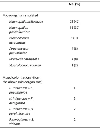
patients. Eight subjects yielded more than one PPM in their sputum. enzae made up 72% of all bacterial isolates.Haemophilus influenzae and H. parainflu-There were significant differences in cigarette con-sumption, cough, dyspnoea, comorbidities, COPD exac-erbations and hospitalisations in the previous year, and sputum colour between patients with and without bacte-rial colonisation (Table 3).
The distribution of colonised patients according to spu-tum colour is presented in Figure 1. Samples with colour 1 (white) were predominantly sterile, whereas in the sam-ples with colours 3 to 5 (yellow to greenish) the preva-lence of colonisation was higher than 80%. Colour number two (light yellow) was not discriminative between colonised and non-colonised.
When colonised patients were divided according to bacterial load, 36 patients had a high bacterial load (>105
CFU/mL) and the remaining 22 had a low bacterial load (≤105 CFU/mL). The characteristics of colonised patients
with a high bacterial load (n = 36) were compared with a group formed by non-colonised patients (n = 61) and those with a low bacterial load (n = 22) considered together (n = 83). Statistically significant differences between the two groups in smoking (pack-years), cough, grade of dyspnoea, hospitalisations in the previous year and sputum colour persisted when patients with high bacterial loads were compared with the remaining
Table 1: Clinical characteristics of the study population
Data Frequency
Subjects, no. 119
Sex, men, no. (%) 112 (92.5) Age, years, mean (SD) 68.1 (9.1) Current smokers, no. (%) 11 (9.2) Smoking, pack-years,
mean (SD)
40 (21.1)
Cardiovascular morbidity, no. (%)
36 (29.7)
Exacerbations in the previous year, mean (SD)
1.3 (0.5)
Requiring hospital admission
0.3 (0.5)
Post-bronchodilator spirometry, mean (SD)
FVC, mL 2790 (942)
FVC, % 68.9 (19.2)
FEV1, mL 1406 (493)
FEV1, % 46.4 (14.1)
Table 2: Potentially Pathogenic Microorganisms (PPMs) isolated in colonised COPD patients.
No. (%)
Microorganisms isolated
Haemophilus influenzae 21 (42)
Haemophilus parainfluenzae
15 (30)
Pseudomonas aeruginosa
5 (10)
Streptococcus pneumoniae
4 (8)
Moraxella catarrhalis 4 (8)
Staphylococcus aureus 1 (2)
Mixed colonisations (from the above microorganisms)
H. influenzae + S. pneumoniae
1
H. influenzae + P. aeruginosa
3
H. influenzae + H. parainfluenzae
2
P. aeruginosa + S. viridans
Table 3: Differences between stable COPD patients with and without bacterial colonisation
Variables Colonised (n = 58) Not colonised (n = 61) P value
Sex, men, no. (%) 54 (93.1) 55 (90.2) 0.74
Age, years, mean (SD) 68.3 (8.3) 67.6 (9.8) 0.67
Current smokers, no. (%) 7 (12.1) 4 (6.6) 0.35
Smoking, pack-years, mean (SD) 46.7 (25.1) 34.2 (23.4) 0.006
Cardiovascular morbidity, no. (%) 22 (37.9) 20 (32.8) 0.23
Comorbid conditions, mean (SD) 1.06 (0.99) 0.61 (1.02) 0.025
Use of inhaled steroids, no. (%) 46 (79.3) 47 (77.1) 0.83
Symptoms, no. (%)
Dyspnoea 56 (96.5) 58 (95.1) 0.72
Cough 44 (75.9) 56 (91.8) 0.024
Expectoration 57 (98.3) 58 (95.1) 0.62
Grade of dyspnoea, mean (SD) 1.78 (0.92) 1.15 (0.54) <0.001
Exacerbations in the previous year, no. (%)
Number 0.021
≤2 31 (53.4) 47 (77.1)
>2 27 (46.6) 14 (22.9)
Requiring hospital admission 0.007
None 36 (62.1) 51 (83.6)
≤1 16 (27.6) 10 (16.4)
>1 6 (10.3) 0
Lung function tests, mean (SD)
FVC, mL 2852.7 (979.1) 2710.9 (911.5) 0.41
FVC, % 70.5 (19.5) 66.7 (18.9) 0.28
FEV1, mL 1411.4 (511.7) 1380.0 (433.1) 0.72
FEV1, % 47.4 (15.2) 45.1 (13.1) 0.38
FEV1/FVC 50.4 (11.8) 52.6 (13.9) 0.43
Sputum analysis
Colour, mean (SD) 2.94 (1.0) 1.56 (0.8) <0.001
Pro-inflammatory cytokines, median (IQR) in pg/mL
IL-1, n = 53 14 (4-432) 168 (49-758) 0.82
IL-6, n = 53 258 (76-653) 112 (33-368) 0.62
IL-8, n = 61 13480 (1335-43400) 5390 (252-14335) 0.27
TNF-alpha, n = 54 45 (20-94) 35 (10-183) 0.12
FVC = forced vital capacity; FEV1 = forced expiratory volume in the first second; IQR = interquartile range; IL= interleukin; TNF = tumour necrosis factor.
patients (Table 4). Sufficient sputum for inflammatory analysis was available from only 61 subjects, all from spontaneous sputum. Sputum concentrations of inflam-matory markers showed a great inter-individual variabil-ity and did not follow a normal distribution. There were no significant differences in sputum concentrations for any of the inflammatory markers analysed between
The results of the multivariate analysis were very simi-lar when identifying the factors significantly associated with the presence of PPMs or on classifying the popula-tion according to bacterial load. In both cases, only the degree of dyspnoea and sputum colour were significantly and independently associated with the presence of PPMs and with high bacterial load. Sputum colour was a stron-ger indicator of the presence of positive cultures for PPMs than its load (Table 5).
Discussion
In the present study, bacterial colonisation of the airways by PPMs, mainly H. influenzae and H. parainfluenzae, was reported in 49% of patients with stable COPD. This finding adds evidence to a high prevalence of bacterial colonisation of airways in stable COPD reported by oth-ers [4,5,9-12]. Interestingly, our results using sputum samples are quite similar to those obtained in other stud-ies with the use of the PSB technique or bronchial lavage for microbiologic assessment of the lower airways in COPD [4,5,11,12,20,21]. The possibility of sputum collec-tion along a maximum of three monthly clinical visits and the use of the induced sputum technique in selected cases may have accounted for this high diagnostic yield of the sputum. However, most of our patients were able to pro-duce a valid sputum sample for microbiological examina-tion and inducexamina-tion of sputum was necessary in only 5 cases. A previous study by our group demonstrated that spontaneous and induced sputum yielded equivalent results in terms of frequency of bacterial colonisation and species recovered [22]. A pooled analysis of data from studies that used PSB demonstrated that a PPM load ≥102
CFU/mL should be considered abnormal and allowed the estimation that at least one quarter of the patients with stable COPD were colonised by PPMs [5]. Furthermore, most patients with exacerbated COPD had concentration
of PPMs > 105 [4,5]. Since there is no universally accepted
cut-off for high bacterial load in sputum samples, a 105
CFU/mL concentration was used in our study [4,8]. With this value, 30% of our total population and almost two thirds of the colonised patients in our study had a high PPM load.
Bacterial colonisation in our study was related to cumu-lative consumption of cigarette smoking, history of exac-erbations in the previous year and sputum colour. Exacerbations in the previous year leading to hospitalisa-tion were associated with increased bacterial load, although this relationship disappeared on multivariate analysis. In other studies, current smoking and severe air-flow obstruction have been identified as predisposing factors for bacterial colonisation in stable COPD [11,12]. However, we did not observe significant differences in lung function between colonised and non-colonised patients. The relationship between lung function and fre-quency of colonisation is not clear, since a lack of associa-tion between FEV1 and colonisation has also been observed in other studies [8,12,21,23] and may be due, at least in part, to the under-representation of mild patients in most series as well as in the current study. Interest-ingly, the only two factors identified in multivariate analy-sis to be significantly and independently associated with both presence of bacterial colonisation and high bacterial load were a more severe degree of dyspnoea and a darker colour of sputum. The degree of dyspnoea is a marker of severity of COPD and being a categorical variable with a wider distribution in our population probably contrib-uted to its demonstrated association with colonisation, in contrast to the severity of FEV1 impairment.
Regarding bronchial inflammation, it should be noted that we did not find increased sputum concentrations of pro-inflammatory cytokines in patients with bacterial colonisation. Different reasons may explain this finding, including a small number of patients with valid samples for analysis, the inter-individual variability in the sputum concentrations of the cytokines was very large [24], and there was a large number of patients with low bacterial loads. In fact, Hill et al. [8] have demonstrated that mark-ers of inflammation increased progressively with increas-ing bacterial load in patients with stable COPD. Consequently, when our colonised patients were catego-rized according to high or low bacterial load, besides the persistence of the clinical differences already observed between the colonised and non-colonised groups (i.e., cigarette smoking, hospitalisations in the previous year, grade of dyspnoea and sputum colour) a non-significant trend towards higher sputum concentrations of inflam-matory markers (except IL-6) was observed in patients with high bacterial load. Our results concur with previ-ous observations regarding the lack of association
Figure 1 Percentage of bacterial colonisation according to spu-tum colour (differences statistically significant at P < 0.001).
Table 4: Differences between colonised and non-colonised COPD patients according to bacterial load
Variables High bacterial load (≥105)
(n = 36)
Low bacterial load (<105)
and not colonised (n = 83)
P value
Sex, men, no. (%) 33 (91.7) 76 (91.6) 0.98
Age, years, mean (SD) 68.6 (6.9) 67.7 (9.9) 0.63
Current smokers, no. (%) 6 (16.7) 5 (6) 0.086
Smoking, pack-years, mean (SD)
48.5 (22.5) 36.7 (25.2) 0.017
Cardiovascular morbidity, no. (%)
11 (30.6) 31 (37.3) 0.34
Comorbid conditions, mean (SD)
0.86 (0.99) 0.83 (1.02) 0.88
Use of inhaled steroids, no (%) 29 (80.6) 64 (77.1) 0.81
Symptoms, no. (%)
Dyspnoea 36 (100) 78 (93.9) 0.66
Cough 25 (69.4) 75 (90.4) 0.007
Expectoration 36 (100) 79 (95.2) 0.31
Grade of dyspnoea, mean (SD) 1.86 (0.83) 1.28 (0.73) <0.001
Exacerbations in the previous year, no. (%)
Number 0.32
≤2 20 (55.6) 58 (69.9)
>2 16 (44.4) 25 (30.1)
Requiring hospital admission
0.003
None 20 (55.6) 67 (80.7)
≤1 11 (30.6) 15 (18.1)
>1 5 (13.9) 1 (1.2)
Lung function tests, mean (SD)
FVC, mL 2936.9 (975.7) 2712 (927.6) 0.23
FVC, % 71.9 (21.0) 67.1 (18.4) 0.21
FEV1, mL 1423.9 (536.5) 1382 (443.1) 0.66
FEV1, % 47.3 (15.9) 45.8 (13.4) 0.61
FEV1/FVC 48.9 (11.2) 52.7 (13.5) 0.15
Sputum analysis
Colour, mean (SD) 2.97 (0.94) 2.01 (1.08) <0.001
Pro-inflammatory cytokines, median (IQR) in pg/mL
IL-1, n = 53 47 (5-593) 29 (4-255) 0.14
IL-6, n = 53 134 (39-381) 169 (34-415) 0.18
IL-8, n = 61 8060 (460-31400) 4890 (201-15025) 0.09
TNF-alpha, n = 54 76 (11-269) 38 (11-71) 0.96
between colonisation and increased IL-6 [9,10] but are discordant with other works showing significantly increased bronchial IL-8 and TNF-alpha in colonised patients, particularly with H.influenzae [9,10,21,23,25]. Therefore, our data, if confirmed in a larger sample of patients, would also suggest a dose-response relationship between bacterial load and bronchial inflammation and that a threshold of bacterial load might be necessary to elicit a significant inflammatory reaction in the airways [5,6,26]. In contrast, Sehti et al. [27] examined whether the increase in bacterial concentrations functions as a separate mechanism of exacerbation induction, indepen-dent of a new strain acquisition. In a prospective longitu-dinal cohort of COPD patients assessed during exacerbations and stable disease, sputum concentrations of pre-existing strains of H. influenzae and H. haemolyti-cus were not significantly different in exacerbation versus stable disease. Concentrations of M. catarrhalis and S.
pneumoniae were even lower during exacerbations
com-pared with stable periods. However, concentrations of new strains of H. influenzae and M. catarrhalis were increased during exacerbations, but the differences were small. These authors speculate that change in bacterial load was unlikely to be a major primary mechanism of exacerbation induction in COPD [27,28]. This hypothesis is a matter of debate, because the interpretation of what a significant increase in bacterial load is when measured in a logarithmic scale is not clear [10], and when trans-formed to a non-logarithmic scale, the differences in absolute bacterial counts were of a very high magnitude [29].
The identification of bronchial colonisation has clinical implications. Patel et al. [9] demonstrated that the pres-ence of lower airway bacterial colonisation in stable COPD was significantly related to exacerbation fre-quency and severity. In the study of Rosell et al. [5], again high bacterial loads were associated with exacerbation and showed a statistically significant dose-response rela-tionship between bacterial load and exacerbation after adjustment for covariates. In our study colonised patients had significantly more exacerbations and hospital
admis-sions the year previous to the study compared with non-colonised patients, but the significance disappeared on multivariate analysis. It should be taken into account that our study was neither designed nor powered to demon-strate differences in exacerbation or hospitalisation rates between colonised and non-colonised COPD patients. Therefore, the identification of patients colonised by PPMs using a non-invasive and relatively inexpensive technique such as the analysis of sputum may play an important role in the management of severe and very severe COPD, particularly if intervention studies with antibiotics demonstrate improved clinical outcomes [13]. To facilitate the diagnosis of bronchial colonisation the use of a surrogate marker could be of interest. Purulence (colour) of sputum graded by the investigator with a sim-ple scale from 1 to 5 revealed significant differences in colour between colonised and non-colonised patients. Patients with colour 3 or higher (dark yellow to green sputum) had a prevalence of bacterial colonisation greater than 80%. The relevance of sputum colour has been already described and validated for exacerbated patients in which yellowish or greenish sputum is signifi-cantly associated with a bacterial exacerbation compared with white (non-bacterial) sputum [30,31] but the rela-tionship between sputum colour and bacterial colonisa-tion in stable COPD has deserved little attencolonisa-tion [8].
The present results should be interpreted taking into account some limitations of the study, particularly the small sample size may not have allowed determination of sputum concentrations of inflammatory markers in all samples, in most cases due to the small recovery of spu-tum that did not provide enough supernatant for the quantification of inflammatory mediators. The cross-sec-tional design did not allow the dynamics and time course of bacterial colonisation and airway inflammation during exacerbations to be examined. Patients with negative bronchodilator test were included to exclude individuals with asthma who are less likely to be colonised, but the results may not be extrapolated to partially reversible COPD patients. High concentrations of PPMs in sputum samples, however, is a simple parameter that may help to
Table 5: Results of multivariate analysis of factors associated with presence of bacteria in sputum and with high bacterial load.
Factor OR 95% CI P value
Factors associated with bacteria in sputum
Degree of dyspnoea 2.63 1.53 - 5.09 0.004
Sputum colour 4.11 2.30 - 7.29 <0.001
Factors associated with high bacterial load as opposed to no bacteria and low bacterial load
Degree of dyspnoea 2.01 1.17 - 3.46 0.012
select candidates to participate in antibiotic trials of sta-ble COPD in order to demonstrate bacterial eradication and potentially prolong time to exacerbation [6,32,33].
Conclusions
Almost half of a population of ambulatory moderate to very severe COPD patients carry PPMs in their airways. Colonised patients had more severe dyspnoea, and spu-tum colour allows the identification of patients most likely to be colonised by PPMs.
List of abbreviations
CFU: colony-forming units; CI: confidence interval; COPD: Chronic obstructive pulmonary disease; CT: computed tomography; FEV1: forced expiratory volume in one second; FVC: forced vital capacity; IQR: inter-quartile range; 1: interleukin-1; 6: interleukin-6; IL-8: interleukin-8; OR: odds ratio; PPMs: potentially patho-genic microorganisms; PSB: protected specimen brush; SD: standard deviation; TNF-alpha: tumour necrosis fac-tor-alpha.
Competing interests
Marc Miravitlles has received honoraria for consultancy and speaking at scien-tific meetings from Bayer Schering, GlaxoSmithKline, Boehringer Ingelheim and AstraZeneca. Cristian de la Roza is fully employed in the Medical Depart-ment of Bayer Schering Pharma. Antoni Torres has received honoraria for con-sultancy and speaking at scientific meetings from Bayer and Covidien. Alicia Marín, Eduard Monsó, Sara Vilà, Ramona Hervás, Cristina Esquinas, Marian García, Laura Millares and Josep Morera have no conflict of interest to disclose.
Authors' contributions
MM designed the study, participated in the analysis and interpretation of data and wrote the manuscript. EM, JM and AT designed the study, and participated in the analysis and interpretation of data. AM and SV recruited the patients, col-lected data and participate in the design and analysis. CR participated in the design and analysis of the study. CE and RH collected and processed the sam-ples, and created and cleaned the database. LM and MG perfomed the micro-biological investigations. All authors read and approved the final manuscript.
Acknowledgements
This study was funded by unrestricted grants from Fundación Respira-SEPAR and La Marató de TV3 and Bayer Schering Pharma. We thank Marta Pulido, MD, for providing an outline for this manuscript and support in editing and journal styling. Bayer Schering Pharma was the source of funding for medical writing. The funding bodies had no role in study design, data analysis, interpretation and writing of the manuscript, and in the decision to submit the manuscript for publication.
Author Details
1Fundació Clínic. Institut D'Investigacions Biomèdiques August Pi i Sunyer (IDIBAPS). Ciber de Enfermedades Respiratorias (CIBERES), Barcelona, Spain, 2Department of Pneumology, Hospital Germans Trias i Pujol. Autonomous University of Barcelona; Ciber de Enfermedades Respiratorias (CIBERES), Barcelona, Spain, 3Department of Pneumology, Hospital Germans Trias i Pujol, Ciber de Enfermedades Respiratorias (CIBERES), Badalona, Barcelona, Spain, 4Medical Department, Bayer Schering Pharma, Sant Joan Despi, Barcelona, Spain and 5Department of Pneumology, Institut Clínic del Tòrax (IDIBAPS), Hospital Clínic, Ciber de Enfermedades Respiratorias (CIBERES), Barcelona, Spain
References
1. Miravitlles M, Murio C, Guerrero T, Gisbert R: Pharmacoeconomic evaluation of acute exacerbations of chronic bronchitis and COPD.
Chest 2002, 121:1449-1455.
2. Donaldson GC, Seemungal TA, Bhowmik A, Wedzicha JA: Relationship between exacerbation frequency and lung function decline in chronic obstructive pulmonary disease. Thorax 2002, 57:847-852.
3. Soler-Cataluña JJ, Martínez-García MA, Román P Sánchez, Salcedo E, Navarro M, Ochando R: Severe acute exacerbations and mortality in patients with chronic obstructive pulmonary disease. Thorax 2005,
60:925-931.
4. Monsó E, Ruiz J, Rosell A, Manterola J, Fiz J, Morera J, Ausina V: Bacterial infection in chronic obstructive pulmonary disease. A study of stable and exacerbated outpatients using the protected specimen brush. Am J Respir Crit Care Med 1995, 152:1316-1320.
5. Rosell A, Monsó E, Soler N, Torres F, Angrill J, Riise G, Zalacaín R, Morera J, Torres A: Microbiologic determinants of exacerbation in chronic obstructive pulmonary disease. Arch Intern Med 2005, 165:891-897. 6. Miravitlles M: Exacerbations of chronic obstructive pulmonary disease:
when are bacteria important? Eur Respir J 2002, 20(Suppl 36):1s-11s. 7. Papi A, Bellettato CM, Braccioni F, Romagnoli M, Casolari P, Caramori G,
Fabbri LM, Johnston SL: Infections and airway inflammation in chronic obstructive pulmonary disease severe exacerbations. Am J Respir Crit Care Med 2006, 173:1114-1121.
8. Hill AT, Campbell EJ, Hill SL, Bayley DL, Stockley RA: Association between airway bacterial load and markers of airway inflammation in patients with stable chronic bronchitis. Am J Med 2000, 109:288-295. 9. Patel IS, Seemungal TA, Wilks M, Lloyd-Owen SJ, Wedzicha JA:
Relationship between bacterial colonisation and the frequency, character, and severity of COPD exacerbations. Thorax 2002,
57:759-764.
10. Wilkinson TM, Patel IS, Wilks M, Donaldson GC, Wedzicha JA: Airway bacterial load and FEV1 decline in patients with chronic obstructive pulmonary disease. Am J Respir Crit Care Med 2003, 167:1090-1095. 11. Zalacain R, Sobradillo V, Amilibia J, Barrón J, Achótegui V, Pijoan JI,
Llorente JL: Predisposing factors to bacterial colonisation in chronic obstructive pulmonary disease. Eur Respir J 1999, 13:343-348. 12. Monsó E, Rosell A, Bonet G, Manterola J, Cardona PJ, Ruiz J, Morera J: Risk
factors for lower airway bacterial colonisation in chronic bronchitis.
Eur Respir J 1999, 13:338-342.
13. Miravitlles M, Marín A, Monsó E, Vilà S, de la Roza C, Hervás R, Esquinas C, García M, Millares L, Morera J, Torres A: Efficacy of moxifloxacin in the treatment of bronchial colonisation in COPD. Eur Respir J 2009,
34:1066-1071.
14. Sanchis J y Grupo de trabajo de la SEPAR: Normativa para la práctica de la espirometría forzada. Arch Bronconeumol 1989, 25:132-142. 15. Pin I, Gibson PG, Kolendowicz R, Girgis-Gabardo A, Denburg JA, Hargreave
FE, Dolovich J: Use induced sputum cell counts to investigate airway inflammation in asthma. Thorax 1992, 47:25-29.
16. Pizzichini E, Pizzichini MM, Efthimiadis A, Evans S, Morris MM, Squillace D, Gleich GJ, Dolovich J, Hargreave FE: Indices of airway inflammation in induced sputum: reproducibility and validity of cell and fluid-phase measurements. Am J Respir Crit Care Med 1996, 154:308-317. 17. Aaron SD, Angel JB, Lunau M, Wright K, Fex C, Le Saux N, Dales RE:
Granulocyte inflammatory markers and airway infection during acute exacerbation of chronic obstructive pulmonary disease. Am J Respir Crit Care Med 2001, 163:349-355.
18. Angrill J, Agustí C, de Celis R, Rañó A, Gonzalez J, Solé T, Xaubet A, Rodriguez-Roisin R, Torres A: Bacterial colonisation in patients with bronchiectasis: microbiological pattern and risk factors. Thorax 2002,
57:15-19.
19. Cabello H, Torres A, Celis R, El-Ebiary M, Puig de la Bellacasa J, Xaubet A, González J, Agustí C, Soler N: Bacterial colonisation of distal airways in healthy subjects and chronic lung disease: a bronchoscopic study. Eur Respir J 1997, 10:1137-1144.
20. Weinreich UM, Korsgaard J: Bacterial colonisation of lower airways in health and chronic lung disease. Clinical Respiratory Journal 2008,
2:116-122.
21. Soler N, Ewig S, Torres A, Filella X, Gonzalez J, Xaubet A: Airway inflammation and bronchial microbial patterns in patients with stable chronic obstructive pulmonary disease. Eur Respir J 1999, 14:1015-1022.
Received: 8 January 2010 Accepted: 14 May 2010 Published: 14 May 2010
This article is available from: http://respiratory-research.com/content/11/1/58 © 2010 Miravitlles et al; licensee BioMed Central Ltd.
This is an Open Access article distributed under the terms of the Creative Commons Attribution License (http://creativecommons.org/licenses/by/2.0), which permits unrestricted use, distribution, and reproduction in any medium, provided the original work is properly cited.
22. Marin A, García M, Badorrey I, Sabrià M, Morera J, Monsó E: Spontaneous sputum production as a marker of bacterial colonisation in stable COPD. Eur Respir J 2005, 26(Suppl 49):232. abstract
23. Sethi S, Maloney J, Grove L, Wrona C, Berenson CS: Airway inflammation and bronchial bacterial colonisation in chronic obstructive pulmonary disease. Am J Respir Crit Care Med 2006, 173:991-998.
24. Sapey E, Bayley D, Ahmad A, Newbold P, Snell N, Stockley RA: Inter-relationships between inflammatory markers in patients with stable COPD with bronchitis: intra-patient and inter-patient variability.
Thorax 2008, 63:493-503.
25. Bresser P, Out TA, van Alphen L, Jansen HM, Lutter R: Airway inflammation in nonobstructive and obstructive chronic bronchitis with chronic Haemophilus influenzae airway infection. Comparison with noninfected patients with chronic obstructive pulmonary disease. Am J Respir Crit Care Med 2000, 162:947-952.
26. Bresser P, van Alphen L, Habets FJ, Hart AA, Dankert J, Jansen HM, Lutter R:
Persisting Haemophilus influenzae strains induce lower levels of interleukin-6 and interleukin-8 in H292 lung epithelial cells than nonpersisting strains. Eur Respir J 1997, 10:2319-2326.
27. Sethi S, Sethi R, Eschberger K, Lobbins P, Cai X, Grant BJ, Murphy TF:
Airway bacterial concentration and exacerbations of chronic obstructive pulmonary disease. Am J Respir Crit Care Med 2007,
176:356-361.
28. Sethi S, Evans N, Grant BJ, Murphy TF: New strains of bacteria and exacerbations of chronic obstructive pulmonary disease. N Engl J Med
2002, 347:465-471.
29. Abusriwil H, Stockley RA: Bacterial load and exacerbations of COPD. Am J Respir Crit Care Med 2008, 177:1048-1049.
30. Stockley RA, O'Brien C, Pye A, Hill SL: Relationship of sputum colour to nature and outpatient management of acute exacerbations of COPD.
Chest 2000, 117:1638-1645.
31. Soler N, Agustí C, Angrill J, Puig de la Bellacasa J, Torres A: Bronchoscopic validation of the significance of sputum purulence in severe exacerbations of chronic obstructive pulmonary disease. Thorax 2006,
62:29-35.
32. Wilson R: Using antibiotics to delay exacerbations of chronic obstructive pulmonary disease. Hot Topics Respir Dis 2006, 2:21-26. 33. Chodosh S: Clinical significance of the infection-free interval in the
management of acute bacterial exacerbations of chronic bronchitis.
Chest 2005, 127:2231-226.
doi: 10.1186/1465-9921-11-58
Cite this article as: Miravitlles et al., Colour of sputum is a marker for