Original Research Article
Correlation of neonatal and maternal clinico-hematological parameters
as predictors of early onset neonatal sepsis
Champa Panwar
1, Shyam L. Kaushik
1*, Rajni Kaushik
2, Arvind Sood
1INTRODUCTION
Neonatal sepsis is one of the major health problems throughout the world and one of the commonest causes of neonatal mortality. It accounts for 30-50% of the total
neonatal deaths in developing countries.1,2 Overall incidence of sepsis is 1-5/1000 live births and mortality rate of untreated sepsis can be as high as 50%.3,4 In India incidence of sepsis is 38 per 1000 live births in tertiary care institutes and it contributes to 36% of deaths in
ABSTRACT
Background: Neonatal sepsis is one of the commonest causes of neonatal mortality and accounts for 30-50% of the total neonatal deaths in developing countries. Diagnosis of neonatal sepsis is still a great challenge since there is no single laboratory test with 100% sensitivity and specificity. Certain maternal and neonatal characteristics are predictive of Early Onset neonatal Sepsis (EOS) (onset < 72 hours of birth). The objective of this study was to evaluate the correlation of neonatal and maternal clinical and haematological manifestations with EOS and to identify the reliable specific parameters for early diagnosis of neonatal sepsis.
Methods: This prospective study enrolled 95 newborns with suspected EOS and their mothers. Perinatal history, clinical profile, symptoms and laboratory data of mother and baby were recorded in each case. The neonatal sepsis screen included TLC, peripheral blood smears (PBS), DLC, ANC, I: T ratio, micro - ESR and platelet count. The maternal haematological parameters were TLC and platelet count. These parameters were evaluated based on the standard reference values. A blood culture was the standard indicator for proven sepsis.
Results: Out of 95 newborns presenting with EOS, 46(48.4%) had positive blood culture. EOS was seen predominantly in preterm, males, LBW and neonates delivered vaginally. Among the various neonatal haematological parameters toxic granules, raised Micro–ESR, I/T Ratio >0.2, thrombocytopenia remained significant markers for early diagnosis of culture positive EOS (p <.05). Majority of EOS was caused by Gram negative organisms with
E.Coli being the commonest. Important adverse perinatal factors associated with EOS were seen in 64.2% mothers. Maternal total leukocyte count was the most important parameter for predicting EOS in their newborns with a sensitivity of 67.2%, specificity of 77.5%, PPV of 80.4% , NPV of 63.2% though not found statistically significant (p =0.065).
Conclusions: Risk factors for EOS include both maternal and neonatal factors. It is very essential to diagnose the sepsis in early phase and it is also important to rule out sepsis to prevent irrational use of antibiotics. A high index of suspicion along with simple, cost effective hematological screening parameters is sensitive and satisfactory tools in predicting early onset neonatal sepsis.
Keywords: EOS, Haemotological parameters, Preterm
1Department ofPediatrics, IGMC, Shimla-171001, Himachal Pradesh, India
2
Department ofPathology, IGMC, Shimla-171001, Himachal Pradesh, India
Received: 02 December 2016
Accepted: 14 December 2016
*Correspondence:
Dr. Shyam L. Kaushik,
E-mail: shyam.kaushik@live.in
Copyright: © the author(s), publisher and licensee Medip Academy. This is an open-access article distributed under the terms of the Creative Commons Attribution Non-Commercial License, which permits unrestricted non-commercial use, distribution, and reproduction in any medium, provided the original work is properly cited.
hospitals.5,6 The major concern of the clinicians is its non-specific presentation, sometimes the rapid progression of sepsis and the unavailability of tests with a high positive predictive value (PPV). Therefore, the early detection of neonatal sepsis is a vexing problem. Though blood culture is considered as gold standard, it is time consuming with limitation in preterm and has high false negative rates.7-10
The susceptibility of the newborn to infection is related to immaturity of both the cellular and humoral immune systems at birth. This feature is particularly evident in preterm neonates. Early-onset sepsis (EOS) syndrome is also associated with acquisition of microorganisms from the mother through blood-borne transplacental infection of the fetus, ascending infection, and infection upon passage through an infected birth canal or exposure to infected blood at delivery.11-13
For the same reason, several rapid haematological tests are done as part of sepsis screen for early diagnosis of neonatal sepsis.
The present study was aimed to evaluate the neonatal and maternal clinical manifestations and their hematological parameters, individually and in combination for rapid identification of early onset neonatal sepsis (EOS).
METHODS
This is a cross-sectional study conducted for one year (May 2012 to April 2013) at the Neonatal Intensive Care Units of Indira Gandhi Medical College, Shimla. Newborns of >30 weeks gestation and >1000grams weight with features suggestive of EOS (onset < 72 hours of life) constituted the study cohort.
Each neonate was examined by a pediatrician and signs and symptoms of the neonate were recorded. The gestational age was assessed by period of gestation (POG) and if not known by New Ballard Scoring System.12
A detailed history including adverse perinatal factors; intra-partum fever (>37.50C), chorioamnionitis, Prolonged rupture of membrane (>18 hours) and unclean vaginal examination were recorded and detailed clinical examination was performed according to designed proforma in each case.12
Blood samples of the neonates were collected at the time of admission and before initiation of antibiotic therapy. Blood sampling was done under all aseptic precautions in the NICU. Soon after admission two ml blood sample was taken in EDTA vacutainer and processed for TLC and platelet count by MS -9 (3-part) Coulter hematology autoanalyser. TLC < 5000 or >20,000 /mm3 were considered abnormal.15
Peripheral blood smears were drawn on clean slides and stained by Giemsa stain. A differential leukocyte count (DLC) was done to obtain the total neutrophil count (TNC), immature neutrophil count (IM), including bands and stabs; and mature neutrophil count.16
Neutrophils were classified as band forms when there were no nuclear segmentation or when the width of the nucleus at any constriction was not less than one third the width of its widest portion. Band forms together with less mature cell forms were classified as immature polymorphonuclear (PMN) leukocytes. Using these values, I/M and I/T ratios were computed. Neutrophils were further examined for degenerative changes such as toxic granulation, Dohle bodies, and vacuolization on PBS by Giemsa stain.15Immature to total neutrophil ratio (IT Ratio) of >0.2 was taken significant.17 Micro - ESR was done by capillary method. (µ - ESR) ≥15 mm at the end of 1 hour was taken significant.15 In addition blood for CRP was also tested.
Another 1 ml blood sample was inoculated into 5 ml of culture media - brain heart infusion (BHI) broth in all the cases of suspected sepsis prior to starting antibiotics and were observed for at least 72 hours before they were reported as sterile.18
Maternal investigations included complete haemogram with peripheral blood smear. Maternal TLC count >12,000/mm3 and platelet count <1.5 lac/mm3 were taken as significant.19 Endocervical swabs and urine samples for culture and sensitivity were collected in all mothers with newborns suspected to have EOS.19
After detailed investigation and culture reports neonates were further categorized into culture positive sepsis or culture negative sepsis. The clinical manifestations and hematological parameters were compared, individually and in combination, with the blood culture result.
Statistical analysis
Sensitivity, specificity, positive and negative predictive values (NPVs) were calculated for each parameter. P values were also calculated for different parameters. Data was statistically analyzed using SPSS software.
RESULTS
During the study period there were 6964 live births .Of the total nursery admissions, 95 newborns were clinically suspected of sepsis within 72 hrs of life with or without maternal history of one or more risk factors for sepsis.Of all the neonates suspected of EOS, 46 (48.4%) were culture proven, giving 0.7% incidence of culture positive sepsis .
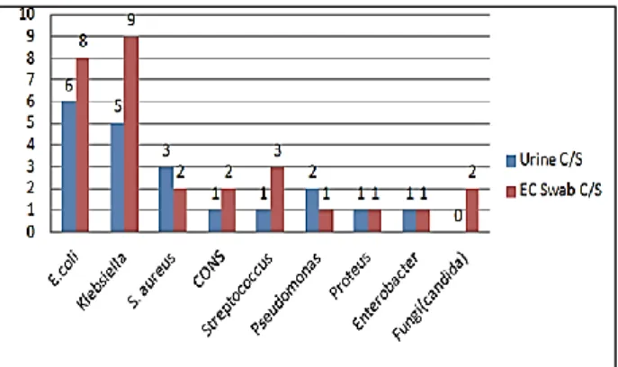
(8.6%), Candida 2 (4.3%). Streptococcus, Pseudomonas,
Proteus sp., Enterococcus, Enterobacter and NLF in 1 (2.1%) each. Among the neonates with blood culture positive EOS, E. coli 6 (13%) was the commonest micro-organism in maternal urine culture whereas Klebsiella
accounted for 9/46(19.5%) of the maternal EC swab cultures. Maternal culture organisms were similar to those found in neonates (Figure 1). A high incidence of EOS was seen in preterm (58.6%), males (65.3%), LBW (52.1%) and newborns delivered vaginally (80.4%) but none of these were statistically significant between culture positive and culture negative sepsis ( p >0.05, Table 1).
Figure 1: Maternal urine and EC swab culture profile in newborns with blood culture positive EOS (n = 46).
Table 1: Distribution of demographic profile in newborns with EOS (n = 95).
Neonatal profile Blood C/S (+) (n = 46) Blood C/S (-) (n = 49) Total ( n = 95) p-value
Gestational age (weeks)
≤ 37 27 (58.6%) 26 (49.1%) 53 (55.7%)
0.212
≥ 37 19 (41.4%) 23 (54.7%) 42 (44.3%)
Sex
Male 30 (65.3%) 26 (46.5%) 56 (58.9%)
0.5
Female 16 (34.7%) 23 (58.9%) 39 (41.1%)
Birth weight (gms)
≥2500 7 (15.3%) 11 (22.5%) 18 (27.3%)
0.661 2499-1500 24 (52.1%) 20 (40.8%) 44 (42.1%)
<1500 15 (32.6%) 18 (36.7%) 33 (30.6%)
Mode of delivery
Vaginal 37 (80.4%) 27 (55.2%) 64 (67.4%)
0.053 Caesarean section 9 (19.6%) 22 (44.8%) 31 (32.6%)
C/S: Culture/Sensitivity.
Table 2: Distribution of clinical manifestations in newborns with EOS (n = 95).
Neonatal clinical manifestations n = 95
Fever, refusal to feed and lethargy, each 39 (41%)
Pneumonia 49 (31.4%)
Disseminated intravascular coagulation 19 (12.1%) Upper GI bleed and feed intolerance 19 (12.1%)
Birth asphyxia 19 (12.1%)
Shock 18 (11.5%)
Respiratory failure 15 (9.6%)
Neonatal jaundice 15 (9.6%)
Necrotising 12 (7.6%)
Apnea 12 (7.6%)
Hypoglycemia 12 (7.6%)
Meconium aspiration syndrome 11 (7%)
Skin lesions (SSSS,pustules and abscess) 10 (6.4%)
Seizure 10 (6.4%)
Pulmonary hemorrhage 7 (4.4%)
Sclerema 7 (4.4%)
Hyperglycemia 5 (3.2%)
Hypothermia 5 (3.2%)
Urinary tract infection 5 (3.2%)
Clinical manifestations of the newborns with early onset sepsis included fever, refusal to feed and lethargy in 41% each, pneumonia in 31.4%, shock in 11.5%, DIC, UGI
bleed, feed intolerance and birth asphyxia in 12.1% each (Table 2).
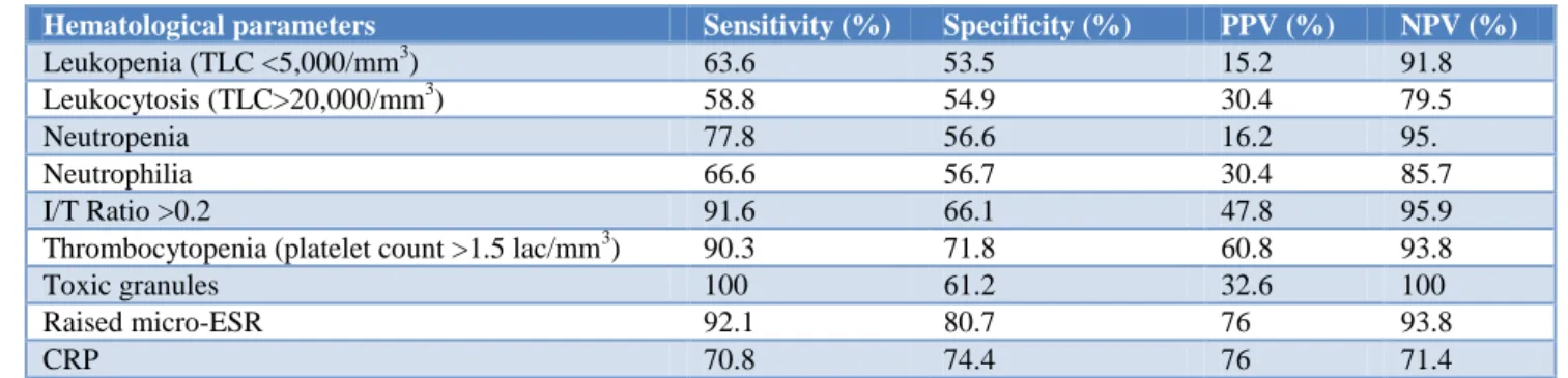
Table 3: Sensitivity, Specificity, PPV and NPV of various hematological parameters in EOS.
Hematological parameters Sensitivity (%) Specificity (%) PPV (%) NPV (%)
Leukopenia (TLC <5,000/mm3) 63.6 53.5 15.2 91.8 Leukocytosis (TLC>20,000/mm3) 58.8 54.9 30.4 79.5
Neutropenia 77.8 56.6 16.2 95.
Neutrophilia 66.6 56.7 30.4 85.7
I/T Ratio >0.2 91.6 66.1 47.8 95.9
Thrombocytopenia (platelet count >1.5 lac/mm3) 90.3 71.8 60.8 93.8
Toxic granules 100 61.2 32.6 100
Raised micro-ESR 92.1 80.7 76 93.8
CRP 70.8 74.4 76 71.4
CRP: C-reactive protein, I/T Ratio: immature to total neutrophil ratio; ESR: Erythrocyte sedimentation rate, PPV: Positive predictive value; NPV: Negative predictive value, TLC: Total leucocyte count.
Table 4: Distribution of various hematological parameters in newborns with EOS (n = 95).
Neonatal haematological parameters Blood C/S (+) (n = 46) Blood C/S (-) (n = 49) p value
TLC (<5,000/mm3 or >20,000/mm3)
Leukopenia -7 (15.2%) Leukopenia - 4 (8.1%)
0.006 Normal TLC - 25 (54.3%) Normal TLC - 35 (71.5% )
Leukocytosis - 14 (30.5%) Leukocytosis - 10 (20.4%)
Total PMNs Neutropenia - 7 (15.2%) Neutropenia - 2 (4.08%) 0.085 Neutrophilia -14 (30.4%) Neutrophilia -7 (14.2%)
I/T Ratio >0.2 22 (47.8%) 2 (4.08%) 0.000
Immature PMNs 21 (45.6%) 5 (10.2%) 0.072
Thrombocytopenia 28 (60.8%) 3 (6.1%) 0.000
Toxic granules 15 (32.6%) 0 0.000
Raised µ-ESR 35 (76%) 3 (6.1%) 0.000
CRP 34 (73.9%) 14 (28.5%) 0.067
C/S: Culture/Sensitivity, I/T Ratio: Immature to Total neutrophil ratio; ESR: Erythrocyte sedimentation rate, PMNS: polymorphonuclear leukocytes.
CRP: C-reactive protein, I/T Ratio: Immature to Total neutrophil ratio; ESR: Erythrocyte sedimentation rate, PPV: Positive predictive value; NPV: Negative predictive value, TLC: Total leucocyte count. Among the neonatal haematological parameters in EOS, toxic granules were found to be the most important with a sensitivity of 100%, specificity of 61.2%, PPV of 32.6% and a high NPV of 100% (Table 3).
Neonatal TLC, IT Ratio, toxic granules, platelet count and micro–ESR were found to be statistically significant in early diagnosis of culture positive EOS (p < 0.05, Table 4).
Maternal data and investigations were collected in 95 newborns who presented with suspected EOS. Significant risk factors observed were PROM > 18 hours (61.1%), fever within 2weeks of delivery (26.3%), instrumental delivery (23.1%), foul smelling liquor (16.3%),
prolonged labour (6.3%) and history of unclean vaginal examination (5.2%).
Among the maternal hematological parameters TLC was the most reliable. In blood culture positive EOS newborns group 37 (80.4%) mothers had leukocytosis (TLC> 12,000/mm3) with a sensitivity of 67.2%, specificity of 77.5%, PPV of 80.4% and NPV of 63.2% (p = 0.065). Thrombocytopenia was seen in 13.1% with culture positive sepsis (p = 0.3) with sensitivity of 33.3%, specificity of 48.1%, PPV of 13% and NPV of 75.5%. Though maternal TLC was more specific than platelet count in predicting EOS, but none of these were statistically significant (p > 0.05, Table 5).
Table 5: Distribution of maternal haematological parameters in newborns with EOS (n = 95).
Maternal Parameters Blood C/S (+)
(n = 46)
Blood C/S (-)
(n = 49) p value Sensitivity Specificity PPV NPV
Maternal leukocytosis
(TLC >12,000/mm3) 37 (80.4%) 18 (36.7%) 0.065 67.2% 77.55% 80.4% 63.2% Thrombocytopenia
(platelet count <1.5 lac) 6 (13.1%) 1 (2.04%) 0.3 33.3% 48.1% 13% 75.5%
C/S: Culture/Sensitivity; PPV: Positive predictive value; NPV: Negative predictive value; TLC: Total leucocyte count.
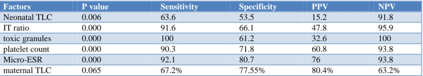
Table 6: Correlation of neonatal and maternal factors with neonatal sepsis.
Factors P value Sensitivity Specificity PPV NPV
Neonatal TLC 0.006 63.6 53.5 15.2 91.8
IT ratio 0.000 91.6 66.1 47.8 95.9
toxic granules 0.000 100 61.2 32.6 100
platelet count 0.000 90.3 71.8 60.8 93.8
Micro-ESR 0.000 92.1 80.7 76 93.8
maternal TLC 0.065 67.2% 77.55% 80.4% 63.2%
DISCUSSION
The overall incidence of EOS has improved over last two decades in our institution, 6.6/1000 live births in present study versus 17.3/1000 births by Chaudhary et al. It is also lower than that reported by Mondal et al (15.5/1000 live births).20,21 This can be explained by the availability of better health care facilities in our institution over the time. Among EOS, culture positive sepsis was seen in 48.4% neonates in the present study which is well in agreement with reported range of 8-73% in various studies.9,10,19,22-24 EOS was more common among male neonates with an overall M:F ratio of 1.9:1 which is similar to other studies (1.6:1 to 1.8:1).25-27 It was more common in preterm (58.6%) in our study, similar to observations by Antoniette et al (64.7%) and Shirazi et al (69%).19,23 In our study 84.7% of the septicemic neonates with EOS were LBW which was quite high as compared to 51% reported by Waseem et al.28
Out of 95 mothers included in our study, 64.2% had one or more risk factors for neonatal sepsis which was higher than a study by Antoniette et al (19.4%).19 The incidence of PROM >18 hours in a study by Khair et al was 75% which was higher compared ours (61.1%).24
Fever and refusal to feed were the common symptoms noticed in 41% septicemic neonates followed by pneumonia in 49 (31.4%). Similar observations were mentioned by Chaudhary et al, Buch et al, Khatua et al, Dawodu et al, Jaswal et al, Waliulla et al, whereas Antoniette et al found respiratory problems to be the commonest.19,21,27,29-32
In the present study, among the blood culture positive neonates, markers of sepsis which were significantly raised included I/T ratio, toxic granules, micro-ESR and CRP. Toxic granules had highest sensitivity and NPV of
100%, specificity of 61.2% and PPV of 32.6%. IT ratio had a sensitivity of 91.6% and specificity of 66.1%, PPV 47.8% and NPV of 95.9% which was similar to that observed by Sunit et al who reported a sensitivity of 100%, specificity of 86%, PPV of 69% and NPV of 100% for toxic granules.33
In this study neonatal thrombocytopenia also had high sensitivity of 90.3%, NPV of 93.8%, specificity of 71.8% and PPV of 60.8% which is higher than the observation by Khair et al (sensitivity of 60% and NPV 94%, specificity of 82% and PPV of 31% ) and Antoniette et al (sensitivity of 35.3%, specificity of 89.2%, PPV of 40% and NPV of 87.1% ) for neonatal thrombocytopenia.19,24 Though TLC was not raised in majority of the neonates but it was statistically significant amongst culture proven sepsis patients. This was comparable with study by Antoniette et al.19
Hematological profile of mothers with neonates having EOS
Similar to the findings by Antoniette et al we also observed statistically significant association of maternal leukocytosis (p = 0.006) in culture positive neonates. However no such correlation could be established for thrombocytopenia. 19
Bacteriological profile of mothers and neonates having EOS
Bacteriological profile in mothers and neonates in EOS varies from place to place. This was evident in a study by Asindi et al who contrary to our observation of E.coli in urine and kleibsella in EC swabs has found CoNS,
Klebsiella pneumonia, Pseudomonas aeruginosa and
EOS in various studies has been found to be caused mainly by gram negative organisms which are also an observation in the present study. However E. coli was the commonest micro-organism in our subjects whereas others have found Kleibsella and CoNS as the commonest.21, 26, 35-37
CONCLUSION
Neonatal sepsis is a serious illness associated with high mortality so a high index of suspicion is important in the diagnosis and treatment of neonatal infection because it is hampered by vague and nonspecific clinical manifestations. Neonatal haematological parameters like toxic granules, IT ratio >0.2, raised micro-ESR, thrombocytopenia, TLC and maternal TLC are sensitive and specific in early diagnosis of neonatal sepsis while awaiting blood culture reports. Therefore, neonatal and maternal, clinical and haematological profile can be relied as early indicators of EOS even in resource poor countries
.
Funding: No funding sources Conflict of interest: None declared
Ethical approval: The study was approved by the Institutional Ethics Committee
REFERENCES
1. Bang AT, Bang RA, Bactule SB, Reddy HM, Deshmukh MD. Effect of home - based neonatal care and management of sepsis on neonatal mortality: field trial in rural India. Lancet. 1999;354:1955-61.
2. Stoll BJ. The global impact of neonatal infection. Clin Perinatol. 1997;24:1-21.
3. Baley J, Goldfarb J. Neonatal Infections. In: Klaus MH, Fanaroff AA, editors. Care of the high risk neonate. 4th edition. Philadelphia: WB Saunders Company; 1993.
4. Bellig L, Ohning B. Neonatal sepsis. E Med J Peadiatr/Neonatol. 2003;4(1):6.
5. Report of the National Neonatal Perinatal database National Neonatology Forum 2000. Available at http://www.newbornwhocc.org/nnpd-India_South-Asia.html. Accessed on 1st November 2016.
6. Zaidi AK, Huskins WC, Thaver D, Bhutta ZA, Abbas Z, Goldmann DA. Hospital-acquired neonatal infections in developing countries. Lancet. 2005;365:1175-88.
7. Liu L, Johnson HL, Cousens S, Perin J, Scott S, Lawn JE, et al. Global, regional, and national causes of child mortality: an updated systematic analysis for 2010 with time trends since 2000. Lancet. 2012;379:2151-216.
8. Oestergaard MZ, Inoue M, Yoshida S, Mahanani WR, Gore FM, Cousens S, et al. Neonatal mortality levels for 193 countries in 2009 with trends since 1990: a systematic analysis of progress, projections, and priorities. PLoS Med. 2011;8:e1001080.
9. Kuruvilla KA, Pillai S, Jesudason, Jana AK. Bacterial profile of sepsis in a neonatal unit in south India. Indian Pediatrics. 1998;35:851-8.
10. Ayengar V, Madhulika, Vani SN. Neonatal sepsis due to vertical transmission from maternal genital tract. Indian J Pediatr. 1991;58:661-4.
11. Linder N, Ohel G, Gazit G, Keider D, Tamir I, Reichman B. Neonatal sepsis after prolonged rupture of premature membranes. J Perinatol. 1995;15:36-8.
12. Marlowe SE, Greenwald J, Anwar M, Haitt M, Hegyi T. Prolonged rupture of membranes in the term newborn. Am J Perinatol. 1997;14:483-6. 13. Botet F, Cararach V, Sentis J. Premature rupture of
membranes in early pregnancy. Neonatal prognosis. J Perinatol Med.1994;22:45-52.
14. Autraliasian study group for neonatal infections. Early onset group B streptococcal infections in Aboriginal and non-aboriginal infants. Med J Aust. 1995;163:302-6.
15. Manroe BL, Weinberg AG, Rosenfeld CR, Browne R. The neonatal blood count in health and disease. J Pediatr. 1979;95(1):89-98.
16. Rodwell RL, Leslie AL, Tudehope DI. Early diagnosis of neonatal sepsis using a hematologic scoring system. J Pediatr. 1988;112(5):761-7. 17. Fanaroff AA, Stoll BJ, Wright LL, Carlo WA,
Ehrenkranz RA, Stark AR, et al. nichd neonatal research network. Trends in neonatal morbidity and mortality for very low birthweight infants. Alam J Obstet Gynecol. 2007;196(2):147-8.
18. Gerdes JS, Polin R. Early diagnosis and treatment of neonatal sepsis. Indian J Pediatr. 1998;65:63-78. 19. Mayuga WAB, Isleta PFD. Clinical correlation of
neonatal and maternal hematological parameters as predictors of neonatal sepsis. PIDSP J. 2005;9:(2)36-42.
20. Mondal GP, Raghavan M, Bhat BV. Srinivasan S. Neonatal septicaemia among inborn and outborn babies in referra hospital. Indian J Pediatr. 1991;58:529-33.
21. Chaudhary S. Neonatal septicemia: a clinic-etiological study. MD (thesis). Shimla: Himachal Pradesh University; 1989.
22. Mane AK, Nagdeo NV, Thombare VR. Study of neonatal septicemia in a tertiary care hospital in rural Nagpur. J Recent Adv Appl Sci. 2010;25:19-24.
23. Shirazi H, Riaz S, Tahir R. Role of the hematological profile in early diagnosis of neonatal sepsis. Ann Pak Inst Med Sci. 2010;6(3):152-5. 24. Khair KB, Rahman MA, Sultana T, Roy CK,
Rahman Md. Q, Shahidullah M, et al. Role of hematologic scoring system in early diagnosis of neonatal septicemia. BSMMU J. 2010;3(2):62-7. 25. Trotman H, Bell Y, Thame M, Nicholson AM,
26. Gheibi S, Fakoor Z, Karamyyar M, Khashabi J, Iikhanizaden B, Sana FA, et al. Coagulase negative staphylococcus; the most common cause of neonatal septicaemia in Uremia, Iran. Iran J Pediatr. 2008;18:237-43.
27. Buch AC, Srivastava V, Kumar H, Jadhav PS. Evaluation of haematological profile in early diagnosis of clinically suspected cases of neonatal sepsis. In J Basic Appl Med Sci. 2011;1:1-6. 28. Waseem R, Shah AA, Khan MQ, Qureshi AW.
Indicators of early outcome in neonatal sepsis. Biomedica. 2005;21:117-20.
29. Khatua SP, Das AK, Chatterjee BD. Neonatal septicaemia. Ind J Pediatr. 1986;53:509-14.
30. Dawodu A, Al Umran K, Twum DK. Case control study of neonatal sepsis. Experience from Saudi Arabia. J Trop Pediatr. 1997;43:84-8.
31. Jaswal RS, Kaushal RK, Goel A, Pathania K. Role of C-reactive protein in deciding duration of antibiotic therapy in neonatal septicaemia. Indian Pediatr. 2003;40:880-3.
32. Walliullah MS, Ishan MN, Siddika M, Hossain MK, Hossain MA. Risk factor, clinical manifestation bacteriological profile of neonatal sepsis a tertiary
level pediatric hospital, Mymensingh. Med J. 2009;18:66-72.
33. Sunit S, Kumar V. Predictors of serious bacterial infection in infants up to 8 weeks of age. Indian Pediatr. 1994;31:171-80.
34. Asindi AA, Archibong EI, Mannan NB. Mother-infant colonization and neonatal sepsis in prelabor rupture of membranes. Saudi Med J. 2002;23:1270-4.
35. Kumar GD, Ramchandran VG, Gupta P. Bacteriological analysis of blood culture isolates from neonates a tertiary care hospital in India. J Health Popul Nutr. 2002;20:343-34.
36. Chaudhary HR, Hassan MA, Habibullah M. Neonatal sepsis and use of antibiotic in a tertiary care hospital. Park J Med Sci. 2007;23:78-81. 37. Jain NK, Seth D, Mangal V. A clinicomicrobial
association in neonatal septicaemia. Pediatric Oncall J 2010;7:97-9.