R E S E A R C H
Open Access
TNF
α
-blockade stabilizes local airway
hyperresponsiveness during TLR-induced
exacerbations in murine model of asthma
Magnus Starkhammar
1,2,4, Susanna Kumlien Georén
1,2, Sven-Erik Dahlén
2,3, Lars-Olaf Cardell
1,2,4and Mikael Adner
2,3,5*Abstract
Viral infections are a common cause of asthma exacerbation. These maladies are sometimes complicated by bacterial infections. Toll-like receptors (TLRs) are in the forefront of our microbial defence, with TLR3 responding to viral and TLR4 to bacterial stimulation. The present study was designed to evaluate the effect of concomitant TLR3 and TLR4 stimulation in a murine model of allergic asthma.
BALB/c mice were stimulated intranasally with a combination of poly(I:C) and LPS activating TLR3 and TLR4,
respectively. This resulted in the development of airway hyperresponsiveness (AHR) in the proximal part of the lung, along with signs of neutrophilic inflammation. Analysis of the bronchioalveolar lavage fluid (BALF) revealed a marked increase in TNFα. In contrast, the allergic airway inflammation induced by ovalbumin administration to sensitized mice caused AHR in the whole lung along with an increase in eosinophils and lymphocytes in the BALF and lung.
When poly(I:C) + LPS were given to mice with an ongoing allergic airway inflammation induced by ovalbumin, the AHR was further increased in the peripheral lung and neutrophils appeared together with eosinophils and lymphocytes in the BALF and lung. Treatment with the TNFα-blocking antibody infliximab blunted the AHR increase, without affecting the cells influx in BALF.
To conclude; a combined TLR3- and TLR4-stimulation, representing a concomitant viral and bacterial infection, causes an AHR that is further exaggerated during an ongoing allergic inflammation. The airway stabilizing effect of infliximab indicates the possible future use of TNFαblockade in treatment of microbial induced exacerbations of allergic asthma.
Keywords:Airway mechanics, Allergy, Asthma, Innate immunity, Viral infection, Bacterial infection
Introduction
Viral infections, such as respiratory syncytial virus (RSV) and human rhinovirus (HRV) are believed to be the primary cause of asthmatic exacerbations in adults [1, 2]. Haemophilius influenzae and Moraxella cattar-halis are bacteria known to play an opportunistic role following respiratory viral infections, with their own ability to trigger exacerbations [3]. Moreover, especially
Haemophilius influenza is frequently found together
with RSV and HRV [4]. Thus, combined bacterial and viral infestations are not an uncommon problem in asthma.
Pattern recognition receptors (PRRs) is an umbrella term for several receptor families all with their specific ability to recognizing various microbes, initiating an innate host defence reaction [5, 6]. The Toll-like receptors (TLRs) are the most well characterized PRR family comprised of 10 members (13 in mice) [5]. TLR3 are known to identify viruses like RSV and HRV, whereas TLR4 recognizes bacteria like Haemophilius influenza.During experimental conditions poly(I:C) and LPS, ligands for TLR3 and TLR4, can be used to mimic the innate immune effects of viruses
* Correspondence:mikael.adner@ki.se
2Division of ENT diseases, CLINTEC, Karolinska Institutet, Stockholm, Sweden 3
Institute of Environmental Medicine, Karolinska Institutet, Stockholm, Sweden
Full list of author information is available at the end of the article
(i.e. RSV/HRV) and bacteria (i.e. Haemophilius influenza), respectively.
As a consequence of respiratory infections patients with allergic asthma generally suffer from prolonged and more severe symptoms than patients without established allergy [7, 8]. The mechanistic interplay between on-going allergy inflammation, microbial infections and TLR activation is complex and still poorly understood. Airway hyperrespon-siveness (AHR) is closely related to the asthmatic exacer-bation [2, 9]. In the present study AHR was evaluated in ovalbumin (OVA) mouse model of allergic asthma. The effect of a combined TLR3 and TLR4 activation was stud-ied by adding poly(I:C) and LPS together once daily dur-ing four consecutive day, just after the mice had completed their sensitization protocol. The purpose was to mimic the situation when asthmatic subjects with an al-lergic airway inflammation are exposed to a concomitant viral and bacterial infection for a time period representing occasional infection. In addition to changes in AHR, alter-ations in the local respiratory tract cell and mediator com-position were assessed in bronchioalveolar lavage fluid (BALF). Special attention was given to the role of TNFα which has been implicated in many aspects of airway pathology in asthma [10]. In the present study infliximab, a well-established TNFαantibody was used to investigate the involvement of TNFαin AHR.
Methods Animals
Female BALB/c mice (approximately 20 g, 8–12 weeks old) were obtained from Charles River (Sulzfeld, Germany). The mice were housed in plastic cages with adsorbent bedding material in a conventional animal house with 12-hours dark/light cycles. Water and pelleted food were provided
ad libitum. All animal procedures were approved by the regional committee of animal experimentation eth-ics (Stockholm norra djurförsöksetiska nämnd).
Treatment protocol
In the first set of experiments the mice were given 20μg poly(I:C) (lyophilized; Sigma-Aldrich, St Louis, MO, USA) and 2 μg LPS (from Escherichia coli 0127:B8; Sigma-Aldrich, St Louis, MO, USA) solved in 20μl PBS [11] or 20μl PBS as control intranasally during isoflur-ane anaesthesia on four consecutive days. In the second set of experiments mice were first sensitized with 10 μg ovalbumin (OVA) (grade II; Sigma-Aldrich, St Louis, MO, U.S.A.) and 1 mg Al(OH)3 (Sigma-Aldrich)
sus-pended in 200μl PBS, given as an intraperitoneal injec-tion (i.p.) on days 0 and 7. On days 15, 16 and 17 the animals were anaesthetized by isoflurane inhalation and 20 μl OVA (50 μg) or PBS were administered intrana-sally. On the subsequent four days (18–21) the animals were given poly(I:C) + LPS in the same regimen as the
first set of experiments. PBS was used as vehicle control. For the second set of experiments the animals were treated with a monoclonal antibody against TNF-α (200μl, 0.5 mg/ml infliximab (Infliximab, Centocor B.V, Leiden, The Netherlands) or PBS i.p. one hour before each Poly(I:C)/LPS administration. Lung mechanics to-gether with collection of tissues and bronchoalveolar lavage fluid (BALF) were assessed 24 h after the last treatment (day 22) (Fig. 1).
Lung mechanics
The mice were anaesthetised (pentobarbital sodium, 90 mg/kg bw given i.p.), put on a heating pad (body temperature, 37 °C), tracheotomised (18-gauge cannula) and connected to the flexivent animal ventilator (Scireq, Montreal, Canada). After ventilation was started, the mice were monitored as described earlier [11]. After a five mi-nutes resting period, methacholine (MCh; Sigma-Aldrich) was injected through the tail vein in increasing doses (0.01, 0.03, 0.1, 0.3, 1, 3 and 10 mg/kg · body weight), in order to induce AHR.
For the first experiments, lung resistance and compli-ance were measured by assuming a single-compartment linear model and multiple linear regressions at a sinus-oidal frequency of 2.5 Hz every eighth breath for 3 min after each injection. For the second experiment, lung me-chanics were measured using a forced oscillation tech-nique [12]. The parameters obtained were the Newtonian resistance (RN), a close approximation of resistance in the
conducting airways; tissue resistance (G), which reflects energy dissipation in the lung tissue consisting of airway closure and heterogeneity of airway distribution; and tis-sue elastance (H), which characterises tissue stiffness and reflects the energy storage within the tissue.
Bronchoalveolar lavage
Directly after lung function measurements BALF was collected (1 mL ice cold PBS containing 0.6 mM ethy-lendiaminetetraacetic acid (EDTA) was lavaged three times in the lung). The fluid was centrifuged at +4 °C, 1200 rpm, for 10 min and the supernatant was stored at −80 °C until use. Lysis buffer (150 mM NH4Cl,
10 mM KHCO3, 0.1 mM EDTA, pH 7.2), was used for
2 min, to lyse the red blood cells, followed by washing in PBS. Total cell number was counted and expressed as cells/ml BALF. Differential cell counts were performed on May-Grünwald/Giemsa stained cytospins, counting a minimum of 300 cells, in a blinded manner.
Measurement of cytokines
simultaneous immunoassay was carried out according to manufacturer’s instructions.
Lung histology
Lungs were removed and immersed with 4 % buffered formaldehyde. They were then embedded in paraffin, sectioned and stained in haematoxylin and eosin. Histo-logical findings with a focus on inflammatory cells, such as peribronchial/perivascular and parenchymal cell infil-tration, were semi-quantitatively graded in a blinded manned as 0 to 4 (zero to abundant cell infiltration) and summed together. The cells were identified according to their morphology in 1000x magnification.
Statistical analysis
Data were analysed using Graph Pad Prism®, version 5.01, software (GraphPad Software Inc., San Diego, CA, U.S.A.). Results are presented as mean ± standard error of mean (SEM) andnequals’number of subjects. For comparison of airway reactivity, two-way analysis of variance (ANOVA) was followed by Bonferroni’s Multiple Comparison Test. BALF cell data and lung histology data were analysed with Kruskal-Wallis analysis of variance, followed by Dunn’s test for between group comparisons. A p value of less than 0.05 was considered significant.
Results
Intranasal administration of poly(I:C) + LPS for four consecutive days induces airway hyperresponsiveness with concomitant influx of inflammatory cells and release of a number of inflammatory mediators
To define the specific effects dual TLR activation in-duces on allergic airways, the effect on non-allergic airways was needed as comparison. The impact of the combined TLR3 and TLR4 stimulation on airway func-tion was assessed by measuring the increased resistance induced by cumulative administration of methacholine. For mice given poly(I:C) + LPS, the amplitude of the lung resistance (4.88 ± 0.43 cmH2O.s.mL−1) was
increased more than 2-fold when compared to the one measured in control mice (2.05 ± 0.11 cmH2O.s.mL−1)
which received PBS vehicle (Fig. 2a). When evaluating the cells in BALF from the same study groups, there was a strong increase in macrophages, neutrophils and lym-phocytes in the poly(I:C) + LPS treated mice compared to the control mice (Fig. 2b). No eosinophils were found in any of the groups. The BALF from these mice were processed by measuring the levels of inflammatory me-diators. Ten (IL-1α, IL-5, IL-12, IL-17, TNFα, CCL2, CCL3, CCl5 CXCL9 and VEGF) out of 21 selected medi-ators were increased in BALF from the poly(I:C) + LPS treated mice when compared to the PBS-treated mice (Fig. 2c). For five mediators (IL-2, IL-6, IL-13, CXCL1 and CXCL10) no difference between the groups were observed and for six (IL-1β, IL-4, IL-10, IFNγ, FGF and GM-CSF) the levels were below the detection limit in both groups.
TNFαblockade inhibits the poly(I:C) + LPS induced exaggerated airway hyperresponsiveness
TNFα, one of the mediators that was increased after the treatment with poly(I:C) + LPS, was not increased in a previous study when exposing the mice separately to the two TLR agonists with a similar protocol [11]. Thus, to investigate the role for TNFα, further experiments were performed using treatment with infliximab, an antibody against TNFα, administered i.p. one hour before the TLR-agonists were given. With this intervention, the role of TNFαwas investigated both in non-allergic con-ditions and on an established allergic airway inflamma-tion. In order to delineate the localization of the effect both of TLR stimulation and the TNFα action, forced oscillation techniques were used measuring the airway responses both in the conducting airways (RN) and in
the peripheral parts, defined as tissue damping (G) and tissue elastance (H).
poly(I:C) + LPS caused AHR. However, this TLR-induced AHR was only found in the proximal region
RN (Fig. 3a) and not in the distal partsG (Fig. 3b) and H(Fig. 3c). Treatment with infliximab did not influence the TLR-induced increase ofRN.
As predicted, ovalbumin sensitization and challenge caused an AHR in both the proximal (Fig. 3d) and distal
parts of the lung (Fig. 3e-f ) [12]. When poly(I:C) + LPS was administered after an allergic airway inflammation was established, the treatment did not cause a further in-crease in AHR in RN. In contrast, the TLR treatment
caused a marked increase in AHR in both the distal pa-rametersGand H. This increased effect of the TLRs on
GandHon the established allergic airway inflammation Fig. 2Pulmonary reactions to concomitant stimulation of TLR3 and TLR4. Responses were measured after one daily intranasal administration of 20μg poly(I:C) + 2μg LPS or PBS as control for four days.aAirways resistance after methacholine provocation.bCells in BALF.cCytokines measured in BALF. Results are shown as mean ± S.E.M. Lines between circles or columns representP< 0.05;n= 10–12
was markedly inhibited by infliximab whereas the inflixi-mab treatment did not affect theRN.
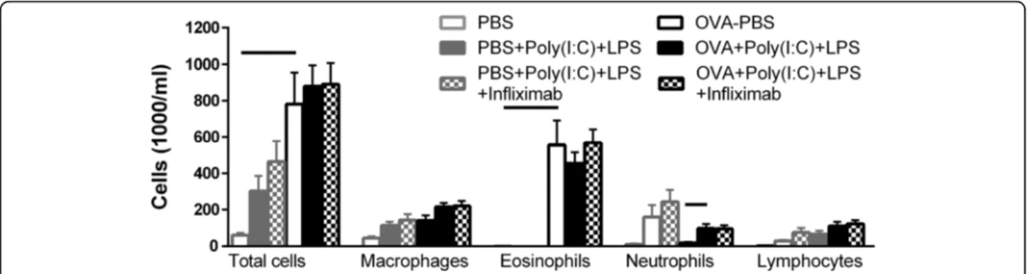
TNFαblockade does not inhibit the poly(I:C) + LPS induced exaggerated cellular influx in bronchoalveolar lavage fluid
As described in Fig. 2, administration of poly(I:C) + LPS to non-sensitized mice again increased the neutrophils, mac-rophages and lymphocytes. This TLR-induced increase was not affected by concomitant infliximab treatment (Fig. 4). OVA-exposed mice showed a marked increase in eosinophils. Poly(I:C) + LPS administration to the mice with the established allergic inflammation did not influ-ence the eosinophils, but caused an increase in neutro-phils and a trend to increasing both macrophages and lymphocytes. This augmentation by TLR-stimulation was not affected by the infliximab treatment.
TNFαblockade does not affect the poly(I:C) + LPS induced increase in inflammatory cell infiltration in lung tissue
Administration of poly(I:C) + LPS to non-sensitized mice also caused a strong influx of cells in the lung which not was altered by infliximab-treatment (Fig. 5a, b). The in-flux consisted of neutrophils and lymphocytes (Fig. 5c). A strong influx of cells in the lung tissue was also seen in OVA-exposed mice (Fig. 5a, b). The main cell types were eosinophils and lymphocytes (Fig. 5c). In the lung from mice with the established allergic inflammation and exposed to Poly(I:C) + LPS a similar strong influx of cells was shown although with additional neutrophils (Fig. 5a-c) This influx was not affected by the infliximab treatment.
Discussion
The present study showed that intranasal installation of poly(I:C) together with LPS for four days in mice trig-gered an AHR and airway inflammation with increased influx of macrophages, neutrophils and lymphocytes,
and enhanced release of several pro-inflammatory cyto-kines including TNFαin BALF. These TLR-induced ef-fects were not affected by treatment with the anti-TNF antibody infliximab. In mice with established allergic airway inflammation, characterized by an AHR and an eosinophilic influx in BALF, the administration of poly(I:C) + LPS caused a further increase of AHR in the peripheral lung; tissue damping and tissue elastance. This TLR agonist-induced increase of AHR was blocked by infliximab, but it did not inhibit the additional influx of macrophages, neutrophils and lymphocytes in BALF. Thus, the present study indicates that TNFα causes an AHR in the peripheral lung in allergic inflammatory airways which not is linked to cellular influx in BALF.
Four days intranasal administration of poly(I:C) to-gether with LPS, mimicking an effect of a combined viral and bacterial stimulation during a time period represent-ing occasional infection, caused a marked AHR in mice. In our earlier studies, where these two TLR agonists were given separately in a similar regimen, both of them induced AHR [11, 13]. However, the mean maximal ef-fect on the lung resistance (RL) was clearly higher for
the combination of poly(I:C) together with LPS in this study (4.9 cmH2O.s/ml) compared to the effect when
ei-ther of the TLR agonist were given alone which did not exceed 3.5 cmH2O.s/ml. Isolated mouse airways, which
express both TLR3 and TLR4 in the smooth muscle layer, become hyperreactive when exposed to either poly(I:C) or LPS [14]. In the same study it was shown that when the two TLR agonists were given together this caused a synergistic increase of the hyperreactivity [14]. Thus, it is possible that the combined effect of intranasal challenge with poly(I:C) or LPS cause is due to a direct action of TLR3 and TLR4 on airway smooth muscle layer which causes AHR.
The combined treatment with poly(I:C) and LPS also caused an inflammation shown both by the influx of cells in BALF and lung tissue as well as by the recorded
release of inflammatory mediators in the airways. Mac-rophages, neutrophils and lymphocytes were found to be increased in BALF. As it has previously been shown that poly(I:C) induces lymphocytes and LPS induces neutro-phils [11, 13], the increase of both neutroneutro-phils and lym-phocytes in BALF in the present study suggests additive effects for the TLR agonists. The infiltration in lung was also dominated by neutrophils and lymphocytes. As an
combined TLR agonist treatment. In addition, the levels of IL-1α, IL-17, TNFα, CCL3 and CCL5 were only in-creased when poly(I:C) and LPS were given together. This synergistic effect of poly(I:C) + LPS in causing TNFα se-cretion has previously been shown in macrophages and osteoclast precursors [15, 16]. As TNFαrelease is a sup-posed to be an early event and it can stimulate the induc-tion of various inflammatory genes [17], it is possible that several of the other mediators are induced due to the ef-fect of TNFα. Thus, it appears that the TLR agonists both induce an effect by themselves and their combined activa-tion causes an interacactiva-tion that generates further effects.
Although clinical studies using antibodies against TNFαto treat asthma has shown variable [18], the initial clinical studies using TNFα-antibodies showed improved effect specifically on AHR [10, 19]. Interestingly, a sub-group of severe asthmatic patients also suffering from rhinosinuitis responded very well to anti-TNFα treat-ment [20]. To evaluate the importance of the TLR-induced effect by TNFα on the development of AHR in this study, the mice were treated with the TNFα anti-body infliximab during the four days of intranasal ad-ministration of poly(I:C) + LPS. In these experiments a forced oscillation technique was used to localize which parts of the airway were affected. As in the first experi-ment, the combination of the TLR agonists induced AHR. However this was found to be localized in the conducting airways (RN) and not in the peripheral tissue
damping (G) and tissue elastance (H). Previous experi-ments also investigating the pulmonary resistance using a forced oscillation technique showed the same pattern for the TLR9-agonist CpG oligodeoxy-nucleotide 2006 which only caused an increase ofRNwith no effect onG
and H[21]. As the conductive airways is the part of the lung that contain smooth muscle cells it further strengthen the suggestion above that the TLR-agonists given in non-allergic healthy conditions cause a hyper-contractile smooth muscle a poly(I:C) + LPS [14, 22] which is responsible for the AHR. However, the effects by the dual TLR stimulation onRN, the cellular influx in
BALF and infiltration of cells in the lung were not af-fected by infliximab indicating that TNFα has only a minor effect on smooth muscle and cellular trafficking in this model.
The sensitization and challenge of OVA also induced an AHR which atRNwas similar to that induced by the
administration of poly(I:C) + LPS but it caused a higher AHR in both the peripheral parameters. This level of AHR was observed five days after the last OVA chal-lenge, indicating a persistent allergic airway inflamma-tion as described previously [13, 21]. When poly(I:C) + LPS were given for four days on an established allergic inflammation, the AHR was further increased as mea-sured by both the peripheral parameters G and H but
the TLR-agonists did not affect the conducting airways (RN). An increase in lung resistance has been shown in
response to both poly(I:C) and LPS when they were given separately on a similar allergic airway inflamma-tion [13]. However comparison of the level of response is not possible as the effect in different compartments of the lung was not determined in that study. It has been shown that when GandH increase in a similar manner it depends on airway closure [23] which is a major com-ponent of hyperresponsiveness in allergically inflamed airways [24]. One important factor for airway closure is the production of surfactant which has shown to be de-creased by both viral [25] and TLR4 activation [26]. Thus, the increase of the combined response to TLR3 and TLR4 suggests that there may be a specific effect of combined viral and bacterial infection on peripheral dys-function of the lung during allergic conditions. Consid-ering that there is a substantial amount of evidence that inflammation of the small airways contributes to the clinical expression of asthma [27], the markedly differ-ence of AHR caused by TLR activation during allergic (peripheral) and non-allergic (central) may be one rea-son for the severity for asthmatic subjects during infections.
When infliximab was given on the established allergic inflammation one hour before the TLR-agonist adminis-tration the increased peripheral AHR was markedly re-duced, without any effect on the influx of cells in the BALF or the infiltration of cells in lung. These results in-dicate that TNFα has a specific effect on the peripheral AHR, which has been suggested to be dependent on het-erogeneous airway closure due to protein interference with surfactant [28]. Through the separation of the lung mechanics it could be shown that the response onRNof
TLR-stimulation on non-provoked airways differed from the responses when the TLR-agonists were given on an established allergic airway inflammation. The reason may be that the latter condition induces a resistance close to the maximal capacity of this parameter. On the other hand, that the TLR-agonist-induced increase of AHR in the peripheral parameters was caused by TNFα may be of specific importance since airway closure is linked to excessive bronchoconstriction [29], recurrent exacerbations [30] and asthma severity [31]. It is possible that several other inflammatory mediators also are re-duced secondary to the reduction of TNFα. The inter-pretation is further complicated by the effects TNFα by itself has on the airways [32]. It is also important to keep in mind that secondary stimulus like IL-1β [33] can be directly linked to TNFα.
likely that there is a synergistic interplay between the two innate receptor subtypes that contributes to the worsening of the situation for the mice as reflected in an increased AHR and elevated levels of a multitude of BALF mediators. It is interesting to notice that the TLR-induced increase of AHR in our model was dampened when the mice were treated with infliximab suggesting a pivot role for TNFα in microbial induced exacerbation of allergic asthma. The outcome of previous attempts to use various TNFα blocking agents in asthma has been meagre [18]. Most of these drug evolutions have been dealing with asthma from a more general perspective, and it could be that the role of TNFα is of specific im-portance only during the circumstances reflected in the present set up. It should be stated that the experimental set-up in this study was performed to define the action of TNF during infection and not to investigate the effect as add-on therapy for asthmatic subjects exposed to in-fections. Hence, further studies needs to outline whether it might be a future for specific anti-TNFαtrials focusing on exacerbations of allergic asthma.
Competing interests
The authors declare that they have no competing interests.
Authors’contributions
MS conducted the animal experiments, analysed the data and wrote the manuscript, SGK conducted the animal experiments, analysed the data and wrote the manuscript; SED designed the study, supervised the project and wrote the manuscript, LOC designed the study, supervised the project and wrote the manuscript and MA designed the study, analysed the data, supervised the project and wrote the manuscript. All authors read and approved the final manuscript.
Acknowledgements
We would like to acknowledge the support from the Swedish Medical Research Council, the Swedish Heart-Lung foundation, Konsul Th C Berghs research foundation, the Stockholm County Council Research Funds (ALF), Karolinska Institutet, the ChAMP project, COST BM1201, the Swedish Society of Medicine and the Centre for Allergy Research at Karolinska Institutet.
Author details
1Centre for Allergy Research, Karolinska Institutet, Stockholm, Sweden. 2
Division of ENT diseases, CLINTEC, Karolinska Institutet, Stockholm, Sweden.
3Institute of Environmental Medicine, Karolinska Institutet, Stockholm,
Sweden.4Department of ENT Diseases, Karolinska University Hospital, Stockholm, Sweden.5Unit of Experimental Asthma and Allergy Research,
Institute of Environmental Medicine, Scheeles väg 1, Karolinska Institutet, SE-171 77 Stockholm, Sweden.
Received: 5 March 2015 Accepted: 9 October 2015
References
1. Kurai D, Saraya T, Ishii H, Takizawa H. Virus-induced exacerbations in asthma and COPD. Front Microbiol. 2013;4:293.
2. Busse WW, Lemanske Jr RF, Gern JE. Role of viral respiratory infections in asthma and asthma exacerbations. Lancet. 2010;376:826–34.
3. Kraft M. The role of bacterial infections in asthma. Clin Chest Med. 2000;21:301–13.
4. van den Bergh MR, Biesbroek G, Rossen JW, de Steenhuijsen Piters WA, Bosch AA, van Gils EJ, et al. Associations between pathogens in the upper respiratory tract of young children: interplay between viruses and bacteria. PLoS One. 2012;7:e47711.
5. Kumar H, Kawai T, Akira S. Pathogen recognition by the innate immune system. Int Rev Immunol. 2011;30:16–34.
6. Gon Y. Toll-like receptors and airway inflammation. Allergol Int. 2008;57:33–7.
7. Corne JM, Marshall C, Smith S, Schreiber J, Sanderson G, Holgate ST, et al. Frequency, severity, and duration of rhinovirus infections in asthmatic and non-asthmatic individuals: a longitudinal cohort study. Lancet.
2002;359:831–4.
8. Olenec JP, Kim WK, Lee WM, Vang F, Pappas TE, Salazar LE, et al. Weekly monitoring of children with asthma for infections and illness during common cold seasons. J Allergy Clin Immunol. 2010;125:1001–1006 e1001. 9. Micillo E, Bianco A, D’Auria D, Mazzarella G, Abbate GF. Respiratory
infections and asthma. Allergy. 2000;55 Suppl 61:42–5.
10. Berry MA, Hargadon B, Shelley M, Parker D, Shaw DE, Green RH, et al. Evidence of a role of tumor necrosis factor alpha in refractory asthma. N Engl J Med. 2006;354:697–708.
11. Starkhammar M, Kumlien Georen S, Swedin L, Dahlen SE, Adner M, Cardell LO. Intranasal administration of poly(I:C) and LPS in BALB/c mice induces airway hyperresponsiveness and inflammation via different pathways. PLoS One. 2012;7:e32110.
12. Swedin L, Ellis R, Kemi C, Ryrfeldt A, Inman M, Dahlen SE, et al. Comparison of aerosol and intranasal challenge in a mouse model of allergic airway inflammation and hyperresponsiveness. Int Arch Allergy Immunol. 2010;153:249–58.
13. Starkhammar M, Larsson O, Kumlien Georen S, Leino M, Dahlen SE, Adner M, et al. Toll-like receptor ligands LPS and poly (I:C) exacerbate airway hyperresponsiveness in a model of airway allergy in mice, independently of inflammation. PLoS One. 2014;9:e104114.
14. Bachar O, Adner M, Uddman R, Cardell LO. Toll-like receptor stimulation induces airway hyper-responsiveness to bradykinin, an effect mediated by JNK and NF-kappa B signaling pathways. Eur J Immunol. 2004;34:1196–207. 15. Bagchi A, Herrup EA, Warren HS, Trigilio J, Shin HS, Valentine C, et al.
MyD88-dependent and MyD88-independent pathways in synergy, priming, and tolerance between TLR agonists. J Immunol. 2007;178:1164–71. 16. Krisher T, Bar-Shavit Z. Regulation of osteoclastogenesis by integrated
signals from toll-like receptors. J Cell Biochem. 2014;115:2146–54. 17. Chen G, Goeddel DV. TNF-R1 signaling: a beautiful pathway. Science.
2002;296:1634–5.
18. Matera MG, Calzetta L, Cazzola M. TNF-alpha inhibitors in asthma and COPD: we must not throw the baby out with the bath water. Pulm Pharmacol Ther. 2010;23:121–8.
19. Howarth PH, Babu KS, Arshad HS, Lau L, Buckley M, McConnell W, et al. Tumour necrosis factor (TNFalpha) as a novel therapeutic target in symptomatic corticosteroid dependent asthma. Thorax. 2005;60:1012–8. 20. Wenzel SE, Barnes PJ, Bleecker ER, Bousquet J, Busse W, Dahlén SE, et al. T03
Asthma Investigators. Am J Respir Crit Care Med. 2009;179:549–58. 21. Adner M, Starkhammar M, Georen SK, Dahlen SE, Cardell LO. Toll-like
receptor (TLR) 7 decreases and TLR9 increases the airway responses in mice with established allergic inflammation. Eur J Pharmacol. 2013;718:544–51. 22. Safholm J, Lovdahl C, Swedin L, Boels PJ, Dahlen SE, Arner A, et al.
Inflammation-induced airway smooth muscle responsiveness is strain dependent in mice. Pulm Pharmacol Ther. 2011;24(4):361–6. 23. Backus-Hazzard GS, Howden R, Kleeberger SR. Genetic susceptibility to
ozone-induced lung inflammation in animal models of asthma. Curr Opin Allergy Clin Immunol. 2004;4:349–53.
24. Lundblad LK, Thompson-Figueroa J, Allen GB, Rinaldi L, Norton RJ, Irvin CG, et al. Airway hyperresponsiveness in allergically inflamed mice: the role of airway closure. Am J Respir Crit Care Med. 2007;175:768–74.
25. Krishnan S, Halonen M, Welliver RC. Innate immune responses in respiratory syncytial virus infections. Viral Immunol. 2004;17:220–33.
26. Glasser SW, Maxfield MD, Ruetschilling TL, Akinbi HT, Baatz JE, Kitzmiller JA, et al. Persistence of LPS-induced lung inflammation in surfactant protein-C-deficient mice. Am J Respir Cell Mol Biol. 2013;49:845–54.
27. van den Berge M, ten Hacken NH, Cohen J, Douma WR, Postma DS. Small airway disease in asthma and COPD: clinical implications. Chest. 2011;139:412–23. 28. Kaminsky DA. Peripheral lung mechanics in asthma: exploring the outer
limits. Pulm Pharmacol Ther. 2011;24:199–202.
29. Gibbons WJ, Sharma A, Lougheed D, Macklem PT. Detection of excessive bronchoconstriction in asthma. Am J Respir Crit Care Med. 1996;153:582–9. 30. in’t Veen JC, Beekman AJ, Bel EH, Sterk PJ. Recurrent exacerbations in severe
31. Sorkness RL, Bleecker ER, Busse WW, Calhoun WJ, Castro M, Chung KF, et al. Lung function in adults with stable but severe asthma: air trapping and incomplete reversal of obstruction with bronchodilation. J Appl Physiol (1985). 2008;104:394–403.
32. Bachar O, Adner M, Cardell LO. Toll-like receptor activation in airway smooth muscle: dual actions via separate MAPK pathways. Am J Physiol. 2006;291:L322–3.
33. Zhang Y, Adner M, Cardell LO. IL-1beta-induced transcriptional up-regulation of bradykinin B1 and B2 receptors in murine airways. Am J Respir Cell Mol Biol. 2007;36:697–705.
Submit your next manuscript to BioMed Central and take full advantage of:
• Convenient online submission
• Thorough peer review
• No space constraints or color figure charges
• Immediate publication on acceptance
• Inclusion in PubMed, CAS, Scopus and Google Scholar
• Research which is freely available for redistribution