Original Research Article
Correlation between histological grading, LVI and PNI of carcinoma
oral tongue to lymph node metastasis
Sumana C. Viswanatha
1*, Naveen Hedne
2, Suhel Hasan
1INTRODUCTION
Overall, 57.5% of global head and neck cancers, excluding oesophageal cancers occur in Asia, especially in India, for both sexes.1 In India, Head and neck cancers account for 30% of all cancers in males, except Dibrugarh in Assam (49.6%). They constitute 11 to 16% of all cancers in females. Among them, tongue and mouth in males contribute to more than one-third of the total cancers, except in Dibrugarh where hypopharynx was the major contributor. Among females, mouth cancer was the leading cause.2
Oral cavity cancer is the 8th most common cancer in developed countries and 3rd most common cancer in developing countries.3 Squamous cell carcinoma (SCC) is the most common malignancy in the oral cavity, accounting for 95% of all oral cavity malignant lesions.4 The most common sites are tongue, gingivo-buccal mucosa complex, lower lip and floor of the mouth. When compared to other oral cancers, squamous cell carcinoma of the oral tongue has a higher predisposition to produce metastasis in lymph nodes (incidence 15-75%) depending on the extension of the primary lesion.5,6
ABSTRACT
Background: Squamous cell carcinoma (SCC) of oral tongue has a higher predisposition to lymph node metastasis which reduces survival by 50%. In clinical practice, TNM classification is used for treatment planning which does not provide information on the biological characteristics of the tumor.
Methods: This prospective cross sectional observational study included 30 patients with T1 to T3, N0/+ oral tongue SCC from 1st March 2014 to 30th April 2015. Incisional biopsy was taken from the primary tumour, pathological evaluation for differentiation of the tumour and assessment of lymphovascular (LVI) and perineural invasion(PNI) was carried out. Post operative histopathological examination included differentiation, LVI and PNI. The pathological findings were correlated using chi square test.
Results: Majority presented with T2 stage. 27% had nodal metastasis. There was higher incidence of lymph node metastasis in moderately differentiated (MD) and poorly differentiated (PD) SCC which was not statistically significant. Significant correlation was seen between LVI and PNI to lymph node metastasis (p≤0.001).
Conclusions: There is a higher incidence of lymph node metastasis seen in moderately and poorly differentiated SCC of oral tongue, which can be assessed on the preoperative biopsy, guiding us to be more aggressive in the management of cervical nodes in early tongue cancer.LVI and PNI are good predictors of nodal metastasis, help in assessing aggressiveness and prognosis of the disease, and are soft indicators for considering adjuvant / concurrent RT.
Keywords: Carcinoma oral tongue, Cervical lymph node metastasis, Histologic grade, Lymphovascular-perineural invasion
1
Department of ENT, 2Department of Head and Neck Surgical Oncology, Mazumdar Shaw Medical Centre, Narayana Health City, Bangalore, Karnataka, India
Received: 08 October 2018
Accepted: 15 November 2018
*Correspondence:
Dr. Sumana C. Viswanatha, E-mail: sumanacv84@gmail.com
Copyright: © the author(s), publisher and licensee Medip Academy. This is an open-access article distributed under the terms of the Creative Commons Attribution Non-Commercial License, which permits unrestricted non-commercial use, distribution, and reproduction in any medium, provided the original work is properly cited.
Regional metastatic disease is the most reliable prognostic indicator of treatment outcomes in patients with squamous cell carcinoma of the oral tongue.7-10 Survival decreases by 50% with the presence of regional lymphadenopathy.7
In clinical practice, the treatment plan and prognosis of oral tongue squamous cell carcinoma is mainly based on the TNM (primary tumour, regional lymph node metastasis, distant metastasis) classification. However, this system does not provide any information on the biological characteristics of the tumor. Information about characteristics is important to obtain new objective prognostic factors that may provide information about the aggressiveness of oral tongue cancer.9,10
The aim of this study was to assess the correlation between the histologic grade of carcinoma of oral tongue, lymphovascular and perineural invasion with cervical lymph node metastasis.
METHODS
This prospective cross sectional observational study was conducted at Mazumdar Shaw Medical Centre, Narayana Health City, Bangalore. 30 patients with T1 to T3, N0/+ oral tongue squamous cell carcinoma were included in the study during the period from 1st March 2014 to 30th April 2015.
Inclusion criteria
cT1/2/3 primary squamous cell carcinoma of tongue, N0 N+, non-metastatic M0, previously untreated and those planned for a curative intent surgery.
Exclusion criteria
cT4 lesions, previously treated (Chemotherapy-Radiotherapy), patients with induction chemotherapy, neoadjuvant chemotherapy (NACT), palliative intent treatment/ distant metastasis, patients with history of other cancer and second primary cases.
Methodology
Institutional review board and ethical committee clearance was taken for the study. This prospective study enrolled 30 patients with previously untreated squamous cell carcinoma of oral tongue, undergoing curative intent surgery. Informed consent in their regional language was taken from all the patients. Detailed history regarding use of tobacco agents, alcohol consumption were noted. Clinical and radiological examination of the tumour regarding size, nodal involvement and metastasis was done and clinical TNM staging was noted. Incisional biopsy was taken from the primary tumour, pathological evaluation for differentiation of the tumour (well-differentiated/ moderately differentiated/ poorly
differentiated), and assessment of lymphovascular and perineural invasion was done.
All the patients were planned for curative intent surgery after multidisciplinary tumor board review. Patients underwent adequate glossectomy and selective neck dissection from level I-IV followed by reconstruction with local or free flap as indicated. The primary specimen was sent for pathological evaluation after orientation along with the neck nodal levels sent separately. Specimens underwent detailed histopathological examination by a competent head and neck pathologist, which included differentiation, lymphovascular invasion, and perineural invasion. The pathological findings were correlated.
Statistical methods
The statistical analysis was performed by STATA 11.2 (College Station TX USA). The demographic details of age, gender, histopathological evaluation for differentiation, lymphovascular invasion, perineural invasion were analyzed. Chi-square test was used to measure the association between the histological grades of tumor, lymphovascular invasion, perineural invasion with cervical lymph node metastasis. P<0.05 was considered as statistically significant.
RESULTS
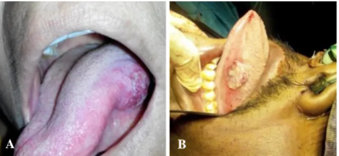
The present study included 30 patients of previously untreated T1-3 N0/+ M0, oral tongue cancer, ranging from 26 to 75 years of age with a mean age of 53.4 years. There were 24 males (80%) and 6 females (20%). Patients were clinically and radiologically (CECT/MRI) evaluated. The primary tumour and cervical lymph nodes were staged according to the TNM staging. Majority of the patients were T2 (63%) (Figure 1 A, B).
Figure 1 (A, B): cT2 left lateral border oral tongue.
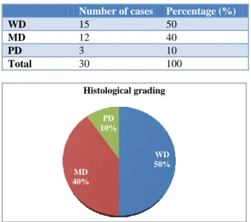
Incisional biopsy was taken from the tumour and pathological evaluation was done for differentiation of the tumour and assessment of lymphovascular and perineural invasion. Table 1 and Figure 2, show the distribution of histological grades. Majority of them were well differentiated (50%), while only 10% were poorly differentiated.
Table 1: Distribution of histological grades (WD-well differentiated, MD-moderately differentiated,
PD-poorly differentiated).
Number of cases Percentage (%)
WD 15 50
MD 12 40
PD 3 10
Total 30 100
Figure 2: Distribution of histological grades (WD-well differentiated, MD-moderately differentiated,
PD-poorly differentiated).
Lymphovascular invasion and perineural invasion could not be identified in any of the pretreatment biopsy specimen.
Figure 3: Adequate glossectomy- intraoperative pic and the specimen.
Following a curative intent surgery of the primary and neck (Figure 3), the neck nodes dissected from different levels were marked and sent separately for pathological evaluation which included differentiation, lymphovascular invasion and perineural invasion.
Pathological T stage assessment
Majority of the patients had T2 tumours (50%), followed by T1 (33.33%).
Pathological neck node staging
73% of cases had no neck node metastasis pathologically (pN0), while 27% had positive metastasis. Majority of
them had N2b stage disease, followed by N1. Only one patient had N2c which was also noted clinically.
Correlation between histological grading to lymph node metastasis
Histological grade which was obtained by pretreatment biopsy was correlated to post treatment pathological lymph node metastasis. In our study, as shown in Table 2, poorly differentiated squamous cell carcinoma had more lymph node metastasis (67%), when compared to well differentiated squamous cell carcinoma (13%). Though there were more nodal metastasis in moderately differentiated and poorly differentiated, it was not statistically significant (p=0.112). Among the 8 nodal metastasis, 50% were moderately differentiated. However, in the node negative group, majority were well differentiated tumours (59.09%) followed by moderately differentiated (36.36%).
Table 2: Correlation between histological grades to lymph node metastasis.
Differentiation N0 (%) N+ (%) Total P value
Well
differentiated 13 (87) 2 (13) 15
0.112
Moderately
differentiated 8 (67) 4 (33) 12 Poorly
differentiated 1 (33) 2 (67) 3
Total 22 8 30
Table 3: Correlation between lymphovascular invasion (LVI) to lymph node metastasis.
LVI N0 (%) N+ (%) Total P value
Present 1 (17) 5 (83) 6(20)
<0.001
Absent 21 (87.5) 3 (12.5) 24(80)
Total 22 8 30
Lymphovascular invasion seen in the post treatment biopsy specimen was correlated to lymph node metastasis (Table 3). 20% of cases had lymphovascular invasion, out of which 83% had positive nodal metastasis. In comparison, out of the 80% of cases without lymphovascular invasion, only 12.5% had nodal metastasis, which was statistically significant (p<0.001). Out of the 8 positive nodal metastasis 62.5% had lymphovascular invasion, whereas among the 22 pN0 cases, only 4.5% had lymphovascular invasion.
Correlation between perineural invasion to lymph node metastasis is shown in Table 4. 63% of cases had a positive perineural invasion, out of which 42% had positive lymph node metastasis. While in the remaining, 37% who had no perineural invasion, none of them had positive nodal metastasis which was statistically significant (p<0.001).
WD 50% MD
40% PD 10%
Histological grading
Table 4: Correlation between perineural invasion (PNI) to lymph node metastasis.
PNI N0 (%) N+ (%) Total P value
Present 11 (58%) 8 (42%) 19(63%) <0.001
Absent 11 (100%) 0 (0%) 11(37%)
Total 22 8 30
Thus, all those patients who had no perineural invasion, did not have nodal metastasis, while all the 8 positive nodal disease cases had positive perineural invasion, although 50% of N0 disease had a positive perineural invasion.
DISCUSSION
Oral cavity cancer is one of the most common cancers in our country, tongue being the most commonly affected subsite. Tongue is a complex anatomical site and its form and function are crucial for efficient swallowing, speech and appreciation of taste. In the past, various methods of treatment have been employed, but tongue has remained a difficult area to assess and treat. The survival in carcinoma of the tongue is poor compared to the other subsites in oral cavity.7,11
Most Indian patients with oral tongue cancer present in advanced stage, and show high propensity for cervical lymph node metastasis. From many studies it is shown that, lymph node metastasis is one of the most important prognostic factors. Survival reduces by 50% in the presence of cervical lymph nodes.7
For many years, treatment plan and prognosis was mainly based on TNM staging, which does not include any biological characteristics of the tumor. Hence, histopathological characteristics are more important in assessing prognosis, planning treatment and to provide information about aggressiveness of the disease.
Figure 4: (A-C) Microscopic picture–well, moderately and poorly differentiated SCC.
Only 10% of our patients had poorly differentiated squamous cell carcinoma (Figure 4 A-C). There was a higher occurrence of nodal metastasis among poorly differentiated squamous cell carcinoma, but it was not statistically significant (p=0.112). A similar observation was seen in the retrospective study done by Rikardsen et al among 133 patients of oral squamous cell carcinoma. In their study, 42% had well-differentiated and 8% poorly differentiated squamous cell carcinoma.12 Akhter et al, in
their study on 50 patients of oral squamous cell carcinoma, 18% of well-differentiated squamous cell carcinoma had nodal metastasis, 26% among moderately differentiated and all patients of poorly differentiated were positive for nodal metastasis.13 There were also other studies which showed significant correlation between histological grading and lymph node metastasis, like in the study done by Kurokawa et al, which included 50 patients with carcinoma tongue, 3.1% of well differentiated squamous cell carcinoma had positive nodal metastasis, while 33.3% of moderately differentiated were positive for nodal metastasis. Thus, their study showed a statistically significant correlation with p<0.01.14 From the above mentioned studies, we can say that higher occurrence of nodal metastasis can be predicted amongst the moderately and poorly differentiated squamous cell carcinoma.
Among the various parameters used to predict the outcome of malignant disease in oral squamous cell carcinoma, lymphovascular invasion (LVI) and perineural invasion (PNI) are both widely used as indicators of aggressive behaviour.15,16 They have been associated with increased risk of local recurrences and lymph node metastasis.
Lymphovascular invasion is classified according to the presence or absence of neoplastic cells located in the wall or lumen of blood or lymphatic vessels. It has been demonstrated to be a good prognostic tool, and it has been found to be correlated with low rates of survival and high risk of recurrence.17 However, due to the difficulty in defining and recognizing this parameter with certainty, some grading systems have omitted this factor.15
Perineural invasion is a tropism of tumour cells for nerve bundles in the surrounding tissues. It is well known as an independent predictor of poor outcome in colorectal carcinoma and salivary gland malignancies. However, in oral squamous cell carcinoma, there is no consensus among authors about the real prognostic impact.16
Figure 5 (A, B): Microscopic picture showing LVI and PNI (20x view).
In our study, 20% of cases had positive lymphovascular invasion (Figure 5A), out of which 83% had positive nodal metastasis. There was a significant correlation between lymphovascular invasion and lymph node metastasis (p<0.001).
A A
B C
We noted that out of the 8 positive nodal metastasis, 62.5% had positive lymphovascular invasion, whereas amongst the 22 pN0 cases, only 4.5% had positive lymphovascular invasion. Similar observation are also seen in studies done by Sharma et al, Jardim et al and Michikawa et al.18-20 Jardim et al, in their study had 40.8% positive lymphovascular invasion and among the positive nodal metastasis, 50.5% had positive lymphovascular invasion.19 But, Lim et al showed no correlation between lymphovascular invasion and nodal metastasis in their study. Out of the 16% positive lymphovascular invasion, 30% had nodal metastasis, while among 84% of negative lymphovascular invasion, 44% had positive nodal metastasis.21
63% of our cases had a positive perineural invasion (Figure 5B). 42% of these had positive lymph node metastasis, which was statistically significant (p<0.001). Significant correlation (p=0.019) was also seen in the study done by Jardim et al whereas the study done by Lim et al, did not show significant correlation (p>0.99) between positive perineural invasion and lymph node metastasis.19 In their study out of the 12.5% who had positive perineural invasion, 28% had nodal metastasis. And out of the 87.5% negative perineural invasion, 20% had nodal metastasis.21
All the patients with no perineural invasion, did not have nodal metastasis in our study. All the 8 positive nodal disease cases had a positive perineural invasion. Half the patients with node negative disease had a positive perineural invasion.
We could thus summarise that lymphovascular invasion and perineural invasion are good predictors of nodal metastasis and help in assessing the aggressiveness and prognosis of the disease, and are soft indicators for considering adjuvant radiotherapy/ concurrent radiotherapy.
CONCLUSION
We conclude from our study, that there is a higher incidence of cervical lymph node metastasis seen in moderately and poorly differentiated SCC of oral tongue, which can be assessed on the preoperative biopsy, guiding us to be aggressive in the management of cervical nodes in early tongue cancer.
LVI and PNI have a significant correlation to lymph node metastasis. We can thus assess the aggressiveness of the tumor, predict the prognosis and plan an appropriate management for the neck, and also plan adjuvant treatment for patients with early tongue cancer.
Funding: No funding sources Conflict of interest: None declared Ethical approval: Not required
REFERENCES
1. Chaturvedi P. Head and Neck Surgery. J Can Res Ther. 2009;5:143.
2. National Cancer Registry Programme (NCRP, ICMR). Consolidated report of population based cancer registries 2004-2005. Bangalore: NCRP; 2008.
3. Tumuluri VR. A retrospective analysis of cell proliferation in human oral squamous cell carcinoma. Queen Elizabeth Research Institute for Mothers and Infants: University of Sydney; 1998. 4. Dantas DDL, Ramos CCF, Costa ALL, Souza LB,
Pinto LP. Clinical-pathological parameters in squamous cell carcinoma of the tongue. Braz Dent J. 2003;14(1):22-5.
5. Grandi C, Alloisio M, Moglia D, Podrecca S, Sala L, Salatori P, et al. Prognostic significance of lymphatic spread in head and neck carcinomas: therapeutic implications. Head Neck Surg. 1985;8:67–73.
6. Kalnins IK, Leonard AG, Sako K, Razack MS, Shedd DP. Correlation between prognosis and degree of lymph node involvement in carcinoma of the oral cavity. Am J Surg. 1977;134:450–4. 7. Schuller DE, McGuirt WF, McCabe BF, Young D.
The prognostic significance of metastatic cervical lymph nodes. Laryngoscope. 1980;90:557–70. 8. Snow GB, Annyas AA, van Slooten EA, Bartelink
H, Hart AA. Prognostic factors of neck node metastasis. Clin Otolaryngol. 1982;7:185–92. 9. Dib LL, Sabba LMB, Marques LA, Araújo NS.
Fatores prognósticos em carcinoma de borda de língua: análise clínica e histopatológica. Acta Oncol Bras. 1994;14:88-93.
10. Hiratsuka H, Miykawa A, Nakamori K, Kido Y, Sunakawa H, Kohama G. Multivariate analysis of occult lymph node metastasis as a prognostic indicator for patients with squamous cell carcinoma of the oral cavity. Cancer.1997;80:351-6.
11. American Cancer Society. Cancer Facts and Figures 2005. Atlanta: American Cancer Society; 2005. 12. Rikardsen OG, Bjerkli IH, Uhlin-Hansen L,
Hadler-Olsen E, Steigen SE. Clinicopathological characteristics of oral squamous cell carcinoma in Northern Norway: a retrospective study. BMC Oral Health. 2014;14:103.
13. Akhter M, Hossain S, Rahman QB, Molla MR. A study on histological grading of oral squamous cell carcinoma and its co-relationship with regional metastasis. J Oral Maxillofac Pathol. 2011;15(2):168–76.
14. Kurokawa H, Yamashita Y, Takeda S, Zhang M, Fukuyama H, Takahashi T. Risk factors for late cervical lymph node metastases in patients with stage I or II carcinoma of the tongue. Head Neck. 2002;24(8):731–6.
16. Binmadi NO, Basile JR. Perineural invasion in oral squamous cell carcinoma: a discussion of significance and review of the literature. Oral Oncol. 2011;47:1005–10.
17. Jones HB, Sykes A, Bayman N, Sloan P, Swindell R, Patel M, Musgrove B. The impact of lymphovascular invasion on survival in oral carcinoma. Oral Oncol. 2009;45(1):10-5.
18. Sharma P, Shah SV, Taneja C, Patel AM, Patel MD. A prospective study of prognostic factors for recurrence in early oral tongue cancer J Clin Diagn Res. 2013;7(11):2559–62.
19. Jardim JF, Franciso ALN, Gondak R, Damascena A, Kowalski LP. Prognostic impact of perineural invasion and lymphovascular invasion in advanced stage oral squamous cell carcinoma. Inter. J Oral Maxillo Facial Surg. 2015;44(1):23–8.
20. Michikawa C, Uzawa N, Kayamori K, Sonoda I, Ohyama Y, Okada N, et al. Clinical significance of lymphatic and blood vessel invasion in oral tongue squamous cell carcinomas. Oral Oncol. 2012;48(4):320-4.
21. Lim SC, Zhang S, Ishii G, Endoh Y, Kodama K, Miyamoto S, et al. Predictive markers for late cervical metastasis in stage I and II invasive squamous cell carcinoma of the oral tongue. Clin Cancer Res. 2004;10,166–72.
Cite this article as: Viswanatha SC, Hedne N, Hasan S.Correlation between histological grading, LVI and PNI of carcinoma oral tongue to lymph node