Original Research Article
Interlay versus underlay type 1 tympanoplasty: a comparative study of
the techniques in 100 cases
Nitin Sharma
1,
Pritosh Sharma
1*, V. P. Goyal
1, Kumar Gourav Sharma
2INTRODUCTION
Perforation of the tympanic membrane is most commonly the end result of chronic middle ear infection. Other causes include trauma or iatrogenic causes. A majority of these perforations eventually heal spontaneously but the remaining chronic non healing perforations result in recurrent ear discharge and decreased hearing and will subsequently need tympanoplasty.1
Tympanoplasty is one of the most commonly done otological procedure. It was introduced by Berthold and further developed by Wullstein and Zollner.2-4 Type I tympanoplasty is surgically used to repair the tympanic membrane perforation along with improvement of hearing level in the patient.5
In literature many surgical techniques of tympanoplasty are described. To name a few most commonly used techniques are underlay, interlay, overlay, sandwich and double breasting techniques.6-11 Out of these techniques the most commonly used techniques are underlay, interlay, and overlay. Each technique has its own advantages as well as disadvantages. As per the available literature the end result of tympanoplasty depending on the graft take up rates and the hearing improvement varies between 75 to 98%. These results are dependent on the surgical skill of the operating surgeon as well as on the surgical technique employed.
In underlay type I tympanoplasty the graft is placed below all the three layers. Thus there are chances of graft medialization as well as danger of residual epithelium and anterior blunting in underlay technique.12,13 Underlay
ABSTRACT
Background: Type 1 tympanoplasty is a surgical technique used to restore the integrity of tympanic membrane as well as to improve hearing level in patients with large central perforations (inactive mucosal chronic otitis media).
Methods: This is a randomized prospective study of 12 month duration from January 2017 to December 2017 in 100 patients of chronic otitis media inactive mucosal type with large central perforation admitted in the E.N.T department at Geetanjali Medical College and Hospital, Udaipur.
Results: The graft uptake rate in the present study was found to be 96% and 90% respectively in interlay and underlay techniques. Post operatively after 12 weeks mean air bone gap was maximum reduced in Interlay tympanoplasty.
Conclusions: The present study showed that Interlay technique had a better graft take rate as well as hearing improvement in large central perforation of chronic otitis media than the underlay technique.
Keywords: Chronic otitis media, Interlay, Underlay, Type 1 tympanoplasty
1
Department of ENT & Head and Neck Surgery, 2Department of Audiology and Speech Therapy, Geetanjali Medical College, Udaipur, Rajasthan, India
Received: 28 October 2018
Revised: 28 November 2018
Accepted: 29 November 2018
*Correspondence:
Dr. Pritosh Sharma,
E-mail: [email protected]
Copyright: © the author(s), publisher and licensee Medip Academy. This is an open-access article distributed under the terms of the Creative Commons Attribution Non-Commercial License, which permits unrestricted non-commercial use, distribution, and reproduction in any medium, provided the original work is properly cited.
technique is considered easier and faster technique with a high graft uptake rate.13
Interlay technique has many advantages upon the underlay technique. In it the graft is kept in between the mucosal and the fibrous layers which eventually grow on the inner and the outer surface of the graft leading to closure of the perforation. Thus this mucosal and fibrous layer plane is the most physiological plane for keeping the graft. It obviously prevents medialization as well as lateralization of the graft since graft is supported on both the sides by the outer fibrous and the inner mucosal layer. It also prevents any reduction of the middle ear space as well as the operating time and the healing times are short. There are very less chances of residual epithelium and anterior blunting. The interlay technique has more than 90% graft take up rate.14-18
Aim
The aim of the present study is to analyse and compare the results of the two most commonly used type I tympanoplasty techniques, underlay and the interlay technique in chronic otitis media with mucosal disease in large central perforation, in terms of graft uptake and hearing improvement.
METHODS
Study design and setting
The present study is a randomized prospective study of 12 month duration from January 2017 to December 2017 in 100 patients of chronic otitis media inactive mucosal type with large central perforation admitted in the E.N.T department at Geetanjali Medical College and Hospital, Udaipur. The study was conducted after getting ethical clearance from the Ethical committee. Patients and the attendants were informed and counselled regarding the disease process, surgical procedure involved and the expected outcomes, complications and alternative treatments available. Written and informed consent was taken from the patient as well as the attendant.
Study period
The duration of study was one year from January 2017 to December 2017. The follow up period was 3 months.
Sample size
100 patients from the outpatient department of E.N.T at the Geetanjali Medical College and Hospital, Udaipur. Randomly 50 patients were included in each group i.e. underlay group (Group 1) and interlay group (Group 2).
Results were calculated in terms of graft take up rate and hearing improvement.
Inclusion criteria
Cases of chronic otitis media with inactive mucosal disease with a large central perforation and pure conductive hearing loss were included in the study. The ear was dry for at least 6 weeks. Both males and females in the age group of 10 to 60 years of age were included in the study.
Exclusion criteria
Following patients were excluded from the study- Patients with active mucosal disease; patients with squamosal disease; patients with ossicular discontinuity/ necrosis; patients with tympanosclerosis; patients with sensorineural and mixed hearing loss; patients below 10 years and above 60 years were excluded from the study; patients with diabetes mellitus, hypertension and other systemic diseases; patients with active focus of infection in throat, nose and oral cavity; patients with recurrent disease (revision cases); patients who fail to follow-up for at least 3 months.
Standard statistical analysis was done and statistical package for the social sciences (SPSS) Software was used to analyse the data.
Procedure
All these cases were undergone a detailed workup which included relevant history of the disease, thorough clinical examination of ear, nose, and throat along with routine laboratory investigations.
Ear examination including examination under
microscope, tuning fork tests, pure tone audiometry, and radiological tests (X-ray mastoid - Schuller’s view) were done in all cases. Informed consent was obtained from all the patients as well as the attendants.
Pre-operatively all patients had pure tone audiogram with an average of four frequencies 0.5, 1, 2, 4 Khz calculated for both the air and the bone conduction.
All the cases were performed under general anesthesia, through postaural approach and using temporalis fascia graft. In all these cases, posterior meatotomy was done followed by freshening of margins.
Underlay technique was done in 50 randomly selected patients having dry ear. Surgery was done by conventional method using temporalis fascia graft. After conforming ossicular mobility graft was placed under the remnant of drum after elevating the tympanomeatal flap along with the annulus.
tympanomeatal flap fibrous annulus was taken out of the bony sulcus all around, thus fibrous and squamoual layers were lifted off the tympanic membrane remnants along with the annulus, keeping the mucosal layer intact. Ossicular mobility was checked and confirmed.
Temporalis fascia graft was harvested dried in room air. It was grafted, under the malleus handle, and resting on the bony canal walls all around and supported by the remnant mucosal layer below. The tympanomeatal flap was reposited back. The graft was covered with blood and antibiotic solution soaked gelfoams and a medicated ointment pack was kept in the external auditory canal for 1 week.
All the patients were followed up on a regular basis, i.e., after end of 1st week, four weeks, eight weeks, and 12 weeks. At every follow up, oto-microscopy was done to check the graft uptake and complication (if any).
Post-operatively a pure tone audiogram using (0.5/1/2/4 Khz) was performed at the end of three months. Hearing results
were assessed by comparing pre-operative and
postoperative pure tone averages as well as closure of the air-bone gap.
RESULTS
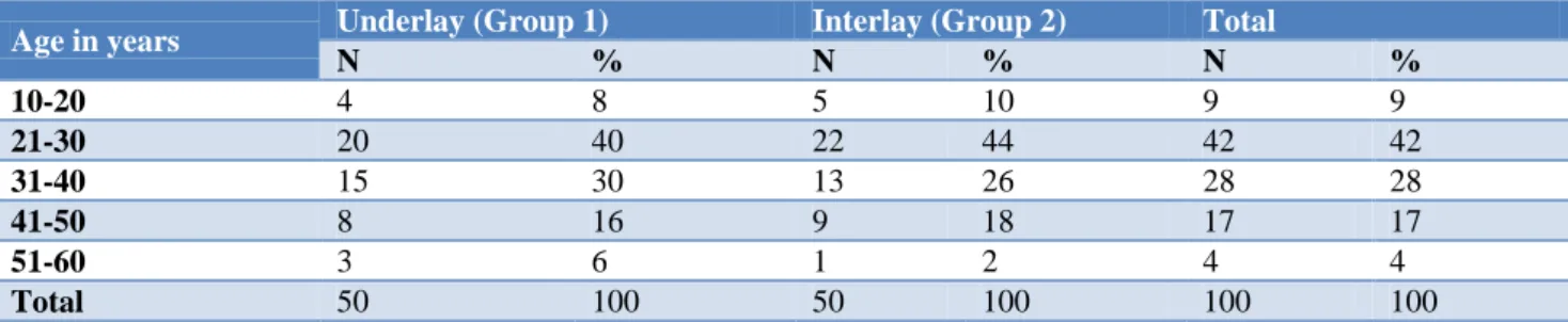
Out of 100 cases enrolled in the study, randomly 50 patients were taken for underlay and 50 were taken for interlay technique and comprised group 1 and 2 of the study respectively. Age of patients ranged from 10 to 60 years.
Left ear was operated in 55 (55%) patients while right ear was operated in the remaining 45 (45%) cases.
Mean age of patients in groups 1 and 2 were 33.12±10.294 and 31.86±10.11 years respectively.
Table 1: Age distribution of study subjects.
Age in years Underlay (Group 1) Interlay (Group 2) Total
N % N % N %
10-20 4 8 5 10 9 9
21-30 20 40 22 44 42 42
31-40 15 30 13 26 28 28
41-50 8 16 9 18 17 17
51-60 3 6 1 2 4 4
Total 50 100 50 100 100 100
Chi-square =1.408 with 4 degrees of freedom; p=0.842 (NS).
Table 2: Preoperative air-bone gap of the patients.
Preoperative air-bone gap (dB)
Group 1 (underlay) Group 2 (interlay)
No. % No. %
10-20 13 26 14 28
21-30 20 40 22 44
31-40 12 24 11 22
>40 5 10 3 6
Table 3: Postoperative air-bone gap of the patients.
Postoperative air-bone gap (dB)
Group 1 (underlay) Group 2 (interlay)
No. % No. %
<10 20 40 24 48
11-20 15 30 17 34
21-30 10 20 6 12
31-40 4 8 2 4
>40 1 2 1 2
Table 4: Change in air bone gap in both groups at 12 weeks follow up.
Groups Preoperative Post-operative Change Significance of change
Mean SD Mean SD Mean P value Group 1 (Underlay) 26.3 8.50 17.60 8.762 -8.7 <0.001
Table 5: Outcome of graft at 12 weeks follow up.
Parameter/ variable Group 1 (underlay) (n=50) Group 2 (interlay) (n=50)
No. % No. %
Rejected/failed 5 10 2 4
Accepted/successful 45 90 48 96
In group1, a total of 26 (52%) patients were males and remaining 24 (48%) were females and in group 2, 29 (58%) patients were males and remaining 21 (42%) were females showing a slight male preponderance but it was statistically insignificant. [Chi-square =1.36 with 1degree of freedom; p=0.05 (NS)]
Mean hearing gain (Closure in air-bone gap)
Preoperative mean air bone gap in groups 1 and 2 was 26.3±8.50, and 25.5±8.16 dB and postoperative mean air bone gap was 17.6±8.762 and 13.5±5.369 dB (Table 4).
In both the groups a significant mean reduction in air bone gap was observed. Mean reduction was maximum in group 2. Statistically, intergroup difference in reduction in air bone gap was highly significant (p<0.001)
Graft status (accepted or rejected)
In the present study Graft failure was observed in 7 cases (7%) resulting in a residual perforation. Success rate was 90% and 96% in Underlay (group1) and Interlay (group 2) respectively. Least graft failure rate (4%) was found in group 2 (Table 5).
DISCUSSION
India is one of the countries with highest load of chronic otitis media.19 It is important being a curable cause of deafness. It is the end result of acute otitis media and is characterized by a persistent discharge from the middle ear through a tympanic membrane perforation.
Tympanoplasty is the operative procedure performed to repair the perforation in ear drum by repairing the tympanic membrane.3 It is a beneficial procedure to protect the middle ear and inner ear from future damage. Improvement in hearing sensitivity is also observed.20
There are many approaches to perform this procedure. Out of these the Underlay technique is widely used.
Recently a new technique, Interlay has also emerged and is being successfully used worldwide with very good results. The differences in immediate success rates and resultant hearing gain are comparable in both the techniques.8,12
In our study, in Underlay technique graft rejected in 5
and postoperatively it came to be 17.60 dB. Postoperatively there was an 8.70 dB mean hearing gain after 12 weeks.
In interlay technique graft rejected in 2 patients. Preoperative mean air bone gap was 25.5 dB which comes to be 13.50 dB. Postoperative there was change in 12.0 dB mean hearing gain after 12 weeks.
No complication was noticed in any of the two procedures. The final success rate was 90% in Underlay and 96% in Interlay technique.
Thus in present study as far as resolution of air bone gap is concerned, Interlay technique showed a statistically significant better outcome as compared to the underlay group. The findings in present study showed a better graft take in Interlay method which coupled with a better post-operative air bone gap provided a better overall outcome.
Preoperative mean air bone gap in groups 1 and 2 was 26.30±8.50 dB and 25.50±8.16 dB and postoperative mean air bone gap was 17.60±8.762 dB and 13.50±5.369 dB (Table 4).
In our study graft uptake as well as hearing gain was better in the interlay technique than the underlay technique which was consistent with most of the studies. Jain et al had graft uptake rate to be 96.6% in interlay technique and 95.4% of the patients, reported an improvement in terms of hearing.7 Kawatra et al concluded Interlay technique has a significantly better graft uptake and hearing improvement as compared to underlay technique.8 Komune et al reported a 94.2% graft uptake rate.14 Guo et al had a better graft uptake rate and hearing improvement in interlay technique than in underlay technique.15 Patil et al had a graft uptake rate of 96% and a significant haring gain in interlay technique.18
CONCLUSION
The present study showed that Interlay technique had a better graft take and hearing improvement than the underlay technique. These results indicate that interlay is a better approach than the underlay type I tympanoplasty in chronic otitis media with large central perforation of mucosal variety.
Funding: No funding sources Conflict of interest: None declared
REFERENCES
1. Galdstone HB, Jackler RK, Varav K. Tympanic
Membrane Wound Healing. An Overview.
Otolaryngol Clin North Am. 1995;28(5):913–32.
2. Berthold E. Overlay myringoplasty Wier Med Bull.
1878;1:627.
3. Wullstein H. Theory and practice of tympanoplasty. Laryngoscope. 1956;66:1076-93.
4. Zollner F. The principles of plastic surgery of the sound-conducting apparatus. J Laryngol Otol. 1955;69:637-52.
5. Aslam MA, Aslam MJ. Comparison of
Over-Underlay and Underlay Techniques Of
Myringoplasty. Pak. Armed Forces Med J. 2009;59(2):155-6.
6. Shea JJ Jr. Vein graft closure of eardrum
perforations. J Laryngol Otol. 1960;74:358-62. 7. Jain S, Gupta N, Gupta R, Roy A. Interlay type 1
tympanoplasy in large central perforations: Analysis of 500 cases. IJO. 2017;23:32-5.
8. Kawatra R, Maheshwari P, Kumar G. A
comparative study of the techniques of
myringoplasty – overlay, underlay & interlay. IOSR J Dent MED Sci. 2014;13:12-6.
9. House WF. Myringoplasty. AMA Arch Otolaryngol.
1960;71:399-404.
10. Karlan MS. Gelatin film sandwich in
tympanoplasty. Otolaryngol Head Neck Surg. 1979;87:84-6.
11. Juvekar MR, Jurekar RV. The double breasting technique of tympanoplasty: A study of 200 cases. Indian J Otol. 1999;5:145-8.
12. Kartush JM, Michaelides EM, Becvarovski Z,
Larouere MJ. Over-Under Tympanoplasty.
Laryngoscope. 2002;112:802-7.
13. Singh M, Rai A, Bandyopadhyay S, Gupta SC. Comparative study of underlay and overlay techniques of myringoplasty in large and subtotal
perforations of the tympanic membrane. J Laryngol Otol. 2003;117(6):444-8.
14. Komune S, Wakizono S, Hisashi K, Uemura T, Interlay Method For Myringoplasty. Larynx Auris Nasus. 1992;19(1):17-22.
15. Guo M, Huang Y, Wang J. Report Of
Myringoplasty with interlay method in 53 ears perforation of tympani. Lin Chuang Er Bi Yan Hou Ke Za Zhi. 1999;13(4):147-9.
16. Vishal US. A One-Year Prospective Study To
Evaluate The Results Of Superiorly Based Tympanomeatal Flap In Endoscopic Myringoplasty Conducted In District Hospital, Belgaum And KLES And MRC, Belgaum During July 2003 To July 2004, Dissertation, MS (ENT), RGUHS, Karnataka. 2006.
17. Hay A, Blanshard J, The anterior interlay
myringoplasty: Outcome and hearing results in
anterior and subtotal tympanic membrane
perforations, Otol Neurotol. 2014;35(9):1569-76.
18. Patil BC, Misale PR, Mane RS, Mohite AA,
Outcome Of Interlay Grafting In Type 1 Tympanoplasty For Large Central Perforation.
Indian J Otolaryngol Head Neck Surg.
2014;66(4):418-24.
19. World Health Organization. Chronic Suppurative Otitis Media, Burden of illness and management options, WHO Child and Adolescent Health Department, Prevention of Blindness and Deafness, Geneva, 2004.
20. Hussain A, Yousaf N, Khan AR. Outcome of
Myringoplasty. J Postgrad Med Inst. 2004;18:693-6.
Cite this article as: Sharma N, Sharma P, Goyal VP, Sharma KG.Interlay versus underlay type 1