One-year results of photorefractive keratectomy
and laser in situ keratomileusis for myopia
using a 213 nm wavelength solid-state laser
Nikolaos S. Tsiklis, MD, MSc, George D. Kymionis, MD, PhD, George A. Kounis,
Aristofanis I. Pallikaris, MSc, Vasilios F. Diakonis, MD, Spyridon Charisis, MD,
Marinos M. Markomanolakis, MD, Ioannis G. Pallikaris, MD, PhD
PURPOSE:To study the long-term results of photorefractive keratectomy (PRK) and laser in situ keratomileusis (LASIK) in low to moderate myopic corrections using the Pulzar Z1 system (CustomVis), a 213 nm wavelength solid-state laser.
SETTING:University refractive surgery center.
METHODS:This prospective noncomparative case series comprised 20 patients (40 eyes) who had refractive surgery using the Pulzar Z1 laser system. Manifest refraction, uncorrected visual acuity, best spectacle-corrected visual acuity (BSCVA), safety, predictability, stability, and confocal microscopy images were evaluated.
RESULTS:Ten patients (20 eyes) had PRK and 10 patients (20 eyes) had LASIK. The mean follow-up was 13.9 monthsG1.1 (SD) (range 12 to 17 months) and 14.6G1.2 months (range 12 to 18
months) in the PRK group and LASIK group, respectively. No eye lost a line of Snellen BSCVA during the follow-up period; 2 eyes (10%) gained 2 Snellen lines. There was a statistically significant de-crease in spherical equivalent manifest refraction postoperatively in both groups (P<.05). Refractive stability was obtained during the first postoperative month and remained stable during the follow-up period, with no significant changes between any interval in both groups (P>.05). At the last follow-up, 95% of all eyes were withinG1.00 diopter of emmetropia. No late postoperative complications
were observed.
CONCLUSION:Refractive surgery using the Pulzar Z1 213 nm wavelength solid-state laser was a safe, effective procedure in the treatment of low to moderate myopia.
J Cataract Refract Surg 2007; 33:971–977Q2007 ASCRS and ESCRS
Photoablation of the cornea within the range of 190 to 220 nm (ultraviolet wavelength) offers a high degree of precision with minimum collateral damage1 and a small risk for cellular inactivation and mutation.2–4 Over the past years, solid-state lasers have been gain-ing popularity for refractive surgery because many reports5–14indicate they can be a reliable alternative to traditional excimer laser systems with a 193 nm wavelength. In vitro5 and in vivo6 studies with a 213 nm solid-state laser found smooth ablation sur-faces with a clinical course and histopathological findings similar to those of excimer laser photorefrac-tive keratectomy (PRK).15Moreover, newly developed solid-state laser platforms have been used in myopic or irregular astigmatism treatments with promising results.12–14
The present study evaluated the long-term results of PRK or laser in situ keratomileusis (LASIK) using the Pulzar Z1 213 nm wavelength solid-state laser (CustomVis).
PATIENTS AND METHODS
This prospective noncomparative case series comprised patients who had refractive surgery with the Pulzar Z1 laser system. Before their participation in the study, all patients gave informed consent in accordance with institutional guidelines and the Declaration of Helsinki.
The Pulzar Z1 laser system is a solid-state, quintupled, diode-pumped neodymium:YAG (Nd:YAG) device generat-ing a 213 nm wavelength (one fifth of a standard Nd:YAG laser). It uses a 0.6 mm diameter scanning flying spot with a Gaussian profile and a pulse repetition rate of 300 Hz. The solid-state eye-tracking system locks onto the limbus
with a detection rate of 5 KHz, an eye-tracker delay time of 0.5 ms, and a closed-loop response of 1 KHz.
All patients had a complete ophthalmic examination pre-operatively to exclude ocular disease. Exclusion criteria were active anterior segment disease; residual, recurrent, or active ocular disease; previous intraocular or corneal surgery; history of herpes keratitis; diagnosed autoimmune disease; systemic connective tissue disease or atopic syndrome; and corneal topographic findings suspicious for keratoconus.
Preoperative assessments included uncorrected visual acuity (UCVA), best spectacle-corrected visual acuity (BSCVA), manifest and cycloplegic refractions, slitlamp examination with fundus evaluation, applanation tonome-try, corneal topography (C-Scan, Technomed Arnold-Sommerfeld-Ring1), ultrasonic pachymetry (Pac Scan 300P, Sonomed), and confocal microscopy (Heidelberg Engineer-ing GmbH). The latter was performed with a modified confocal scanning laser ophthalmoscope (HRT II/Rostock Cornea Module, Heidelberg Engineering). The principle of operation of the modified scanning laser ophthalmoscope has been described.16In-depth qualitative evaluation of the central corneal images was performed, with special attention paid to the subepithelial nerve plexus, ablated zone, and deep stroma.
All procedures were performed by the same surgeon (I.G.P.) using an identical technique. In the PRK group, deep-ithelialization was achieved using a brush. In the LASIK group, a 110 mm head Carriazo-Pendular microkeratome (Schwind GmbH) was used in the manual mode to create a superiorly hinged flap. A soft contact lens was applied to all PRK eyes until the epithelium healed completely. The targeted correction in all eyes was emmetropia.
Postoperatively, the PRK patients received sodium flurbi-profen 0.03% drops (Ocuflur) 4 times a day for 2 days, dexa-methasone–tobramycin drops (TobraDex) 4 times a day until the removal of the contact lens, and then fluorometholone drops (FML) 4 times a day tapered over the following month. Sodium hyaluronate 0.18% drops (Vismed) were used hourly at first and then, depending on patients’ needs, for a month thereafter. The LASIK patients received Ocuflur drops 4 times a day for 2 days, TobraDex drops 4 times a day for 2 weeks, and Vismed drops hourly at first and then, depending upon patients’ needs, for a month thereafter.
Follow-up examinations were performed 1, 3, 6, and 12 months postoperatively. The following were assessed at each visit: UCVA, BSCVA, corneal topography, slitlamp examination, and confocal microscopy.
Statistical analysis was performed using the Studentttest for continuous variables. Differences in categorical variables were tested by the chi-square test. Results are presented as meanGstandard deviation. APvalue less than 0.05 was considered statistically significant.
RESULTS
Initially, 25 patients (50 eyes) participated in a clinical trial for the safety, efficacy, and predictability of re-fractive surgery (PRK or LASIK) using the Pulzar Z1 solid-state laser. Twenty of the patients had primary bilateral PRK (10 eyes; 4 men and 6 women) or LASIK (10 eyes; 5 men and 5 women) for myopic corrections (compound or not with less than 1.00 diopter [D] astig-matism) and completed at least a 1-year follow-up. These patients were included in the study. Table 1 shows the patients’ demographic and refractive data.
Uncorrected Visual Acuity
The mean UCVA significantly improved from finger counting preoperatively to 20/40 or better at the last follow-up examination. One month after PRK or LASIK, 18 eyes (90%) achieved a UCVA of 20/30 or better and 11 eyes (55%) of 20/20 or better. At the
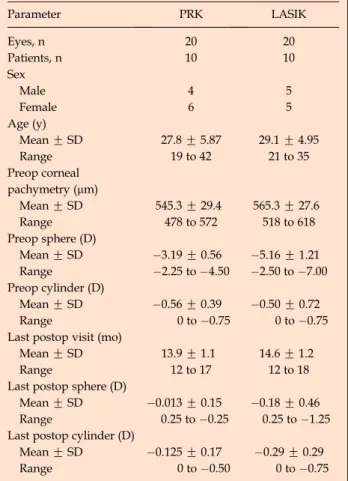
Table 1. Patients’ demographic and refractive data. Parameter PRK LASIK Eyes, n 20 20 Patients, n 10 10 Sex Male 4 5 Female 6 5 Age (y) MeanGSD 27.8G5.87 29.1G4.95 Range 19 to 42 21 to 35 Preop corneal pachymetry (mm) MeanGSD 545.3G29.4 565.3G27.6 Range 478 to 572 518 to 618 Preop sphere (D) MeanGSD 3.19G0.56 5.16G1.21 Range 2.25 to 4.50 2.50 to 7.00 Preop cylinder (D) MeanGSD 0.56G0.39 0.50G0.72 Range 0 to 0.75 0 to 0.75 Last postop visit (mo)
MeanGSD 13.9G1.1 14.6G1.2
Range 12 to 17 12 to 18 Last postop sphere (D)
MeanGSD 0.013G0.15 0.18G0.46
Range 0.25 to 0.25 0.25 to 1.25 Last postop cylinder (D)
MeanGSD 0.125G0.17 0.29G0.29
Range 0 to 0.50 0 to 0.75 LASIKZlaser in situ keratomileusis; PRKZphotorefractive keratectomy
Accepted for publication February 27, 2007.
From the Department of Ophthalmology (Tsiklis, Kymionis, Chari-sis, I.G. Pallikaris) and Institute of Vision and Optics (Tsiklis, Kymionis, Kounis, A.I. Pallikaris, Diakonis, Markomanolakis, I.G. Pallikaris), University of Crete, Crete, Greece.
No author has a financial or proprietary interest in any material or method mentioned.
Corresponding author: Nikolaos S. Tsiklis, MD, MSc, Institute of Vision and Optics, University of Crete, Medical School Department of Ophthalmology, 71003 Heraklion, Crete, Greece. E-mail:ntsiklis@ hotmail.com.
last postoperative examination, all PRK eyes achieved a UCVA of 20/25 or better and 19 (95%) of 20/20 or better and 18 LASIK eyes (90%) achieved a UCVA of 20/25 or better and 17 (85%) of 20/20 or better.
Best Spectacle-Corrected Visual Acuity
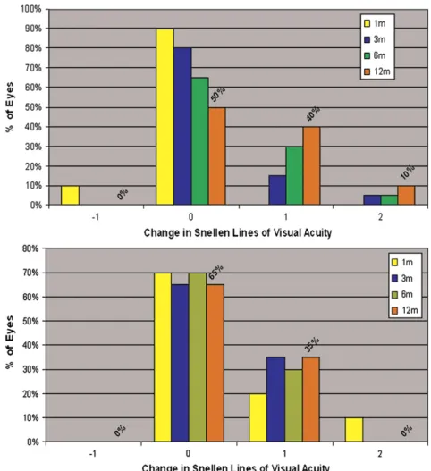
Figure 1 shows the changes in the BSCVA during the follow-up. In the PRK group, 1 eye lost 1 Snellen line by the 1-month postoperative examination as a re-sult of severe dry eye. No eye lost a line of Snellen BSCVA; 8 eyes (40%) gained 1 line and 2 eyes (10%) gained 2 lines by the last follow-up. In the LASIK group, no eye lost a line of Snellen BSCVA line; 7 eyes (35%) had gained 1 line by the last postoperative examination.
Refractive Error
In the PRK group, the mean spherical equivalent (SE) refraction was 3.46 D G 0.52 (SD) preopera-tively. It decreased to 0.24 G 0.45 D at 1 month,
0.04 G 0.31 D at 3 months, 0.05 G 0.29 D at
6 months, and 0.08G0.21 D at the last postoperative examination (P!.05). Refractive stability was obtained
by the first postoperative month, and the mean resid-ual refractive error remained stable during all the follow-up examinations with no significant changes between any interval (PO.05) (Figure 2, A). One 1 eye (5%) had a change in SE of more than 0.50 D be-tween 6 months postoperatively and the last examina-tion. One month after surgery, the postoperative SE refraction was withinG0.50 D of emmetropia in 16 eyes (80%) and withinG1.00 D in 19 eyes (95%). At the last follow-up, all eyes in the PRK group were within G0.50 D of emmetropia. The correlation be-tween the attempted myopic correction and achieved myopic correction at the last follow-up was highly lin-ear, with 14 eyes (70%) at G0.25 D and all eyes at G0.50 D (Figure 3,A).
In the LASIK group, the mean SE refraction ( 5.41G 1.21 D) was 5.41G1.21 D (range 2.62 to 7.25 D) preoperatively. It decreased to 0.45 G 0.55 D at 1 month, 0.40G0.59 D at 3 months, 0.35G0.48 D at 6 months, and 0.32G0.49 D at the last follow-up (P!.05). There were no significant changes in SE re-fractive error between any interval (PO.05) (Figure 2, B). The SE refraction did not change by more than
Figure 1. Change in BSCVA 1, 3, 6, and 12 months after PRK (top) and LASIK (bottom).
0.50 D between 6 months postoperatively and the last examination in any eye. One month after surgery, the postoperative SE refraction was within G0.50 D of emmetropia in 12 eyes (60%) and withinG1.00 D in 17 eyes (85%). At the last follow-up, predictability showed that 11 eyes (55%) were withinG0.25 D and 18 eyes (90%) were withinG0.50 D (Figure 3,B). One 1 patient (2 eyes; 10%) had an SE refraction greater than 1.00 D of emmetropia.
Confocal Microscopy Analysis
Corneal images in the PRK group and LASIK group showed normal epithelial structures. The subepithelial nerve plexus in both groups appeared regenerated af-ter 1 year of surgery (Figures 4 and 5, top left). In-creased scattering was observed at the ablation site in all PRK-treated corneas, even 1 year after surgery (Figure 4, top right). Highly reflective particles and
scattering were observed at the flap interface in LA-SIK-treated corneas, even 1 year after surgery (Figure 5, top right). Moreover, keratocyte activation and abnormal scattering of the nuclei of the kerato-cytes were observed at the stromal layers immediately posterior to the ablation site (Figures 4 and 5,bottom left). In contrast, deeper stromal layers had normal keratocyte activation and scattering of the nuclei of the keratocytes with an oval shape (Figures 4 and 5, bottom right). Finally, the endothelial cells appeared normal in shape and size (cell density within normal range) in both groups.
Adverse Effects and Postoperative Complications
No eye had intraoperative, early, or late postopera-tive complications. The epithelium healed completely within 3 to 5 days after surgery in all PRK-treated eyes. Trace haze was documented in 2 PRK-treated
Figure 2.Stability of intended correction during the follow-up period after PRK (top) and LASIK (bottom).
eyes 3 months postoperatively and in 1 eye at the last follow-up examination.
DISCUSSION
Solid-state lasers provide a technical platform that could eliminate some of the excimer laser system’s dis-advantages such as the absence of toxic gas with the solid-state laser. Moreover, the Pulzar Z1 laser system has characteristics that may prove useful in refractive surgery (especially in customized treatments). The small spot size (0.6 mm diameter, 2.5 times smaller than a typical excimer laser spot), uniform Gaussian intensity beam distribution, high pulse-to-pulse
stability, and ultra-fast tracking system contribute to the accurate transfer of the planned treatment onto the corneal stroma, creating a smooth ablation surface. Another potential benefit of the smaller spot size is less mechanical stress on the cornea during ablation,17 which might cause fewer cellular alterations and less damage to the corneal structure.18In addition, the Pul-zar Z1 laser system emits at a 213 nm wavelength, which is close to the absorption peak of corneal colla-gen,1and it is less sensitive (1 to 4 orders of magnitude) to corneal hydration than at 193 nm.19Thus, corneal hydration20 and environmental humidity,21 factors that affect the final outcomes with conventional exci-mer laser systems, are limited with the solid-state laser. Despite the theoretical advantages of solid-state laser systems, there are few clinical studies of them and all were of the results after PRK.6,12–14The current study found promising results after PRK14as well as after LASIK using the solid-state laser over a minimum follow-up of 1 year. Of the 40 study eyes (20 PRK, 20 LASIK), 38 (95%) had a UCVA of 20/25 and 36 (90%) achieved 20/20 or better by the last follow-up. No eye had lost a line of Snellen BSCVA at the last postoperative examination. Predictability improved during the follow-up period, from 20 eyes (70%) withinG0.50 D of emmetropia at the 1-month postop-erative examination to 38 eyes (95%) withinG0.50 D of emmetropia at the last follow-up. Epithelial healing time22and the haze rate23were similar to those found after PRK using the excimer laser.
Confocal microscopy images showed findings simi-lar to those in previous studies that used the excimer laser.24–27 Subepithelial nerve plexus regeneration has been observed even 7 days after PRK treatments22 and 3 months after LASIK surgery,24while morpho-logical recovery of corneal innervation may take more than 12 months with either procedure.24,25 More-over, previous studies found keratocyte activation, morphological changes of keratocyte nuclei, and depo-sition of extracellular matrix (ECM) near the ablation site in PRK patients25or near the lamellar cut in LASIK patients.26 Nonetheless, the increased scattering caused by the keratocyte activation and/or deposition of ECM was not of clinical importance as UCVA was not influenced by haze in any patient in the present study. Finally, the small reflective particles at the flap interface have been observed in previous studies. These particles have been described as small metal particles from the microkeratome blade and salt pre-cipitates from the tear film.
This study had limitations that preclude a viable conclusion. These were the small sample of treated eyes, the small range of the attempted SE correction ( 2.50 to 7.00 D), and the lack comparison with a group having excimer laser treatment. Whether the
Figure 3.Correlation between the attempted and the achieved myo-pic correction at the last follow-up examination (12 months) after PRK (top) and LASIK (bottom).
Figure 4.Confocal microscopy in PRK patients.Top left: Regenerated subepithelial nerve plexus. Top right: Surface ablation site.Bottom left: Stromal layer immediately posterior to ablation site.Bottom right: Deep stromal layer.
Figure 5. Confocal microscopy in LASIK patients. Top left: Regenerated subepithelial nerve plexus.Top right: Flap interface with highly reflective particles. Bottom left: Stromal layer immediately posterior to the ablated zone.Bottom right: Deep stromal layer.
theoretical advantages of the solid-state laser platform over excimer laser systems have a practical impact on corneal healing, and therefore on the patient’s quality of vision, remains to be studied.
In conclusion, refractive surgery with the Pulzar Z1 213 nm solid-state laser system seemed to be a safe and effective procedure for treating low to moderate myopia, despite the limitations of this study.
REFERENCES
1. Lembares A, Hu X-H, Kalmus GW. Absorption spectra of corneas in the far ultraviolet region. Invest Ophthalmol Vis Sci 1997; 38:1283–1287
2. Ito T, Ito A, Hieda K, Kobayashi K. Wavelength dependence of in-activation and membrane damage to Saccharomyces cerevisiae cells by monochromatic synchrotron vacuum-UV radiation (145-190 nm). Radiat Res 1983; 96:532–548
3. Hieda K, Ito T. Action spectra for inactivation and membrane damage of Saccharomyces cerevisiae cells irradiated in vacuum by monochromatic synchrotron UV radiation (155-250 nm). Photochem Photobiol 1986; 44:409–411
4. Munakata N, Hieda K, Kobayashi K, et al. Action spectra in ultraviolet wavelengths (150-250 nm) for inactivation and mu-tagenesis of Bacillus subtilis spores obtained with synchro-tron radiation. Photochem Photobiol 1986; 44:385–390 5. Ren Q, Simon G, Parel J-M. Ultraviolet solid-state laser (213-nm)
photorefractive keratectomy; in vitro study. Ophthalmology 1993; 100:1828–1834
6. Ren Q, Simon G, Legeais J-M, et al. Ultraviolet solid-state laser (213-nm) photorefractive keratectomy; in vivo study. Ophthal-mology 1994; 101:883–889
7. Gailitis RP, Ren QS, Thompson KP, et al. Solid state ultraviolet laser (213 nm) ablation of the cornea and synthetic collagen lenticules. Laser Surg Med 1991; 11:556–562
8. Dair GT, Pelouch WS, van Saarloos PP, et al. Investigation of corneal ablation efficiency using ultraviolet 213-nm solid state laser pulses. Invest Ophthalmol Vis Sci 1999; 40:2752–2756 9. Krueger RR, Seiler T, Gruchman T, et al. Stress wave
ampli-tudes during laser surgery of the cornea. Ophthalmology 2001; 108:1070–1074
10. Ren Q, Gailitis RP, Thompson KP, Lin JT. Ablation of the cornea and synthetic polymers using a UV (213 nm) solid-state laser. IEEE J Quantum Electron 1990; 26:2284–2288
11. Caughey TA, Cheng F-C, Trokel SL, et al. An investigation of laser-tissue interaction of a 213 nm laser beam with animal corneas. Lasers Light Ophthalmol 1993/1994; 6:77–85 12. Anderson I, Sanders DR, van Saarloos P, Ardrey WJIV.
Treat-ment of irregular astigmatism with a 213 nm solid-state, diode-pumped neodymium:YAG ablative laser. J Cataract Refract Surg 2004; 30:2145–2151
13. Roszkowska AM, Korn G, Lenzner M, et al. Experimental and clinical investigation of efficiency and ablation profiles of new solid-state deep-ultraviolet laser for vision correction. J Cataract Refract Surg 2004; 30:2536–2542
14. Roszkowska AM, De Grazia L, Ferreri P, Ferreri G. One-year clinical results of photorefractive keratectomy with a solid-state laser for refractive surgery. J Refract Surg 2006; 22: 611–613
15. L’Esperance FA Jr, Taylor DM, Warner JW. Human excimer laser keratectomy: short-term histopathology. J Refract Surg 1988; 4:118–124
16. Kymionis GD, Tsiklis NS, Pallikaris AI, et al. Long-term follow-up of Intacs for post-LASIK corneal ectasia. Ophthalmology 2006; 113:1909–1917
17. Krueger RR, Seiler T, Gruchman T, et al. Stress wave ampli-tudes during laser surgery of the cornea. Ophthalmology 2001; 108:1070–1074
18. Kermani O, Lubatschowski H. Struktur und Dynamik photoakus-tischer Schockwellen bei der 193 nm Excimerlaserphotoablation der Hornhaut. [Structure and dynamics of photo-acoustic shock-waves in 193 nm excimer laser photo-ablation of the cornea.] Fortschr Ophthalmol 1991; 88:748–753
19. Dair GT, Ashman RA, Eikelboom RH, et al. Absorption of 193- and 213-nm laser wavelengths in sodium chloride solution and balanced salt solution. Arch Ophthalmol 2001; 119: 533–537
20. Dougherty PJ, Wellish KL, Maloney RK. Excimer laser ablation rate and corneal hydration. Am J Ophthalmol 1994; 118: 169–176
21. Walter KA, Stevenson AW. Effect of environmental factors on myopic LASIK enhancement rate. J Cataract Refract Surg 2004; 30:798–803
22. Fagerholm P. Wound healing after photorefractive keratectomy. J Cataract Refract Surg 2000; 26:432–447
23. Hersh PS, Stulting RD, Steinert RF, et al. Results of phase III excimer laser photorefractive keratectomy for myopia; the Summit PRK Study Group. Ophthalmology 1997; 104: 1535–1553
24. Calvillo MP, McLaren JW, Hodge DO, Bourne WM. Corneal reinnervation after LASIK: prospective 3-year longitudinal study. Invest Ophthalmol Vis Sci 2004; 45:3991–3996
25. Moilanen JA, Vesaluoma MH, Muller LJ, Tervo TMT. Long-term corneal morphology after PRK by in vivo confocal microscopy. Invest Ophthalmol Vis Sci 2003; 44:1064–1069
26. Perez-Gomez I, Efron N. Change to corneal morphology after refractive surgery (myopic laser in situ keratomileusis) as viewed with confocal microscope. Optom Vis Sci 2003; 80:690–697
27. Vesaluoma MH, Pe´rez-Santonja J, Petroll WM, et al. Corneal stromal changes induced my myopic LASIK. Invest Ophthalmol Vis Sci 2000; 41:369–376; erratum, 2027
First author:
Nikolaos S. Tsiklis, MD, MSc
Department of Ophthalmology and Institute of Vision and Optics, University of Crete, Crete, Greece