Received 13 Apr 2016
|
Accepted 11 Nov 2016
|
Published 21 Dec 2016
A live RSV vaccine with engineered thermostability
is immunogenic in cotton rats despite high
attenuation
Christopher C. Stobart
1,2,
*
,
w
, Christina A. Rostad
1,2,
*, Zunlong Ke
3
, Rebecca S. Dillard
1,2
, Cheri M. Hampton
1,2
,
Joshua D. Strauss
1,2
, Hong Yi
4
, Anne L. Hotard
1,2,
w
, Jia Meng
1,2,
w
, Raymond J. Pickles
5
, Kaori Sakamoto
6
,
Sujin Lee
1,2
, Michael G. Currier
1,2
, Syed M. Moin
7
, Barney S. Graham
7
, Marina S. Boukhvalova
8
, Brian E. Gilbert
9
,
Jorge C.G. Blanco
8
, Pedro A. Piedra
9,10
, Elizabeth R. Wright
1,2,4
& Martin L. Moore
1,2
Respiratory syncytial virus (RSV) is a leading cause of infant hospitalization and there
remains no pediatric vaccine. RSV live-attenuated vaccines (LAVs) have a history of safe
testing in infants; however, achieving an effective balance of attenuation and immunogenicity
has proven challenging. Here we seek to engineer an RSV LAV with enhanced
immuno-genicity. Genetic mapping identifies strain line 19 fusion (F) protein residues that correlate
with pre-fusion antigen maintenance by ELISA and thermal stability of infectivity in live RSV.
We generate a LAV candidate named OE4 which expresses line 19F and is attenuated by
codon-deoptimization of non-structural (NS1 and NS2) genes, deletion of the small
hydro-phobic (SH) gene, codon-deoptimization of the attachment (G) gene and ablation of the
secreted form of G. OE4 (RSV-A2-dNS1-dNS2-DSH-dG
m-Gs
null-line19F) exhibits elevated
pre-fusion antigen levels, thermal stability, immunogenicity, and efficacy despite heavy
attenuation in the upper and lower airways of cotton rats.
DOI: 10.1038/ncomms13916
OPEN
1Department of Pediatrics, Emory University School of Medicine, Atlanta, Georgia 30322, USA.2Children’s Healthcare of Atlanta, Atlanta, Georgia 30322,
I
n the 1960s, a formalin-inactivated RSV (FI-RSV) vaccine
primed for enhanced illness in infants on natural infection
1.
This
phenomenon
was
replicable
in
animal
models
and considered dependent on RSV naive status
2. Subsequent
studies
using
subunit-based
vaccines
also
primed
for
immunopathology in animals
3,4. These early RSV vaccines
encouraged development of LAVs, which do not prime for
enhanced disease in animals or seronegative infants
2,5. However,
development of pediatric RSV LAV strains with sufficient
attenuation and immunogenicity has been difficult
6. To address
these dual challenges, newer RSV LAVs have incorporated genetic
modifications
rationally
designed
to
retain
or
enhance
immunogenicity compared with wild-type virus
7–9because
natural infection may be suboptimally immunogenic for LAVs
derived by classic attenuation methods.
Recent elucidation of the structure of the pre-fusion
conformation of RSV F protein (pre-F
10) and discovery of
its importance as a natural immunogen
11has had implications
for RSV vaccine development. The high capacity of pre-F to
elicit neutralizing antibody titres has been demonstrated in
multiple vaccine platforms, including purified proteins
12–14,
virus-like particles
15, and recombinant parainfluenza viruses
16.
Use of pre-F in passive immunization, either by anti-pre-F
monoclonal antibody (mAb) prophylaxis or by boosting
RSV neutralizing antibody (nAb) titres in pregnant mothers
with pre-F protein-based vaccines, holds promise for reducing
RSV disease in the youngest infants
14. Nevertheless, active
immunization of infants with a replicating RSV vaccine
could potentially have a large child health benefit if protection
spanned beyond the persistence of passively acquired maternal
Ab. Since natural RSV infection induces anti-pre-F nAb
11,
we hypothesized that RSV with enhanced pre-F expression
would have increased LAV immunogenicity.
Here we first identified a chimeric RSV strain A2-line19F
with enhanced pre-fusion antigen levels, thermostability and
immunogenicity compared with parental strain A2. We then
incorporated line19F into an RSV LAV candidate ‘OE4’ with the
genotype
RSV-A2-dNS1-
dNS2-
D
SH-dG
m-Gs
null-line19F.
We found that OE4 exhibited elevated pre-fusion antigen levels,
thermal
stability,
immunogenicity,
and
efficacy
despite
heavy attenuation in the upper and lower airways of cotton rats.
Results
Pre-fusion F ELISAs
. Metastable pre-F undergoes a dynamic
transition to form a thermodynamically stable six-helix
post-fusion bundle that facilitates viral and host membrane post-fusion
10,14.
Since both pre-F and post-F are present on RSV virions
in prepared virus stocks
17,18, we evaluated the relative amount
of pre-F antigen in RSV stocks using an ELISA-based approach
to compare MPE8 with motavizumab antibody binding. MPE8
is a human monoclonal antibody that preferentially binds to
two highly conserved anti-parallel
b
-strands on pre-F, which
are rearranged in the post-fusion conformation to render
them less accessible to antibody binding
19. Motavizumab,
in contrast, stably binds to both pre- and post-fusion F. We
found that strain A2-line19F, which expresses the F protein
of strain line 19 in the background of the prototypical
A2 strain
20,21, exhibited significantly higher relative binding to
MPE8 than did strain A2 (Fig. 1a). We confirmed this finding
using the human monoclonal antibody D25, which binds to a
distinct antigenic site on pre-F (antigenic site Ø)
10with
even greater specificity than MPE8 (ref. 22). We found that
A2-line19F exhibited higher relative binding to D25 than A2,
which was similar in magnitude and correlated with MPE8
binding (Fig. 1b).
Five unique amino-acid residues distinguish line 19F from
A2 F: M79, R191, K357, Y371 and I557 (Supplementary Fig. 1)
20.
We generated A2-line19F mutants by substituting A2 residues in
place of the unique line 19F residues
20. To determine the
effects of these residues on pre-F antigen levels in virus stocks, we
performed MPE8 and motavizumab ELISAs on the recombinant
A2-line19F mutant viruses. We found that residues M79,
K357 and Y371 contributed to line 19 F pre-F antigen levels
(Fig. 1a). These results were consistent with previous data
showing that the K357/Y371 residues together impeded
A2-line19F fusion activity
in vitro
20.
Thermal stability assays
. We next evaluated the thermal stability
of A2-line19F compared with A2. RSV is known to be a
heat-labile virus, and elevated temperatures can trigger the
transition
from
pre-
to
post-fusion
F
23.
We
therefore
hypothesized that RSV with enhanced pre-F levels would be
more resistant to temperature inactivation. We analysed
thermostability at 4 and 37
°
C because thermostability at
4
°
C may have implications for retention of vaccine potency in
cold chain, whereas thermostability at 37
°
C has more relevance
for physiological conditions. We found that RSV A2-line19F
infectivity was more thermostable over time than A2 at
both temperatures (Fig. 2a,b), a phenotype that was mediated
in part by the residues K357 and Y371 (Fig. 2c). We then
introduced K357 and Y371 into the F of a genetically divergent
vaccine strain DB1, which expresses a consensus F gene of the
antigenic subgroup B ‘Buenos Aires’ (BAF) clade. We previously
described
the
generation
of
DB1,
which
also
contains
codon-deoptimized non-structural protein genes and deleted
SH gene, with a genotype RSV-A2-dNS1-dNS2-
D
SH-BAF
9. DB1
expressed low levels of pre-F antigen and was thermally unstable;
however, incorporation of the K357 and Y371 residues to
generate DB1–357/371 enhanced MPE8 binding (Fig. 1a)
and partially restored thermal stability (Fig. 2d). These data
demonstrated that residues 357 and 371 modulated not only
MPE8 binding, a correlate of pre-F antigen levels, but also viral
resistance to thermal inactivation in viral stocks.
Generation of RSV live-attenuated vaccine OE4
. We next
generated a novel RSV LAV called OE4, by incorporating line
** ** **** ****
* NS 2.5
2.0
1.5
1.0
0.5
0.0
Ratio of MPE8/MOTA
normalized to A2
A2
A2-line19F
A2
A2-line19F
OE4 OE4
DB1-357/371
DB1
357/371
557
371
357
191
79
Ratio of D25/MOTA normalized to A2
2.5
2.0
1.5
1.0
0.5
0.0
a
b
19F into a multi-component vaccine designed to achieve
attenuation, improved immunogenicity and genetic stability.
We previously codon-deoptimized the NS1 and NS2 genes, which
encode two nonstructural proteins of RSV that suppress
host innate immunity by targeting interferon pathways and
suppressing apoptosis
24,25. Codon deoptimization of NS1 and
NS2 genes was genetically stable and reduced NS1 and
NS2 protein expression, resulting in virus attenuation with
slightly enhanced immunogenicity in mice
8. We subsequently
deleted the small hydrophobic (SH) protein gene with the goal of
increasing the transcription of downstream viral genes, including
F, by altering their proximity to the viral leader
26. The deletion of
SH is also mildly attenuating in mice and chimpanzees,
but conferred no apparent attenuation in a vaccine candidate in
children
26–28. Last, we codon-deoptimized the RSV attachment
(G) glycoprotein gene and ablated the secreted form of G by
a point mutation. RSV expresses a membrane-bound form
(G
m) and a secreted form (G
s) of G, which are not required for
viral replication in immortalized cell lines
29–32. RSV G is capable
of eliciting protective neutralizing antibodies
33. However, G is
less conserved than F and suppresses the innate immune response
through chemokine mimicry
34,35. G
sfunctions as an antigen
decoy and can alter dendritic cell signalling and activation
through interactions with C-type lectins
36,37. The resulting
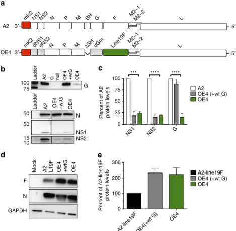
genotype of the OE4 vaccine candidate was
RSV-A2-dNS1-dNS2-
D
SH-dG
m-Gs
null-line19F (Fig. 3a). Using western blotting,
we demonstrated that OE4 had decreased expression levels
of NS1, NS2 and G as expected compared with parental
A2 (Fig. 3b,c). We additionally found that OE4 had higher
levels of F expression than A2-line19F, likely attributable to the
deletion of SH (Fig. 3d,e).
Analysis of OE4 surface glycoproteins
. We analysed the MPE8
and D25 binding of OE4 and measured vaccine thermal stability
at 4 and 37
°
C. Similar to A2-line19F, OE4 exhibited high relative
pre-F antigen levels by antibody binding (Fig. 1a,b) and thermal
stability (Fig. 2a,b) consistent with its expression of the line
19F protein. We further explored this relationship by quantifying
pre-F stability as measured by MPE8 binding of OE4 and
A2 from virus stocks incubated at 4
°
C over time. Relative pre-F
antigen levels declined in both viruses over a period of 8 days
(Supplementary Fig. 2a). Therefore, the kinetics of thermal
stability of A2 and OE4 infectivity did not correlate with the
decay of pre-F antigen levels. However, OE4 maintained greater
than twice the levels of pre-F antigen levels at each time point
compared with A2 (Supplementary Fig. 2b), and a minimal
threshold of pre-F may be sufficient to maintain infectivity.
In order to assess the overall structure of the virions
and glycoprotein incorporation into RSV A2 and OE4, we then
performed thin-section transmission electron microscopy (TEM),
native immuno-TEM, and cryo-electron tomography (cryo-ET)
of viruses budded from BEAS-2B cells, an immortalized human
bronchial epithelial cell line. In all cases, virus-infected cells
and released virions were analysed following minimal sample
processing to maximize preservation of the native structure of
the virions. First, native immunogold labelling combined with
thin-section TEM was performed using mAbs that preferentially
bound pre-F (MPE8), post-F (131-2A), total F (motavizumab) or
G (131-2G) (Fig. 4a)
38. The density of gold particles per
membrane
length
was
quantified
for
each
virus
and
immunolabel (Fig. 4b)
39. OE4 virus particles exhibited a greater
density of incorporated pre-F and total F than A2, potentially due
to the deletion of SH. There was no significant difference in the
amount of post-F detected on the surfaces of A2 and
OE4
particles.
G
protein
density
on
OE4
particles
was significantly reduced, as was expected in the setting of
codon-deoptimization of the G gene.
When visualized by cryo-ET, A2 and OE4 virus particles
were morphologically similar and formed filaments with
1** ** ** ** ** * * *
*
* ** *
0.001 0.01 1 10
100 **
* **
** ** ** ** ** ** 100
a
b
c
d
10
0.1
0.01
0.001
0.0001
0.00001
1 100
10
0.1
0.01
0.001
0.0001
0.00001
% of initial titre (FFU ml
–1
) Day 0
Day 1 Day 3 Day 5 Day 7
Day 0 Day 1 Day 3 Day 5 Day 7
Day 0
Day 1 Day 3
Day 5 Day 7
A2
A2-line19F
OE4 A2
A2-line19F OE4
% of initial titre (FFU ml
–1
)
0.001 0.01 0.1 1 10 100
** *
**
** **
* Day 0
Day 3 Day 7
% of initial titre (PFU ml
–1
)
A2-line19F
A2 79 191 357 371 557
357/371
% of initial titre (FFU ml
–1
)
0.1
A2
A2-line19F DB1
DB1-357/371
abundant glycoprotein spikes on the surface, matrix protein
lining the inside of the viral membrane, and ribonucleoprotein
complex in the interior of the virions (Fig. 5a–c)
17,18,38.
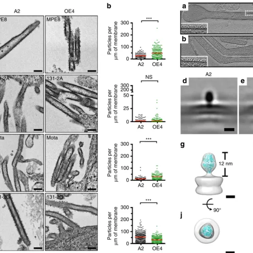
To further investigate the conformations of RSV F on the
surfaces of A2 and OE4 virions in their native states, we then
calculated subvolume averages of F structures from the cryo-ET
data. These studies demonstrated that the majority of F proteins
on both viruses in their native states immediately after budding
was in the pre-F conformation (Fig. 5). The application of
heat (55
°
C for 30 min) triggered the conformational change from
pre- to post-F, providing direct evidence of the relationship
between temperature and pre-F stability (Fig. 5c,f,i,j).
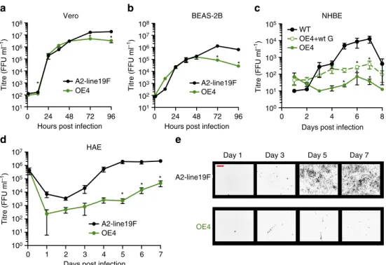
Characterization of OE4 in cell culture and primary cells
.
We next characterized the OE4 vaccine candidate
in vitro
by
measuring attenuation levels in immortalized cells and in primary
human airway epithelial cells (Fig. 6). In Vero cells, which
were used for virus stock generation, OE4 grew to titres slightly
below the parental unattenuated A2-line19F (Fig. 6a). OE4 was
more attenuated relative to wild type in BEAS-2B cells (Fig. 6b).
We then evaluated OE4 growth in primary human airway
epithelial cells, which are an established system for approximating
RSV
LAV
attenuation
in
seronegative
children
40.
We
implemented two models, NHBE-ALI and HAE-ALI, and
found that OE4 was significantly attenuated in both models
(Fig. 6c,d) and exhibited deficiency in spreading through
the cultures (Fig. 6e). The codon deoptimization of G in
OE4 contributed significantly to the level of attenuation
compared with OE4 expressing wild-type G (OE4
þ
wtG) in
NHBE-ALI (Fig. 6c), likely due to the previously described
attachment role of G in primary cells
30.
Characterization of OE4 in BALB/c mice
. To measure relative
levels of attenuation
in vivo
, we inoculated mice intranasally (i.n.)
and measured lung viral loads on days 2, 4, 6 and 8 post infection.
We found that OE4 was moderately attenuated compared
with A2 and A2-line19F in this model (Fig. 7a). We compared
lung viral loads of mice inoculated with OE4 with and without
the mKate2 gene and found that reporter had no effect on
viral attenuation (Supplementary Fig. 3), consistent with
pre-viously published results
41. We sequenced the NS1, NS2, G and
F genes of OE4 from virus recovered from mouse lungs on day
6 post infection, and there were no mutations in these genes.
To analyse immunogenicity, we then vaccinated mice and
measured nAb titres on days 35, 70 and 100 and found that
OE4 elicited nAb titres equivalent to A2-line19F and higher than
A2 at each time point post infection (Fig. 7b). Following
i.n. inoculation, the mice were then challenged on day 102 with
5′ 3′
A2
OE4 3′ 5′
G
NS1 NS2 N
A2 OE4 +wtG OE4
A2 G null OE4 +wtG OE4
0 25 50 75 100
A2 *** **** ****
F
N
GAPDH
OE4 +wtG
A2- L19F OE4
Mock
0 100 200 300
OE4
Ladder
100 75
50
50
15 10
Ladder
mK2
mK2 dNS1dNS2 N P M ΔSH dGm Line19F NS1NS2 N P M SH G F M2–1M2–2
M2–1 M2–2
L
L
Percent of A2 protein levels
OE4 (+wt G) OE4
NS1 NS2 G
Percent of A2-line19F
protein levels
OE4 (+wt G) A2-line19F
A2-line19F OE4(+wt G) OE4
a
b
c
d
e
A2-line19F, and the OE4-vaccinated mice were completely
protected against the challenge (Fig. 7c). We next measured
OE4 mucus production in the lungs of mice. We previously
demonstrated that A2-line19F induces increased airway mucin
expression, a measure of pathogenicity in this model
20,21.
However, OE4, which also expresses line 19F, induced
lower levels of airway mucin expression than A2-line19F in
mice (Fig. 7d and Supplementary Fig. 4), indicating that
the attenuating genetic modifications in OE4 modulated the
mucogenic phenotype. Subsequently, we compared OE4-induced
mucin expression with a reconstituted RSV mutant containing a
deletion of M2-2 (A2-del-M2-2), the primary genetic
modifi-cation of a clinically advanced LAV candidate
7. The deletion of
M2-2 results in reduced viral replication and elevated
transcription of downstream RSV genes (for example, NS1,
NS2, SH, G, F and so on), which represents a different
attenua-tion strategy than OE4. It should be noted that our reconstituted
A2-del-M2-2 is not identical to MEDI-
D
M2-2 due to minor
genetic differences between the A2 backbones. Because the
deletion of M2-2 results in increased levels of non-essential
virulence proteins, we hypothesized that A2-del-M2-2 would
be mucogenic in mice. Compared with OE4, we found that
A2-del-M2-2 induced significantly more airway mucin expression
(Fig. 7d and Supplementary Fig. 4).
Characterization of OE4 in cotton rats
. OE4 attenuation
and immunogenicity were next evaluated in cotton rats, a more
permissive model of RSV infection. In cotton rats, OE4 was
highly attenuated in the upper and lower respiratory tracts, and
more attenuated than A2-del-M2-2 (Fig. 7e,f). Despite significant
attenuation, OE4 induced relatively high levels of serum
nAb against a panel of RSV strains representing RSV diversity
(Fig. 7g). OE4-vaccinated cotton rats were completely protected
300300 200
200 50
25
0 100
0
300
200
100
0
A2 OE4 A2
MPE8
131-2A 131-2A
Mota Mota
131-2G 131-2G MPE8
OE4
A2 OE4
A2 OE4
A2 OE4 *** NS
*** ***
Particles per
μ
m of membrane
Particles per
μ
m of membrane
Particles per
μ
m of membrane
300
200
100
0
Particles per
μ
m of membrane
a
b
Figure 4 | Immunogold labelling of RSV surface glycoproteins F and G. (a) Representative TEM images of BEAS-2B cells infected at an MOI of 10 with A2 (black) or OE4 (green) and labelled with MPE8 (pre-F mAb), 131-2A (post-F mAb), Motavizumab (total F mAb) or 131-2G (G mAb) and probed with gold-labelled secondary antibodies. (b) Quantification of the amount of immunogold particles per measured membrane length per virion. For each labeling condition, more than 100 virions (graph data points) were evaluated for each virus. The red lines represent the mean particle densities along the membrane for each condition. Significant differences are indicated by ***Po0.0005 determined byt-test with Welch’s correction. Scale bars represent 200 nm.
A2
A2
12 nm
90° 90° 90°
12 nm 16 nm
OE4
OE4
A2-heat
A2-heat
a
c
b
d
e
f
g
h
i
j
k
l
against RSV challenge, not only in lungs (Fig. 7h) but also in
the upper respiratory tracts (Fig. 7i). Thus, OE4 established
effective mucosal immunity despite being highly attenuated in
cotton rats.
Last, a primary concern highlighted by the failure of the
FI-RSV vaccine candidate is the potential for vaccine-enhanced
priming for disease on natural RSV infection. Although RSV
LAV candidates have not been shown to cause enhanced illness,
we evaluated whether the novel vaccination strategy employed
by OE4 would prime for enhanced disease upon challenge in
cotton rats. Results demonstrated that RSV challenge did not
cause
enhanced
histopathology
following
infection
with
OE4 compared with mock (Fig. 8). In contrast, FI-RSV
did result in enhanced disease associated with elevated
peribronchiolar infiltration and alveolitis.
Discussion
We
identified
a
chimeric
RSV
strain
A2-line19F
that
had increased relative MPE8 binding, increased thermal stability
in viral stocks and increased immunogenicity
in vivo
compared
with parental strain A2. A2-line19F differs from A2 by only
five unique residues within the F protein. Incorporation of two
of these residues (357/371) into a heterologous vaccine strain
DB1 conferred increased relative MPE8 binding and increased
thermal stability at 4
°
C. To exploit these properties in an
RSV LAV, we incorporated the line 19F protein into a rationally
designed vaccine candidate OE4 with the genotype
RSV-A2-dNS1-dNS2-
D
SH-dG
m-G
Snull-line19F. Like A2-line19F, OE4 had
increased relative MPE8 and D25 binding and increased thermal
stability compared with RSV A2. OE4 was also immunogenic
and highly efficacious in BALB/c mice and cotton rats, despite
significant
levels
of
attenuation
in
vitro
and
in
vivo
.
The mutations incorporated into OE4 were genetically stable
in virus recovered from BALB/c mice. Furthermore, lung
histopathologic staining demonstrated that OE4 was not
mucogenic in mice, nor did it cause enhanced histopathology
following RSV challenge in cotton rats.
One inherent limitation of our study is that neither mice
nor cotton rats fully recapitulate RSV disease in humans. In our
study, for example, we observed a difference in the attenuation
levels of OE4 in our two animal models. Whereas OE4 was highly
attenuated in cotton rats and in human primary airway epithelial
cells, it was less attenuated in BALB/c mice. We also found
that OE4 was more immunogenic in BALB/c mice than in cotton
rats. We suspect these discrepancies were attributable to
strain-specific differences in the attachment and infectivity of the line
19F protein and the differential effects of codon-deoptimized
G protein in cotton rats compared with mice. For example,
Teng
et al.
demonstrated that deletion of G from an RSV clinical
stain was completely attenuating in cotton rats
32, whereas
Widjojoatmodjo
et al.
42found that RSV-
D
G was only
moderately
attenuated
in
mice.
Nevertheless,
OE4
was
significantly attenuated in both animal models and was capable
of inducing protective neutralizing antibodies.
A second limitation of our study relates to the utilization
of MPE8 and D25 ELISAs to quantify pre-F antigen levels in
viral stocks. Both MPE8 and D25 are monoclonal antibodies
that preferentially bind to the pre-fusion conformation of
F; however, the conformational specificities of these two
antibodies have not been fully validated. MPE8, in particular,
competes with palivizumab
19and binds only 10–20 times better
to pre- than post-fusion F
22. D25, in contrast, binds at the apex
of the pre-fusion trimer at the antigenic site Ø which undergoes
0 2 4 6 8 WT
0 1 2 3 4 5 6 7 0
107
106
105
104
103
102
101
107
106
105
104
103
102
101
100
A2-line19F
OE4
Day 1 Day 3 Day 5 Day 7 HAE
*
* *
* * *
*
*
* *
* * * NHBE 108
Titre (FFU ml
–1
) 107
106
105
104
103
102
101 100
105
104
103
102
101
108
Titre (FFU ml
–1)
Titre (FFU ml
–1
)
Vero
A2-line19F OE4
A2-line19F OE4 BEAS-2B
96 24 48 72 Hours post infection
0 24 48 72 96 Hours post infection
OE4+wt G OE4
Days post infection
Titre (FFU ml
–1
)
A2-line19F OE4
Days post infection
a
b
c
d
e
marked structural rearrangement upon transition to post-F
(ref. 10). Thus, D25 binds with even greater specificity to
pre-than post-F (100-fold); ref. 22. Nevertheless, limited
cross-reactivity with post-F has been observed, and a monomeric
form of F has also been identified which retains
pre-fusion-specific epitopes
13,43. Despite these limitations, both MPE8 and
D25 demonstrate relatively high pre-F specificity, and generated
consistent results among the viruses analysed in this study.
Native immunogold labelling combined with thin-section
TEM also demonstrated increased pre-F and total F on
the surface of OE4 compared with A2. We suspect the increased
incorporation of total F into OE4 was attributable to the
* * * * * * * * * 22 24 26 28 210 212 105 104 103 102106 A2
* ** *** * ** *** * 20 22 28 A2 OE4 * * * * * * * * * * ** * 0 1 2 3 4 5 6 **** **** 0 1 2 3 4 5 6 **** **** 0 20 40 60 80 100 **** **** **** 1 2 3 4 5 6 * ** *** *** 1 2 3 4 5 6 * ** *** ***
Lung viral load (FFU g
–1
)
Days post infection 8 6 4 2 OE4 A2-line19F nAb EC50 210 26 24 100 70 35 100 70 35 100 70 35
Days post vaccination A2-line19F
Lung viral load
after challenge (PFU g
–1) 102 103 104 105 Vaccination strain OE4 OE4 Mock Mock A2 A2 A2-line19F OE4 Mock A2 A2-line19F OE4 Mock A2 A2-line19F A2-line19F A2-del-M2-2 OE4 A2-del-M2-2 A2 OE4 A2-del-M2-2
PAS positive area (%)
Lung viral load (log
10
PFU g
–1
)
Nasal wash viral load
(log 10 PFU g –1) nAb EC50 Mock A (Tracy) OE4 A2-del-M2-2 (B) TX1156 (B) NH (B) 9320 (A) 91-09 (A) 12-21 (A) A2-2-20F/G
Lung viral load after challenge
(log
10
PFU g
–1
)
Nasal viral load after challenge
(log 10 PFU g –1 )
a
b
c
d
e
f
g
h
i
deletion of the SH gene, which shifted the F gene towards
the viral promoter. The vast majority of F in both OE4 and
A2 was in the pre-fusion conformation, likely because the virions
were maintained in their native states and not subjected to
viral harvesting and stock preparation. Subvolume averaging of
the F structures confirmed that the majority of F was in the
pre-fusion conformation. However, application of heat to
A2 triggered the conformational change to post-F, clearly
demonstrating a relationship between temperature and pre-F
stability. Although these results demonstrate that heat triggers the
transition from pre- to post-fusion F, the relationship between
temperature and pre-F stability remains incompletely defined.
Similarly, the favourable immunogenicity to attenuation profile of
OE4 is likely multifactorial and cannot be attributed specifically
to the expression of pre-fusion F or to thermostability.
In conclusion, we identified key molecular determinant
positions of RSV line 19F which were associated with both
thermal stability and the availability of the pre-F antigen.
Genetically modifying these residues to thermally stabilize and
boost immunogenicity of RSV LAVs represents a promising new
approach to next-generation RSV vaccine design. Using reverse
genetics, we rationally designed a novel RSV LAV OE4 that
incorporated line 19F into the genotype
RSV-A2-dNS1-dNS2-D
SH-dG
m-G
Snull-line19F. In addition to being thermally and
genetically stable, OE4 was also highly immunogenic and
efficacious despite significant attenuation
in vitro
and
in vivo
.
These data demonstrate that we fundamentally altered RSV
immunogenicity and generated a promising LAV candidate that
merits further investigation.
Methods
Cells and animals
.
HEp-2 (ATCC CCL-23) and Vero (ATCC CCL-81) were cultured in minimal essential medium (MEM) containing 10% fetal bovine serum (FBS) and 1mg ml1penicillin, streptomycin and amphotericin B (PSA)8.BSR-T7/5 (a gift from Ursula Buchholz, National Institutes of Health,
Bethesda, MD) were cultured in Glasgow’s minimal essential medium (GMEM) containing 10% FBS and 1mg ml1PSA supplemented with 1 mg ml1
Geneticin with every other passage8. BEAS-2B cells (ATCC CRL-9609) were cultured in RMPI containing 10% FBA and 1mg ml1PSA44. The cell lines were not authenticated, and they were negative for mycoplasma using the LookOut Mycoplasma detection kit (Sigma). Normal human bronchial epithelial (NHBE) cells were purchased from Lonza and differentiated 4–6 weeks at air–liquid interface (ALI) as described8. Prior to infection, NHBE-ALI cultures exhibited
trans-epithelial resistance. Human airway epithelial (HAE) cells from airway specimens of patients without defined lung disease were isolated by the University of North Carolina (UNC) Marsico Lung Institute Tissue Culture Core45. Patients
provided informed consent under UNC at Chapel Hill Institutional Review Board-approved protocols from the National Disease Research Interchange (NDRI, Philadelphia, PA). Primary cells were cultured initially in a cell culture-treated flask before being seeded at a density of 3105cells per Transwell disk.
Similar to NHBE cells, HAE cells were cultured at ALI for 4–6 weeks forming differentiated polarized cultures45.
Six- to eight-week-old female BALB/c mice (The Jackson Laboratory or Charles River) were maintained under pathogen-free conditions until the time of use. The Emory University Institutional Animal Care and Use Committee (IACUC) approved the mouse studies. Male and femaleSigmodon hispiduscotton rats were bred and housed in the vivarium in Baylor College of Medicine. These cotton rats wereB75 to 150 g of body weight at the start of the experiments, and all experimental protocols were approved by the Baylor College of Medicine’s IACUC. InbredSigmodon hispiduscotton rats at Sigmovir Biosystems, Inc. (Rockville, MD) were utilized in a challenge study approved by the Sigmovir IACUC. All mouse and cotton rat experiments were conducted in accordance with the Guide for Care and Use of Laboratory Animals of the National Institutes of Health, as well as local, state and federal laws. Mice and cotton rats were randomly assigned to groups based on sequential selection from an inventory, and investigators were not blinded to outcome assessment.
Assembly and rescue of recombinant RSV viruses
.
The following recombinant viruses were previously described: A2, A2-line19F, A2-line19F(M79I),A2-line19F(R191K), A2-line19F(K357T), A2-line19F(Y371N), A2-line19F(I557V), A2-line19F(K357T/Y371N) and A2-mKate2-2-20F/G20,41,46. The bacterial artificial chromosome (BAC) construct for OE4 was generated through modification of the published BAC containing A2-mKate2-line19F(I557V)41. The gene for monomeric
Katushka 2 (mKate2, K), a far-red fluorescent reporter, is in the first gene position of the RSV antigenomic cDNA. Inclusion of mKate2 in this position did not attenuate RSVin vitroor in mice41. Deletion of SH (DSH) was performed by
recombination-mediated mutagenesis (recombineering)41. The following 0
1 2 3 4
Mock FI-RSV *
** NS
NS
OE4
Histopathology score
Alveolitis Interstitial Peri-vasc Peri-bronchio
a
b
c
d
oligonucleotides (Integrated DNA Technologies/IDT) were used to PCR-amplify thegalKcassette such that the amplicon termini are homologous to the target site to replace SH with galK: dSH50f (50-AGATCTAGTACTCAAATAAGTTAATAA AAAATATACACATGGACGTCCATCCTGTTGACAATTAATCATCGGCA-30), where the underlined portions represent the 50 nt immediately upstream of the SH gene start in the BAC, and dSH50r (50-GTCTTAGCGGTGCGTTGGTCCTTGTT TTTGGACATGTTTGCATTTGCCCCTCAGCACTGTCCTGCTCCTT-30), where the underlined portion represents the complement of 50 nt beginning with the G gene start in the BAC. The non-underlined portions of the primers are specific to thegalKcassette, as described41. Recombination inE coliresulted in replacing SH,
from the beginning of the gene start to the end of the SH-G intergenic region, with thegalKcassette. The following complementary oligonucleotides were annealed and used for removing thegalKcassette in the second step of recombineering: dSH100f (50-AGATCTAGTACTCAAATAAGTTAATAAAAAATATACA CATGGACGTCCATGGGGCAAATGCAAACATGTCCAAAAACAAGGAC-CAACGCACCGCTAAGAC-30) and dSH100r (50-GTCTTAGCGGTGCGTTGG TCCTTGTTTTTGGACATGTTTGCATTTGCCCCATGGACGTCCATGTGTA-TATTTTTTATTAACTTATTTGAGTACTAGATCT-30). Precise deletion of SH was confirmed by sequencing, yielding A2-K-DSH-line19F(I557V) BAC. The human codon-deoptimized NS1 (Supplementary Fig. 5) and NS2 (Supplementary Fig. 6) coding regions were digested from the BAC used for recovery of A2-dNSh previously described and ligated into the A2-K-DSH-line19F(I557V) BAC yielding an A2-K-dNSh-DSH-A2-K-DSH-line19F(I557V)8. This
construct was used for recovery of OE4þwild-type A2 G (termed OE4þwtG). Codon deoptimization of G was performed through substitutionin silicoof all codons least frequently used based on human codon usage bias into the RSV G sequence of A2. A point mutation (M48I) was introduced to ablate the secreted form of G (Supplementary Fig. 7). The coding region of codon-deoptimized G (dG) was synthesized by GenScript and cloned by restriction digestion and ligation into the A2-K-dNSh-DSH-line19F(I557V) BAC yielding A2-K-dNSh-DSH-dG-line19F(I557V) yielding the recovery BAC for OE4. The rescue of DB1 was previously described9. DB1–357/371 was generated through introduction of the line 19F residues K357 and Y371 into the DB1 coding sequence. The BAC for rescue of A2-del-M2-2 was generated by recombineering. We deleted 234 nt (from the seventh codon to the stop codon) of M2-2, as had been done previously for RSVDM2-2 (ref. 47). The following oligonucleotides were used to PRC-amplify thegalKcassette for the first step of recombineering, delM2-1-f (50-TTAGTGA TACAAATGACCATGCCAAAAATAATGATACTACCTGACAAATACCTGTT-GACAATTAATCATCGGCA-30) and delM2-2-r (50-ATTGTTTGAATTA ATAATGTAACGATGTGGTGAGTGTTAGAATTGAGTGTTCAGCACTGTC CTGCTCCTT-30). The following complementary oligonucleotides were annealed and used for the second recombineering step, M22_100f (50-TTAGTGATACAAA TGACCATGCCAAAAATAATGATACTACCTGACAAATAACACTCAATTC-TAACACTCACCACATCGTTACATTATTAATTCAAACAAT-30), and M22_100r (50-ATTGTTTGAATTAATAATGTAACGATGTGGTGAGTGT TAGAATTGAGTGTTATTTGTCAGGTAGTATCATTATTTTTGGCATGGT-CATTTGTATCACTAA-30). Precise deletion of the targeted 234 nt was confirmed by sequencing. A version of OE4 without the mKate2 gene was also generated from pSynkRSV-dNS1-dNS2-DSH-line19F by excising the coding region containing the mKate2 gene with KpnI and AvrII. The resultant fragment containing mKate2 flanked by identical BlpI sites was then excised using BlpI, and the flanking fragments were ligated to generate pSynkRSV-dNS1-dNS2-DSH-line19F without mKate2. Recombinant viruses described in this paragraph were rescued in BSR-T7/5 cells41, and virus stocks were propagated in Vero cells.
The panel of RSV strains used for quantification of RSV nAb titres in cotton rat anti-sera were generated by first having cDNAs of F and G genes of the following A and B strains synthesized (GeneArt, Invitrogen): RSVA/1998/12-21 (JX069802), Riyadh A/91/2009 (JF714706/JF714710); and RSV B strains NH1276 (JQ680988/JQ736678), 9320 (AY353550), and TX11-56 (JQ680989JQ736679). The G and F gene segments were cloned into the A2-K BAC by restriction digestion and ligation, and the reporter viruses were recovered by transfection into BSR-T7/5 cells, followed by propagation of stocks in HEp-2 cells as was previously described9.
RSV thermal stability.Virus aliquots were thawed at room temperature, pooled in 15 ml conical tubes, and mixed 1:1 with serum-free MEM before vortexing for 30 s. After vortexing, 1.5 ml of each virus suspension was transferred to replicate tubes to be incubated at either 4 or 37°C in water baths. At designated time points, the tubes containing virus suspensions were removed, vortexed for 30 s each, and 300ml was transferred to tubes then frozen in liquid nitrogen and stored at
80°C. Quantification of titre was either determined by counting fluorescent focus units (FFU), if the virus was mKate2-expressing, or by plaque assay. For quantification by FFU titre, after all time points had been collected, samples were thawed on ice, vortexed 30 s, and serially diluted by 10-fold reductions in serum-free MEM in a 96 well plate. Once serially diluted, 50ml of each dilution in triplicate was transferred to a 96-well plate containing confluent Vero cells. The virus was spinoculated onto the cells at 2,900gfor 30 min at 4°C before being overlaid with a 0.75% methylcellulose suspension in complete MEM. The plates were incubated at 37°C for 2 days before FFU were counted. The methods for plaque assay have been previously described20. Plates of Vero cells infected for
immunoplaque assay were incubated at 37°C for 6 days prior to processing.
Pre-F antigen ELISAs
.
Virus aliquots were thawed and diluted in MEM to yield high-titre stock suspensions. Then 100ml of each virus stock suspension was added in triplicate to wells in a 96-well Costar Assay Plate, High Binding (Corning). The plates were covered and incubated at room temperature overnight. The next day, the virus suspension was dumped from the plate, and the plate was washed once with 150ml per well of PBS-Tween (PBST, 0.05% Tween20 in PBS) followed by addition of 150ml of 5% BSA (in PBS) per well for blocking. The plate was incubated at room temperature for 2 h. Pre-F-specific mAb MPE8 (ref. 19) was generated in HEK293-X2FreeStyle cells (U-Protein Express, BV) using human codon-optimized VHand VLsequences. Motavizumab mAb which binds pre-F andpost-F was kindly provided by Nancy Ulbrandt (MedImmune/AZ). MPE8 and motavizumab antibodies were prepared by diluting the antibodies to 1mg ml1in PBS before further dilution of 1:10,000 to 1:320,000 by serial dilutions in 1% BSA. Following blocking, the plate was washed once again with 150ml per well of PBST before 100ml of the serially diluted primary antibodies were applied to the wells. The plate was incubated for 2 h at room temperature before being dumped and washed three times with 150ml per well of PBST. After washing, 100ml of a 1:10,000 dilution of anti-human-HRP antibody in 1% BSA was applied and the plate incubated for 1 h at room temperature. Then the plate was dumped and washed three times with 150ml of PBST before 100ml of a pre-mixed reactive substrate reagent mixture (R&D Systems) was applied to catalyse a colorimetric reaction. The plate was covered and incubated forB10 min before the reaction was quenched by the addition of 100ml of 0.2 N sulfuric acid. The plate was read at 450 nm on an ELISA plate reader. Background absorbance levels were subtracted from the test sample absorbance readings and plotted to a curve. The ratio of the area under the curve for MPE8 (pre-F) to the area under the curve for motavizumab (pre-F and post-F, total F) was calculated to determine pre-F level normalized to total F. The identical ELISA procedure was replicated using D25 instead of MPE8 as an additional measure of pre-F antigen levels.
To determine the stability of pre-F in OE4 compared with A2 at 4°C over time, we incubated vials of virus for 0, 3 or 7 days and similarly applied 100ml of each virus stock suspension in triplicate to wells in a 96-well Costar plate. We incubated the plates at 4°C overnight, such that the final time points at time of measurement were 1, 4 and 8 days, respectively. We then performed ELISAs using MPE8 and motavizumab as above, but kept the plates and substrates at 4°C or on ice for the remainder of the experiment.
Western blotting
.
Western blots were performed on infected Vero cell lysates harvested in RIPA buffer as described8using polyclonal rabbit antisera specificto NS1 (1:5,000) and NS2 (1:400; gifts from Michael Teng, USF Health), D14 (1:5,000; anti-RSV N; gift from Edward Walsh, University of Rochester), 131-2G (1:2,000; anti-RSV G, MAB858-2-5; Millipore), motavizumab (1:5,000; anti-F; gift from Nancy Ulbrant) or GAPDH (1:5,000), followed by peroxidase-conjugated anti-rabbit, anti-mouse or anti-human secondary antibodies (1:10,000; Jackson ImmunoResearch) (Fig. 3 and Supplementary Fig. 8). Densitometry analyses were performed using Image Lab software (Bio-Rad).
Viral replication in immortalized and primary cell cultures
.
The media from 70% confluent Vero or BEAS-2B cells in six-well plates was aspirated, and 0.5 ml of virus at a multiplicity of infection (MOI) of 0.01 was added to replicate wells for each of the time points to be acquired for each virus strain. The plates were rocked at room temperature for 1 h. Following infection, the virus was carefully aspirated and the monolayers washed twice with 1 ml of PBS before 2 ml of pre-warmed complete E-MEM (Vero) or RPMI (BEAS-2B) was added. The plates were incu-bated at 37°C and 5% CO2for the duration of the time courses. Time points wereacquired at 1, 12, 24, 36, 48, 72 and 96 h post infection. At each time point, the monolayers were scrapped into the supernatant, vortexed briefly and flash frozen in liquid nitrogen before storage at 80°C. NHBE cells from two donors were differentiated at ALI and the monolayers washed with PBS before being infected apically with 100ml of virus at an MOI of 2.6. The virus was left to incubate for 2 h at 37°C before removal and three subsequent washes with PBS. At designated time points, 150ml of differentiated medium without inducer was incubated on the apical surface for 10 min at 37°C before harvesting and transfer into micro-centrifuge tubes. The process was repeated to yield a total of 300ml of pooled apical wash, which was frozen in liquid nitrogen and stored at80°C for later titration. Similar to the NHBE infection, HAE cells from two donors were differentiated at ALI, the apical surface washed with PBS, and infected with an initial MOI of 6.7. Following 2 h incubation at 37°C, the virus inoculum was aspirated, the apical layer washed three times with PBS and the culture incubated at 37°C. For each designated time point, the apical layers were washed with 425ml of media for 30 min at 37°C and the supernatant stored at 80°C. FFU titration was per-formed for all analyses as described above on either HEp-2 or Vero cells.
lung homogenate from day 6 post infection to sequence the genes of interest after passage in mice. We first isolated viral RNA directly
from lung homogenate using Nucleospin RNA purification kit (Macherey-Nagel) and performed reverse transcription using SuperScript III reverse transcriptase (Thermo-Fisher). We then amplified regions of interest using PfuTurbo DNA polymerase (Agilent) and obtained sequences via GenHunter Corp.
For determination of serum nAb titres and challenge studies, 7-week-old female BALB/c mice (Jackson) were infected i.n. with 100ml of virus in serum-free MEM. On days 35, 70 and 100, the mice were sedated and serum samples obtained via submandibular vein bleeding. Sera were stored at 80°C until quantification by a FFU microneutralization assay8. Neutralization titres were determined by co-incubating heat-inactivated (56°C, 30 min) sera, which had been two-fold serially diluted with 50–100 FFU of virus for 1 h at 37°C. The serum-virus mixtures were then spinoculated onto HEp-2 monolayers in 96-well plates at 2,900gfor 30 min at 4°C before being overlaid with 0.75% methylcellulose in complete MEM. FFU per well were counted 2 days later, and EC50 titres were determined by nonlinear regression analysis (GraphPad Prism). To challenge the mice after vaccination, the mice were sedated on day 102 post inoculation and infected i.n. with 105PFU A2-line19F. After 4 days, the viral load was determined on the left lung by plaque assay on HEp-2 cells.
Histopathology in mice
.
Female 8-week-old BALB/c mice (Jackson) were sedated and infected intranasally with either mock solution or 105FFU of A2, A2-line19F, OE4 or A2-del-M2-2. After 8 days, the lungs were harvested, fixed, sectioned and stained with Periodic acid-Schiff (PAS). Morphometric quantification of airway PAS positivity was performed on digitized slides using a Mirax digital pathology system (Zeiss) and Histoquant software as previously described20. All airways in the sections were analysed.Attenuation and efficacy in cotton rats
.
For determination of viral load in naive animals, 8- to 10-week-old male and female cotton rats were sedated and inocu-lated i.n. with 105FFU of virus in 100ml in serum-free MEM at Baylor College ofMedicine. On day 4 post infection, the cotton rats were killed. For acquisition of lung lavage washes, the left lobe of the lung was excised and transpleurally lavaged with 3 ml of Iscove’s media with 15% glycerin mixed
with 2% FBS-MEM (1:1). For acquisition of nasal wash, the jaws were first disarticulated and the head was removed. A solution of 1 ml of Iscove’s media with 15% glycerin mixed with 2% FBS-MEM (1:1) was washed through each nare for a total of 2 ml of volume. Titration was performed by plaque assay on HEp-2 cell monolayers. For determination of nAb titres, 8- to 10-week-old male and female cotton rats at Baylor College of Medicine were sedated and inoculated i.n. with 105FFU of virus in 100ml of serum-free MEM. On day 42, serum was obtained
via the orbital plexus of the cotton rats and stored at 80°C until analysed as described above. To assess efficacy, cotton rats were challenged on day 42 post infection (following i.n. vaccination with 3105FFU) with 1106FFU i.n. of
RSV strain A2-line19F at Sigmovir Biosystems Inc. On day 4 post challenge, the nasal turbinates were homogenized in 3 ml of HBSS supplemented with 10% SPG, and the left lung was homogenized in 3 ml of HBSS supplemented with 10% SPG. The nasal and lung tissue titres were determined by plaque assay as described above.
Enhanced disease study in cotton rats
.
Groups of five 6- to 8-week-old female cotton rats were vaccinated intranasally with either 105FFU of OE4, MEM(mock treatment), or intramuscularly with FI-RSV (lot 100; 1:125) at Sigmovir, Inc. On day 21, FI-RSV-vaccinated rats were boosted with a second identical vaccination. On day 42, all cotton rats were challenged intranasally with 1.35105PFU of A2-line19F. On day 48 (day 6 post challenge), the cotton rats
were killed and the lungs excised, perfused with 10% formalin and sections of paraffin-embedded inflated lungs were stained with hematoxylin-eosin (H&E). The slides were scored by a pathologist blinded to the groups on a scale of 0 to 4 based on increasing severity of peribronchial mononuclear inflammatory cell infiltration, perivascular mononuclear inflammatory cell infiltration, interstitial pneumonitis and alveolitis.
Cryo-ET
.
BEAS-2B cells were seeded on gold R2/1 Quantifoil TEM grids in MatTek dishes and were infected when subconfluent (30–40%) at an MOI of 10 using A2 and OE4 strains. Twenty-four hours post infection, infected cells on gold Quantifoil TEM grids were plunge frozen using a Gatan CryoPlunge 3 apparatus (Gatan, Inc., Pleasanton, CA). For the A2 strain that was heat-treated, BEAS-2B cells were seeded on gold R2/1 Quantifoil TEM grids in MatTek dishes and were infected when subconfluent (30–40%) at an MOI of 10 with the A2 strain. Twenty-four hours post infection, the MatTek dishes containing TEM grids were incubated for 30 min at 55°C (ref. 23). Immediately after heat treatment, infected cells on gold Quantifoil TEM grids were plunge frozen using a CryoPlunge 3 apparatus. In all instances, 4ml of BSA-conjugated 10 nm gold nanoparticles was applied onto the TEM grid prior to cryoplunging. Cryogrids were stored in liquid nitrogen prior to imaging with a JEOL JEM-2200FS TEM at 200 kV (JEOL Ltd., Japan), which is equipped with a field emission gun, anin-column Omega energy filter with a slit width of 20 eV. Tilt series were recorded
semi-automatically using the SerialEM package from65°to 65°at 2°increment step, 6mm defocus, with a total dose ofB120 eÅ2(refs 17,38,48,49). Images were recorded on a Direct Electron DE-20 camera (Direct Electron, LP, San Diego, CA) at 12 frames per second at a nominal magnification of 10,000 resulting in a pixel size of 0.614 nm.
Tilt series frames were motion corrected prior to tomographic reconstruction using python scripts provided by the manufacturer (Direct Electron, LP). Motion corrected frames were used for tomographic reconstruction in the IMOD software package using the weighted back-projection algorithm, and the 10 nm gold nanoparticles were used as fiducials to align frames at the different tilt angles50.
Reconstructed three-dimensional volumes (unbinned and binned by a factor of 2) were also CTF-corrected by inversing the phase and de-noised by nonlinear anisotropic diffusion.
Subvolume averaging and model fitting
.
Subvolumes of RSV glycoproteins were manually selected (3,827, 2,567 and 1,313 subvolumes for A2, OE4 and A2-heat, respectively) from tomograms binned by a factor of 2, using EMAN2 e2spt_boxer.pyscript51. Initially, two-fold binned data were used in the subvolumeaveraging process. Alignments and averaging were performed in PEET 1.11.0 Alpha version, and each subvolume was normalized (‘flgNormalize¼1’) prior to alignment and averaging52. Initial orientations of the subvolumes were determined using SpikeInit. Particles were considered duplicates if the centre-to-centre distance waso60 Å for A2 and OE4 and 40 Å for A2-heat samples; only the ones with the highest cross-correlation coefficient values were kept. The initial reference was a previously published post-fusion F glycoprotein (EMDB-2393) low-pass filtered to 60 Å. A soft-edged cylinder mask was applied during alignment to eliminate contributions from the neighbouring particles. Using the two-fold binned data, six iterations were run with missing wedge compensation (eight weight groups) and the resulting averages indicated three-fold symmetry, consistent with the crystal structures. Thus, we imposed C3 symmetry by creating a three-fold symmetric data set: the first set are the aligned particles, the second and third sets have all the same tilt angles and positions as the first set, but with either 120°or 240°of twist rotation along theyaxis applied usingmodifyMotiveListin PEET. The initial subvolume averages were used as references for refinements with C3 symmetry imposed, and three more iterative refinements were run with smaller transitional and angular search ranges and increasing high-frequency cutoff values. The respective translation information from the two-fold binned data were scaled by a factor of two to match the unbinned tomograms, and were used as input MotiveList for three more iterative refinements on the unbinned data. The final subvolume averages (final pixel size of 6.14 Å, unbinned) with C3 symmetry were reconstructed from 2,268, 1,687 and 823 subvolumes, for A2, OE4 and A2-heat, respectively. The final density maps of F were low-pass filtered to FSC¼0.143 cutoff calculated in PEET, and masked using a soft edged cylinder generated using SPIDER53. The atomic crystal structures of pre-fusion and post-fusion F glycoprotein (PDB IDs 4JHW and 3RRT, respectively) were manually fitted into the final electron density maps using Chimera54.
Immuno-TEM
.
BEAS-2B cells were seeded on Alcar disks in 24-well plates and were infected when subconfluent (50–70%) at an MOI of 10 using A2 and OE4 strains. Twenty-four hours post infection, anti-pre-F (MPE8)19, anti-post-F(131-2A)33,55, anti-F (motavizumab, gift from Nancy Ulbrant) and anti-G (131-2G,
MAB858-2-5; Millipore), primary antibodies were added to RPMI-1640 medium at a final concentration of 5mg ml1. After primary antibody incubation for 1.5 h at 37°C, cells were washed four times with RPMI-1640 medium, and then incubated for 1.5 h at 37°C with goat anti-human (used for MPE8 and motavizumab) or goat anti-mouse (used for 131-2A and 131-2G) secondary antibody conjugated to 6 nm gold particles in RPMI-1640 medium at a final concentration of 10–20mg ml1.
Following additional medium washes, cells were fixed in 2.5% glutaraldehyde at 4°C overnight. The next day, fixed cells on Aclar disks were washed with 0.1 M phosphate butter (pH 7.4) followed by pre-fixation with 1% OsO4in 0.1 M
phosphate buffer for 1 h. The cells were then washed with deionized water before dehydration at 5 min intervals in graded concentrations of ethanol (25, 50, 75, 95 and 100%). The cells were then treated with a 1:1 resin mixture of 100% ethanol and Eponate 12 for 1 h, followed by polymerization with 100% Eponate 12 resin overnight in the oven. Ultrathin sections were cut between 60 and 80 nm in thickness, and then stained using 5% uranyl acetate and 2% lead citrate. Sections were imaged as montages using SerialEM software on a JEOL JEM-1400 TEM (JEOL Ltd., Japan) equipped with a Gatan US1000 2 k2 k CCD camera (Gatan) at 8,000 nominal magnification17,18,56.
immuno-TEM were selected based on the average gold particle intensities along the membrane.
Statistical analyses
.
All statistical analyses were computationally performed using GraphPad Prism. The number of replicates and type of statistical analysis performed are described for all experiments in the figure legends. No statistical methods were used in predetermining sample sizes.Data availability
.
All relevant data are available from the authors upon request.References
1. Kim, H. W.et al.Respiratory syncytial virus disease in infants despite prior administration of antigenic inactivated vaccine.Am. J. Epidemiol.89,422–434 (1969).
2. Schickli, J. H., Dubovsky, F. & Tang, R. S. Challenges in developing a pediatric RSV vaccine.Hum. Vaccin.5,582–591 (2009).
3. Connors, M.et al.Cotton rats previously immunized with a chimeric RSV FG glycoprotein develop enhanced pulmonary pathology when infected with RSV, a phenomenon not encountered following immunization with vaccinia--RSV recombinants or RSV.Vaccine10,475–484 (1992).
4. Murphy, B. R., Sotnikov, A. V., Lawrence, L. A., Banks, S. M. & Prince, G. A. Enhanced pulmonary histopathology is observed in cotton rats immunized with formalin-inactivated respiratory syncytial virus (RSV) or purified F glycoprotein and challenged with RSV 3-6 months after immunization.Vaccine 8,497–502 (1990).
5. Wright, P. F.et al.The absence of enhanced disease with wild type respiratory syncytial virus infection occurring after receipt of live, attenuated, respiratory syncytial virus vaccines.Vaccine25,7372–7378 (2007).
6. Collins, P. L. & Melero, J. A. Progress in understanding and controlling respiratory syncytial virus: still crazy after all these years.Virus Res.162,80–99 (2011).
7. Karron, R. A.et al.A gene deletion that up-regulates viral gene expression yields an attenuated RSV vaccine with improved antibody responses in children.Sci. Transl. Med.7,312ra175 (2015).
8. Meng, J., Lee, S., Hotard, A. L. & Moore, M. L. Refining the balance of attenuation and immunogenicity of respiratory syncytial virus by targeted codon deoptimization of virulence genes.MBio5,e01704–e01714 (2014). 9. Rostad, C. A.et al.A recombinant respiratory syncytial virus vaccine candidate
attenuated by a low-fusion F protein is immunogenic and protective against challenge in cotton rats.J. Virol.90,7508–7518 (2016).
10. McLellan, J. S.et al.Structure of RSV fusion glycoprotein trimer bound to a prefusion-specific neutralizing antibody.Science340,1113–1117 (2013). 11. Ngwuta, J. O.et al.Prefusion F-specific antibodies determine the magnitude of
RSV neutralizing activity in human sera.Sci. Transl. Med.7,309ra162 (2015). 12. Krarup, A.et al.A highly stable prefusion RSV F vaccine derived from
structural analysis of the fusion mechanism.Nat. Commun.6,8143 (2015). 13. Palomo, C.et al.Influence of respiratory syncytial virus F glycoprotein
conformation on induction of protective immune responses.J. Virol.90, 5485–5498 (2016).
14. McLellan, J. S.et al.Structure-based design of a fusion glycoprotein vaccine for respiratory syncytial virus.Science342,592–598 (2013).
15. McGinnes Cullen, L., Schmidt, M. R., Kenward, S. A., Woodland, R. T. & Morrison, T. G. Murine immune responses to virus-like particle-associated
pre- and postfusion forms of the respiratory syncytial virus F protein.J. Virol. 89,6835–6847 (2015).
16. Liang, B.et al.Enhanced neutralizing antibody response induced by respiratory syncytial virus prefusion F protein expressed by a vaccine candidate.J. Virol. 89,9499–9510 (2015).
17. Kiss, G.et al.Structural analysis of respiratory syncytial virus reveals the position of M2-1 between the matrix protein and the ribonucleoprotein complex.J. Virol.88,7602–7617 (2014).
18. Liljeroos, L., Krzyzaniak, M. A., Helenius, A. & Butcher, S. J. Architecture of respiratory syncytial virus revealed by electron cryotomography.Proc. Natl Acad. Sci. USA110,11133–11138 (2013).
19. Corti, D.et al.Cross-neutralization of four paramyxoviruses by a human monoclonal antibody.Nature501,439–443 (2013).
20. Hotard, A. L.et al.Identification of residues in the human respiratory syncytial virus fusion protein that modulate fusion activity and pathogenesis.J. Virol.89, 512–522 (2015).
21. Moore, M. L.et al.A chimeric A2 strain of respiratory syncytial virus (RSV) with the fusion protein of RSV strain line 19 exhibits enhanced viral load, mucus, and airway dysfunction.J. Virol.83,4185–4194 (2009).
22. Gilman, M. S.et al.Characterization of a prefusion-specific antibody that recognizes a quaternary, cleavage-dependent epitope on the RSV fusion glycoprotein.PLoS Pathog.11,e1005035 (2015).
23. Yunus, A. S.et al.Elevated temperature triggers human respiratory syncytial virus F protein six-helix bundle formation.Virology396,226–237 (2010).
24. Ling, Z., Tran, K. C. & Teng, M. N. Human respiratory syncytial virus nonstructural protein NS2 antagonizes the activation of beta interferon transcription by interacting with RIG-I.J. Virol.83,3734–3742 (2009). 25. Spann, K. M., Tran, K. C. & Collins, P. L. Effects of nonstructural proteins
NS1 and NS2 of human respiratory syncytial virus on interferon regulatory factor 3, NF-kappaB, and proinflammatory cytokines.J. Virol.79,5353–5362 (2005).
26. Bukreyev, A., Whitehead, S. S., Murphy, B. R. & Collins, P. L. Recombinant respiratory syncytial virus from which the entire SH gene has been deleted grows efficiently in cell culture and exhibits site-specific attenuation in the respiratory tract of the mouse.J. Virol.71,8973–8982 (1997).
27. Karron, R. A.et al.Identification of a recombinant live attenuated respiratory syncytial virus vaccine candidate that is highly attenuated in infants.J. Infect. Dis.191,1093–1104 (2005).
28. Whitehead, S. S.et al.Recombinant respiratory syncytial virus bearing a deletion of either the NS2 or SH gene is attenuated in chimpanzees.J. Virol.73, 3438–3442 (1999).
29. Karron, R. A.et al.Respiratory syncytial virus (RSV) SH and G proteins are not essential for viral replication in vitro: clinical evaluation and molecular characterization of a cold-passaged, attenuated RSV subgroup B mutant.Proc. Natl Acad. Sci. USA94,13961–13966 (1997).
30. Meng, J.et al.The respiratory syncytial virus attachment glycoprotein contribution to infection depends on the specific fusion protein.J. Virol.90, 245–253 (2015).
31. Techaarpornkul, S., Barretto, N. & Peeples, M. E. Functional analysis of recombinant respiratory syncytial virus deletion mutants lacking the small hydrophobic and/or attachment glycoprotein gene.J. Virol.75,6825–6834 (2001).
32. Teng, M. N., Whitehead, S. S. & Collins, P. L. Contribution of the respiratory syncytial virus G glycoprotein and its secreted and membrane-bound forms to virus replicationin vitroandin vivo.Virology289,283–296 (2001). 33. Anderson, L. J., Bingham, P. & Hierholzer, J. C. Neutralization of respiratory
syncytial virus by individual and mixtures of F and G protein monoclonal antibodies.J. Virol.62,4232–4238 (1988).
34. Polack, F. P.et al.The cysteine-rich region of respiratory syncytial virus attachment protein inhibits innate immunity elicited by the virus and endotoxin.Proc. Natl Acad. Sci. USA102,8996–9001 (2005).
35. Tripp, R. A.et al.CX3C chemokine mimicry by respiratory syncytial virus G glycoprotein.Nat. Immunol.2,732–738 (2001).
36. Bukreyev, A.et al.The secreted form of respiratory syncytial virus G glycoprotein helps the virus evade antibody-mediated restriction of replication by acting as an antigen decoy and through effects on Fc receptor-bearing leukocytes.J. Virol.82,12191–12204 (2008).
37. Johnson, T. R., McLellan, J. S. & Graham, B. S. Respiratory syncytial virus glycoprotein G interacts with DC-SIGN and L-SIGN to activate ERK1 and ERK2.J. Virol.86,1339–1347 (2012).
38. Yi, H.et al.Native immunogold labeling of cell surface proteins and viral glycoproteins for cryo-electron microscopy and cryo-electron tomography applications.J. Histochem. Cytochem.63,780–792 (2015).
39. Mayhew, T. M. Mapping the distributions and quantifying the labelling intensities of cell compartments by immunoelectron microscopy: progress towards a coherent set of methods.J. Anat.219,647–660 (2011). 40. Wright, P. F.et al.Growth of respiratory syncytial virus in primary
epithelial cells from the human respiratory tract.J. Virol.79,8651–8654 ð2005Þ:
41. Hotard, A. L.et al.A stabilized respiratory syncytial virus reverse genetics system amenable to recombination-mediated mutagenesis.Virology434, 129–136 (2012).
42. Widjojoatmodjo, M. N.et al.A highly attenuated recombinant human respiratory syncytial virus lacking the G protein induces long-lasting protection in cotton rats.Virol. J.7,114 (2010).
43. Swanson, K. A.et al.A monomeric uncleaved respiratory syncytial virus F antigen retains prefusion-specific neutralizing epitopes.J. Virol.88, 11802–11810 (2014).
44. Stokes, K. L.et al.Differential pathogenesis of respiratory syncytial virus clinical isolates in BALB/c mice.J. Virol.85,5782–5793 (2011). 45. Zhang, L., Peeples, M. E., Boucher, R. C., Collins, P. L. & Pickles, R. J.
Respiratory syncytial virus infection of human airway epithelial cells is polarized, specific to ciliated cells, and without obvious cytopathology.J. Virol. 76,5654–5666 (2002).
46. Zhao, H.et al.A novel pregnane-type alkaloid from Pachysandra terminalis inhibits methicillin-resistantStaphylococcus aureus in vitroandin vivo. Phytother. Res.29,373–380 (2015).
47. Jin, H., Cheng, X., Zhou, H. Z., Li, S. & Seddiqui, A. Respiratory syncytial virus that lacks open reading frame 2 of the M2 gene (M2-2) has altered growth characteristics and is attenuated in rodents.J. Virol.74,74–82 (2000). 48. Mastronarde, D. N. Dual-axis tomography: an approach with alignment
49. Mastronarde, D. N. Automated electron microscope tomography using robust prediction of specimen movements.J. Struct. Biol.152,36–51 (2005). 50. Kremer, J. R., Mastronarde, D. N. & McIntosh, J. R. Computer visualization
of three-dimensional image data using IMOD.J. Struct. Biol.116,71–76 ð1996Þ:
51. Galaz-Montoya, J. G., Flanagan, J., Schmid, M. F. & Ludtke, S. J. Single particle tomography in EMAN2.J. Struct. Biol.190,279–290 (2015).
52. Nicastro, D.et al.The molecular architecture of axonemes revealed by cryoelectron tomography.Science313,944–948 (2006).
53. Frank, J.et al.SPIDER and WEB: processing and visualization of images in 3D electron microscopy and related fields.J. Struct. Biol.116,190–199 (1996).
54. Pettersen, E. F.et al.UCSF Chimera--a visualization system for exploratory research and analysis.J. Comput. Chem.25,1605–1612 (2004).
55. McLellan, J. S., Yang, Y., Graham, B. S. & Kwong, P. D. Structure of respiratory syncytial virus fusion glycoprotein in the postfusion conformation reveals preservation of neutralizing epitopes.J. Virol.85,7788–7796 (2011). 56. Wang, H.et al.Controlled growth of few-layer hexagonal boron nitride
on copper foils using ion beam sputtering deposition.Small11,1542–1547 (2015).
Acknowledgements
This work was supported by R01 AI087798 (M.L.M.), U19 AI095227 (M.L.M.), T32 AI074492 (C.C.S.), K12 HD072245 (C.A.R.), and the Emory Children’s Center for Childhood Infections and Vaccines (CCIV). This work was also supported in part by Emory University, Children’s Healthcare of Atlanta, and the Georgia Research Alliance to E.R.W.; the Center for AIDS Research at Emory University (P30 AI050409); the James B. Pendleton Charitable Trust to E.R.W.; public health service grants R21 AI101775, R01 GM114561, to E.R.W. from the NIH, and NSF grant 0923395 to E.R.W., and S10 RR025679. We thank Ursula Buchholz (National Institute of Allergy and Infectious Diseases, Bethesda, MD) for the BSR-T7/5 cells used for virus recovery; Michael Teng (University of South Florida, Tampa, FL) for the rabbit antisera against NS1 and NS2 used for Western blot analysis; Nancy Ulbrandt (MedImmune, Inc., Gaithersburg, MD) for motavizumab; Jeannette Taylor (Emory University, Atlanta, GA) for her assistance and guidance with the TEM immunolabeling experiments; John Heumann (University of Colorado, Boulder, CO) for his advice using PEET; and the Robert P. Apkarian Integrated Electron Microscopy Core of Emory University for microscopy services. The cotton rat studies performed at Baylor College of Medicine were supported by the
Division of Microbiology and Infectious Diseases (DMID) of the National Institute of Allergy and Infectious Diseases (NIAID) (HHSN272201000004I).
Author contributions
C.C.S., C.A.R., Z.K., R.S.D., H.Y., A.L.H., J.M., R.J.P., K.S., S.L., M.G.C., M.S.B., J.C.G.B. and M.L.M. performed experiments. S.M.M. and B.S.G. provided reagents and advice. C.C.S., C.A.R., Z.K., J.C.G.B., P.A.P., E.R.W. and M.L.M. designed the experiments and analysed data. C.C.S., C.A.R., E.R.W. and M.L.M. wrote the paper.
Additional information
Supplementary Informationaccompanies this paper at http://www.nature.com/ naturecommunications
Competing financial interests:M.L.M. co-founded Meissa Vaccines, Inc. and serves as Chief Scientific Officer for the Company. M.L.M., C.C.S., A.L.H., J.M. and C.A.R. are co-inventors of RSV vaccine technology subject to evaluation in this paper. The vaccine technology has been optioned to Meissa by Emory University. The remaining authors declare no competing financial interests.
Reprints and permissioninformation is available online at http://npg.nature.com/ reprintsandpermissions/
How to cite this article:Stobart, C. C.et al.A live RSV vaccine with engineered thermostability is immunogenic in cotton rats despite high attenuation.Nat. Commun. 7,13916 doi: 10.1038/ncomms13916 (2016).
Publisher’s note:Springer Nature remains neutral with regard to jurisdictional claims in published maps and institutional affiliations.
This work is licensed under a Creative Commons Attribution 4.0 International License. The images or other third party material in this article are included in the article’s Creative Commons license, unless indicated otherwise in the credit line; if the material is not included under the Creative Commons license, users will need to obtain permission from the license holder to reproduce the material. To view a copy of this license, visit http://creativecommons.org/licenses/by/4.0/