Received 7 Oct 2016
|
Accepted 24 May 2017
|
Published 19 Jul 2017
A shape-shifting redox foldase contributes
to
Proteus mirabilis
copper resistance
Emily J. Furlong
1
, Alvin W. Lo
2,3
, Fabian Kurth
1,
w
, Lakshmanane Premkumar
1,2,
w
, Makrina Totsika
2,3,
w
,
Maud E.S. Achard
2,3,
w
, Maria A. Halili
1
, Begon
˜a Heras
4
, Andrew E. Whitten
1,
w
, Hassanul G. Choudhury
1,
w
,
Mark A. Schembri
2,3
& Jennifer L. Martin
1,5
Copper resistance is a key virulence trait of the uropathogen
Proteus mirabilis
. Here we show
that
P. mirabilis
ScsC (PmScsC) contributes to this defence mechanism by enabling swarming
in the presence of copper. We also demonstrate that PmScsC is a thioredoxin-like disulfide
isomerase but, unlike other characterized proteins in this family, it is trimeric. PmScsC
trimerization and its active site cysteine are required for wild-type swarming activity in the
presence of copper. Moreover, PmScsC exhibits unprecedented motion as a consequence of a
shape-shifting motif linking the catalytic and trimerization domains. The linker accesses
strand, loop and helical conformations enabling the sampling of an enormous folding
land-scape by the catalytic domains. Mutation of the shape-shifting motif abolishes disulfide
isomerase activity, as does removal of the trimerization domain, showing that both features
are essential to foldase function. More broadly, the shape-shifter peptide has the potential for
‘plug and play’ application in protein engineering.
DOI: 10.1038/ncomms16065
OPEN
1Institute for Molecular Bioscience, University of Queensland, St Lucia, Queensland 4072, Australia.2School of Chemistry and Molecular Biosciences,
T
he
Escherichia coli
periplasmic Dsb protein family is the
best characterized bacterial oxidative folding system.
E coli
DsbA is a monomeric thioredoxin-fold oxidase
1that
introduces disulfide bonds into protein substrates
2, whereas
E. coli
DsbC is a dimeric V-shaped thioredoxin-fold protein
disulfide isomerase
3that proof-reads and shuffles incorrect
disulfide bonds
4.
E. coli
DsbG is also a dimeric V-shaped
thioredoxin-fold protein disulfide isomerase and cysteine
reducing system
5that protects cysteines of periplasmic proteins
from inappropriate oxidation
6.
Here, we characterize a putative DsbA-like protein from
P. mirabilis
and show it plays a key role in virulence by enabling
swarming during copper stress. We find that it is not DsbA-like,
as it does not catalyse disulfide bond formation. Rather, it is a
disulfide isomerase that shuffles incorrect disulfide bonds in
proteins. Further, we show that this thioredoxin fold protein is
unlike any other characterized to date—it is trimeric—and we
demonstrate that its function depends on a shape-shifting motif
that could potentially be used as a ‘plug and play’ peptide module.
Results
Proteus mirabilis
UniProt C2LPE2 is a predicted thioredoxin-fold
protein, and the purified protein exhibits redox properties
characteristic of the thioredoxin fold family. It is highly oxidizing
(
108 mV), has an acidic active site cysteine (p
K
a3.1; compared
to a typical cysteine thiol pKa of
B
8–9) and the oxidized disulfide
form of the active site destabilizes the protein compared with the
reduced form (Supplementary Fig. 1a–c).
The encoded protein plays an important role in
P. mirabilis
virulence. Copper intoxication is a component of nutritional
immunity having a number of detrimental effects on bacterial
cells. Copper can react with host-generated hydrogen peroxide to
generate free radicals, which damage biological molecules
7;
cycling between Cu(I) and Cu(II) can lead to detrimental redox
reactions in bacteria; and copper can bind to protein thiols in
place of other metal co-factors
8,9. Overcoming these antibacterial
effects of copper is a key bacterial defence mechanism. The gene
encoding
P. mirabilis
C2LPE2 is located within a cluster of four
predicted
s
uppressor of copper
s
ensitivity (Scs) genes and we
proposed this the encoded protein (hereafter called PmScsC)
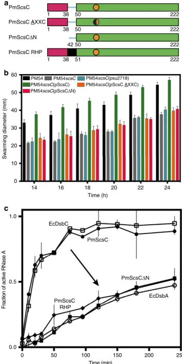
would contribute to bacterial copper resistance. Indeed,
inactivation of
scsC
in two independent
P. mirabilis
clinical
isolates significantly inhibited swarming motility (a key virulence
trait of
P. mirabilis
) in the presence of copper (Fig. 1b,
Supplementary
Figs
2–4).
This
phenotype
could
be
complemented by introduction of a plasmid encoding wild-type
PmScsC, but not a plasmid encoding an inactive PmScsC
(Fig. 1a,b).
Although it is annotated DsbA-like, PmScsC is a powerful
protein disulfide isomerase (Fig. 1c) that is able to refold and
reactivate the scrambled disulfide form of the model substrate
RNase A just as rapidly as the archetypal disulfide isomerase
E. coli
DsbC. It has negligible DsbA-like dithiol oxidase activity
(Supplementary Fig. 1d).
The best-characterized protein disulfide isomerases—EcDsbC
and eukaryotic protein disulfide isomerase (PDI)—each have two
thioredoxin-fold catalytic domains. DsbC is a dimer, and PDI is a
modular 4-domain protein with two catalytically active
thior-edoxin domains. The presence of two thiorthior-edoxin catalytic
domains is thought to contribute to highly efficient disulfide
shuffling activity
10,11. The amino acid sequence of PmScsC
encodes a single thioredoxin fold, and we therefore expected that
its disulfide isomerase activity would be a consequence of
dimerization to generate the necessary two catalytic domains.
We also expected that the protein would adopt the same dimeric
b
a
14 16 18 20 22 24
0 10 20 30 40 50
Sw
ar
ming diameter (mm)
Time (h)
0 50 100 150 200 250
1.0
0.5
0.0
PmScsC EcDsbC
PmScsCΔN
Time (min)
Fraction of active RNase A
60
EcDsbA
c
PmScsC RHP 1
50 PmScsC RHP
PmScsCΔN
38 50 222
222
PM54scsC(psu2718)
PM54scsC(pScsC) PM54scsC(pScsC AXXC)
PM54scsC(pScsCΔN)
PM54 PM54scsC
1 38 50 222
1 38 51 222
PmScsC
PmScsC AXXC
42
Figure 1 | PmScsC function.(a) Linear representation of the domain organization of PmScsC mutants. The trimerization domain is coloured magenta, the catalytic domain is green, with the CXXC motif represented as an orange circle, and the linker region is shown in cyan. The rigid helical linker is shown in black. (b) Swarming motility of wild typeP. mirabilisstrain PM54 (black), PmScsC deletion mutant PM54scsC(grey) and PM54scsC
containing control and complementation plasmids: PM54scsC(pSU2718) (vector control; cyan), PM54scsC(pScsC) (wild-type; green), PM54scsC
architecture as V-shaped EcDsbC. However, we were wrong on
both counts.
Unexpectedly,
evidence
from
chemical
cross-linking
(Supplementary Fig. 5), small angle X-ray solution scattering
(Fig. 2a,b) and multi-angle light scattering (Fig. 2c) all indicated
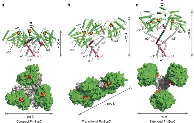
that PmScsC is trimeric. We confirmed that PmScsC is a trimer
by determining three independent crystal structures. The three
crystal structures—which we refer to as compact, transitional and
extended (Fig. 3a–c, Table 1)—reveal an extraordinary range of
motion in this protein. Importantly, the compact and transitional
structures have multiple protomers in the asymmetric unit that
adopt the same overall structures in each case (8 compact trimers,
RMSD
B
1.5 Å for 645 C
a
; two transitional trimers, RMSD 0.5 Å
for 653 C
a
) so that crystal symmetry does not appear to impact
on the observed conformation. Moreover, the compact and
transitional conformations were determined from crystals grown
under similar conditions (2.85 M sodium malonate pH 5.8, 20
°C
with either 0.1 M cobalt or copper added). Nevertheless,
comparison of the eight compact and the two transitional trimers
in these two crystal structures, reveals an unprecedented level of
conformational re-arrangement (RMSDs
4
20 Å). The third
structure was determined from crystals grown in the presence
of 2 mM copper chloride (32% Jeffamine M-600, 0.1 M HEPES
pH 8, 20
°C) and reveals a fully extended trimer with one
protomer in the asymmetric unit.
PmScsC also exhibits dynamic conformational flexibility in
solution. No single PmScsC crystal structure is consistent with the
experimentally determined SAXS scattering curve. However, an
ensemble of structures fits the experimental data closely
(Fig. 2a,b) supporting the notion that the trimer is highly
dynamic in solution. Moreover, the solution scattering curve is
unchanged by modification of the pH (range 6.0–8.0) or ionic
strength (150–1,500 mM NaCl) (Supplementary Fig. 6) indicating
the dynamic motion is an inherent property of the protein.
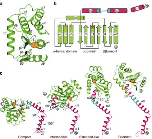
We can pinpoint the region responsible for the extraordinary
range of motion in this protein, to an 11-residue peptide linking
the trimerization and catalytic domains (residues
39-KADEQ-QAQFRQ-49) (Fig. 3). The 31 crystallographically characterized
PmScsC protomers have the same thioredoxin-fold catalytic
domains (Fig. 4a,b) (RMSD 0.2–1.2 Å for 129 C
a
). Similarly, all
31 PmScsC protomers share the same trimeric right-handed
coiled coil stalk formed from the N-terminal residues (Fig. 3a–c).
At the secondary structure level, it is only the 11-residue linker
peptide that changes significantly across the crystal structures
(Fig. 4c, Supplementary Movie 1).
The linker is a shape-shifting peptide that can adopt helical,
strand or loop conformations. In the 24 protomers present in the
compact PmScsC crystal structure, the linker forms a loop that
positions the three catalytic domains close to the trimerization
stalk (Figs 3a and 4c). In the extended crystal structure the linker
is helical, rotating and translating the catalytic domain away from
the trimerization domain relative to the compact structure
(Figs 3c and 4c). In the transitional PmScsC crystal structure,
each of the two trimers incorporates protomers in three
conformations:
compact,
extended-like
and
intermediate
(Fig. 3b). The two compact protomer conformations of the
transitional structure are similar to those in the compact crystal
structure (RMSD 0.9–1.8 Å for 188 C
a
). The two extended-like
protomer conformations of the transitional structure are similar
to the protomer in the extended crystal structure though they
bend at different points in the linker helix, giving rise to
somewhat different catalytic domain placements (Fig. 4c). The
two intermediate conformations in the transitional crystal
structure have a linker that forms a short
b
-strand, and positions
the catalytic domain directly above the trimerization stalk
(Fig. 4c).
Molecular mass (kDa)
Refractive index
Retention time (min) 15
16 25 40 63 100
10
20 25 30
PmScsC (73.9 kDa)
PmScsCΔN (18.3 kDa)
c
b
a
0 50 100
0.0 0.5 1.0
0.05
0.00 0.10 0.15 0.20 0.25 10–1
10–2
10–3
10–4
r (Å)
p(r)
(10
–12
cm)
q (Å–1)
l(q)
(cm
–1
) 0.0 0.5 1.0
–0.6 –0.4 –0.2
q2(10–3 Å–2)
ln
l
(
q
) /
l
(0)
Figure 2 | SAXS and MALLS of PmScsC.(a) Small-angle X-ray scattering data collected from wild type PmScsC (grey) and the calculated scattering profile of the ensemble model overlayed in black (SASBDB: SASDB94). The predicted scattering profile of each of the crystal structures is also shown (dashed lines: PDB: 4XVW compact, red; PDB: 5IDR transitional, blue; PDB: 5ID4 extended, green). The agreement between the experimental data and the ensemble model is excellent, yieldingw2¼1.0 (compared tow2¼863.9 (compact);w2¼1222 (transitional);w2¼348.2 (extended)). The Guinier region (inset) of the scattering data is linear, consistent with a
a
α1’’
α2’’
α1’ α1
α2 α3’
α3’
α3
α3’’
~ 50 Å
~ 70 Å
α1
α2’
α1’
α1’’
α3’’
α2
α3
α2’’
b
c
~ 80 Å
α1’’
α1
α1’
α3
α3’
α2”
α3”
α2 α2’
Compact PmScsC Transitional PmScsC Extended PmScsC ~ 105 Å
~ 80 Å ~ 80 Å
α2’
Figure 3 | PmScsC crystal structures.(a) Compact (PDB: 4XVW), (b) Transitional (PDB: 5IDR) and (c) Extended (PDB: 5ID4) crystal structures of PmScsC. Upper panels: side view, secondary structure with catalytic domains in green, 11-residue peptide linker in cyan and trimerization domains in magenta or white. Height in this orientation is indicated. In each case, one trimerization domain is shown in magenta for comparison of the conformational changes across the three crystal structures. Lower panels: top view, surface representation (catalytic TRX fold domains green, trimerization domains white), maximum dimension in this orientation is labelled. Active site positions are indicated in orange for each protomer.
Table 1 | PmScsC crystal structure statistics.
Compact (4XVW) Transitional (5IDR) Extended (5ID4) Data collection
Space group P 21 I4 H32
Cell dimensions
a, b, c(Å) 137.5, 163.9, 181.9 193.1, 193.1, 105.8 86.7, 86.7, 330.9
a,b,g(deg) 90, 90, 90 90, 90, 90 90, 90, 120
Resolution (Å) 91.15–2.60 (2.74–2.60) 136.51–2.56 (2.57–2.56) 110.29–2.92 (2.93–2.92)
Rmerge 0.072 (0.617) 0.083 (0.741) 0.059 (0.625)
I/sI 11.0 (2.0) 14.9 (2.2) 14.2 (2.8)
Completeness (%) 98.6 (95.4) 99.4 (100.0) 99.2 (100.0)
Redundancy 3.8 (3.7) 4.1 (4.1) 4.1 (4.2)
Refinement
Resolution (Å) 91.15–2.60 42.82–2.56 40.36–2.92
No. reflections 243409 62069 10652
Rwork/Rfree(%) 24.8/28.2 17.1/22.2 25.1/26.3
No. atoms
Protein 40850 10262 1720
Ligand/ion NA NA NA
Water 281 82 0
B factors(Å2)
Protein 59.7 50.6 122.2
Ligand/ion NA NA NA
Water 41.5 43.0 NA
RMS deviations
Bond length (Å) 0.006 0.008 0.010
Bond angles (deg) 1.21 1.05 1.17
As a consequence of the linker peptide flexibility, the three
crystal structures describe extraordinarily different PmScsC
quaternary shapes and dimensions. The eight compact PmScsC
trimers are mushroom-shaped with dimensions 80
80
50 Å
3(Fig. 3a). The two intermediate PmScsC crystal structures are
asymmetric flat triangles with dimensions 105
40
70 Å
3(Fig. 3b). The extended PmScsC crystal structure is
three-leaf-clover shaped with dimensions 75
80
80 Å
3(Fig. 3c).
We hypothesized that the dynamic motion suggested by the
crystal structures, and supported by solution data, is critical to the
mechanism of PmScsC. Indeed, replacement of the shape-shifting
peptide
with
a
rigid
helical
peptide
(PmScsC
RHP,
39-AEAAAKEAAAKA-50)
12reduced the flexibility of the
protein
as
evidenced
by
small
angle
scattering
data
(Supplementary Fig. 7), and abolished activity in the isomerase
assay (Fig. 1c). Furthermore, we showed that trimerization of
PmScsC is critical for activity because removal of the N-terminal
residues (PmScsC
D
N) also abolishes
in vitro
disulfide isomerase
activity (Fig. 1c) and
in vivo
complementation assays (swarming
in the presence of copper) (Fig. 1b).
Curiously, Nature has also performed the ScsC
D
N experiment.
PmScsC shares 58% sequence identity with
Salmonella enterica
serovar Typhimurium StScsC
13(Supplementary Fig. 8) and both
proteins contribute to copper resistance. Whereas we showed
above that PmScsC is required for swarming motility under
copper stress, StScsC is required for growth of
S. Typhimurium
in
rich media in the presence of copper. By contrast, deletion of
scsC
in
P. mirabilis
had no effect on growth in rich media whether
copper was present or absent (Supplementary Fig. 2c). These
findings imply that the two proteins have different target
substrates and/or different molecular functions. Indeed, StScsC
and PmScsC are very different proteins. StScsC lacks the
N-terminal residues of PmScsC, it is monomeric rather than
trimeric, and it has no isomerase activity. Intriguingly, the
catalytic domains of the two proteins (as assessed by crystal
structures) are very similar (PDB: 4GXZ) (RMSD 1.0–1.6 Å for
129 C
a
). On the basis of this comparison, we predicted that
removal of the N-terminal trimerization residues from PmScsC
would not only convert the trimer into a monomer and abolish
isomerase activity, it might also convert the enzyme into a dithiol
oxidase. Indeed, we confirmed that this was the case
(Supplementary Fig. 1d).
These two proteins, trimeric disulfide isomerase PmScsC and
monomeric dithiol oxidase StScsC, are encoded in similar loci
and are both associated with copper resistance. Yet, they have
very different architectures, and different molecular and cellular
functions. To examine if the region encoding the N-terminal
extension of PmScsC is broadly conserved in
P. mirabilis
, the
scsC
gene was PCR amplified from 25 randomly selected clinical
isolates and sequenced. We found that the Pm
scsC
sequence was
conserved in all 25 isolates—including the region encoding the
trimerization domain and shape-shifting motif—except for a
single synonymous mutation in the signal sequence. This high
degree of conservation suggests a highly conserved functional
role.
Caulobacter crescentus
ScsC also has an extended
N-terminal region and is predicted to be dimeric
14. Altogether
with our work, this suggests that the N-terminal region of
this protein is necessary for oligomerization and that there are
at least two very different molecular and functional classes of
ScsC.
α4 α5
α6 α7
α8
α9 β1
β2 β3
β4
β5 C
α1 α1 α1
α2
α2 α2
90°
α3 α3
β’
120°
85° 75°
155° 60°
α3
α1 α2 α3
a
c
Compact Intermediate Extended-like Extended
N
N N
N N
C
C C
C
α-helical domain βαβ-motif ββα-motif
N
C
α7 α6 α5 β1 β3 α4 α9
α1
α8
b
α3 α2
β5
β4
β2
How does the trimeric and highly dynamic molecular
architecture of PmScsC compare with other structurally
char-acterized disulfide isomerases? Until this point, highly efficient
disulfide isomerases were thought to function through the
presence of two TRX domains (each with a CXXC active site)
that can simultaneously interact with two substrate cysteines
15.
The two active sites of V-shaped DsbC and U-shaped PDI are
embedded within a dynamic framework that enable shuffling of
substrate disulfides
16–19(Fig. 5a,b). Trimeric PmScsC also has the
potential
to
interact
with
multiple
substrate
cysteines
simultaneously. However, the evidence presented here shows a
much more expansive range of motion than observed previously
for
DsbC
or
PDI,
as
depicted
in
morphing
movies
(Supplementary Movies 2–4) and static structural comparison
(Fig. 5c). For example, the distance between active sites of DsbC
(PDB: 1JZD-1EEJ)
3,16or PDI (PDB: 4EKZ-4EL1)
18in reported
crystal structures ranges between 31–39 Å (26% increase) and
28–40 Å (43%), respectively. By comparison, this distance ranges
from 16 to 52 Å (225%) in PmScsC.
Discussion
Why is it that other characterized disulfide isomerases do not
have the highly flexible, trimeric structure of PmScsC? How does
the
monomeric
S.
Typhimurium
ScsC—not
a
disulfide
isomerase—confer copper resistance? How do the very diverse
ScsC proteins (monomeric, dimeric or trimeric) form redox relay
systems with a predicted partner protein ScsB
14that appears to be
highly conserved in bacterial Scs gene clusters? The likely
explanation for the diversity in architecture and flexibility of
ScsC proteins may relate to differences in their target specificity.
The structural diversity among ScsC homologues may simply
reflect the fact that each ScsC homologue has evolved to optimize
disulfide reduction and/or isomerization of a distinct protein
substrate or set of substrates that are sensitive to copper-induced
damage. Identifying and characterizing these specific substrates,
and understanding why these are not refolded or reduced by
other periplasmic TRX-fold proteins are key questions that will
inform our understanding of bacterial copper resistance
mechanisms.
In summary, PmScsC is a unique and highly dynamic disulfide
isomerase. The N-terminal residues bring three catalytic domains
together into a trimer, and a unique flexible linker enables
extraordinary twisting and extending motions that impact on the
catalytic domain placement. Crystal structure snapshots
sup-ported by X-ray scattering data from solution, indicate an almost
doubling in length of the protein in one dimension, in concert
with considerable catalytic domain rotation. The combination,
extent and diversity of these motions would enable mis-folded
substrates bound to PmScsC to explore a broad folding landscape,
consistent with the redox foldase activity of this key
P. mirabilis
copper resistance protein. These findings show in much greater
detail than ever before how redox proteins with multiple active
sites are able to shuffle incorrectly folded substrates. It is unclear
whether PmScsC directly interacts with copper or how it interacts
with other proteins required for virulence under copper stress.
Nevertheless, the structural data may provide a suitable basis for
drug discovery targeting a central defence mechanism of an
important human pathogen
20,21. Importantly, the shape-shifting
motif may also represent a modular component for dynamic
motion that could be used in ‘plug and play’ protein engineering
applications.
Methods
Bacterial strains and growth conditions
.
BLAST searches of DsbA homologues were performed on the genome ofProteus mirabilisHI4320 (ref. 22). Clinical isolates ofP. mirabiliswere cultured from the urine or blood of patients with urinary tract infection or bacteremia. The isolates were sourced from the Princess Alexandra Hospital or Sullivan Nicolaides Pathology (Brisbane, Australia).E. coliTop10 (Invitrogen) was used for plasmid manipulations.E. coliandP. mirabilis
were cultured in Luria-Bertani (LB) broth (10 g l1tryptone, 5 g l1yeast extract, 10 g l1NaCl) on LB agar (LB medium containing 15 g l1agar). To prevent swarming ofP. mirabilis, LB medium containing 30 g l1agar and without NaCl
was used. Media were supplemented with chloramphenicol (30mg ml1),
ampicillin (100mg ml1) or kanamycin (25mg ml1) as required.
Construction ofProteus mirabilis scsCmutants
.
Mutation of thescsCgene inP. mirabilisPM38 and PM54 was performed using the TargeTron gene knockout system (Sigma-Aldrich) as per the manufacturer’s instructions. Briefly, optimal intron insertion sites and primer sequences (5827, 50-aaaaaagcttataattatc cttaag-tatcgaagatgtgcgcccagatagggtg, 5828, 50 -cagattgtacaaatgtggtgataacagataagtcgaa-gatgctaacttacctttctttgt, 5829, 50-tgaacgcaagtttctaatttcgattatacttcgatagagg aaagtgtct) were predicted using the Sigma TargeTron online algorithm, followed by a retar-geting PCR and cloning of the amplicon into the pACD4-K-C shuttle vector, resulting in the generation of plasmid pACD4-K-CscsC.Plasmid pAC4K-CscsC
and pAR1219 (helper plasmid) were transformed into PM38 and PM54; induction and retargeting of the intron containing the kanamycin-resistance gene cassette intoscsCwas performed according to the manufacturer’s instructions. Transfor-mants with the correct intron insertion were selected by growth in the presence of kanamycin. Mutants with the correct insertion were confirmed by PCR and nucleotide sequencing using primers 5914 (50-atgaaaaaaatttggctggcgttagc) and 5915 (50-ttattttttcatcagtaattgattgacgacatcagaataag), and referred to as PM38scsCand PM54scsC, respectively. The reprogrammed intron was inserted between nucleo-tide 528 and 529 in the sense strand ofscsC. Plasmids pACD4-K-CscsCand pAR1219 were cured by passaging on nonselective medium followed by screening for loss of chloramphenicol and ampicillin resistance. Plasmid pScsC was con-structed by amplifying thescsCgene from PM54 using primers PMscsC-pSU2718F (50- gcgtctagattaactattctttcagaggctaaaggagcc) and PMscsCpSU2718R (50 -cgaagcttcgttattttttcactttcgccagttgttctttcac), digestion with XbaI-HindIII and ligation into XbaI-HindIII digested pSU2718. Mutation of the CXXC active site (C82A; plasmid pScsC AXXC) and deletion of residues 1–42 (plasmid pScsCDN) was performed by PCR; all constructs were confirmed by sequencing. These mutants retained the periplasmic signal sequence. Complementation was performed by hPDI
EcDsbC
a
b
c
PmScsC
Top Side
introducing plasmids pScsC, pScsC AXXC, pScsCDN or pSU2718 (vector control) into PM54scsC.
Swarming motility in the presence of CuSO4
.
Overnight cultures ofP. mirabilis were diluted in fresh LB broth to an OD600of 0.1. Swarming on LB agar was examined in the absence or presence of CuSO4(final concentration of 1.5 mM). For assays performed in the presence of CuSO4, a 10ml sample of the dilutedP. mirabilisPM54 and PM54scsCstrains, respectively, was inoculated onto the centre of an agar plate and allowed to soak into the agar (resulting in equal diameter for each sample). The plates were incubated at 37°C. The diameter of the swarming zone was measured after 14 h incubation, and every one or two hours thereafter for a total of 10 h. Swarming of PM54scsCcontaining pSU2718 (vector control) or complementation plasmids (pScsC, pScsC AXXC and pScsCDN) was performed as described above, but with the addition of chloramphenicol (30mg ml1) and 1 mM isopropylb-D-1-thiogalactopyranoside for ScsC induc-tion. Swarming motility at 37°C on LB agar was measured after 6 and 8 h of incubation. Three replicate experiments were performed for all assays. Swarming motility of PM38 wild-type, mutant and complemented strains in the presence and absence of copper was performed in the same manner.
Western blotting
.
ScsC expression inP. mirabiliswild-type, mutant and com-plemented strains was examined by western blot analysis. Crude cell lysates were prepared from standardized overnight cultures. Samples were resolved by SDS– polyacrylamide gel electrophoresis (SDS–PAGE) and transferred onto a poly-vinylidene difluoride membrane. Blots were probed using a PmScsC specific polyclonal antiserum raised in rabbits (WEHI Antibody Facility) at 1:1,000 dilution in phosphate buffer saline with 0.05%v/vTween-20.Protein production
.
Codon-optimized PmScsC (see Supplementary Note 1 for the sequence) lacking the first 21 amino acids (which correspond to the predicted secretion signal) or PmScsCDN (lacking the first 63 residues, starting at A1Q2F3y)was inserted into pMCSG7 (Midwest Center for Structural Genomics) by ligation-independent cloning. PmScsC and PmScsCDN were expressed in BL21(DE3)pLysS cells (Novagen) at 30°C for 16–20 h using ZYP-5052 autoinduction media23. Selenomethionine-labelled PmScsC (mature PmScsC contains four native methionine sites) was expressed in BL21(DE3)pLysS cells grown in minimal media (M63) supplemented with 0.05 mg ml1D/L-selenomethionine. Recombinant protein expression was induced by 0.1 mM isopropylb-D-1-thiogalactopyranoside overnight at 30°C.
After expression, all cells were collected by centrifugation then resuspended in lysis buffer (25 mM Tris pH 7.4, 150 mM NaCl) with added protease inhibitor (1:1,000 dilution into lysate, BioPioneer, Inc., USA) and DNase I (3.3mg ml1final
concentration, Sigma-Aldrich). The bacterial cells were processed through a Constant Systems (LTD, UK) TS-Series cell disruptor twice, at pressures of 22 and 24 kpsi. The lysate was clarified by centrifugation (40,000g, 30 min, 4°C, rotor JA-25.5, Beckman Coulter, Brea, CA) and the soluble fraction was run twice over 23 ml of equilibrated Talon (Clontech) resin to bind the His-tagged protein. The resin was washed with six column volumes of lysis buffer before the protein was eluted with 25 mM Tris pH 7.4, 150 mM NaCl, 250 mM imidazole. To remove the His6-tag, Tobacco Etch Virus (TEV) protease cleavage was performed at room temperature for 2 h with 1 mg of TEV protease per 50 mg of His-tagged protein. The protein mixture was then desalted using a Sephadex G-25 fine 16/60 column connected to an A¨ KTA FPLC system (GE Healthcare) and reverse IMAC (22 ml of Talon resin) was used to separate cleaved protein from the His-tagged contaminants. PmScsC and PmScsCDN were reduced or oxidized with a 25-fold molar excess of DTT, or a 10-fold molar excess of copper(II)/1,10-phenanthroline, respectively. The final step of purification for the proteins was size-exclusion chromatography using a Superdex S75 column with 10 mM HEPES pH 7.4, 150 mM NaCl. Proteins were concentrated using Amicon Ultra centrifugal filter devices with a 10-kDa cutoff (Merck Millipore, USA). Yields for PmScsC were B80 mg of purified protein per litre of culture. SDS–PAGE (NuPAGE 4–12% BisTris gel, Invitrogen, Australia) with Coomassie blue stain was used to assess the protein quality. Concentration of protein samples was determined using the A280of the sample (read using a Thermo Scientific NanoDrop 2000c spectrophotometer) and calculated extinction coefficients from ProtParam24.
E. coliDsbA andE. coliDsbC, lacking the periplasmic leader signal were expressed and purified, as described for PmScsC.
Design of PmScsC rigid helical peptide mutant
.
The 11-amino-acid flexible linker peptide in PmScsC (residues 39-KADEQQAQFRQ-49) was replaced with a 12-amino-acid rigid helical peptide (39-AEAAAKEAAAKA-50)12using overlapextension PCR25with the primers, 50-gcagaagcagcggccaaagaggctgcggctaaagccgca ctggctagcgaacatgatgcc and 50- ggctttagccgcagcctctttggccgctgcttctgctttcgtctgcagagcca tgattgc. The mutated gene was then reinserted into pMCSG7 by ligation-independent cloning. The construct was confirmed by sequencing and the protein expressed and purified, as described for wild-type PmScsC.
PmScsC sequence conservation
.
All clinical isolates were de-identified, and individual informed consent was not required. Genomic DNA isolation was per-formed using the UltraClean Microbial DNA Isolation Kit (MO-BIO Laboratories, #12224) according to the manufacturer’s instructions. Yields varied between 100 and 500 ngml1of DNA. PCR amplification of thescsCgene fromP. mirabilisstrains was performed using a sequence specific primer pair (50-gtgccgttt aacca-gatttatg) and (50-cgtagataaatcagtaagttctg) in combination with Phusion High-Fidelity DNA Polymerase (New England BioLabs Inc., MA, USA). DNA was initially denatured in a single step at 98°C for 30 s followed by 30 cycles of denaturation (98°C, 10 s), annealing (50°C, 20 s) and elongation (72°C, 30 s). In a final step fragment elongation was completed at 72°C for 7 min. PCR samples were examined by electrophoresis and subsequent DNA sequencing.
Chemical cross-linking
.
In total 150mM of purified PmScsC (in 50 mM HEPES buffer pH 8.5) was reacted with 3 mM homobifunctional dithiobis(sulfosuccini-midylpropionate) (DTSSP) (Pierce) for different time intervals (30 s–30 min) at 23°C. DTSSP contains one amine-reactiveN-hydroxysulfosuccinimide (sulfo-NHS) ester at each end of its eight carbon spacer arm. The sulfo-NHS group reacts preferentially with amines (from lysine residues) between pH¼7 and 9, to form a stable amide bond. DTSSP contains a disulfide bond in the spacer arm, which can be readily cleaved with dithiothreitol (DTT). Aliquots were removed at various time points and mixed with (NH4)HCO3(50 mM final), to stop the reaction, through hydrolysis of remaining functional ester groups in DTSSP. The samples were then run on a non-reducing SDS–PAGE and subjected to Coomassie staining. As control, 50 mM DTT was added to selected samples, which led to cleavage of the internal disulfide bond in DTSSP, and consequently disruption of cross-links between ScsC protomers.MALLS
.
A combined approach, using analytical size exclusion chromatography (SEC) and multiangle laser light scattering (MALLS) was utilized to determine and compare the stoichiometry of PmScsC with PmScsCDN in solution. The setting consisted of an LC20 high-performance liquid chromatography (HPLC) system (Shimadzu, Rydalmere, Australia) and a DAWN HELEOS II laser light detector connected to an Optilab T-rEX refractive index detector (Wyatt Technology, Dernbach, Germany). Either a Superdex 200 or Superdex 75 10/300 GL analytical column (GE Healthcare, USA) was connected to the LC20 HPLC system and equilibrated with 25 mM Tris and 150 mM NaCl, pH 7.5 overnight. Purified pro-teins were injected (500ml of 3 mg ml1at a flow rate of 1.0 ml min1) into theSEC-MALLS system for analysis. To calibrate the detector, 500ml of 5 mg ml1 bovine serum albumin (BSA) (Sigma-Aldrich, Australia) was used in 25 mM Tris and 150 mM NaCl, pH 7.5 at a flow rate of 1.0 ml min1. Wyatt Astra V software
was used for data collection and analysis.
SAXS
.
Small angle X-ray scattering data were collected on the SAXS-WAXS beamline at the Australian Synchrotron26. Immediately before loading, all samples were centrifuged at 10,000gto remove large particles from the solution, and radiation damage was minimized by flowing samples (B100ml) past the beam in 1.5 mm quartz capillaries (Hampton Research). Data reduction was carried out using the Australian Synchrotron Scatterbrain software27correcting for sample transmission and solvent scattering.Scattering data were collected on reduced PmScsC wildtype and RHP mutant proteins in 25 mM HEPES pH 7.5, 150 mM NaCl, 1 mM DTT. Data were also collected on oxidized wildtype PmScsC, but as no significant differences in the scattering were observed, only data from the reduced forms are presented. To confirm that the pH and ionic strength of the crystallization conditions do not induce conformational changes in PmScsC, scattering data were collected from wildtype PmScsC prepared at 0.50 mg ml1in a gradient of 25 mM HEPES pH
(6.0, 6.5, 7.0, 7.5 and 8.0) and NaCl (150, 300, 600, 900 and 1,500 mM). No significant systematic change in the structural parameters was observed at any point of the gradient, hence, it was concluded that neither ionic strength nor pH influence the conformation of the complex in the ranges measured.
Data quality was assessed by inspection of the linearity of the Guinier region of the data (qRgo1.3), estimated molecular mass of the protein complex, and concentration dependence of the scattering. The estimated molecular mass was determined as outlined in28, where the contrast and partial specific volume were estimated from the protein sequence29. The pair-distance distribution function (p(r)) was generated from the experimental data usingGNOM30from whichI(0), RgandDmaxwere determined. Rigid body modelling of the scattering data from PmScsC wildtype and mutant proteins was performed usingCORAL31.C3 symmetry was assumed, and the starting model was oriented such the 3-fold axis was parallel with thez-axis, and passed through the centre of the oligomization domain. Two rigid bodies were then defined for each monomer: residues 3–44 (oligomerization domain); 47–224 (catalytic domain). The position of the oligomerization domain was fixed, and the position and orientation of the catalytic domain was then optimized against the measured scattering data. The program was run 16 times for each protein, but the models with the lowest penalty function still showed small systematic deviations from the experimental data (Wildtype: w2¼3.4; CorMap test32, 182 points,C¼51,P¼0.000; RHP mutant:w2¼5.6;
significant structural diversity, it is likely that these systematic deviations arise from the fact that there is an ensemble of structures present in solution. Hence, ensemble optimization was also performed with the program EOM33. From an initial pool of
1,000 structures (the oligomerization domain possessedC3symmetry in all structures, but the entire trimer was permitted to adopt eitherC1orC3symmetry), a final ensemble of 6 structures was obtained that yielded an excellent fit to the data (Wildtype:w2¼1.0; CorMap test, 182 points,C¼12,P¼0.021; Mutant:w2¼1.8;
CorMap test, 171 points,C¼11,P¼0.076). The ensemble for the wildtype protein is composed of six structures and spans a diverse range of conformations. Thus, the SAXS data supports the notion that the PmScsC trimer is highly dynamic in solution. The ensemble for the mutant protein is composed of four structures, all in extended conformations. The initial pool of random structures was the same for both WT and RHP optimizations, and the number of structures in the final ensemble was not artificially limited. The reduction in size of the ensemble (from 6 for WT to 4 for RHP) is consistent with the RHP mutation rigidifying the flexible helix, and reducing the conformational space sampled by the protein. Details of the data collection and structural parameters are summarized in Supplementary Table 1.
Crystallization and structure determination
.
Crystallization screenings were performed at the UQ ROCX facility at the University of Queensland (uqrocx.im-b.uq.edu.au) using commercial screens and the hanging-drop vapour diffusion method.Compact structure
.
Selenomethionine-labelled PmScsC crystals were grown at 20°C from a drop comprising 200 nl of 30 mg ml1purified protein in 10 mMHEPES pH 7.4 and 200 nl of well solution, 2.85 M sodium malonate pH 5.8 containing 0.1 M cobalt(II)chloride hexahydrate. Crystals grew over a period of 2–3 days. A data set at the Se-edge (wavelength¼0.9792 Å) was measured under cryogenic conditions (100 K) at the Australian Synchrotron MX2 beamline using the BluIce interface34. Data were processed in XDS35and scaled to space group
P21212 in AIMLESS36. A manually guided molecular replacement approach was performed using StScsC as the template (PDB ID: 4GXZ) in MOLREP37. Manual
selection of solutions based on observed packing, allowed placing of six molecules in the asymmetric unit. This solution was improved in the subsequent MR runs in MOLREP37, allowing the addition of eight molecules in the second trial and a total
of nine molecules in the third run. A SAD-MR approach in PHENIX38, by
combining SAD phasing and the partial MR solution (nine molecules), and subsequent density modifications resulted in an electron density map with a skew value of 0.82 (1,826 residues (190 side chain built) in 250 fragments). Successive AutoBuild runs in PHENIX38built 1,557 residues (806 side chain) and dropped the R and Rfree values to 38% and 41.5%, respectively. Extensive manual building was required to complete one copy of PmScsC. Molecular replacement using this model as template in PHASER39allowed placing of 12 molecules in the asymmetric unit. After several rounds of manual building in Coot40and PHENIX38refinements R/ Rfree values dropped to 29.1 and 32.3%, but further refinement cycles were unsuccessful. Careful examination of Ctruncate41indicated the presence of nearly
perfect pseudo-merohedral twinning (-h,-k,l) and led to a reconsideration of space group. Reprocessing the data set to a resolution of 2.6 Å in space groupP21yielded lower R-merge (0.072 versus 0.097 for data processed inP21212). A new molecular replacement run using the reprocessed data inP21 space group and the PmScsC template with a resolution cutoff set to 3.2 Å finally identified 24 copies in PHASER39. Rigid body refinement in REFMAC42showed that refinement
considering the twin operator (-h,-k,l) produced better R-factors (37 versus 42%, no twinning). After manual correction of two chains that were placed inaccurately during molecular replacement by PHASER39, and several rounds of refinement in
PHENIX38and REFMAC42the final R/Rfree values are 24.8/28.2% at 2.6 Å
resolution. The Ramachandran favoured/outlier statistics for the structure are 95%/0.9%. A stereo image of representative electron density for this structure is show in Supplementary Fig. 9a.
Transitional structure
.
Crystals were grown at 20°C from drops containing 1ml of 48 mg ml1oxidized PmScsC in 10 mM HEPES pH 7.4 and 1ml of 2.85 M sodium malonate pH 5.8 containing 0.1 M copper (II) chloride. Crystals formed after 2–3 days and were cryoprotected with 3.4 M sodium malonate pH 5.8 and flash frozen in liquid nitrogen. Data were collected using the BluIce software34on the MX2 beamline of the Australian Synchrotron at a wavelength of 0.9792 Å under cryogenic conditions (100 K). The data were processed with XDS35and scaled in AIMLESS36using the autoPROC framework43. The space group was determined to beI4 and molecular replacement using PHASER39, as implementedin the CCP4 suite44, was performed with a single catalytic domain from the
compact structure (residues 46–224). Five molecules were initially found in the asymmetric unit and after assessing the crystal packing one of these was deleted and a subsequent round of PHASER39was run with the altered solution as an
additional input model. This resulted in the placement of 6 molecules in the asymmetric unit, which is consistent with the presence of two trimers. Manual building of the trimerization domain was performed in Coot and rounds of autobuild using Buccaneer45in CCP4 and refinement using REFMAC42and
BUSTER46,47were performed to improve the structure. Several rounds of manual adjustment in Coot40and refinement in PHENIX38were performed to yield final
R/Rfree values of 17.1 and 22.2%. Validation in MolProbity48was used throughout
the refinement process, and the final Ramachandran favoured/outlier statistics for
the structure are 98%/0.5%. A stereo image of representative electron density for this structure is show in Supplementary Fig. 9b.
Extended structure
.
Crystals were grown at 20°C from drops containing 1ml of 20 mg ml1oxidized PmScsC in 10 mM HEPES pH 7.4, 150 mM NaCl and 2.5 mM copper (II) chloride (added to protein solution immediately before crystallization) and 1ml of 32% Jeffamine M-600 pH 7 in 0.1 M HEPES pH 8. Crystal formation occurred within minutes of set up and crystals continued to grow until day two. Crystals were cryoprotected with 32% Jeffamine M-600 pH 7, 0.1 M HEPES pH 8 and 20% ethylene glycol and frozen in liquid nitrogen. Data were collected using the BluIce software34on the MX2 beamline of the Australian Synchrotron at a wavelength of 0.9537 Å under cryogenic conditions (100 K). Using the autoPROC framework43the data were processed with XDS35and scaledin AIMLESS36. The Wilson B factor for the data was relatively high (Bdouble that of the data for the other two crystal structures). The space group was determined to beH32and molecular replacement using PHASER39, as implemented in the CCP4 suite44, was performed with a single catalytic domain from the compact structure
(residues 46–224). Only one molecule was found in the asymmetric unit (the trimer is generated by crystal symmetry). Manual building of the trimerization domain was performed in Coot with rounds of autobuild (Buccaneer45) and refinement
(BUSTER47) performed to improve the structure. Several rounds of manual adjustment in Coot40and refinement in PHENIX38, including the use of TLS, were performed to yield final R/Rfree values of 25.1 and 26.3%. Validation in MolProbity48was used throughout the refinement process and the final Ramachandran favoured/outlier statistics for the structure are 98%/0.5%. A stereo image of representative electron density for this structure is show in Supplementary Fig. 9c.
Residues in all three crystal structures were numbered based on their position after the TEV cleavage site in the construct. MUSTANG-MR49,50was used for structure alignment and RMSD calculations and PyMOL was used to create images of the structures and perform other measurements. The distance range between active sites in DsbC, PDI and PmScsC structures was measured using the Cb positions of the more N-terminal of the two catalytic cysteines. Supplementary Movie 1 was created in Pymol by generating morphs between each of the PmScsC crystal structures, as well as morphs between each of the available structures for
E. coliDsbC (PDB: 1JZD-1EEJ) and human PDI (PDB: 4EKZ-4EL1).
Measurement of PmScsC redox potential and pKavalues
.
The redox potentialof PmScsC was determined by incubating 2mM PmScsC with 1 mM GSSG in combination with varying concentrations of GSH (4–15 mM) forB12 h at 24°C in 100 mM sodium phosphate buffer, pH 7.0 and 1 mm EDTA. Trichloroacetic acid, 10%v/v, was used to precipitate the protein samples and the pellets were washed with ice-cold acetone. The samples were then treated with 2 mM 4-acetamido-40 -maleimidylstilbene-2,20-disulfonic acid, in 50 mM Tris–HCl, pH 8.0, and 1% SDS, which blocks free thiols and increases the weight of the reduced protein by B1 kDa. The reduced and oxidized forms of PmScsC were separated on a 12% non-reducing SDS–PAGE (NuPAGE) and stained with Coomassie Brilliant Blue. The fraction of reduced protein,R, was determined via densitometric analysis of the stained SDS–PAGE gel using ImageJ51, and the equilibrium constantKeqwas calculated viaR¼([GSH]2/[GSSH])/(Keqþ([GSH]2/[GSSH]))52. The Nernst equation, E0¼E0
GSH/GSSG–(RT/nF)lnKeq, whereE0GSH/GSSGis the standard potential of 240 mV (ref. 53), was used to calculate the redox potential of PmScsC.
The pH-dependent absorbance of the catalytic thiolate anion of PmScsC was determined at 240 nm (ref. 54) using a CARY 50 UV/VIS spectrophotometer (Agilent Technologies, USA). The pH titration measurements of oxidized or reduced PmScsC (40mM) were conducted at 22°C in 2 ml reaction buffer (10 mM Tris, 10 mM sodium citrate, 10 mM K2HPO4, 10 mM KH2PO4, 200 mM KCl and 1 mM EDTA). Absorbance (l¼240 and 280 nm) was measured between pH 6.5 and 1.5 in 0.25 increments. The pKavalue was calculated from the fitted curves of three replicates using the Henderson–Hasselbalch equation (pH¼pKalog ((A240/A280)red/(A240/A280)oxid)). Redox potential and pKaexperiments were each repeated three times.
Relative stability of oxidized and reduced PmScsCDN
.
Unfolding of PmScsCDN was monitored using the change in the far-ultraviolet circular dichroism (CD) signal55. The largest difference in molar ellipticity for oxidized and reduced enzymes was calculated from initial far-ultraviolet CD spectra (from 250 to 190 nm) recorded at 25 and 95°C using a Jasco J-810 circular dichroism (CD) spectropolarimeter (Jasco, USA), respectively. The unfolding of oxidized and reduced protein (PmScsCDNox¼222.5 nm, PmScsCDNred¼219.5 nm) was monitored at a constant heat rate of 1°C min1starting from 25°C and increasingProtein disulfide isomerase assay
.
The bovine pancreatic ribonuclease RNase A contains four disulfide bonds that are necessary for its enzymatic function. Starting with ‘scrambled’ RNase A (in which the cysteines have formed non-native disulfide bonds), one can determine the ability of other enzymes to restore its function by spectroscopically monitoring the hydrolysis of 30,-50-cyclic monophosphate (cCMP), a synthetic substrate of the ribonuclease. Thus,in vitrodisulfide isomerase activity of PmScsC was monitored by using scrambled RNase A as substrate56.Inactive scrambled RNase A was produced by treating 70 mg of native RNase A (Sigma-Aldrich) with 6 M guanidinium chloride and 150 mM DTT in 50 mM Tris pH 8, incubated at room temperature for 16 h. After adjusting the solution to pH 8, the unfolded protein was then desalted and buffer exchanged into 100 mM acetic acid/NaOH pH 4. The presence of reduced cysteines was confirmed
spectrophotometrically by mixing an unfolded RNase A sample with eight-fold higher molar concentration of 5,5-dithio-bis-(2-nitrobenzoic acid) (DTNB or Ellman’s reagent), which reacts with thiols to absorb at 412 nm. The unfolded RNase A was then air-oxidized over 5 days in 6 M guanidinium chloride, 50 mM Tris pH 8.5 at room temperature in the dark. After concentration of the protein and readjustment of the pH to 8.5, the scrambled RNase A was buffer exchanged into 100 mM acetic acid/NaOH pH 4 and concentrated to 11.5 mg ml1for use in assays. The absence of reduced cysteines was confirmed, as described above. The total reaction volume for the scrambled RNase A assay was 750ml containing 100 mM sodium phosphate buffer pH 7.0, 1 mM EDTA, 8.2mM DTT and 10mM PmScsC, PmScsCDN, PmScsC RHP, EcDsbC or EcDsbA and 40mM of scrambled RNase A. At various time intervals, 50ml sample was removed and mixed with 150ml of 3 mM cytidine 30,-50-cyclic monophosphate (cCMP) to measure RNase A activity. Hydrolysis of the cyclic phosphate ester bond in cCMP was monitored at 296 nm using a Synergy H1 multimode plate reader (BioTek, USA). Samples containing native RNase A and scrambled RNase A in the absence of any other enzyme served as positive and negative controls, respectively. The disulfide isomerase assays were repeated twice for each protein.
PmScsC dithiol oxidation activity
.
The potential of PmScsC/PmScsCDN to cat-alyse disulfide bond formationin vitrowas assessed using a synthetic model peptide (CQQGFDGTQNSCK), as described before57. Dithiol oxidation wasdetermined fluorometrically, using a Synergy H1 multimode plate reader (BioTek Instruments, USA) with the excitation wavelength set to 340 nm and emission to 615 nm. A 150ms delay before reading and 100ms reading time were used for time-resolved fluorescence. The reaction buffer contained 50 mM MES, 50 mM NaCl and 2 mM EDTA at pH 5.5. The reaction volume was 50ml in each well of a white 384-well plate (Perkin Elmer OptiPlate-384, Part #: 6007290), including increasing concentrations (80–320 nM) of EcDsbA, EcDsbC, PmScsC or PmScsCDN and 2 mM glutathione (GSSG) as oxidant. At last, 8mM peptide substrate was added to initiate the reaction. EcDsbA served as positive control. As negative control, reactions in the absence of proteins were used. The initial linear portion of the raw data was used to calculate the rate of oxidation as fluorescence increase per minute. The dithiol oxidation activity experiments were repeated three times for each protein.
Data availability
.
The UniProt accession code for theP. mirabilusScsC protein described in this paper is C2LPE2 and the accession codes for other proteins described in this study are H9L4C1 (StScsC), P0AEG4 (EcDsbA) and P0AEG6 (EcDsbC). Coordinates and structure factors for the three ScsC crystal structures were deposited in the protein data bank (PDB) with accession codes 4XVW (compact), 5IDR (transitional) and 5ID4 (extended). In addition to the PDB accession codes listed above, PDB accession code 4GXZ (StScsC) was used as a molecular replacement template and 1EEJ and 1JZD (EcDsbC), and 4EKZ and 4EL1 (hPDI) were used to generate Fig. 5. Scattering data and models have been deposited in SASBDB58with accession codes SASDB94 (wildtype PmScsC) andSASDBW6 (RHP mutant). All other data are available from the corresponding authors upon reasonable request.
References
1. Martin, J. L., Bardwell, J. C. & Kuriyan, J. Crystal structure of the DsbA protein required for disulfide bond formationin vivo.Nature365,464–468 (1993). 2. Inaba, K.et al.Crystal structure of the DsbB-DsbA complex reveals a
mechanism of disulfide bond generation.Cell127,789–801 (2006). 3. McCarthy, A. A.et al.Crystal structure of the protein disulfide isomerase,
DsbC, fromEscherichia coli.Nat. Struct. Biol.7,196–199 (2000).
4. Missiakas, D., Georgopoulos, C. & Raina, S. TheEscherichia colidsbC (xprA) gene encodes a periplasmic protein involved in disulfide bond formation.
EMBO J.13,2013–2020 (1994).
5. Heras, B., Edeling, M. A., Schirra, H. J., Raina, S. & Martin, J. L. Crystal structures of the DsbG disulfide isomerase reveal an unstable disulfide.Proc. Natl Acad. Sci. USA101,8876–8881 (2004).
6. Depuydt, M.et al.A periplasmic reducing system protects single cysteine residues from oxidation.Science326,1109–1111 (2009).
7. Achard, M. E.et al.Copper redistribution in murine macrophages in response to Salmonella infection.Biochem. J.444,51–57 (2012).
8. Djoko, K. Y., Ong, C. L., Walker, M. J. & McEwan, A. G. The role of copper and zinc toxicity in innate immune defense against bacterial pathogens.J. Biol. Chem.290,18954–18961 (2015).
9. Hodgkinson, V. & Petris, M. J. Copper homeostasis at the host-pathogen interface.J. Biol. Chem.287,13549–13555 (2012).
10. Sun, X. X. & Wang, C. C. The N-terminal sequence (residues 1-65) is essential for dimerization, activities, and peptide binding ofEscherichia coliDsbC.J. Biol. Chem.275,22743–22749 (2000).
11. Zhao, Z., Peng, Y., Hao, S. F., Zeng, Z. H. & Wang, C. C. Dimerization by domain hybridization bestows chaperone and isomerase activities.J. Biol. Chem.278,43292–43298 (2003).
12. Chen, X., Zaro, J. L. & Shen, W. C. Fusion protein linkers: property, design and functionality.Adv. Drug Deliv. Rev.65,1357–1369 (2013).
13. Shepherd, M.et al.Structural and functional characterization of ScsC, a periplasmic thioredoxin-like protein from Salmonella enterica serovar Typhimurium.Antioxid. Redox Signal.19,1494–1506 (2013). 14. Cho, S. H.et al.A new family of membrane electron transporters and
its substrates, including a new cell envelope peroxiredoxin, reveal a broadened reductive capacity of the oxidative bacterial cell envelope.mBio.3,
1–11 (2012).
15. Pan, J. L. & Bardwell, J. C. The origami of thioredoxin-like folds.Protein Sci.15,
2217–2227 (2006).
16. Haebel, P. W., Goldstone, D., Katzen, F., Beckwith, J. & Metcalf, P. The disulfide bond isomerase DsbC is activated by an immunoglobulin-fold thiol oxidoreductase: crystal structure of the DsbC-DsbDalpha complex.EMBO J. 21,4774–4784 (2002).
17. Tian, G.et al.The catalytic activity of protein-disulfide isomerase requires a conformationally flexible molecule.J. Biol. Chem.283,33630–33640 (2008). 18. Wang, C.et al.Structural insights into the redox-regulated dynamic
conformations of human protein disulfide isomerase.Antioxid. Redox Signal. 19,36–45 (2013).
19. Tian, G., Xiang, S., Noiva, R., Lennarz, W. J. & Schindelin, H. The crystal structure of yeast protein disulfide isomerase suggests cooperativity between its active sites.Cell124,61–73 (2006).
20. Adams, L. A.et al.Application of fragment-based screening to the design of inhibitors ofEscherichia coliDsbA.Angew. Chem. Int. Ed. Engl.54,2179–2184 (2015).
21. Duprez, W.et al.Peptide inhibitors of theEscherichia coliDsbA oxidative machinery essential for bacterial virulence.J. Med. Chem.58,577–587 (2015). 22. Pearson, M. M.et al.Complete genome sequence of uropathogenic Proteus
mirabilis, a master of both adherence and motility.J. Bacteriol.190,4027–4037 (2008).
23. Studier, F. W. Protein production by auto-induction in high-density shaking cultures.Protein Exp. Purif.41,207–234 (2005).
24. Gasteiger, E.et al.inThe Proteomics Protocols Handbook(ed. Walker, J. M.) 571–607 (Humana Press, 2005).
25. Heckman, K. L. & Pease, L. R. Gene splicing and mutagenesis by PCR-driven overlap extension.Nat. Protoc.2,924–932 (2007).
26. Kirby, N. M.et al.A low-background-intensity focusing small-angle X-ray scattering undulator beamline.J. Appl. Crystallogr.46,1670–1680
(2013).
27. Scatterbrain—Software for acquiring, processing and viewing SAXS/WAXS data at the Australian Synchrotron. (Australian Synchrotron, Clayton, VIC). 28. Orthaber, D., Bergmann, A. & Glatter, O. SAXS experiments on absolute scale
with Kratky systems using water as a secondary standard.J. Appl. Crystallogr. 33,218–225 (2000).
29. Whitten, A. E., Cai, S. & Trewhella, J. MULCh: modules for the analysis of small-angle neutron contrast variation data from biomolecular assemblies.J. Appl. Crystallogr.41,222–226 (2008).
30. Svergun, D. I. Determination of the regularization parameter in indirect-transform methods using perceptual criteria.J. Appl. Crystallogr.25,495–503 (1992).
31. Petoukhov, M. V.et al.New developments in the program package for small-angle scattering data analysis.J. Appl. Crystallogr.45,342–350 (2012). 32. Franke, D., Jeffries, C. M. & Svergun, D. I. Correlation Map, a goodness-of-fit
test for one-dimensional X-ray scattering spectra.Nat. Methods12,419–422 (2015).
33. Tria, G., Mertens, H. D., Kachala, M. & Svergun, D. I. Advanced ensemble modelling of flexible macromolecules using X-ray solution scattering.IUCrJ2,
207–217 (2015).
34. McPhillips, T. M.et al.Blu-Ice and the distributed control system: software for data acquisition and instrument control at macromolecular crystallography beamlines.J. Synchrotron Radiat.9,401–406 (2002).
35. Kabsch, W. XDS.Acta Crystallogr. D Biol. Crystallogr.66,125–132 (2010). 36. Evans, P. R. & Murshudov, G. N. How good are my data and what is the
resolution?Acta Crystallogr. D Biol. Crystallogr.69,1204–1214 (2013). 37. Vagin, A. & Teplyakov, A. An approach to multi-copy search in molecular
38. Adams, P. D.et al.PHENIX: a comprehensive Python-based system for macromolecular structure solution.Acta Crystallogr. D Biol. Crystallogr.66,
213–221 (2010).
39. McCoy, A. J.et al.Phaser crystallographic software.J. Appl. Crystallogr.40,
658–674 (2007).
40. Emsley, P. & Cowtan, K. Coot: model-building tools for molecular graphics.
Acta Crystallogr. D Biol. Crystallogr.60,2126–2132 (2004).
41. Dauter, Z. Estimation of anomalous signal in diffraction data.Acta Crystallogr. D Biol. Crystallogr.62,867–876 (2006).
42. Vagin, A. A.et al.REFMAC5 dictionary: organization of prior chemical knowledge and guidelines for its use.Acta Crystallogr. D Biol. Crystallogr.60,
2184–2195 (2004).
43. Vonrhein, C.et al.Data processing and analysis with the autoPROC toolbox.
Acta Crystallogr. D Biol. Crystallogr.67,293–302 (2011).
44. Winn, M. D.et al.Overview of the CCP4 suite and current developments.Acta Crystallogr. D Biol. Crystallogr.67,235–242 (2011).
45. Cowtan, K. The Buccaneer software for automated model building. 1. Tracing protein chains.Acta Crystallogr. D Biol. Crystallogr.62,1002–1011 (2006). 46. Smart, O. S.et al.Exploiting structure similarity in refinement: automated NCS
and target-structure restraints in BUSTER.Acta Crystallogr. D Biol. Crystallogr. 68,368–380 (2012).
47. Bricogne, G.et al. BUSTER version 2.10.1(Global Phasing Ltd., 2011). 48. Chen, V. B.et al.MolProbity: all-atom structure validation for macromolecular
crystallography.Acta Crystallogr. D Biol. Crystallogr.66,12–21 (2010). 49. Konagurthu, A. S., Whisstock, J. C., Stuckey, P. J. & Lesk, A. M. MUSTANG: a
multiple structural alignment algorithm.Proteins64,559–574 (2006). 50. Konagurthu, A. S.et al.MUSTANG-MR structural sieving server: applications
in protein structural analysis and crystallography.PLoS ONE5,e10048 (2010). 51. Schneider, C. A., Rasband, W. S. & Eliceiri, K. W. NIH Image to ImageJ: 25
years of image analysis.Nat. Methods9,671–675 (2012).
52. Wunderlich, M. & Glockshuber, R.In vivocontrol of redox potential during protein folding catalyzed by bacterial protein disulfide-isomerase (DsbA).J. Biol. Chem.268,24547–24550 (1993).
53. Gilbert, H. F. Thiol/disulfide exchange equilibria and disulfidebond stability.
Methods Enzymol.251,8–28 (1995).
54. Nelson, J. W. & Creighton, T. E. Reactivity and ionization of the active site cysteine residues of DsbA, a protein required for disulfide bond formation
in vivo.Biochemistry33,5974–5983 (1994).
55. Heras, B.et al.Staphylococcus aureus DsbA does not have a destabilizing disulfide. A new paradigm for bacterial oxidative folding.J. Biol. Chem.283,
4261–4271 (2008).
56. Hillson, D. A., Lambert, N. & Freedman, R. B. Formation and isomerisation of disulfide bonds in proteins: protein disulfide-isomerase.Methods Enzymol.107,
281–294 (1984).
57. Vivian, J. P.et al.Structure and function of the oxidoreductase DsbA1 from
Neisseria meningitidis.J. Mol. Biol.394,931–943 (2009).
58. Valentini, E., Kikhney, A. G., Previtali, G., Jeffries, C. M. & Svergun, D. I. SASBDB, a repository for biological small-angle scattering data.Nucleic Acids Res.43,D357–D363 (2015).
Acknowledgements
We acknowledge use of the UQ ROCX Diffraction Facility and of the MX1, MX2 and SAXS/WAXS beamlines at the Australian Synchrotron. We thank the support teams at
both facilities for advice and expert assistance and we are grateful to G. King for chemical cross-linking support. This work was supported by the Australian Research Council through a Laureate Fellowship (FL0992138, J.L.M.), Future Fellowships (FT100100662 MAS; FT130100580, B.H.) and DECRA Fellowship (DE130101169, M.T.); and the National Health and Medical Research Council (Senior Research Fellowships to J.L.M., GNT455829 and to M.A.S., GNT1106930; project grant to M.A.S., GNT1106590).
Author contributions
E.J.F. and F.K. performed molecular cloning of ScsC constructs for protein purification, produced the ScsC protein for biochemical and structural studies. E.J.F., F.K., L.P. and H.G.C. collected diffraction data and solved the structures, and together with J.L.M. refined the structures and analysed the data. E.J.F., F.K., B.H. and M.A.H. performed the in vitroassays for the wildtype protein. E.J.F. designed and generated the rigid helical linker mutant and performed thein vitroassays for this protein. A.W.L. constructed the P. mirabilismutants, generated the complementation plasmids, performed the motility assays and examined ScsC expression by western blotting. M.E.S.A. tested the growth of wild-type and mutant strains in the presence and absence of copper. F.K. and M.T. evaluated the clinicalP. mirabilisstrains. A.E.W. designed the SAXS experiments and collected, analysed and modelled the SAXS data. J.L.M. and M.A.S. designed and directed the project. H.G.C., L.P., A.E.W., M.A.S. and J.L.M. jointly supervised the research. All authors contributed to writing the paper.
Additional information
Supplementary Informationaccompanies this paper at http://www.nature.com/ naturecommunications
Competing interests:The authors declare no competing financial interests.
Reprints and permissioninformation is available online at http://npg.nature.com/ reprintsandpermissions/
How to cite this article:Furlong, E. J.et al.A shape-shifting redox foldase contributes to Proteus mirabiliscopper resistance.Nat. Commun.8,16065 doi: 10.1038/ncomms16065 (2017).
Publisher’s note:Springer Nature remains neutral with regard to jurisdictional claims in published maps and institutional affiliations.
Open Access This article is licensed under a Creative Commons Attribution 4.0 International License, which permits use, sharing, adaptation, distribution and reproduction in any medium or format, as long as you give appropriate credit to the original author(s) and the source, provide a link to the Creative Commons license, and indicate if changes were made. The images or other third party material in this article are included in the article’s Creative Commons license, unless indicated otherwise in a credit line to the material. If material is not included in the article’s Creative Commons license and your intended use is not permitted by statutory regulation or exceeds the permitted use, you will need to obtain permission directly from the copyright holder. To view a copy of this license, visit http://creativecommons.org/ licenses/by/4.0/