TISSUE INTEGRATION AND ANTIMICROBIAL EFFECTS OF SURFACE-DERIVED
NITRIC OXIDE RELEASE
Scott Philip Nichols
A dissertation submitted to the faculty of the University of North Carolina at Chapel Hill in
partial fulfillment of the requirements for the degree of Doctor of Philosophy in the
Department of Chemistry (Analytical Chemistry)
Chapel Hill
2012
Approved by:
ii
ABSTRACT
SCOTT PHILIP NICHOLS: Tissue Integration and Antimicrobial Effects of Surface-derived
Nitric Oxide Release
(Under the direction of Professor Mark H. Schoenfisch)
The analytical performance of glucose sensors is inhibited by the host’s foreign body
response (FBR) and risk of bacterial infection. To date, no one strategy has circumvented the
physiological reactions to implanted materials. Nitric oxide (NO) is an endogenously
produced free radical that acts to initiate events in the FBR and fight bacterial infection.
Herein, the potential of NO-releasing surfaces to both mitigate the FBR and bacterial
invasion is described.
Evaluation of the performance of NO-releasing surfaces to improve glucose sensor
performance in vivo was carried out through imparting NO release to microdialysis probes.
Perfusion of saturated NO solutions through implanted probes delivered a constant flux of
162 pmol cm
-2s
-1delivering 4.6
µ
mol cm
-2NO each day. The NO-releasing probes
recovered significantly greater concentrations of glucose after 7 d of implantation versus
controls. Histological analysis revealed a thinner collagen capsule and decreased
inflammation adjacent to NO-releasing probes.
iii
of 14 d and large NO payload (9.3
µ
mol cm
-2) were most effective at decreasing the collagen
encapsulation and inflammation adjacent to the implants. Inflammation was only modulated
during active NO release from the implant.
iv
ACKNOWLEDGEMENTS
This work would not be possible without the support I have received. First, I must thank
my advisor Prof. Mark Schoenfisch for all of his guidance and support as well as providing a
collaborative scientific environment which has allowed me to grow as a researcher and a person.
My in vivo research on the foreign body response has offered me the opportunity to
collaborate with Prof. Bruce Klitzman and Nga Le Brown of Duke University. They have both
been extremely helpful by providing their surgical skill, knowledge on implantable materials,
and analysis of tissue histology. I would also like to thank Daniel Riccio for his help with
S
-nitrosothiol chemistry and synthesis of NO-releasing nanoparticles and xerogels. The work
presented herein could not have been completed without the assistance of Ahyeon Koh, Danielle
Slomberg, Bin Sun, and Michael Rose all of whom assisted in the development, characterization,
and in vivo testing of the NO-releasing polyurethane-coated substrates. I also want to thank my
past and present coworkers Alexis Carpenter, BJ Privett, Rebecca Hunter, Katey Reighard, and
Wesley Storm for creating an atmosphere conducive to the research I have been able to pursue.
v
TABLE OF CONTENTS
LIST OF TABLES
ix
LIST OF FIGURES
xi
LIST OF EQUATIONS
xiii
LIST OF ABBREVIATIONS AND SYMBOLS
xiv
CHAPTER 1. OPPORTUNITIES IN NITRIC OXIDE RELEASE MATERIALS:
TOWARDS MORE BIOCOMPATIBLE GLUCOSE SENSORS
1
1.1. Difficulties with implementation of implantable glucose sensors
1
1.2. Biocompatibility of implanted materials
2
1.2.1. The foreign body response and sensor performance
2
1.2.2. Bacterial infection
8
1.3. Animal models for evaluating biocompatibility
9
1.4. Nitric oxide
11
1.4.1. Roles in the foreign body response
11
1.4.2. Antibacterial properties
12
1.4.3. Controllable delivery of nitric oxide
12
1.5. Evaluating the host response via microdialysis
13
1.6. Histological analysis of NO-releasing materials
18
1.7. Effects of exogenous NO on bacterial adhesion and viability
19
1.8. Summary of dissertation research
22
vi
CHAPTER 2. INCREASED IN VIVO GLUCOSE RECOVERY VIA
NITRIC OXIDE RELEASE
33
2.1. Introduction
33
2.2. Methods and materials
36
2.2.1. Measurement of NO release
36
2.2.2. In vitro glucose recovery
37
2.2.3. Implantation and in vivo perfusion of probes
37
2.2.4. Explantation and fixation of capsules
38
2.2.5. Glucose detection
38
2.2.6. Histological analysis
39
2.3. Results and discussion
39
2.3.1. In vitro glucose extraction efficiency
41
2.3.2. Nitric oxide release from microdialysis probes
41
2.3.3. In vivo glucose recovery
46
2.3.4. Histological analysis
48
2.4. Conclusions
51
2.5. References
54
CHAPTER 3. THE EFFECT OF NITRIC OXIDE SURFACE FLUX ON
THE FOREIGN BODY RESPONSE TO SUBCUTANEOUS
IMPLANTS
58
3.1. Introduction
58
3.2. Materials and methods
60
3.2.1. Preparation of NO-releasing scaffolds
61
vii
3.2.4. Silicon elemental analysis
64
3.2.5. Implantation and explantation of wire substrates
64
3.2.6. Histological analysis
65
3.3. Results and discussion
66
3.3.1. Characterization of polyurethane coatings
66
3.3.2. Nitric oxide release from polyurethane films
69
3.3.3. Collagen deposition
74
3.3.4. Inflammatory response
82
3.4. Conclusions
84
3.5. References
86
CHAPTER 4. NITRIC OXIDE FLUX-DEPENDENT ADHESION AND
VIABILITY OF BACTERIA TO FIBRINOGEN
ADSORBED SURFACES
92
4.1. Introduction
92
4.2. Materials and methods
94
4.2.1. Xerogel synthesis
95
4.2.2. Nitrosation of xerogels
95
4.2.3. Poly(vinyl chloride) coating
95
4.2.4. Bacterial adhesion
96
4.2.5. Optical microscopy for imaging of adhered bacteria
96
4.2.6. Adhered bacterial viability
97
4.2.7. Contact angle measurements
97
4.2.8. Nitric oxide measurements
97
viii
4.3.1. Material characterization
98
4.3.2. Bacterial adhesion
99
4.3.3. Bacteria surface viability
104
4.4. Conclusions
114
4.5. References
115
CHAPTER 5. SUMMARY AND FUTURE DIRECTIONS
119
5.1. Summary
119
5.2. Future directions
121
ix
LIST OF TABLES
Table 2.1.
Results of histological analysis from both hematoxylin and
eosin and Masson’s trichrome-stained slides. Data are mean
± standard deviation
49
Table 3.1.
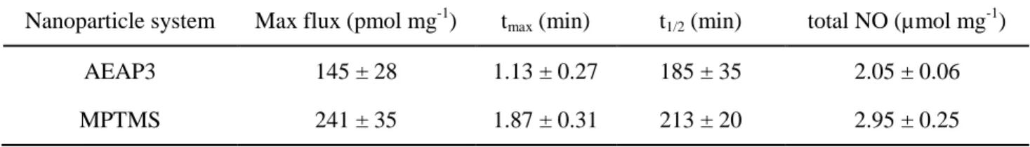
Nitric oxide release properties of silica nanoparticles at
pH 7.4 and 37 °C. Data are mean ± standard deviation
62
Table 3.2.
Water uptake of polyurethane topcoats. Data are mean
± standard deviation
70
Table 3.3.
Nitric oxide release from coatings doped at 18 mg/mL
PROLI/NO as a function of polyurethane topcoat. Data are
mean ± standard deviation
71
Table 3.4.
Nitric oxide release from coatings doped at 18 mg/mL AEAP3
nanoparticles as a function of polyurethane topcoat. Data are mean
± standard deviation
72
Table 3.5.
Nitric oxide release from coatings doped at 36 mg/mL AEAP3
nanoparticles as a function of polyurethane topcoat. Data are mean
± standard deviation
73
Table 3.6.
Nitric oxide release from coatings doped at 18 mg/mL or 36
mg/mL MPTMS nanoparticles with a HPU/TPU polyurethane
topcoat. Data are mean ± standard deviation
75
Table 4.1.
Nitric oxide-release properties of bare and PVC-coated 40%
MPTMS/MTMOS xerogels. Data are ± standard deviation
100
Table 4.2.
Linear regression analysis and NO flux required to inhibit
adhesion of each bacteria strain by 50 and 80% relative to
control (i.e., non-NO-releasing) surfaces. Data are mean ±
standard deviation
103
Table 4.3.
Nitric oxide payloads at 6, 12, and 24 h after the 1 h adhesion
for the eight initial average NO fluxes examined. Data are
mean ± standard deviation
106
Table 4.4.
Relative viability (%) of bacteria adhered to NO-releasing surfaces
after 6 h incubation in bacteriostatic conditions. A relative viability
of 100% represents the viability of bacteria adhered at the initial
x
Table 4.5.
Relative viability (%) of bacteria adhered to NO-releasing surfaces
after 12 h incubation under bacteriostatic conditions. A relative
viability of 100% represents the viability of bacteria adhered at the
initial average NO flux at t = 0. Data are mean ± standard error of
the mean
110
Table 4.6.
Relative viability (%) of bacteria adhered to NO-releasing surfaces
after 24 h incubation under bacteriostatic conditions. A relative
viability of 100% represents the viability of bacteria adhered at the
initial average NO flux at t = 0. Data are mean ± standard error of
the mean
111
Table 4.7.
The necessary total surface-derived NO release to decrease adhered
bacteria viability by 50 and 80% after 24 h incubation under
xi
LIST OF FIGURES
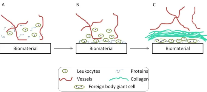
Figure 1.1.
The progression of the foreign body response (FBR) with time.
(A) Initially the biomaterial is implanted into injured native tissue
composed of blood vessels, proteins, and cells. (B) Proteins and cells
adhere to the surface and the adhered cells release chemoattractants
and cytokines to direct the FBR. (C) At 1–2 weeks, macrophages have
fused into foreign body giant cells (FBGCs) and leukocytes have
deposited an organized collagen encapsulation sequestering the material
from the native tissue and blood vessels
3
Figure 1.2.
Two major nitric oxide donors (i.e.,
N
-diazeniumdiolates and
S
-nitrosothiols) and their primary decomposition mechanisms
14
Figure 2.1.
Flow rate-dependent recovery of glucose in vitro to PAES
Microdialysis probes
42
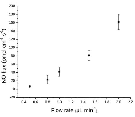
Figure 2.2.
Flow rate-dependent NO flux from microdialysis probes. The
relationship is not linear due to incomplete diffusion through the probe
membrane and leakage through polyurethane microdialysis tubing
43
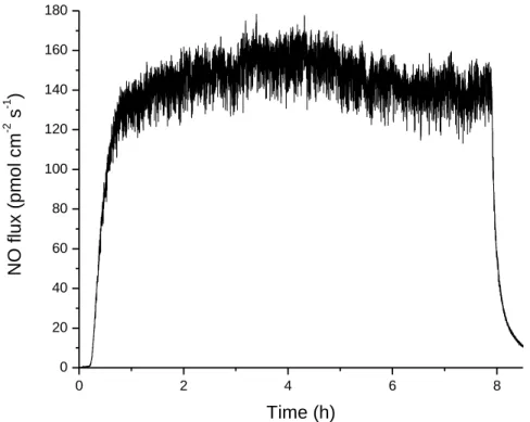
Figure 2.3.
Representative daily NO release from a microdialysis probe over
8 h while flowing PBS-NO at 2.0 µL/min
45
Figure 2.4.
Glucose recovery at various times of implantation for the NO-releasing
(filled, red) and control (empty, black) microdialysis probes. Error bars
are ± standard error of the mean. Significant differences (*) are p<0.05
47
Figure 2.5.
Representative histology slides of cross sections stained with
Masson’s trichrome (A and C) or hematoxylin and eosin (B and D)
of NO-releasing (A and B) and control (C and D) microdialysis probes
explanted at 14 days. Arrows in the hematoxylin and eosin-stained
pictures indicate the probe membrane. Arrows in the Masson’s
trichrome-stainted pictures indicate the implant site, surrounded by
dark-stained inflammatory cells and the collagen capsule. An increased
capsule size and inflammatory response at the membrane surface are
observed at control probes
50
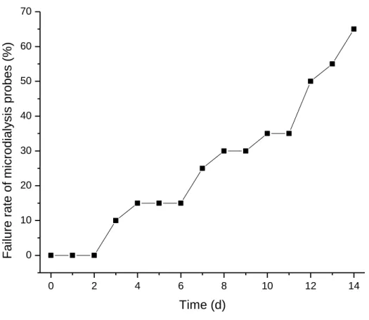
Figure 2.6.
In vivo failure rate of microdialysis probes. All probes remained
functional for 2 d post-implant, but began to fail thereafter. Over
the course of the in vivo study (i.e., 14 d), only 35% of probes
xii
Figure 3.1.
Scanning electron microscope images of polyurethane-coated wire
substrates A) dipcoated four times in 36 mg/mL MPTMS nanoparticles
in 80 mg/mL HPU/TPU before topcoating and B) after topcoating with
a 40 mg/mL HPU/TPU solution. Nanoparticle-induced surface
roughness is masked after topcoating
67
Figure 3.2.
Cumulative leaching from HPU/TPU topcoated 36 mg/mL AEAP3
nanoparticle system over 6 weeks as quantified by ICP-OES. More
than 90% of the total leaching occurs over the first 3 weeks. Over
the maximum length of the in vivo study (i.e., 6 weeks), 4.2% of the
AEAP3 nanoparticles may leach from the polyurethane
68
Figure 3.3.
Collagen capsule thickness surrounding polyurethane-coated wire
substrates at A) 3 and B) 6 weeks. Significant differences between
NO-releasing and relative controls are indicated at p < 0.05 (*).
At 6 weeks, the TP-470 topcoated 18 mg/mL AEAP3 nanoparticle
system was not tested due to low sample size (n = 2). Data are
mean ± standard error of the mean
77
Figure 3.4.
Collagen density index (CDI) of collagen capsules surrounding
polyurethane-coated wire substrates at A) 3 and B) 6 weeks. Significant
differences between NO-releasing and relative controls are indicated at
p < 0.05 (*). At 6 weeks, the TP-470 topcoated 18 mg/mL AEAP3
nanoparticle system was not tested due to low sample size (n = 2).
Data are mean ± standard error of the mean
81
Figure 3.5.
Inflammatory response to polyurethane-coated wire substrates
at A) 3 d and B) 1, C) 3, and D) 6 weeks. Significant differences
between NO-releasing and relative controls are indicated at
p < 0.05 (*). At 6 weeks, the TP-470 topcoated 18 mg/mL
AEAP3 nanoparticle system was not tested due to low
sample size (n = 2). Data are mean ± standard error of the mean
83
Figure 4.1.
The NO flux-dependent relative adhesion of A)
S. aureus
,
B) MRSA, C)
S. epidermidis
, D)
E. faecalis
, E)
E. coli
, and
F)
P. aeruginosa
to Fg-adsorbed PVC-coated xerogels. A relative
adhesion of 100% represents the adhesion of the strain to
control (i.e., non-NO-releasing) substrates. Data are
xiii
LIST OF EQUATIONS
Equation 1.1. The Bungay-Morrision-Dedrick equation relating extraction efficiency
(EE) to flow rate (Qd) and resistance to mass transfer (R)
16
Equation 2.1. The mathematical definition of extraction efficiency (EE).
The EE of a given substance is defined as the relationship of the
concentrations of the molecule in the dialysate (
C
d), perfusate (
C
p),
and external medium (
C
e). The EE can also be related to the flow
rate (
Q
d) and the resistances to mass transfer in the membrane (
R
m),
dialysate (
R
d), biofouling layer (
R
bf), collagen encapsulation (R
ec),
and tissue trauma (
R
tr)
35
Equation 4.1. Calculation for relative viability of bacteria adhered at t = x (x = 6,
12, or 24 h) to a given NO-releasing material ([NO] = 0.5, 1.0, 2.5,
xiv
LIST OF ABBREVIATIONS AND SYMBOLS
~
approximately
°C
degree(s) Celsius
µg
microgram(s)
µL
microliter(s)
µm
micrometer(s)
µM
micromolar
%
percent
λ
wavelength
σ
standard deviation
ε
molar absorptivity
AEAP3
N
-(2-aminoethyl)-3-aminopropyltrimethoxysilane
AFM
atomic force microscopy
Ag
+silver ion
AgNO
3silver nitrate
AHAP3
N
-(6-aminohexyl)aminopropyltrimethoxysilane
AHAP3/NO
N
-diazeniumdiolate-modified AHAP3
Ar
argon gas
ATCC
American Type Culture Collection
atm
atmosphere(s)
bFGF
basic fibroblast growth factor
BSA
bovine serum albumin
BTMOS
isobutyltrimethoxysilane
C. albicans
Candida albicans
xv
CaCl
2calcium chloride
CDI
collagen density index
CFU
colony forming units
cGMP
cyclic guanidine phosphate
cm
centimeter(s)
CO
2carbon dioxide
CVC
central venous catheter
Cys
L-cysteine
CysNO
S
-nitrosocysteine
d
day(s)
DBHD
N
,
N
’-dibutyl-1,6-hexanediamine
DBHD/N2O2
N
-diazeniumdiolate-modified DBHD
DI
deionized
DNA
deoxyribonucleic acid
DTPA
diethylenetriamine pentaacetic acid
E. coli
Escherichia coli
E. faecalis
Enterococcus faecalis
e.g.
for example
EDRF
endothelium-derived relaxation factor
EE
extraction efficiency
EE%
extraction efficiency percentage
ELISA
enzyme-linked immunosorbent assay
eNOS
endothelial nitric oxide synthase
et al.
and others
xvi
FBGC
foreign body giant cell
FBR
foreign body response
FBS
fetal bovine serum
FDA
United States Food and Drug Administration
FEP
fluorinated ethylene propylene
Fg
fibrinogen
Fig.
Figure
h
hour(s)
H&E
hematoxylin and eosin
H
2O
water
HA
hyaluronic acid
HCl
hydrochloric acid
HP 93A
Tecophillic HP-93A-100
HPLC
high performance liquid chromatography
HPU
Hydrothane AL 25-80A
Hz
hertz
i.e.
that is
ICP-OES
inductively couple plasma optical emission spectroscopy
IgG
immunoglobulin G
IL-6
interleukin-6
IL-8
interleukin-8
IL-10
interleukin-10
iNOS
inducible nitric oxide synthase
KCl
potassium chloride
xvii
kDa
kilodalton(s)
kg
kilogram(s)
krpm
kilorevolutions per minute
m
meter(s)
M
molar
MΩ
megaohm(s)
MAHMA
N
,
N
’-dimethyl-1,6-hexanediamine
MAHMA/NO
N
-diazeniumdiolate-modified MAHMA
MBC
minimum bactericidal concentration
MCP-1
monocyte chemoattractant protein-1
MeOH
methanol
mg
milligram(s)
MIC
minimum inhibitory concentration
min
minute(s)
mL
milliliter(s)
mm
millimeter(s)
mM
millimolar
mmol
millimole(s)
mol
mole(s)
MPTMS
3-mercaptopropyltrimethoxysilane
MRSA
methicillin-resistant
Staphylococcus aureus
MTMOS
methyltrimethoxysilane
N
newton
N.A.
numerical aperature
xviii
NaCl
sodium chloride
NH
4OH
ammonium hydroxide
nm
nanometer(s)
nmol
nanomole(s)
nNOS
neuronal nitric oxide synthase
NO
nitric oxide
[NO]
maxmaximum NO flux
[NO]
tNO flux at time t
NOA
Nitric Oxide Analyzer
NOS
nitric oxide synthase
NSAID
non-steroidal anti-inflammatory drug
O
2oxygen gas
ONOO
-peroxynitrite
P. aeruginosa
Pseudomonas aeruginosa
P. mirabilis
Proteus mirabilis
P. vulgaris
Proteus vulgaris
PAES
polyarylethersulfone
PAN
polyacrylonitrile
PBS
phosphate buffered saline, pH 7.4
PC
polycarbonate
PDGF
platelet derived growth factor
PEEK
polyether ether ketone
PES
poly(ether sulfone)
pg
picogram(s)
xix
PLG
poly(lactide-
co
-glycolide)
PLGA
poly(lactide-
co
-glycolic acid)
pM
picomolar
PMMA
poly(methyl methacrylate)
pmol
picomole(s)
ppb
parts per billion
ppm
parts per million
PROLI/NO
N
-diazeniumdiolate-modified
L-proline
PRP
platelet-rich plasma
PU
polyurethane
PVC
poly(vinyl chloride)
Q
dFlow rate of dialysate
R
bfresistance to mass transfer in biofouling layer
R
dresistance to mass transfer in dialysate
R
ecresistance to mass transfer in encapsulation layer
R
mresistance to mass transfer in membrane
R
trresistance to mass transfer in tissue trauma layer
rev
revolution
s
second(s)
S. auerus
Staphylococcus aureus
S. epidermidis
Staphylococcus epidermidis
SdrG
Serine-aspartate repeat G
SEM
Scanning electron microscope/microscopy
SiR
silicone rubber
xx
t
time
t
maxtime to max nitric oxide flux
TEOS
tetraethoxysilane
THF
tetrahydrofuran
TMOS
tetramethoxysilane
TP-470
Tecoplast TP-470-000
TPU
Tecoflex SG-80A
TSA
tryptic soy agar
TSB
tryptic soy broth
U
unit(s)
U.S.
United States
UV
ultraviolet
UV-Vis
ultraviolet-visible spectroscopy
v/v
volume/volume
v%
percent by volume
VEGF
vascular endothelial growth factor
wt%
percent by weight
Chapter 1:
Opportunities in Nitric Oxide Release Materials: Towards More Biocompatible Glucose
Sensors
1.1. Difficulties with implementation of implantable glucose sensors
2
Though the benefits of an implantable glucose sensor are quite clear, CGM devices
are limited by deficiencies in their biocompatibility. Currently, CGM devices approved by
the FDA to work for 5–7 d and are currently intended only to assist a patient in
understanding their glucose level trends.
5These sensors are useful tools, but cannot replace
finger-prick methods for extended glucose monitoring and often require frequent calibration
(via finger pricks). Implantable glucose sensors are primarily limited by the host’s foreign
body response (FBR). Significant research has explored methods to increase the
biocompatibility of in vivo sensors to extend the analytical functionality and provide reliable
and accurate glucose levels.
61.2. Biocompatibility of implanted materials
Many biomedical implants are currently approved for implantation into the human
body. While these biomedical materials are routinely implemented, limitations in
functionality, safety, and lifetime persist due to the host tissue response, termed the foreign
body response (FBR). Even materials considered to be biocompatible elicit a weak FBR,
forming a thin, avascular encapsulation which prevents full integration of the implant into the
surrounding native tissue. Despite the implementation of aseptic procedures, infection
resulting from implanted biomaterials is also commonplace. An implant-associated infection
necessitates device removal with large risk for severe complications including amputation or
even death. Both isolation of glucose sensors through the FBR and bacterial colonization of
the implant site can lead to erratic sensor performance. Thus, significant research has focused
on methods to promote the development of native host tissue around an implant.
3
4
5
extent of capsule development is dependent on all other preceding components of the FBR,
including protein adhesion, cell activation, and cytokine signaling. The collagen
encapsulation will persist for the lifetime of the device, negatively impacting sensor
performance with respect to sensitivity and response (e.g., lag times).
While the individual effects of each step in the FBR on glucose sensor performance
have been postulated, actual outcomes are more difficult to determine. Researchers have long
sought to untangle the complexities that connect various events in the FBR with tissue
integration and glucose sensor performance, as this knowledge could lead to the development
of materials that address the specific tissue responses that most severely inhibit sensor
performance.
6
tissue over both short- (3 h) and long-term (8 d) implantation periods were typically 3 to 5
times greater than that caused by biofouling of the probes, regardless of implantation
period.
22The observed biofouling was found to have only relatively small direct effects on
the overall resistance and glucose extraction efficiency.
22It is important to note however that
biofouling of proteins and cells at the sensor-tissue interface will also affect the tissue
response to a biomaterial as will the composition of the polymers used to fabricate the
sensors and dialysis probes.
Following protein adhesion/biofouling, the FBR proceed with inflammatory cells
responding to the injury, initiating a more profound immune response to the device. Klueh et
al. reported on the effects of mast cells, regulators of inflammation.
23Both mast
cell-sufficient and -deficient mice were implanted with subcutaneous glucose sensors for 28 days.
During this period, glucose sensor performance in mast cell-sufficient mice was erratic with
temporary response loss occurring within the first 3 weeks. Sensor performance in mast
cell-deficient mice was markedly better with reliable sensor function throughout the 28-day
period.
23Histology samples from both experimental groups confirmed that the mast
cell-deficient mice exhibited reduced fibrosis and inflammation at the implantation site. To
further confirm the effect of mast cells on glucose sensor performance, 10
4–10
5mast cells
were injected at the implant site.
23While glucose sensor performance recovered soon after
injection (~15 min), the sensor response to glucose decreased after 1–2 days, further
indicating a link between mast cell action and erratic glucose sensor performance.
237
coworkers implanted non-porous polyvinyl alcohol (PVA) and stainless steel cages into the
subcutaneous tissue of rats.
24Upon careful explantation of the collagen capsules, the
diffusion of sodium fluorescein (376 g/mol) through the capsule was quantified and used to
model small analyte transport.
24Diffusion of the fluorescein through the explanted capsules
was ~50% that of normal (i.e., subcutaneous) tissue.
24Sensor lag times for native tissue was
estimated at ~20 min, but tripled when modeled with decreased diffusion due to a capsule.
24Interestingly, empirical data suggests that lag times range from 10–15 min in vivo, indicating
the integral function of angiogenesis in early granulation tissue. In subsequent experiments,
Dungel et al. examined the effects of encapsulation on sensitivity by evaluation the response
of glucose sensors inserted into polyvinyl alcohol sponges implanted in rats.
25The glucose
sensitivity in vivo peaked at day 7, but then decreased for the duration of the study.
25Glucose
sensitivity correlated well with the collagen encapsulation of the sponges with thicker
collagen resulting in greater sensitivity loss.
25Koschwanez et al. investigated the effects of
vascularity on glucose sensor performance in real-time by using an implanted optical window
over the sensor, microscopy, and laser Doppler flowmetry.
26The vessel length and perfusion
of the vasculature increased during the implantation period (i.e., 14 d). Despite such
increases in vasculature, the sensitivity of the sensor did not increase indicating that
angiogenesis is not the only factor in assessing the biocompatibility of CGM sensors.
268
conclusion is important for sensor design, the increases to mass transfer were not attributed
to any specific part of the FBR and could originate from collagen capsule thickness, blood
vessel density, or other unanticipated factors. To consider major tissue reactions individually,
Novak and coworkers used a mathematical model to examine FBR effects on glucose sensor
performance.
28Using previous histology data, five parameters (i.e., angiogenesis, cellular
glucose consumption, capsule thickness, capsule diffusion coefficient, and capsule porosity)
were used to design a mathematical model that mimicked glucose diffusion from capillaries
to a glucose sensor in order to simulate the effect on sensor lag time and attenuation.
28The
mathematical model treated vessels as sources of glucose and inflammatory cells as glucose
sinks, while the encapsulation properties acted to impede the diffusion of glucose.
28Changes
in cellular glucose uptake and the capsule diffusion coefficient of glucose had little effect on
simulated sensor performance.
28The model was ultimately used to conclude that collagen
capsule thickness, a common histological parameter, was the primary source of sensor lag
time with little impact on sensor response attenuation.
28The positive correlation between lag
time and capsule thickness supported the models described above.
24, 27, 28The two greatest
factors in reducing sensor attenuation were a low capsule density and high degree of
angiogenesis.
28While these are only mathematical models, the results do identify the
histological parameters of key interest when assessing the biocompatibility of materials for
glucose sensors.
9
which bacteria to colonize and proliferate.
30Current biomaterials do little to prevent
implant-associated infections. Furthermore, the administration of systemic antibiotics, the leading
treatment for infections, has lead to a dwindling efficacy and the emergence of
antibiotic-resistant bacterial strains. The development of an implant-associated infection necessitates
the removal of the device and can lead to more serious complications, including death. With
the rise in biomedical implant usage and antibiotic-resistant strains of bacteria, the number of
implant-associated infections will likely increase.
Implanted biomaterials provide conditions that promote bacterial colonization. Upon
implantation, either the native tissue integrates with the implanted material or bacteria adhere
to the surface and colonize.
31Bacteria are most likely to adhere and colonize during the 6 h
directly following implantation.
32This colonization can result in the formation of a persistent
biofilm which is difficult to fully eradicate. Killing of bacteria comprising a biofilm versus
their planktonic counterparts requires up to 1000x increased doses of antibiotics.
33, 34Early
killing and prevention of adhesion would decrease the susceptibility of implantable materials
to bacterial infections. Currently, many clinically approved materials do not sufficiently
prevent bacterial adhesion and biofilm formation on their surface.
351.3. Animal models for evaluating biocompatibility
When implanting in vivo, researchers often use rodent models (e.g., mouse
36or rat
37, 3810
Unfortunately, the degree to which animal models accurately predict the FBR and
analytical performance of glucose sensors in humans has come under scrutiny. For example,
Wisniewski et al. compared the dialysate concentrations of glucose, pyruvate, lactate,
glycerol, and urea at a microdialysis probe-tissue interface implanted in the subcutaneous
space of both rats and humans to evaluate the validity of the cross-species relationship.
44The
dialysate concentrations were significantly different for all metabolites in rats and humans
throughout the first 6 days of the experiment, with the exception of glycerol on day 0.
44Additionally, the ratio of the glucose concentration in the dialysate to that in blood were
significantly different between rats and humans indicating differences in the relative glucose
availability in the tissue surrounding an implanted device.
44Histology samplings from human
subcutaneous tissue showed a greater concentration of adipose cells while rat tissue was
characterized with larger amounts of collagen.
44Taken together, these results necessitate
careful consideration and caution when extrapolating results from animal models to humans.
11
characteristics of diabetic patients. Of note, many diabetic models are available including
both genetic and chemically-induced.
471.4. Nitric oxide
The implantation of a biosensor into a host raises concerns of a lack of
biocompatibility (i.e., collagen encapsulation and inflammation) and the potential to promote
bacterial colonization and subsequent infection. Thus, a strategy to address these primary
concerns has been the release of nitric oxide (NO) from implantable surfaces.
Since NO was identified as the endothelial derived relaxation factor (EDRF) in 1986,
there has been extensive research into the many functions of NO in the body.
48Nitric oxide is
an endogenously produced free radical synthesized from
L-arginine by one of three isoforms
of nitric oxide synthase (NOS): neuronal NOS (nNOS), endothelial NOS (eNOS), and
inducible NOS (iNOS).
49The nNOS isoform is present in many tissues outside the brain
including skeletal muscle and islet cells while eNOS is present almost exclusively in the
endothelium.
50These two isoforms create low concentrations (i.e., nM) of NO. Inducible
NOS can be expressed in any cell when exposed to certain cytokines or lipopolysaccharide
(LPS) and is often expressed by macrophages in response to bacterial infection or
inflammation to synthesize µM concentrations of NO.
5012
avoiding the typical avascular encapsulation that plagues successful glucose sensor
performance. Additionally, NO is believed to control inflammatory cell recruitment in the
early stages of the FBR. Localized NO may cause nitrosation of proteins and down-regulate
pro-inflammatory cytokine expression (e.g., macrophage chemoattractant protein-1).
53Furthermore, NO has been reported to reduce leukocyte adhesion at elevated concentrations
and could thereby prevent localization of inflammatory cells at an implanted glucose
sensor.
54The reduction of leukocytes at the implant aids in the reduction of inflammation and
subsequent FBR at the wound site.
1.4.2. Antibacterial properties.
Macrophages synthesize and release endogenous NO
at concentrations >1 µM in response to bacterial infections.
55, 56Thus, significant research
efforts are focused on harnessing the antimicrobial properties of NO. As a free radical, NO is
very reactive and can cause damage to cells both directly and indirectly through resulting
byproducts (e.g., N
2O
3). At the exterior of a bacterial cell, NO may react with endogenous
superoxide to produce peroxynitrite, which is capable of damaging the cell membrane
through lipid peroxidation.
57Observations via atomic force microscopy confirm that NO
release compromises the bacteria membrane.
58Inside the cell, NO can nitrosate proteins and
cause strand breaks or base changes in DNA.
59Unlike many antibiotics, NO acts through
multiple killing mechanisms which not only increases the probability of bacterial cell death
but also significantly decreases the probability of bacterial resistance evolving.
6013
upon some trigger.
61, 62The most commonly studied donors for implant-derived in vivo NO
delivery are
N
-diazeniumdiolates and
S
-nitrosothiols (Figure 1.2).
N
-diazeniumdiolates are
formed on secondary amines upon exposure to NO in basic conditions.
63In the presence of a
proton source (e.g., water), the
N
-diazeniumdiolate decomposes to release two molecules of
NO, regenerating the parent amine in the process.
63The rate of release from these moieties is
therefore dependent on the pH, ionic strength, and surrounding chemical environment.
63, 64S
-nitrosothiols are formed on thiols exposed to nitrosating conditions (e.g., acidified nitrite)
and degrade when exposed to light, copper (I), or heat, releasing one NO molecule per
thiol.
65Similar to
N
-diazeniumdiolates, the NO release kinetics from
S
-nitrosothiols are
dependent on the NO donor chemical structure. The utility of NO donors for improving
biocompatibility was initially investigated by incorporating low molecular weight (LMW)
NO donors of
N
-diazeniumdiolates (e.g.,
N
-diazeniumdiolated
L-proline) and
S
-nitrosothiols
(e.g.,
S
-nitrosoglutathione) into biomaterials to impart NO release capabilities.
66-68Limitations of LMW NO donors include short release kinetics and cytotoxicity. More
recently, there have been efforts to synthesize novel NO donors to both alter NO release
kinetics and reduce mammalian cell cytotoxicity. To this end, macromolecular scaffolds have
been designed including silica nanoparticles
69-71and silica xerogels.
72-79Nanoparticles and
xerogels materials can be doped into polymer scaffolds or used as coatings, respectively,
permitting NO release from any implanted material. These materials can be used to evaluate
the effects of NO release in vivo.
1.5. Evaluating the host response via microdialysis
14
15
medium occurs and is collected as a dialysate. The extraction efficiency (EE) is obtained by
comparing the concentrations of a given analyte in the perfusate (C
p), dialysate (C
d), and
external medium (C
e) (Equation 1.1). The EE can be further related through the
Bungay-Morrison-Dedrick equation to resistances to mass transfer of the dialysate (R
d), membrane
(R
m), and external medium (R
e), as well as the flow rate of the perfusate (Q
d) (Equation
1.1).
80, 81When implemented in vivo, resistances in the external medium may be the result of
biofouling or other tissue effects (e.g., collagen encapsulation). Therefore, evaluating the EE
over time offers a method to determine the biocompatibility of the probe material. This
method is particularly useful for investigating glucose sensors as the resistances to mass
transfer also offers insight into the effective lag time of a sensor. While microdialysis allows
for simple temporal testing of materials, the materials tested are limited to semi-permeable
membranes.
16
17
EE of perfused vitamin B
12and recovered glucose by the microdialysis probes after 12 d
were reduced by 71.4 and 71.1%, respectively.
Researchers have attempted to improve glucose recovery of microdialysis probes by
implementing more biocompatible materials and releasing anti-inflammatory molecules.
Norton et al. investigated the EE of glucose in rat subcutaneous tissue using both bare
microdialysis probes and those with an overlaying hydrogel interface.
83The additional
diffusion of glucose through the hydrogels resulted in an increase in resistance to mass
transfer in vitro. When tested in vivo, the improved biocompatibility provided by the
hydrogel caused only non-significant improvements in tissue integration (reduction in
resistance to mass transfer of glucose) during the 8 d implantation. Later studies from Mou et
al. investigated the effect of actively releasing monocyte chemoattractant protein-1 (MCP-1;
a pro-inflammatory drug) and dexamethasone (DX; an anti-inflammatory drug) on the
recovery of glucose in rat subcutaneous tissue.
84The delivery of MCP-1 resulted in a
collagen capsule that was 2x the thickness of control probes after 10 d. In contrast, the
DX-releasing probes had very fragile collagen capsules due to the low degree of fibrosis in the
adjacent tissue and could not be processed for histological analysis. The increased FBR to the
MCP-1-releasing probes caused a significant decrease in glucose recovery versus controls 7 d
post-implant. However, even with the observed fragile collagen capsules adjacent to
DX-releasing probes, no significant differences in glucose recovery were observed over versus
controls over the 10 d experiment. However, the DX-releasing probes were functional 2 d
longer than control probes, indicating a potential benefit to the anti-inflammatory molecule.
18
improving the recovery of glucose in vivo. As NO release has been observed to improve
angiogenesis while decreasing collagen encapsulation and inflammation, delivery of the
molecule may be able to minimize the in vivo resistances to mass transfer of glucose caused
by the FBR.
1.6. Histological analysis of NO-releasing materials
Macromolecular NO donors have been implemented to investigate the potentially
positive tissue reactions to NO-releasing materials. Hetrick et al. coated 40%
N
-(6-aminohexyl)aminopropyltrimethoxysilane
(AHAP3)
balance
isobutyltrimethoxysilane
(BTMOS) xerogels onto square silicon rubber implants and chemically modified them with
N
-diazeniumdiolates.
85The substrates were capable of delivering a NO payload of 1.3 µmol
cm
-2over 3 d.
85The materials were then implanted into rat subcutaneous tissue for 1, 3, or 6
weeks and histology of the surrounding tissue was examined.
85The chronic collagen
encapsulation was reduced at 3 and 6 weeks by ~20–25% compared to non-NO-releasing
xerogels.
85In addition to the reduced capsule thickness, the chronic immune response (i.e., 3
and 6 weeks) was reduced while angiogenesis was enhanced at 1 and 3 weeks.
85Indeed,
these results indicate a possible long-term benefit for NO-releasing glucose sensors. A
mitigated immune response could result in reductions in local glucose consumption and
reactive oxygen species, while angiogenesis observed could significantly impact the
observed sensitivity of a glucose sensor and decrease lag time by reducing the distance from
glucose source to implant surface.
19
releasing NO for 18 h.
21The materials were implanted percutaneously into rats for 3 d.
21Subsequent histological analysis of the tissue revealed a decrease in inflammation at 24 h,
but not 48 h.
21The anti-inflammatory benefits correlated well with the NO release duration
indicating that the observed benefits may only persist for the duration of NO release, in
contrast to observations by Hetrick et al. which observed long-term reductions in
inflammation.
21, 85Such results demonstrate the potential of NO as a mediator of the FBR for
glucose sensors.
These studies each focused only on a single NO-releasing material for evaluating the
effect on the FBR. Therefore, the differences observed may not be optimal and the proper
release of NO from an implantable biomaterial could further enhance tissue integration.
Nitric oxide release properties (e.g., maximum NO flux, NO release duration, total NO
release) likely have a significant impact, but the role of each parameters is largely unknown.
Furthermore, these studies have concentrated on rodent models of the FBR, which while
useful, are not the best model of humans. Porcine models may better mimic the response in a
human and provide insight into the necessary NO release properties to improve tissue
integration.
861.7. Effects of exogenous NO on bacterial adhesion and viability
20
85%. Interestingly,
P. mirabilis
adhesion was also reduced by NO release, but not in a NO
flux-dependent manner. Indeed, all NO fluxes examined (ranging from ~10–32 pmol cm
-2s
-1) reduced adhesion of
P. mirabilis
by ~50% to PVC-coated xerogels.
89Charville et al.
examined the effects of NO release with fibrinogen biofouling to more closely mimic in vivo
conditions. Specifically,
S. aureus
was found to adhere ~5.3x more to poly(vinyl chloride)
(PVC) surfaces with adsorbed fibrinogen than PVC alone while
E. coli
and
S. epidermidis
exhibited modest enhancements (1.2–1.8x). Even while bacterial adhesion was promoted by
fibrinogen adsorption to the surface, NO release reduced bacterial adhesion of
S. aureus
and
E. coli
by 96 and 88%, respectively.
90NO-releasing xerogels exhibited a flux-dependent
reduction in bacterial adhesion. Interestingly,
S. epidermidis
was found to have greater
surface coverage even with relatively large NO fluxes (48% reduction at 30 pmol cm
-2s
-1)
and overall was less susceptible to NO-induced reductions in adhesion.
Under flow conditions, substrates that release ~21 pmol cm
-2s
-1have been shown to
reduce adhesion of
P.
aeruginosa
by 65% relative to controls.
73In the same study, Hetrick
and Schoenfisch examined the viability of adhered
P. aeruginosa
to varying payloads of NO
in static conditions (i.e., phosphate buffered saline). After 16 h of incubation, a NO payload
of 750 nmol cm
-2reduced viability of
P. aeruginosa
by 96% compared to substrates that
released only 25 nmol cm
-2, though the viability was not compared to substrates lacking NO
release. These results exemplified both the ability for NO to decrease bacterial adhesion and
a NO payload-dependent killing of bacteria.
21
CFU of either
S. aureus
and
P. aeruginosa
in 1.0 mL to a 1.8 cm
2surface area, the
NO-releasing films achieved close to complete killing after 12 h of exposure, corresponding to a
NO payload of ~40 nmol cm
-2. Recent work by Cai et al. examined the antibacterial and
anti-adhesive properties of a poly(lactic-co-glycolic acid) (PLGA) coating doped with the small
molecule NO donor DBHD/NO.
91A composition of 20% DBHD/NO was capable of
releasing NO for 15 d with NO fluxes from 0.6–3 pmol cm
-2s
-1at 7 d. The materials were
exposed to
S. aureus
or
E. coli
for 7 d to allow adhesion and growth of a biofilm onto the
surface. Confocal microscopy revealed that NO-releasing materials reduced adhesion of both
strains while also causing cell death of those that did adhere. The NO-releasing materials
were capable of reducing biomass of
S. aureus
and
E. coli
adhered to the substrates by
3-orders of magnitude.
22
maximum NO flux reported was ~1 pmol cm
-2s
-1. Studies reported by Holt et al.
93and Nablo
et al.
92proved successful inhibition of infection can be achieved with maximum NO fluxes of
20 and 295 pmol cm
-2s
-1, respectively.
While the works presented have exemplified NO as an active-releasing molecule to
reduce adhesion in vitro and subsequently infection in vivo, the amounts necessary are poorly
understood. The use of various materials, bacterial strains, and experimental conditions
prevents comparisons between the studies and convolutes the data. Additionally, only a few
studies have examined the viability of bacteria that adhere to NO-releasing surfaces.
1.8. Summary of dissertation research
My dissertation research has focused on the evaluation of the effects of
surface-derived nitric oxide release on the processes involved with the implantation of materials (i.e.,
mitigation of the FBR and inhibition of bacterial adhesion and viability) and the implications
of such effects, specifically related to implantable glucose sensors. My specific aims
included:
1) Quantifying the effect of nitric oxide delivery from microdialysis probes in rat
subcutaneous tissue on the in vivo resistances to mass transfer of glucose.
2) Evaluating the influence of both nitric oxide release kinetics and payloads on the
acute and chronic foreign body response in a porcine subcutaneous model as quantified
through collagen encapsulation and the inflammatory response.
3) Determining the necessary nitric oxide surface fluxes and payloads to reduce
bacterial adhesion and to induce cell death of adhered bacteria of a range of bacterial strains.
23
24
1.9 References
(1) Inzucchi, S. E., "Diagnosis of Diabetes." N. Engl. J. Med.2012,367, 542-550.
(2) Newman, J. D., Turner, A. P. F., "Home blood glucose biosensors: a commercial perspective." Biosens. Bioelectron.2005,20, 2435-2453.
(3) Heller, A., "Implanted Electrochemical Glucose Sensors for the Management of Diabetes."
Annu. Rev. Biomed. Eng.1999,1, 153-175.
(4) Wilson, G. S., Hu, Y. B., "Enzyme based biosensors for in vivo measurements." Chem. Rev.
2000,100, 2693-2704.
(5) Buckingham, B., Caswell, K., Wilson, D. M., "Real-time continuous glucose monitoring."
Curr. Opin. Endocrinol. Diabetes Obes.2007,14, 288-295.
(6) Koh, A., Nichols, S. P., Schoenfisch, M. H., "Glucose sensor membranes for mitigating the foreign body response." J. Diabetes Sci. Technol.2011,5, 1052-9.
(7) Anderson, J. M., "Biological responses to materials." Ann. Rev. Mater. Res.2001,31, 81-110.
(8) Anderson, J. M., Rodriguez, A., Chang, D. T., "Foreign body reaction to biomaterials."
Semin. Immunol.2008,20, 86-100.
(9) Wilson, C. J., Clegg, R. E., Leavesley, D. I., Pearcy, M. J., "Mediation of biomaterial-cell interactions by adsorbed proteins: A review." Tissue Eng.2005,11, 1-18.
(10) Luttikhuizen, D. T., Harmsen, M. C., Van Luyn, M. J. A., "Cellular and molecular dynamics in the foreign body reaction." Tissue Eng.2006,12, 1955-1970.
(11) Zhao, Q. H., McNally, A. K., Rubin, K. R., Renier, M., Wu, Y., Rosecaprara, V., Anderson, J. M., Hiltner, A., Urbanski, P., Stokes, K., "Human plasma alpha-2-macroglobulin promotes in vitro oxidative stress cracking of pellethane-2363-80A - In vivo and in vitro correlations."
J. Biomed. Mater. Res.1993,27, 379-389.
(12) Mosser, D. M., Edwards, J. P., "Exploring the full spectrum of macrophage activation." Nat.
Rev. Immunol.2008,8, 958-969.
25
surface-adherent macrophages and foreign body giant cells." J. Biomed. Mater. Res. Part A
2007,83A, 585-596.
(14) Zhao, Q., Topham, N., Anderson, J. M., Hiltner, A., Lodoen, G., Payet, C. R., "Foreign-body giant cells and polyurethane biostability: In vivo correlation of cell adhesion and surface cracking." J. Biomed. Mater. Res.1991,25, 177-183.
(15) Anderson, J. M., Defife, K., McNally, A., Collier, T., Jenney, C., "Monocyte, macrophage and foreign body giant cell interactions with molecularly engineered surfaces." J. Mater.
Sci.-Mater. Med.1999,10, 579-588.
(16) Kao, W. Y. J., Zhao, Q. H., Hiltner, A., Anderson, J. M., "Theoretical analysis of in vivo macrophage adhesion and foreign body giant cell formation on polydimethylsiloxane, low density polyethylene, and polyetherurethanes." J. Biomed. Mater. Res.1994,28, 73-79.
(17) Sieminski, A. L., Gooch, K. J., "Biomaterial-microvasculature interactions." Biomaterials
2000,21, 2233-2241.
(18) Thomé-Duret, V., Gangnerau, M. N., Zhang, Y., Wilson, G. S., Reach, G., "Modification of the sensitivity of glucose sensor implanted into subcutaneous tissue." Diabetes Metab.1996,
22, 174-178.
(19) Gerritsen, M., Jansen, J. A., Kros, A., Vriezema, D. M., Sommerdijk, N., Nolte, R. J. M., Lutterman, J. A., Van Hovell, S., Van der Gaag, A., "Influence of inflammatory cells and serum on the performance of implantable glucose sensors." J. Biomed. Mater. Res.2001,54, 69-75.
(20) Gifford, R., Kehoe, J. J., Barnes, S. L., Kornilayev, B. A., Alterman, M. A., Wilson, G. S., "Protein interactions with subcutaneously implanted biosensors." Biomaterials 2006, 27, 2587-2598.
(21) Gifford, R., Batchelor, M. M., Lee, Y., Gokulrangan, G., Meyerhoff, M. E., Wilson, G. S., "Mediation of in vivo glucose sensor inflammatory response via nitric oxide release." J.
Biomed. Mater. Res. Part A2005,75A, 755-766.
(22) Wisniewski, N., Klitzman, B., Miller, B., Reichert, W. M., "Decreased analyte transport through implanted membranes: Differentiation of biofouling from tissue effects." J. Biomed. Mater. Res.2001,57, 513-521.
26
(24) Sharkawy, A. A., Klitzman, B., Truskey, G. A., Reichert, W. M., "Engineering the tissue which encapsulates subcutaneous implants. I. Diffusion properties." J. Biomed. Mater. Res.
1997,37, 401-412.
(25) Dungel, P., Long, N., Yu, B., Moussy, Y., Moussy, F., "Study of the effects of tissue reactions on the function of implanted glucose sensors." J. Biomed. Mater. Res. Part A2008,
85A, 699-706.
(26) Koschwanez, H. E., Reichert, W. M., Klitzman, B., "Intravital microscopy evaluation of angiogenesis and its effects on glucose sensor performance." J. Biomed. Mater. Res. Part A
2010,93A, 1348-1357.
(27) Jablecki, M., Gough, D. A., "Simulations of the frequency response of implantable glucose sensors." Anal. Chem.2000,72, 1853-1859.
(28) Novak, M. T., Yuan, F., Reichert, W. M., "Modeling the relative impact of capsular tissue effects on implanted glucose sensor time lag and signal attenuation." Anal. Bioanal. Chem.
2010,398, 1695-1705.
(29) Leaper, D., "Healthcare associated infection: novel strategies and antimicrobial implants to prevent surgical site infection." Ann. R. Coll. Surg. Engl.2010,92, 453-458.
(30) Hetrick, E. M., Schoenfisch, M. H., "Reducing implant-related infections: active release strategies." Chem. Soc. Rev.2006,35, 780-789.
(31) Gristina, A. G., "Biomaterial-centered infection - Microbial adhesion versus tissue integration." Science1987,237, 1588-1595.
(32) Emmerson, M., "A microbiologist's view of factors contributing to infection." New Horiz.-Sci. Pract. Acute Med.1998,6, S3-S10.
(33) Ceri, H., Olson, M. E., Stremick, C., Read, R. R., Morck, D., Buret, A., "The Calgary Biofilm Device: New technology for rapid determination of antibiotic susceptibilities of bacterial biofilms." J. Clin. Microbiol.1999,37, 1771-1776.
(34) Williams, I., Venables, W. A., Lloyd, D., Paul, F., Critchley, I., "The effects of adherence to silicone surfaces on antibiotic susceptibility in Staphylococcus aureus." Microbiology-(UK)
1997,143, 2407-2413.
(35) Estivill, D., Arias, A., Torres-Lana, A., Carrillo-Munoz, A. J., Arevalo, M. P., "Biofilm formation by five species of Candida on three clinical materials." J. Microbiol. Methods
27
(36) Klueh, U., Kreutzer, D. L., "Murine Model of Implantable Glucose Sensors: A Novel Model for Glucose Sensor Development." Diabetes Technol. Ther.2005,7, 727-737.
(37) Mang, A., Pill, J., Gretz, N., Kränzlin, B., Buck, H., Schoemaker, M., Petrich, W., "Biocompatibility of an Electrochemical Sensor for Continuous Glucose Monitoring in Subcutaneous Tissue." Diabetes Technol. Ther.2005,7, 163-173.
(38) Koschwanez, H. E., Yap, F. Y., Klitzman, B., Reichert, W. M., "In vitro and in vivo characterization of porous poly-L-lactic acid coatings for subcutaneously implanted glucose sensors." J. Biomed. Mater. Res. Part A2008,87A, 792-807.
(39) Gough, D. A., Kumosa, L. S., Routh, T. L., Lin, J. T., Lucisano, J. Y., "Function of an Implanted Tissue Glucose Sensor for More than 1 Year in Animals." Sci. Transl. Med.2010,
2.
(40) Moussy, F., Harrison, D. J., Rajotte, R. V., "A miniaturized Nafion-based glucose sensor - In vitro and in vivo evaluations in dogs." Int. J. Artif. Organs1994,17, 88-94.
(41) Wagner, J. G., Schmidtke, D. W., Quinn, C. P., Fleming, T. F., Bernacky, B., Heller, A., "Continuous amperometric monitoring of glucose in a brittle diabetic chimpanzee with a miniature subcutaneous electrode." Proc. Natl. Acad. Sci. U. S. A.1998,95, 6379-6382.
(42) Valdes, T. I., Klueh, U., Kreutzer, D., Moussy, F., "Ex ova chick chorioallantoic membrane as a novel in vivo model for testing biosensors." J. Biomed. Mater. Res. Part A 2003, 67A, 215-223.
(43) Valdes, T. I., Kreutzer, D., Moussy, F., "The chick chorioallantoic membrane as a novel in vivo model for the testing of biomaterials." J. Biomed. Mater. Res.2002,62, 273-282.
(44) Wisniewski, N., Rajamand, N., Adamsson, U., Lins, P. E., Reichert, W. M., Klitzman, B., Ungerstedt, U., "Analyte flux through chronically implanted subcutaneous polyamide membranes differs in humans and rats." Am. J. Physiol.-Endocrinol. Metab. 2002, 282, E1316-E1323.
(45) Blakytny, R., Jude, E., "The molecular biology of chronic wounds and delayed healing in diabetes." Diabetic Med.2006,23, 594-608.
28
(47) Le, N. N., Rose, M. B., Levinson, H., Klitzman, B., "Implant healing in experimental animal models of diabetes." J. Diabetes Sci. Technol.2011,5, 605-618.
(48) Ignarro, L. J., "Nitric oxide: A unique endogenous signaling molecule in vascular biology (Nobel lecture)." Angew. Chem.-Int. Edit.1999,38, 1882-1892.
(49) Walford, G., Loscalzo, J., "Nitric oxide in vascular biology." J. Thromb. Haemost.2003, 1, 2112-2118.
(50) Griffith, O. W., Stuehr, D. J., "Nitric oxide synthases: Properties and catalytic mechanism."
Annu. Rev. Physiol.1995,57, 707-736.
(51) Cooke, J. P., "NO and angiogenesis." Atheroscler. Suppl.2003,4, 53-60.
(52) Dulak, J., Jozkowicz, A., "Regulation of vascular endothelial growth factor synthesis by nitric oxide: Facts and controversies." Antioxid. Redox Signal.2003,5, 123-132.
(53) Schwentker, A., Vodovotz, Y., Weller, R., Billiar, T. R., "Nitric oxide and wound repair: role of cytokines?" Nitric Oxide-Biol. Chem.2002,7, 1-10.
(54) Carreau, A., Kieda, C., Grillon, C., "Nitric oxide modulates the expression of endothelial cell adhesion molecules involved in angiogenesis and leukocyte recruitment." Exp. Cell Res.
2011,317, 29-41.
(55) Wink, D. A., Mitchell, J. B., "Chemical biology of nitric oxide: Insights into regulatory, cytotoxic, and cytoprotective mechanisms of nitric oxide." Free Radic. Biol. Med.1998, 25, 434-456.
(56) Mannick, J. B., "Immunoregulatory and antimicrobial effects of nitrogen oxides." Proc. Am. Thorac. Soc.2006,3, 161-165.
(57) Rubbo, H., Radi, R., Trujillo, M., Telleri, R., Kalyanaraman, B., Barnes, S., Kirk, M., Freeman, B. A., "Nitric oxide regulation of superoxide and peroxynitrite-dependent lipid peroxidation. Formation of novel nitrogen-containing oxidized lipid derivatives." J. Biol.
Chem.1994,269, 26066-26075.
(58) Deupree, S. M., Schoenfisch, M. H., "Morphological analysis of the antimicrobial action of nitric oxide on Gram-negative pathogens using atomic force microscopy." Acta Biomater.