MACROPHAGE PHOSPHOINOSITIDE 3-KINASE p110δ REGULATES INTESTINAL HOMEOSTASIS BY DIRECTING ADAPTIVE IMMUNITY AND
ENHANCING MICROBIAL CLEARANCE
ERIN CATHLEEN STEINBACH
A dissertation submitted to the faculty of the University of North Carolina at Chapel Hill in partial fulfillment of the requirements for the degree of Doctor of Philosophy in the
Department of Microbiology and Immunology.
Chapel Hill 2013
© 2013
ABSTRACT
ERIN CATHLEEN STEINBACH: Macrophage Phosphoinositide 3-kinase p110δ Regulates Intestinal Homeostasis by Directing Adaptive Immunity and Enhancing
Microbial Clearance
(Under the direction of Scott E. Plevy, MD)
The human inflammatory bowel diseases (IBDs), Crohn’s disease (CD) and ulcerative colitis, result from an inappropriately directed immune response to enteric microbiota in a genetically susceptible host. IBDs represent an increasing burden on the global health care system, as incidence is increasing and effective therapies remain elusive. Genome-wide association studies highlight the importance of host innate immune cell-microbial interactions in the pathogenesis of IBDs.
PI3K signaling regulates diverse functions, including cell growth, differentiation, proliferation and survival. The Class IA PI3K catalytic subunit p110δ negatively regulates
toll-like receptor signaling in innate immune cells. The importance of p110δ in intestinal homeostasis is shown in a mouse harboring a kinase-dead p110δ (p110δKD) that develops spontaneous Th1/Th17-skewed colitis. We describe a requirement for the enteric
microbiota to drive intestinal inflammation in p110δKD mice. Microbial-innate immune interactions maintain homeostasis through regulation of both protective (IL-10) and inflammatory (IL-12p40) cytokines, and p110δ is a central regulator of this balance. Additionally, p110δ positively regulates eradication of intracellular bacteria in
orchestrates innate immune cell regulation of pathogenic adaptive immune responses. Importantly, in human CD, decreased intestinal PIK3CD gene expression and an inverse correlation with intestinal IL12B:IL10 ratios are demonstrated. Thus, p110δ appears to be a central homeostatic switch in the intestine, governing the critical balance between IL-12/23 and IL-10 induced by the microbiota that determines the subsequent T cell
ACKNOWLEDGEMENTS
To my mentor, Dr. Scott Plevy, I extend heartfelt gratitude for guiding me through the harrowing journey of the PhD. Some parts were messy! But you never gave up on me. Your advice went beyond the science and taught me how to maneuver through the physician scientist’s career while maintaining the drive to help your patients – always your patients were kept at the forefront. For the unending guidance and support during my PhD education, I thank my thesis committee members: Drs. Balfour Sartor, Virginia Miller, Miriam Braunstein, William Goldman, and Christian Jobin who recently moved to another institution. Thank you for your wise advice at all stages of my dissertation. A special thank you to the National Institutes of Health for funding my education (F30 DK089692).
To Drs. Taku Kobyashi and Shehzad Sheikh, I owe you everything. You kept me on the right track day to day and were always willing (and loved!) to talk science. You were there for me during some of my lowest moments to pick me up, and you were always there to celebrate the highs. You are not only physician scientists I admire and hope to emulate someday, but you are dear friends. Thank you for investing so much in me – because you did, I know I will be successful in whatever the future brings.
To my colleagues who journeyed with me, thank you for your support and
MD/PhD program. I am really excited to follow each of your careers, which I know will be as amazing as each of you are. Here’s to OUR future!
To both my sets of parents, who I am incredibly lucky to have: thank you! Thank you, Mom and Dad Klein, for your unconditional love and support. I think you may have always known I was destined to be a lifelong student, but you have taken it in stride with a grace for which I am so grateful. Thank you for raising me to become who I am; it hasn’t always been easy, but where’s the fun in an easy ride? I love you more than I can express! Thank you, Mom and Dad Steinbach. You accepted me into your family without question and have been incredibly supportive, even when I dragged your only son to live far away in North Carolina. Thank you for making me feel like one of your own. Most importantly, thank you for Jeff. I suppose you know how special he is, but it’s because of your strong values and love that he is such a wonderful person.
TABLE OF CONTENTS
LIST OF FIGURES ... xiii
LIST OF ABBREVIATIONS ... xvi
CHAPTER 1 INTRODUCTION ... 1
1.1 Inflammatory Bowel Diseases ... 1
1.2 Macrophages and Dendritic Cells in Innate Immunity ... 3
1.2.1 Macrophages ... 3
1.2.2 Dendritic Cells ... 5
1.3 Recognition of Pathogen-associated Molecular Patterns ... 6
1.3.1 Toll-like Receptors (TLRs) ... 7
1.3.2 Nucleotide-binding Oligomerization Domain (NOD), Leucine-rich Repeat (LRR) Receptors (NLRs) ... 8
1.3.3 C-type Lectin Receptors (CLRs) ... 9
1.3.4 Retinoic Acid-inducible Gene-1 (RIG-I)-like Receptors (RLRs) ... 9
1.3.5 Functional Integration of Pathogen Recognition Receptor Signaling ... 10
1.4 Macrophage Intracellular Bactericidal Functions ... 11
1.5 Lamina Propria Mononuclear Cells in the Healthy Gastrointestinal Tract ... 13
1.5.1 Lamina Propria Mononuclear Cells ... 13
1.5.3 Lamina Propria Dendritic Cells ... 17
1.6 Lamina Propria Mononuclear Cells in IBD ... 20
1.6.1 Murine Experimental IBD ... 20
1.6.2 Human IBDs ... 27
1.7 Phosphoinositide 3-kinases in Immune Responses ... 29
1.7.1 Structure and Signaling Downstream of the Class IA PI3Ks ... 31
1.7.2 PI3K p110δ in Innate Immune Cells ... 32
1.7.3 PI3K p110δ in Adaptive Immune Cells ... 33
1.8 PI3K p110δ in Intestinal Homeostasis ... 34
1.9 Figures ... 37
CHAPTER 2 ALTERED MACROPHAGE FUNCTION CONTRIBUTES TO COLITIS IN MICE DEFECTIVE IN THE PHOSPHOINOSITIDE 3-KINASE SUBUNIT p110δ ... 38
2.1 Personal Contributions to Manuscript ... 38
2.2 Overview ... 38
2.3 Introduction ... 40
2.4 Results ... 41
2.4.1 PI3K p110δKD mice develop chronic colitis ... 41
2.4.2 PI3K p110δKD mice display an exaggerated mucosal and systemic Th1/Th17 cytokine profile ... 42
2.4.3 PI3K p110δKD macrophages are hyperresponsive to TLR signaling ... 43
2.4.4 PI3K p110δKD macrophages display enhanced MAP kinase activation ... 44
2.4.6 The enteric microbiota induces colonic PI3K p110δ
expression in WT but not in colitis-prone Il10-/- mice ... 45
2.4.7 Il10-/-/p110δKD mice exhibit severe colitis at an early age ... 46
2.5 Discussion ... 47
2.6 Materials and Methods ... 51
2.7 Figures ... 56
2.8 Supplemental Figures ... 64
CHAPTER 3 INNATE PI3K p110δ REGULATES TH1/TH17 DEVELOPMENT AND MICROBIOTA-DEPENDENT COLITIS ... 72
3.1 Overview ... 72
3.2 Introduction ... 73
3.3 Results ... 75
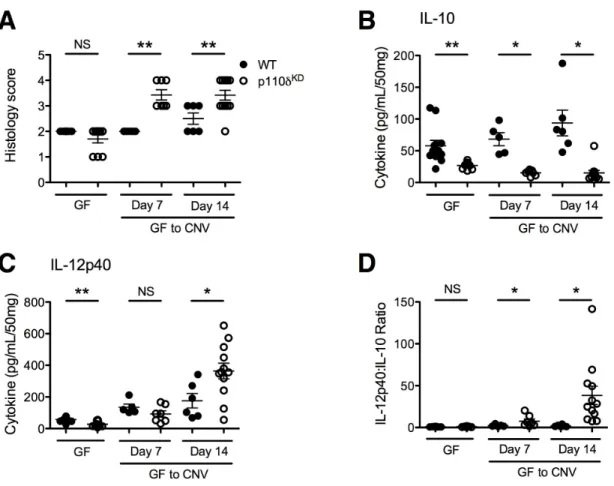
3.3.1 Presence of the enteric microbiota is necessary for the development of colitis in p110δKD mice ... 75
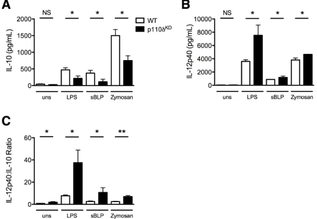
3.3.2 PI3K p110δ regulates macrophage production of IL-10 in response to TLR ligands ... 76
3.3.3 mTOR and GSK-3β act downstream of p110δ in macrophages to regulate cytokine production ... 78
3.3.4 Antigen presenting cell p110δ regulates T cell cytokine production ... 78
3.3.5 Intestinal PIK3CD expression correlates with IL12B:IL10 ratios from patients with CD ... 81
3.4 Discussion ... 81
3.5 Materials and Methods ... 86
3.6 Figures ... 92
3.7 Supplemental Figures ... 102
PI3K p110δ REGULATES BACTERICIDAL ACTIVITY OF
MACROPHAGES... 109
4.1 Overview ... 109
4.2 Introduction ... 109
4.3 Results ... 114
4.3.1 IFN-γ and E. coli-stimulated p110δKD macrophages demonstrate decreased lysosomal activation ... 114
4.3.2 PI3K p110δ regulates reactive oxygen species generation in macrophages ... 115
4.3.3 Phagosome maturation is regulated by p110δ ... 116
4.3.4 Outcome of in vivo infection with Streptomycin-resistant Salmonella is no different between WT and p110δKD mice ... 117
4.4.4 PI3K p110δKD mice demonstrate enteric microbiota dysbiosis ... 118
4.4 Discussion ... 119
4.5 Materials and Methods ... 124
4.6 Figures ... 129
CHAPTER 5 CONCLUSIONS AND FUTURE PERSPECTIVES ... 139
5.1 Overview ... 139
5.2 IBD Heterogeneity and p110δ ... 141
5.3 Development of Novel Tools to Study p110δ Function in Innate Immunity ... 143
5.4 PI3K p110δ Expression and Regulation in IBDs ... 144
5.5 The Enteric Microbiota, p110δ and IBDs ... 145
5.6 Macrophage Intracellular Microbe Eradication and p110δ ... 147
5.8 Conclusion ... 149
LIST OF FIGURES
Figure 1.1. Lamina propria mononuclear cells affect intestinal
homeostasis in health and disease ... 37
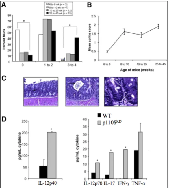
Figure 2.1. PI3K p110δKD mice develop colitis ... 56
Figure 2.2. PI3K p110δKD mice display enhanced expression of
IL-12p40 ... 57
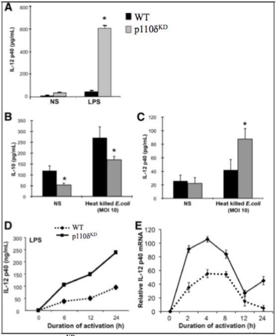
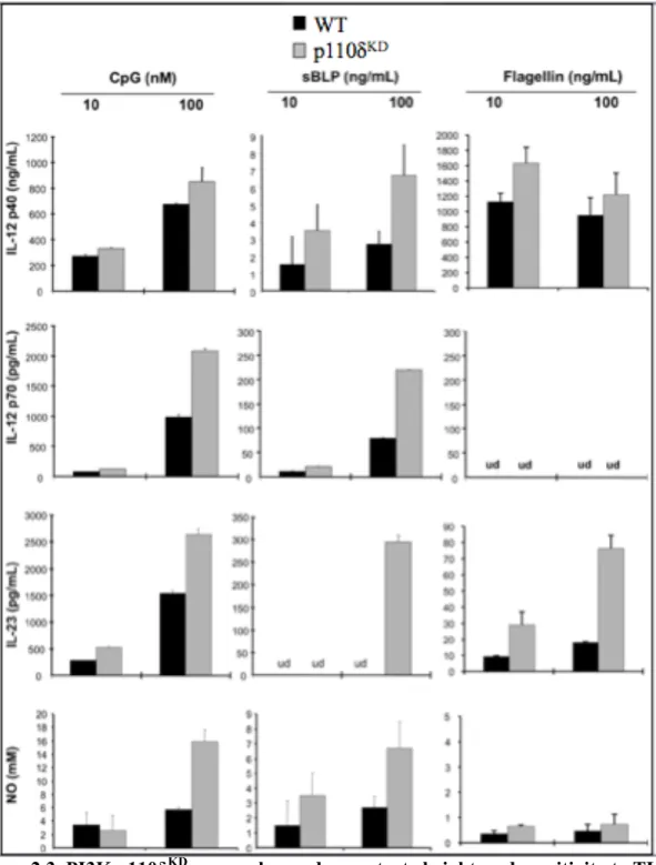
Figure 2.3. PI3K p110δKD macrophages demonstrate
heightened sensitivity to TLR stimulation ... 58
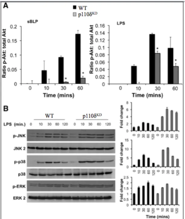
Figure 2.4. PI3K p110δKD macrophages demonstrate altered
kinetics and magnitude of MAPK activation ... 59
Figure 2.5. PI3K p110δKD BMDMs demonstrate defective
bactericidal activity ... 60
Figure 2.6. The enteric microbiota induces colonic p110δ
expression in WT but not colitis-prone Il10-/- mice ... 61
Figure 2.7. Il10-/-/p110δKD mice exhibit severe colitis at an early
age ... 63
Supplemental Figure 2.1. Increased CD3+ IELs in the colonic
crypts of p110δKD mice ... 64
Supplemental Figure 2.2. Colonic explants from p110δKD
secrete elevated chemokines ... 65
Supplemental Figure 2.3. PI3K p110δKD splenocytes secrete
elevated levels TNF-α ... 66
Supplemental Figure 2.4. WT and PI3K p110δKD BMDMs and LPMCs phenotypic and activation marker
expression ... 67
Supplemental Figure 2.5. Further characterization of signal
transduction pathways in p110δKD macrophages ... 68
Supplemental Figure 2.6. Bacterial uptake/phagocytosis is
intact in p110δKD BMDMs ... 69
Supplemental Figure 2.7. Il10-/-/p110δKD mice exhibit severe
Figure 3.1. The enteric microbiota is required for the
development of colitis in p110δKD mice ... 92
Figure 3.2. Defective p110δ activity alters macrophage production of IL-10 and IL-12p40 in response to
bacterial products ... 93
Figure 3.3. A p110δ-specific inhibitor decreases IL-10 and augments IL-12p40 production in WT macrophages
stimulated with bacterial products ... 94
Figure 3.4. IL-10 and IL-12p40 production in macrophages is
mTOR- and GSK-3β-dependent ... 95
Figure 3.5. APC p110δ regulates T cell differentiation ... 96
Figure 3.6. Mild innate mediated colitis develops in
Rag1-/-/p110δKDmice ... 98
Figure 3.7. Adoptive transfer of CD4+CD45RB T cells into
Rag1-/-/ p110δKD recipient mice leads to severe colitis ... 99
Figure 3.8. Human intestinal PIK3CD expression is decreased in patients with CD and inversely correlates with
IL12B:IL10 ratios ... 101
Supplemental Figure 3.1. GF to CNV p110δKD develop
histologic colitis ... 102
Supplemental Figure 3.2. Splenic macrophages and dendritic cells demonstrate increased Il-12p40 and impaired
IL-10 production in response to TLR agonists ... 103
Supplemental Figure 3.3. Inhibition of the Class IA PI3K
isoforms p110α and p110β in WT macrophages has
no effect on IL-10 and IL-12p40 production ... 104
Supplemental Figure 3.4. LPS stimulated splenic macrophages and DCs modulate IL-10 and IL-12p40 expression
in the presence of mTOR and GSK-3β inhibitors ... 105
Supplemental Figure 3.5. WT and p110δKD APCs induce the same amount of antigen-specific CD4+ T cell
proliferation ... 107
Supplemental Figure 3.6. Adoptively transferred T cells localize
Figure 4.1. IFN-γ-activated, E. coli-infected p110δKD BMDMs
demonstrate decreased phagosome acidification ... 129
Figure 4.2. Reactive oxygen species production is diminished in
p110δKD BMDMs ... 130
Figure 4.3. Inhibition of p110δ impairs reactive oxygen species
production in WT BMDMs ... 131
Figure 4.4. K12 E. coli-laden phagosome colocalization with FITC-dextran is decreased in IFN-γ-activated
p110δKD BMDMs ... 132
Figure 4.5. K12 E. coli-laden phagosome colocalization with EEA1 is decreased in IFN-γ-activated p110δKD
BMDMs after 10 minutes ... 133
Figure 4.6. K12 E. coli-laden phagosome colocalization with EEA1 is decreased in IFN-γ-activated p110δKD
BMDMs after 60 minutes ... 134
Figure 4.7. Streptomycin-resistant Salmonella typhimurium
colitis is not worsened in p110δKD mice ... 135
Figure 4.8. Salmonella CFUs recovered from cecal feces, cecal lamina propria and mesenteric lymph nodes of p110δKD mice was decreased compared to that from
WT mice ... 136
Figure 4.9. PI3K p110δKD mice demonstrate unique microbial
LIST OF ABBREVIATIONS
APC, antigen presenting cell
BMDC, bone marrow-derived dendritic cell BMDM, bone marrow-derived macrophage CBL, cecal bacterial lysate
CD, Crohn’s disease
cDC, conventional dendritic cell CFU, colony forming unit
CGD, chronic granulomatous disease CLR, C-type lectin receptor
CNV, conventionalized (murine housing) DC, dendritic cell
DSS, dextran sulfate sodium EEA1, early endosome antigen 1
ELISA, enzyme-linked immunosorbent assay FITC, fluorescein isothiocyanate
Flt3, Fms-related tyrosine kinase 3
Flt3L, Fms-related tyrosine kinase 3 ligand G-CSF, granulocyte colony-stimulating factor GF, germ free
GFP, green fluorescent protein
HKEC, heat-killed Escherichia coli
IBD, inflammatory bowel disease IDO, indoleamine 2,3-dioxygenase IEC, intestinal epithelial cell IEL, intraepithelial lymphocyte IFN, interferon
Ig, immunoglobulin IL, interleukin
IRF, interferon regulatory factor
ITAM, immunoreceptor tyrosine-based activation motif ITIM, immunoreceptor tyrosine-based inhibition motif LP, lamina propria
LPDC, lamina propria dendritic cell LPS, lipopolysaccharide
MAPK, mitogen-activated protein kinase MLN, mesenteric lymph node
MOI, multiplicity of infection
mTOR, mammalian target of rapamycin
MyD88, myeloid differentiation primary response gene 88 NFIL3, nuclear factor, interleukin-3 regulated
NF-κB, nuclear factor kappa-light-chain-enhancer of activated B cells
OVA, ovalbumin
p110δKD, mouse containing a knock-in of the Pik3cd locus with an inactivating point mutation in the kinase domain
PAMP, pathogen-associated molecular pattern pDC, plasmacytoid dendritic cell
PI3K, phosphoinositide 3-kinase
PIP or PI(3)P, phosphoinositide 3-monophosphate PIP2, phosphoinositide 4,5-bisphosphate
PIP3, phosphoinositide 3,4,5-triphosphate
PRR, pattern recognition receptor PUFA, polyunsaturated fatty acid RA, retinoic acid
RKO/δKD, Rag1-/- x p110δKD mouse RLR, RIG-I-like receptor
ROS, reactive oxygen species
RT-PCR, reverse transcriptase polymerase chain reaction sBLP, synthetic bacterial lipoprotein
SCFA, short chain fatty acid SEM, standard error of the mean SNP, single nucleotide polymorphism TGF-β, transforming growth factor β Th, CD4+ T helper cell
TIR, toll-interleukin 1 receptor
TLR, toll-like receptor
TNBS, 2,4,6-trinitrobenzenesulfonic acid TRAM, TRIF-related adaptor molecule Treg, regulatory T cell
TREM-1, triggering receptor expressed on myeloid cells 1 T-RFLP, terminal restriction fragment length polymorphism TRIF, TIR-domain-containing adapter-inducing interferon-β TSLP, thymic stromal lymphopoietin
CHAPTER 1
INTRODUCTION
1.1 Inflammatory Bowel Diseases
The two most common types of IBDs are CD and UC. CD is characterized by chronic, transmural inflammation occurring anywhere along the digestive tract but most commonly affecting the terminal ileum. The disease course tends to be one of relapse and remission, and complications such as stricture and fistula formation often develop. This is in contrast to UC, where ulcerous lesions are typically confined to the superficial layer of the colonic mucosa, extending proximally from the rectum. Histopathologically, UC lesions demonstrate crypt abscesses, goblet cell depletion, and significant infiltration of neutrophils. CD lesions often contain non-caseating granulomas and infiltration of macrophages (Xavier and Podolsky, 2007).
Despite recent advances in identifying IBD susceptibility loci using population-based genome-wide association studies (GWAS), the etiology of IBDs remains elusive. There is a complex interaction of host susceptibility, enteric microbiota, immune system responses and unspecified environmental contributions to IBD pathogenesis. Single nucleotide polymorphisms (SNPs) associated with increased risk of developing IBDs were identified in genes involving microbial sensing (NOD2, IRF5, NFKB1, RELA, REL,
RIPK2, CARD9, and PTPN22) and clearance (ATG16L1, IRGM, NCF4), and integrating antimicrobial adaptive immune responses (IL23R, IL10, IL12, IL18RAP/IL1R1,
Therefore, there is a real and pressing need to understand the pathways involved in IBD pathogenesis in order to develop safer and more effective therapies.
1.2 Macrophages and Dendritic Cells in Innate Immunity
1.2.1 Macrophages
Macrophages are a highly heterogeneous population of cells that demonstrate a continuum of activation states. The wide spectrum of macrophage phenotypes is often somewhat oversimplified into two functional groups: “inflammatory” M1 (high IL-12, low IL-10) and “wound healing” M2 (low IL-12, high IL-10) macrophages (Mosser and Edwards, 2008). Additionally, the recently appreciated subset of macrophages that produces high levels of IL-10 is referred to as “regulatory macrophages.”
to clear intracellular pathogens. While M1 macrophages are essential for the eradication of intracellular infections, they also produce pro-inflammatory cytokines implicated in IBD pathogenesis. Furthermore, unregulated M1 macrophage activity can induce tissue damage, predispose the host to developing neoplastic lesions, and induce insulin
resistance (Sica and Mantovani, 2012; Swann et al., 2008).
M2 macrophages are polarized by IL-4 produced by granulocytes and Th2 cells in response to tissue injury and activation by some fungi and parasites and initiation of SOCS2 signaling (Mosser and Edwards, 2008; Spence et al., 2013). M2 macrophages produce matrix metalloproteases, growth factors, and demonstrate efficient phagocytosis of debris without producing pro-inflammatory cytokines. Th2 responses are aimed at inducing wound healing and clearing parasites, although the exact mechanisms
underlying parasite eradication are unknown. Indeed, the downregulation of microbicidal functions in M2 macrophages can render the host more susceptible to certain infections (Bishop et al., 2008; Harris et al., 2007; Kropf et al., 2005; Muller et al., 2007; Shirey et al., 2008; Tumitan et al., 2007). M2 macrophages are also efficient at recruiting Foxp3+ T regulatory (Treg) cells, which would further downregulate local immune responses (Spence et al., 2013). Furthermore, unregulated M2 macrophage activity can promote the development of fibrotic lesions through elaboration of TGF-β and enhanced allergic responses (Fairweather and Cihakova, 2009; Murray et al., 2011).
macrophages, such as engagement of PRRs by PAMPs. Regulatory macrophages differ from M2 macrophages in that they do not produce extracellular matrix components but express high levels of co-stimulatory molecules (CD80, CD86) necessary for the activation of T cells. Like M2 macrophages, regulatory macrophages produce high amounts of the anti-inflammatory cytokine IL-10 and can render the host more
susceptible to certain infections (Agrawal and Pulendran, 2004; Baetselier et al., 2001; Benoit et al., 2008; Kim et al., 2008; Mahalingam and Lidbury, 2002; Miles et al., 2005; Ruas et al., 2009). Furthermore, unregulated regulatory macrophage activity may also play a role in the induction of neoplastic lesions (Biswas et al., 2006; Lin et al., 2006).
1.2.2 Dendritic Cells
Broadly speaking, dendritic cells (DCs) are professional antigen presenting cells (APCs) with the ability to initiate adaptive immune responses against potential
pathogens. Like macrophages, DCs comprise a heterogeneous population of cells with functional diversity. DCs originate from blood monocytes or a common DC progenitor (CDP) in the bone marrow at steady state. DCs repopulating tissues from monocyte precursors rely on granulocyte-macrophage colony stimulating factor (GM-CSF) for local proliferation (Rutella et al., 2004). Conventional DCs (cDCs) arising from the CDP express high levels of CD11c, varying levels of CD8α and CD11b, and reside in
specialized for antigen presentation. Thus, mature DCs express high levels of co-stimulatory molecules and tend to reside in secondary lymphoid organs where they are ideally positioned to prime antigen-specific T cells (Rescigno and Di Sabatino, 2009). On the other hand, immature DCs demonstrate low surface expression of co-stimulatory molecules and constitutively migrate in low numbers to lymph nodes, perhaps to
maintain tolerizing signals there (Huang et al., 2000; Rescigno and Di Sabatino, 2009).
1.3 Recognition of Pathogen-associated Molecular Patterns
Dysregulation of these pathways can lead to both enhanced susceptibility to infections and development of chronic inflammatory diseases (Kawai and Akira, 2011).
1.3.1 Toll-like Receptors (TLRs)
The best-characterized PRRs are the TLRs (Kawai and Akira, 2010). TLRs are transmembrane proteins with an extracellular LRR domain and an intracellular Toll-interleukin 1 receptor (TIR) domain. Upon binding its ligand to the LRR domain, homo- and heterodimerized TLRs recruit TIR domain-containing adaptor proteins. TLRs signal through one or both of two adapters: myeloid differentiation primary response gene 88 (MyD88) and/or TIR-domain-containing adapter-inducing interferon-β (TRIF) (Akira et al., 2006). TLR3 signaling is TRIF-dependent, whereas TLR4 can signal through both the MyD88- and TRIF-dependent pathways, and the remaining TLRs are MyD88-dependent (Akira et al., 2006). MyD88-dependent signaling activates transforming growth factor-β-activated kinase 1 (TAK1), a kinase of inhibitor of NF-κB (IκB) kinase (IKK). IKK phosphorylates IκB, leading to its degradation and subsequently releasing the
1.3.2 Nucleotide-binding Oligomerization Domain (NOD), Leucine-rich Repeat (LRR) Receptors (NLRs)
NLRs are cytosolic PRRs that can respond to a diverse array of stimuli, from bacteria to viruses and particulates involved in pathogenic states (e.g., monosodium urate crystals in gout and asbestos in mesothelioma and pneumoconiosis) (Kersse et al., 2011). NLRs contain a NACHT (present in NAIP, CIITA, HET-E, and TP-1) oligomerization domain, an LRR domain (except NLRP10 and NAIP), an N-terminal effector domain (except NLRX1), and an N-terminal homotypic protein-protein binding domain. Based on their N-terminal effector domain, the NLRs are divided into four subfamilies: NLRA, NRLB, NLRC, and NLRP. The sole member of NLRA is CIITA, which contains an acidic transactivation domain. NRLB proteins contain a baculovirus inhibitor repeat domain, NRLCs contain a caspase recruitment domain (CARD), and NRLPs contain a pyrin domain. Activation of NLRs can lead to several consequences, from inflammasome assembly and pro-IL-1β and pro-IL-18 processing (Bergsbaken et al., 2009), to caspase-1-dependent pyroptotic cell death (Schroder and Tschopp, 2010), and activation of NF-κB and MAPKs (Ting et al., 2010). Because the array of ligands that activate NLRs is so
diverse, a controversial theory is that NLRs actually respond to a more universal physiologic change within the cell, such as a decrease in intracellular potassium
concentration or an increase in extracellular calcium (Rajamaki et al., 2013; Rossol et al., 2012). Nevertheless, NLRs are important for the clearance of certain intracellular
1.3.3 C-type Lectin Receptors (CLRs)
CLRs are indispensable for immunity against fungal pathogens (Hardison and Brown, 2012). Activation of these transmembrane proteins containing a C-type lectin domain induces binding of the pathogen and phagocytosis, activation of antifungal programs, and production of cytokines and chemokines important in antifungal immunity. Dectin-1, Dectin-2, and Mincle signal through Syk to activate NF-κB, MAPKs, and nuclear factor of activated T cells (NFAT), inducing ROS generation and the production of cytokines and chemokines (Goodridge et al., 2007; Gross et al., 2006; LeibundGut-Landmann et al., 2007; Strasser et al., 2012). In contrast, dendritic cell-specific intercellular adhesion molecule-3-grabbing non-integrin (DC-SIGN) signals through Raf-1 to modulate NF-κB activity (Gringhuis et al., 2007). CLRs demonstrate extensive crosstalk with other PRRs, enhancing antifungal defenses. CLRs are especially important at mucosal surfaces where they induce Th17 responses (Iliev et al., 2012; Vautier et al., 2010).
1.3.4 Retinoic Acid-inducible Gene-1 (RIG-I)-like Receptors (RLRs)
multimerize and interact with the adaptor protein interferon-β promoter stimulator 1 (IPS-1) (Kawai et al., 2005; Meylan et al., 2005; Seth et al., 2005; Xu et al., 2005).
Recruitment of other proteins into the IPS-1 signalosome ultimately leads to type I interferon production through activation of IRF3, IRF7, and NF-κB (Paz et al., 2006). Like CLRs, RLRs demonstrate extensive crosstalk with other PRR signaling pathways.
1.3.5 Functional Integration of Pathogen Recognition Receptor Signaling
responses and how this relates to intestinal homeostasis and IBDs remains incompletely understood.
1.4 Macrophage Intracellular Bactericidal Functions
A central function of macrophages, literally translated as “big eater”, is to engulf and eradicate pathogens and other debris/stimuli. These functions are critical to
maintaining intestinal homeostasis, as demonstrated by IBD-associated SNPs identified in genes regulating bactericidal activity (ATG16L1, IRGM, NCF4) (Jostins et al., 2012). Macrophages can kill or limit the replication of microorganisms through many possible mechanisms. Macrophages can limit the availability of essential nutrients, as well as produce antimicrobial peptides, lysosomal enzymes, and ROS/RNS (Ismail et al., 2002). Oxygen-dependent metabolites are perhaps the most efficient anti-microbial effectors produced by macrophages. NADPH oxidases and associated accessory proteins are therefore essential components of cellular responses to microbial invasion. Hemoprotein complexes of the NADPH phagocyte membrane-bound gp91phox and p22phox subunits, along with the cytosolic p40phox, p47phox, p57phox and Rac proteins, can consume molecular oxygen to produce ROS/RNS (Rada et al., 2008; Robinson, 2008). The cytosolic components stabilize and activate phagocyte NADPH oxidase (Nox2). Compartmentalization of membrane-bound and cytosolic components of Nox2 ensures that the production of cytotoxic oxygen radicals is prevented in resting cells, thus avoiding “collateral damage”.
Another important bactericidal pathway in macrophages following the
the phagolysosome. The nascent phagosome goes through three stages of maturation: early, late, and lysosome-interacting (Fairn and Grinstein, 2012). Rab-family GTPases mediate the maturation process. Early phagosomes are marked by Rab5 decoration, which allows fusion with early endosomes via interactions with endosomal early antigen 1 (EEA1) (Christoforidis et al., 1999; Kinchen et al., 2008). Recruitment of maturation factors such as EEA1 is mediated by the generation of membrane-bound
phosphoinositide 3-phosphate (PIP) molecules by the Class III phosphoinositide 3-kinase (PI3K) vacuolar protein sorting 34 (Vps34) (Kinchen et al., 2008; Scott et al., 2002). GTPase activity on late phagosomes transitions from Rab5 to Rab7, which mediates intracellular trafficking and fusion with lysosomes (Harrison et al., 2003; Johansson et al., 2007). The resulting phagolysosome drives a critical drop in intravesicular pH by pumping H+ into the lumen via V-ATPase (Flannagan et al., 2009). The low pH of
phagolysosomes directly impairs microbe function, activates host hydrolytic enzymes and assists in the generation of superoxide by NADPH oxidase. Furthermore, the H+ gradient is used to pump essential microbial nutrients out of the phagolysosome.
vesicle termed the “autophagosome” (Levine and Deretic, 2007). The autophagosome then fuses with lysosomes to form autolysosomes where degradation of intravesicular contents occurs. Indeed, autophagy is central to intestinal homeostasis, as several IBD-associated SNPs in genes involved in autophagy have been identified (Jostins et al., 2012).
1.5 Lamina Propria Mononuclear Cells in the Healthy Gastrointestinal Tract
1.5.1 Lamina Propria Mononuclear Cells
The gut-associated lymphoid tissue (GALT) represents the largest aggregate of lymphoid tissue in the body. GALT includes various organized collections of immune cells within the gastrointestinal tract, such as Peyer’s patches in the small intestine and cryptopatches in the large intestine; and the diffuse arrangement of intestinal
mononuclear cells within the lamina propria (LP). The close proximity of lamina propria mononuclear cells (LPMCs) to the enteric luminal compartment, separated by an
encountering a danger signal. Thus, LPMCs are integral to directing appropriate immune responses and maintaining intestinal homeostasis in the gut.
There remains active debate about the classification and ontogeny of LP
macrophages and LP dendritic cells (LPDCs). The surface integrins CD11b and CD11c are routinely used to distinguish between macrophages and DCs in peripheral lymphoid tissues (CD11b+CD11c- and CD11b+/-CD11chigh are characterized as macrophages and DCs, respectively). However, the distinction between LP macrophages and LPDCs is less clear, as LP macrophages express both CD11b and CD11c (Mowat and Bain, 2011). It has been proposed that differential expression of CX3CR1 (the receptor for the
chemokine fractalkine, CX3CL1) and CD103 (αEβ7 integrin) reliably distinguish between
LP macrophages and LPDCs (Mowat and Bain, 2011; Schulz et al., 2009). CD103 -CX3CR1hi LP macrophages express the classical macrophage marker F4/80, demonstrate
ultrastructural characteristics of macrophages, and under physiologic conditions do not traffic to draining mesenteric lymph nodes (MLNs) where priming of adaptive immune responses is initiated. However, there is evidence that CX3CR1hi LP macrophages do
travel to MLNs during enteric microbial dysbiosis (Diehl et al., 2013). Conversely, CD103+CX3CR1lo LPDCs are F4/80- and perform functions typically associated with
DCs, including constitutive trafficking to MLNs, antigen presentation to T cells, and inducing gut homing receptors on T cells. Both LP macrophages and LPDCs express high levels of MHC class II, demonstrating their ability to interact with and shape adaptive immune responses. While controversy remains over the exact nature and origin of these LP subsets, for our purposes, we will classify LP macrophages as CD103-CX3CR1hi and
1.5.2 Lamina Propria Macrophages
LP macrophages are unique tissue resident macrophages characterized by the inability to produce inflammatory cytokines in response to microbial stimuli. However, these cells maintain robust phagocytic and microbicidal effector capabilities. The tolerant phenotype of LP macrophages is likely conditioned by locally produced IL-10 and TGF-β (Denning et al., 2007; Smythies et al., 2010). However, the ontogeny of these cells is
unknown. LP macrophage maintenance may depend on local proliferation rather than repopulation from migrating blood monocytes, but this is experimentally difficult to determine due to the extremely low turnover rate of these cells. Additionally, the context during which blood monocytes are recruited to the intestines may determine the final phenotype of the LP macrophages. During non-inflammatory homeostatic conditions, Ly6Chi monocytes almost exclusively repopulate the LP with CD11c+
(F4/80hiCX3CR1hiCD11b+CD103-) LP macrophages (Rivollier et al., 2012). In contrast,
under inflammatory conditions, Ly6Chi monocytes recruited to the LP differentiate into CD103+CX3CR1intCD11b+ DCs that produce high levels of the inflammatory cytokines
IL-12, IL23, iNOS, and TNF-α (Rivollier et al., 2012).
CX3CR1hi LP macrophages extend dendrites between IECs to sample luminal
al., 2005), recent work supports that they may represent a macrophage population (Medina-Contreras et al., 2011). IL-10 produced by LP macrophages promotes the persistence of Foxp3 expression in Treg cells in the intestine (Murai et al., 2009). Additionally, CX3CR1hi LP macrophages participate in the induction of systemic oral
tolerance (Hadis et al., 2011). It has been suggested that CX3CR1hi LP macrophages
sample luminal antigens and deliver them to CD103+ LPDCs, which are then able to traffic to MLN to prime adaptive immune responses (Ruane and Lavelle, 2011).
However, there is recent compelling evidence that CX3CR1hi LP macrophages traffic to
MLNs in a CCR7-dependent manner during dysbiosis of the enteric microbiota (Diehl et al., 2013).
Unique intracellular signaling pathways contribute to the inflammation anergic characteristic of LP macrophages; however, it remains unclear exactly what makes LP macrophages distinct from circulating monocytes and other tissue resident macrophages. Additionally, inflammation anergic LP macrophages are distinct from the more widely studied endotoxin-resistant macrophages. For one, LP macrophages do express PRRs, contrary to conventional thought. Recent studies suggest that the enteric microbiota are not necessary to program LP macrophages to express high amounts of the
IL-10-mediated suppression of IL-12p40 via altering histone acetylation and RNA
polymerase II accessibility to the Il12b promoter (Kobayashi et al., 2012), suggesting that IL-10 directly inhibits the production of pro-inflammatory cytokines in response to PAMP stimulation. IL-10 additionally exerts its anti-inflammatory effects on the innate immune system by regulating transcriptional elongation (Smallie et al., 2010), microRNA induction (McCoy et al., 2010), mRNA stability (Schaljo et al., 2009), and transcriptional repressors and co-repressors (El Kasmi et al., 2007).
Additionally, the PI3K pathway negatively regulates signaling through TLRs in macrophages (Fukao and Koyasu, 2003). In particular, the catalytic subunit of PI3K, p110δ, is enriched in leukocytes and regulates IL-12p40 production in LP macrophages in response to microbial stimulation. PI3K p110δ is indispensable for intestinal
homeostasis as mice harboring an inactivating point mutation in p110δ (p110δ kinase-dead, or p110δKD mice) develop spontaneous colonic inflammation (Uno et al., 2010). LP macrophages from p110δKD mice produce significantly more IL-12p40 and less IL-10 upon stimulation with heat-killed Escherichia coli (Uno et al., 2010). Thus, a loss in the critical negative regulation of TLR signaling results in the disruption of intestinal homeostasis.
1.5.3 Lamina Propria Dendritic Cells
pDCs in the LP are capable of inducing Treg cells and supporting their function (Bilsborough et al., 2003). While the majority of LPDCs are CD11b+CD8α-, CD11b -CD8α+ and CD11b-CD8α- subsets are also present. These DCs weakly stimulate antigen-specific T cell proliferation and constitutively express IL-10 and type I interferons (Chirdo et al., 2005). Furthermore, LPDCs are divided into CD103+ and CD103- (E-cadherin receptor) populations, each demonstrating distinct functions. CD103+ LPDCs are able to induce Foxp3-expressing Treg cells (Coombes et al., 2007; Mucida et al., 2007; Sun et al., 2007), whereas CD103- LPDCs are efficient at inducing Th17 cells when stimulated with flagellin or microbial ATP (Atarashi et al., 2008; Kinnebrew et al., 2012; Uematsu et al., 2008). While the Th17 response is important for antimicrobial immunity, dysregulation of Th17 lymphocytes and cytokines is implicated in a number of autoimmune disorders (Bettelli et al., 2007).
CD103+ LPDCs represent a population of tolerizing innate immune cells that express the enzyme retinaldehyde dehydrogenase (RALDH), which produces retinoic acid (RA) from retinaldehyde, and the important regulatory cytokine TGF-β. Both CD103+ LPDC-produced RA and TGF-β are necessary for the induction of Treg cells in the intestine (Coombes et al., 2007; Mucida et al., 2007; Sun et al., 2007). Additionally, CD103+ LPDCs produce indoleamine 2,3-dioxygenase (IDO), which participates in the induction of Treg cells and suppression of Th cell proliferation (Matteoli et al., 2010).
The induction of CD103 expression in LPDCs is dependent on the vitamin A metabolite retinoic acid (RA) and the local production of factors from IECs and stromal cells. IECs induce CD103 expression in LPDCs in an RA-, TGF-β-, and
constitutively produce prostaglandin E2, which inhibits the production of
pro-inflammatory cytokines in DCs (Newberry et al., 2001). Importantly, thymic stromal lymphopoietin (TSLP) produced by IECs conditions LPDCs to induce Th2 cell
differentiation (Iliev et al., 2009), although its necessity in inducing and maintaining Treg cells is controversial. Nonetheless, TSLP produced by IECs confers a homeostatic
phenotype on LPDCs to protect mice from colitis (Iliev et al., 2009; Liu et al., 2007; Rimoldi et al., 2005; Taylor et al., 2009). CD103+ LPDC differentiation is dependent on Notch2 signaling, as Notch2-/- mice demonstrate a selective loss of CD11b+CD103+ LPDCs (Lewis et al., 2011). Furthermore, the preferential expansion of CD103+ LPDCs depends on the DC differentiating molecule Fms-related tyrosine kinase-3 ligand (Flt3L) (Collins et al., 2012). The function of CD103+ LPDCs depends on several factors.
Dietary vitamin A induces RALDH expression in CD103+ LPDCs (Molenaar et al., 2011) and is necessary for these cells to imprint T cells with gut-homing receptors (Jaensson-Gyllenback et al., 2011; Wang et al., 2011b).
Aside from inducing Th17 differentiation, CD103- LPDCs are involved in the induction of immunoglobulin A (IgA) class switching of B cells, both in the Peyer’s patches and intestinal LP. IgA is abundantly produced in the intestine and prevents
bacterial overgrowth and adhesion to IECs in the intestinal lumen (Mantis et al., 2011). In the isolated lymphoid follicles of the LP, CD70+ LPDCs expressing TLR5 and any of various ATP receptors induce IgA class switching in RA-dependent and T
ligand (APRIL), IL-4, TGF-β, and IL-10, support the induction, maintenance, and expansion of IgA+ plasma cells (Bemark et al., 2012).
LPDCs have a higher turnover rate than LP macrophages due to frequent
trafficking to MLN to present antigen to naïve T cells (Rescigno and Di Sabatino, 2009). Evidence suggests that CD103+CD11b- LPDCs are replenished by DC-committed precursors (pre-cDC) in a Flt3L-dependent manner (Liu et al., 2009b), whereas CD103 -CD11b+ LPDCs are derived from circulating Ly6Chi monocytes in a GM-CSF-dependent manner (Varol et al., 2009). Additionally, the preferential expansion of regulatory
CD103+ LPDCs is also Flt3L-dependent (Collins et al., 2012). The conditions under which precursors are recruited to and the existing microenvironment of the LP likely determine the final phenotype of LPDCs. For instance, under steady-state conditions F4/80loCD103+CD11c+ LPDCs are repopulated from circulating Ly6Chi monocytes (Rivollier et al., 2012). Mice with experimental colitis and reconstituted with Ly6Chi monocytes demonstrated intestinal accumulation of inflammatory CD103
-CX3CR1intCD11b+ LPDCs and exacerbated colitis (Rivollier et al., 2012; Varol et al., 2009).
1.6 Lamina Propria Mononuclear Cells in IBD
1.6.1 Murine Experimental IBD
Jostins et al., 2012). Additionally, the selective depletion of macrophage and DC subsets in mouse models of colitis has been particularly informative about the protective and pathogenic roles innate immune cells play during discrete stages of disease pathogenesis. Lymphocyte deficient mice (severe combined immunodeficiency, SCID) develop colitis upon treatment with the intestinal irritant dextran sodium sulfate (DSS), suggesting that macrophages and DCs are pathogenic in this model in the absence of mature lymphocytes (Dieleman et al., 1994). Depletion of phagocytes in Il10-/- mice (Watanabe et al., 2003), and blocking myeloid cell recruitment in both 2,4,6-trinitrobenzene sulfonic acid (TNBS)-induced (Palmen et al., 1995) and T cell adoptive transfer (Kanai et al., 2006) colitis ameliorate disease, as does selective depletion of LPDCs during DSS colitis (Abe et al., 2007; Berndt et al., 2007). Contrary to these findings, depletion of LP macrophages and LPDCs prior to the induction of DSS colitis results in exacerbated disease (Qualls et al., 2006; Qualls et al., 2009). Furthermore, different subsets of macrophages and DCs have distinct effects on the severity of colitis in animal models. M2 polarized
macrophages protect mice from DSS colitis, whereas M1 polarized macrophages
contribute to disease pathogenesis (Arranz et al., 2012; Hunter et al., 2010; Weisser et al., 2011). Selective expansion of CD103+ LPDCs by Flt3L protects TNFΔARE mice from ileitis (Collins et al., 2012), but E-cadherin-expressing DCs increase colonic pathology in DSS colitis (Siddiqui et al., 2010). Thus, the protective/pathogenic role of distinct
chronic immune stimulation, and (3) by failing to switch from an appropriate pro-inflammatory response to an inflammation-resolving anti-pro-inflammatory response. Here we will discuss each of these defects and how each leads to chronic inflammation and IBDs.
The enteric microbiota is essential for the development of colonic inflammation in most murine models of colitis (Guarner, 2008; Sartor, 2008). Perturbations in the
negative regulation of innate immune responses to stimuli enhance susceptibility to colitis development. The well-characterized Il10-/- murine model of spontaneously developing colitis demonstrates the necessity of the potent anti-inflammatory cytokine IL-10 in the maintenance of intestinal homeostasis (Kuhn et al., 1993). Indeed, LP macrophages derived from germ free (GF) Il10-/- mice produce increased IL-12p40 compared to GF WT LP macrophages at baseline, suggesting that IL-10 is the critical driver of the LP macrophage phenotype (Kobayashi et al., 2012). Furthermore, IL-10 produced by CD11b+ LP macrophages is necessary for the maintenance of Foxp3 expression in Treg cells and protection from colitis (Murai et al., 2009). The important IL-10- and microbiota-inducible nuclear transcription factor, interleukin-3 regulated (NFIL3) negatively regulates IL-12p40 production in LP macrophages and has been recently implicated in intestinal homeostasis (Kobayashi et al., 2011). Thus, studying the regulation of IL-10 production and its downstream signaling effects is crucial to
understanding intestinal homeostasis.
macrophages, B cells, and neutrophils and contains several immunoreceptor tyrosine-based inhibitory motifs (ITIMs) that activate intracellular phosphatases, negatively regulating TLR signaling (Munitz et al., 2010). PIR-B is highly upregulated on LP macrophages following DSS administration in mice. Furthermore, PIR-B-deficient (Pirb -/-) macrophages produce significantly more TNF-α and IL-6 in response to Escherichia coli, and WT mice reconstituted with Pirb-/- macrophages demonstrate increased susceptibility to DSS colitis. PIR-B expression is also important in human intestinal biology, as LP mononuclear cells from both healthy controls and patients with UC express immunoglobulin-like transcript-2/leukocyte Ig-like receptor-1 (ILT-2/LIR-1), a human homologue of PIR-B. Our lab recently described spontaneous colitis development in mice harboring a kinase-dead PI3K catalytic subunit p110δ (p110δKD), a potent
negative regulator of TLR responses in macrophages (Uno et al., 2010). CD11b+ LPMCs from p110δKD mice produced increased pro-inflammatory cytokines (IL-12p40, IL-23) and decreased anti-inflammatory IL-10 in response to enteric microbes compared to CD11b+ LPMCs from WT mice. Conversely, triggering receptor expressed on myeloid cells-1 (TREM-1) amplifies TLR-induced inflammatory responses in macrophages, and blocking its activity attenuates murine colitis (Bouchon et al., 2000; Schenk et al., 2005). Indeed, resident LP macrophages do not express TREM-1 but abundant
The enteric microbiota interacts with host immune cells to induce protective anti-inflammatory responses and maintain intestinal homeostasis. Dysregulation of these protective pathways, either by enteric microbial dysbiosis or intrinsic defects in
macrophage and DC responses to stimuli, may underlie IBD pathogenesis. Short chain fatty acids (SCFAs) are anti-inflammatory metabolites produced by specific phyla of enteric bacteria (Bacteroidetes, Firmicutes) (Cavaglieri et al., 2003). When DSS colitis is induced in immune cell-specific Gpr43-/- mice (a host receptor for SCFAs), colonic inflammation is exacerbated, pointing to the beneficial anti-inflammatory effect of SCFAs in the colon (Maslowski et al., 2009). Interestingly, bacteria also actively suppress intestinal inflammatory responses, although a bacterium can exploit this to promote its pathogenicity. Citrobacter rodentium and Helicobacter pylori express bacterial proteins with domains similar to host ITIMs (Yan et al., 2012). ITIMs
nitrates are produced in abundance (Winter et al., 2013). This suggests that the interplay between host and bacteria actively shapes intestinal homeostasis and participates in IBD pathogenesis.
Both macrophages and DCs actively promote the transition from inflammation to the return to homeostasis after immune system activation, and non-resolving
inflammation is associated with many chronic diseases, including IBDs (Nathan and Ding, 2010). A study found that the pro-resolution mediator prostaglandin D2 was
upregulated only in UC patients who had achieved long-term remission, suggesting that intact pro-resolution pathways are necessary to halt damaging intestinal inflammation (Vong et al., 2010). Additionally, a SNP associated with low expression of the immune cell ectonucleotidase CD39, which generates the pro-resolving mediator adenosine, is associated with CD (Friedman et al., 2009). Immune cells are major contributors of extracellular adenosine at inflammatory sites. Adenosine interacts with its receptor A2B
on macrophages and DCs to inhibit pro-inflammatory cytokine production, expression of co-stimulatory molecules, and induction of T cell proliferation while increasing IL-10 production (Hasko et al., 2009).
gastrointestinal inflammation (Kromann and Green, 1980). PUFA-derived mediators enhance the capacity of macrophages and DCs to promote the resolution of inflammation by inducing efficient phagocytosis of apoptotic granulocytes and debris, preventing further recruitment of neutrophils, inducing anergy or deletion of effector T cells, and promoting repair of local damage (Uddin and Levy, 2011). Treatment with resolvin E1 ameliorates pathology in two experimental murine models of colitis, illustrating the powerful effects of PUFA-derived mediators on resolving inflammation (Arita et al., 2005; Ishida et al., 2010).
Macrophages and DCs additionally respond to resolving mediators by switching to unique “resolution phase” phenotypes. DCs generated in the presence of resolvin E1 demonstrate decreased expression of co-stimulation molecules, TNF-α, and IL-12, while inducing antigen-specific CD4+ T cell apoptosis via IDO production and activation (Vassiliou et al., 2008). A defining distinction of resolution phase DCs from tolerogenic DCs is the continued expression of CCR5, which enhances chemotaxis toward
inflammatory sites, without upregulation of CCR7, which induces chemotaxis to lymph nodes, on resolution phase DCs (Vassiliou et al., 2008). Similarly, resolution phase macrophages demonstrate a distinct phenotype from both M1 and M2 macrophages. Like M2 macrophages, resolution phase macrophages express high levels of molecules
inflammation, and that generation of these local factors or innate immune cell responses to these factors are defective in IBDs.
1.6.2 Human IBDs
In human IBDs, inflammatory lesions demonstrate an increase in accumulation of macrophages that display enhanced expression of co-stimulatory molecules (CD80, CD86) and macrophage activating receptors (CD40) (Rugtveit et al., 1997), TLRs
There is accumulating evidence that inappropriate macrophage and DC responses to the enteric microbiota contribute to human IBD pathogenesis (Xavier and Podolsky, 2007). These include both inadequate protective and enhanced pathogenic responses to such stimuli. Macrophages isolated from both CD and UC patients demonstrate altered cytokine production in response to bacterial challenge: CD macrophages produce more pro-inflammatory IL-23 but less of the protective cytokine IL-10, whereas UC
macrophages constitutively produce high levels of the pro-inflammatory cytokine IL-12 (Campos et al., 2011). This may be in part due to impaired regulation of TLR-induced inflammatory responses in macrophages. For instance, patients with IBDs demonstrate significantly decreased expression of intestinal NFIL3, an IL-10- and microbiota-induced transcriptional repressor of IL-12p40 expression, compared to tissue from healthy, non-inflamed control patients (Kobayashi et al., 2011). Additionally, increased numbers of TREM-1-expressing LP macrophages are found in intestinal tissue from patients with IBDs compared to tissue from control patients (Schenk et al., 2007). TREM-1 critically amplifies TLR-induced inflammatory responses of macrophages and is implicated in IBD pathogenesis. Conversely, LP macrophages from IBD patients produce less of the
cytokine G-CSF, which is protective in experimental models of colitis, in response to the probiotic Lactobacillus rhamnosus GR-1 compared to those from healthy controls (Martins et al., 2009).
polymorphisms associated with NOD2 and ATG16L1 encode proteins involved in the autophagy pathway and lead to defective bacterial clearance (Travassos et al., 2010). Macrophages isolated from patients with CD demonstrate decreased ROS production and impaired eradication of bacteria (Palmer et al., 2009; Smith et al., 2009). Additionally, peripheral blood monocytes isolated from patients with both CD and UC demonstrate decreased phagocytosis and killing of bacteria (Caradonna et al., 2000). Perhaps the most compelling evidence of the link between bacterial persistence and IBD is the long list of primary immunodeficiencies, such as chronic granulomatous disease (CGD), associated with IBD-like clinical manifestations (Diez et al., 2010; Ishii et al., 1987; Marks et al., 2009; Marks et al., 2010; Yamaguchi et al., 2001). Approximately 50% of patients with CGD, in which phagocyte ROS production and bacterial clearance are greatly impaired, develop IBD-like manifestations that share clinical and pathological features of CD (Marks et al., 2009; Segal et al., 2009). Furthermore, a SNP within the first intron of
NCF4 (p40phox) is associated with enhanced susceptibility to IBD (Rioux et al., 2007). Bacterial persistence and chronic stimulation of macrophages and DCs may contribute to IBD development by producing increased pro-inflammatory cytokines that shape
pathogenic adaptive immune responses. Indeed, defects in how macrophages and DCs respond to enteric antigens, eradicate bacteria and induce resolution of inflammation underlie IBD pathogenesis (See Figure 1.1 for summary of pathways and phenotypes).
1.7 Phosphoinositide 3-kinases in Immune Responses
PI3Ks are a group of kinases that regulate diverse cell functions, including
(Vanhaesebroeck et al., 2010). Indeed, dysregulation of PI3K signaling is implicated in various human diseases including diabetes and cancer (Falasca and Maffucci, 2012). PI3Ks initiate intracellular signaling cascades by phosphorylating the 3’-position of the inositol ring on select phosphoinositide molecules embedded in the cell membrane. The consequent phosphoinositide phosphate (PIP), either PI(3)P (e.g., PIP), PI(3,4)P (e.g., PIP2) or PI(3,4,5)P (e.g., PIP3), recruits proteins with recognition domains for PIP, PIP2,
or PIP3 to the cell membrane signaling scaffold.
The PI3K heterodimer consists of a catalytic and regulatory subunit; the regulatory subunit protects the catalytic subunit from degradation, prevents its promiscuous signaling and acts as a protein-binding scaffold (Vanhaesebroeck et al., 2010). PI3K catalytic subunit isoforms are grouped into three classes based on substrate specificity and structure: Class I, II and III. The most abundant phosphoinositide
generated by Class I PI3Ks is PIP3. Class I PI3Ks are further divided into Class IA and IB.
There are three members of the Class IA PI3Ks: p110α, p110β and p110δ. The Class IA
PI3Ks propagate signals downstream of receptor tyrosine kinases, such as TLRs and cytokine receptors. Class IA PI3Ks may also be activated downstream of G
protein-coupled receptors (GPCRs) by Ras (Vanhaesebroeck et al., 2010). The regulatory subunits of Class IA PI3Ks include p85α, p55α, p50α, p85β and p55γ. The sole member
of Class IB PI3Ks is p110γ and is associated with signaling downstream of GPCRs. PI3K
p110γ associates with p101 or p84/p87 regulatory subunits. Whereas p110α and p110β are ubiquitously expressed, p110δ and p110γ are highly enriched in leukocytes (Koyasu, 2003; Papakonstanti et al., 2008), suggesting dominant roles for these subunits in
PI3K-C2β and PI3K-C2γ and preferentially generate PIP. Class II PI3Ks are implicated in cell growth, survival, migration, membrane trafficking and insulin signaling (Falasca and Maffucci, 2012). The Class III PI3K, vacuolar protein sorting 34 (Vps34), associates with the Vps15 regulatory subunit to preferentially generate PIP. Vps34 has been associated with regulation of endocytosis, autophagy, and nutrient homeostasis (Backer, 2008).
1.7.1 Structure and Signaling Downstream of the Class IA PI3Ks
Class I PI3Ks contain a Ras binding domain (RBD), a C2 domain, a helical domain, and a catalytic domain (Vanhaesebroeck et al., 2001). Class IA PI3Ks
additionally have a p85-binding domain. Upon activation of receptor tyrosine kinases or GPCRs, Class IA PI3Ks are recruited to the activated receptor by either Ras or
phosphorylated tyrosine residues, which are recognized by Src homology 2 (SH2) domain of the regulatory p85α subunit (Vanhaesebroeck et al., 2010). Recruitment to the membrane releases p110 from inhibition by p85α, allowing generation of PIP3. Proteins
containing a pleckstrin homology (PH) domain, such as Akt and phosphoinositide-dependent kinase-1 (PDK1), recognize and are recruited to newly generated PIP3. PIP3 is
rapidly dephosphorylated by phosphatase and tensin homolog (PTEN), attenuating signaling (Vanhaesebroeck et al., 2010). Akt is a major signaling molecule activated downstream of Class I PI3K. In turn, Akt regulates multiple signaling pathways,
1.7.2 PI3K p110δ in Innate Immune Cells
The cell-type distribution of the Class IA PI3Ks p110δ and p110γ suggests that
these enzymes regulate immune responses. Indeed, the promoter of PIK3CD, the gene encoding p110δ, contains binding motifs for several immune-specific transcription factors (Kok et al., 2009). PI3K p110δ has emerged as an important negative regulator of TLR signaling in innate immune cells (Fukao and Koyasu, 2003). This was first shown in DCs, as p85α-deficient DCs produced significantly more IL-12p40 and IL-12p70 in response to TLR ligands compared to WT DCs (Fukao et al., 2002). The increased generation of IL-12p70 by DCs led to enhanced Th1 responses. Additionally, p110δ signaling in DCs induces IL-6 production, which limits Th1 responses (Krishnamoorthy et al., 2008). Recently, Aksoy et al., demonstrated p110δ regulates the transition from pro-inflammatory MyD88/TIR domain-containing adaptor protein (TIRAP) signaling downstream of TLRs to anti-inflammatory TRIF/TRIF-related adaptor molecule (TRAM) signaling (Aksoy et al., 2012). The switch to TRIF/TRAM signaling allows the potent anti-inflammatory cytokine IL-10 to be produced. In macrophages, the different Class IA
isoforms perform distinct, non-redundant functions, and p110δ is the dominant isoform responsible for Akt activation downstream of cytokine receptor activation (Papakonstanti et al., 2008). PI3K p110δ signaling in macrophages downstream of TLRs also negatively regulates IL-12p40, IL-12p70 and IL-23 through increased MAPK p38 and JNK
and migration of monocytes and macrophages (Mouchemore et al., 2013). Further
demonstrating a role for p110δ in regulating vesicular trafficking, macrophage trafficking and secretion of TNF-α requires p110δ signaling (Low et al., 2010).
PI3K p110δ also regulates functions of other innate immune cell populations. PI3K p110δ mediates rolling, adhesion, migration of and activation-induced cell morphology changes and cytokine production in eosinophils in circulation, thus
regulating allergic immune responses in vivo (Kang et al., 2012; Tanemura et al., 2009). Neutrophil cytokine production elicited by LPS or TNF-α stimulation and neutrophil migration into tissues is regulated by p110δ (Fortin et al., 2011; Randis et al., 2008). PI3K p110δ signaling in neutrophils recruits p40phox and p47phox, subunits of NADPH oxidase, to the membrane signaling complex, implicating p110δ activation in ROS production (Kanai et al., 2001). Interestingly, pharmacologic inhibition of p110δ in neutrophils and other cell types decreases production of ROS and the respiratory burst (Yamamori et al., 2004). Furthermore, p110δ negatively regulates LPS-induced IL-1β production, but enhances TNF-α and IL-6 production, in mast cells (Hochdorfer et al., 2011). In natural killer cells, p110δ positively regulates maturation as well as secretion of IFN-γ, TNF-α and GM-CSF (Kim et al., 2007).
1.7.3 PI3K p110δ in Adaptive Immune Cells
2007; Haylock-Jacobs et al., 2011; Jou et al., 2002; Okkenhaug et al., 2002; Reif et al., 2004). Furthermore, p110δ positively signals through the T cell receptor (TCR) and B cell receptor (BCR) to induce antigen-specific proliferation of T and B cells (Garcon et al., 2008; Jou et al., 2002; Okkenhaug et al., 2002; Okkenhaug et al., 2006; Ying et al., 2012). PI3K p110δ signaling is also necessary for CD4+CD25+Foxp3+ Treg function (Patton et al., 2006; Patton et al., 2011), antigen-specific cytokine production by both naïve and memory T cells (Liu and Uzonna, 2010; Okkenhaug et al., 2006; Soond et al., 2010), development and function of T follicular helper cells involved in induction of B cell responses (Rolf et al., 2010; So et al., 2013) and regulates antigen-specific T cell homing to inflamed tissues (Jarmin et al., 2008; Liu and Uzonna, 2010; Sinclair et al., 2008). Thus, whereas p110δ negatively regulates many responses to receptor tyrosine kinase activation in innate immune cells, p110δ positively regulates functions
downstream of the TCR and BCR in adaptive immune cells.
1.8 PI3K p110δ in Intestinal Homeostasis
Paradoxically, mice with a germline knock-in of Pik3cd harboring an inactivating point mutation (p110δ kinase-dead; hereafter referred to as “p11δKD”) demonstrate
et al., 2010). Intriguingly, a case study recently described a patient with homozygous germline loss of full-length p85α who lacked B cells and had colitis but did not
demonstrate other pathologic inflammatory processes (Conley et al., 2012). This patient demonstrated normal expression of immune cell p50α and p55α, but greatly reduced expression of p110δ. Both human and murine studies strongly implicate p110δ signaling in the maintenance of intestinal homeostasis. Contrary to prevailing paradigms where p110δ inhibition is a strategic approach in inflammatory diseases driven by adaptive immune defects (Durand et al., 2013; Haylock-Jacobs et al., 2011; Matteoli et al., 2010; Ying et al., 2012), blockade of p110δ in diseases where innate immune processes are central drivers of pathogenesis, such as IBDs, may actually be harmful.
Given the role of the PI3K p110δ subunit in innate immune processes
fundamental to the pathogenesis of IBD, we further characterized host-enteric microbiota and APC-T cell interactions in p110δKD mice. We describe a requirement for the enteric microbiota to drive intestinal inflammation in p110δKD mice. Microbial-innate immune interactions maintain homeostasis through regulation of both protective (IL-10) and inflammatory (IL-12p40) cytokines, and p110δ is a central regulator of this balance. Additionally, p110δ positively regulates eradication of intracellular bacteria in
1.9 Figures
Figure 1.1. Lamina propria mononuclear cells affect intestinal homeostasis in health and disease. LPMCs participate in maintaining intestinal homeostasis and in initiating disease when homeostasis is perturbed. [1] CD103-CX3CR1high LPMCs extend dendrites
across the IEC barrier to sample luminal bacteria and antigens. [2] IECs and stromal cells produce local factors that condition LPMCs to be tolerant. [3] LP macrophages
constitutively produce high levels of IL-10, which is necessary for the maintenance of Foxp3 expression in LP Treg cells. [4] CD103+CX
3CR1low LPDCs produce TGF-β and
retinoic acid (RA) to induce Treg cells and imprint gut homing receptors in adaptive immune cells. [5] CD103+CX3CR1low LPDCs induce IgA class switching in B cells. IgA
is important in controlling the growth and composition of the enteric microbiota. [6] During perturbation of intestinal homeostasis, the enteric microbiota demonstrates dysbiosis. Additionally, the mucous layer just superficial to the IEC layer can break down, exposing IECs to the microbiota and inducing IECs to produce inflammatory cytokines. [7] Defects in intracellular bacterial clearance leads to persistent stimulation of LPMCs and induction of pro-inflammatory cytokines. IL-12 and IL-23 support the maintenance and differentiation of Th1 and Th17 cells, respectively. [8]
CD103+CX
3CR1low cells become inflammatory, producing increased amounts of IL-12,
CHAPTER 2
ALTERED MACROPHAGE FUNCTION CONTRIBUTES TO COLITIS IN MICE DEFECTIVE IN THE PHOSPHOINOSITIDE 3-KINASE SUBUNIT p110δ1
2.1 Personal Contributions to Manuscript
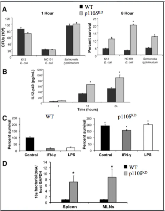
I am a co-author on the manuscript entitled, “Altered macrophage function contributes to colitis in mice defective in the phosphoinositide 3-kinase subunit p110δ,” published in Gastroenterology in 2010 (Uno et al., 2010). I contributed to the manuscript by performing the bacterial assays in which we infected WT and p110δKD bone marrow-derived macrophages (BMDMs) with K12 Escherichia coli, NC101 E. coli or Salmonella
typhimurium and measured bacterial survival and phagocytosis and IL-12p40 produced
by the BMDMs. Additionally, I isolated bacterial DNA from various tissues from WT and p110δKD mice and quantified total bacteria by quantitative RT-PCR. This was a significant contribution to the manuscript, as my work contributed to most of Figure 5.
2.2 Overview
Background and Aims: Innate immune responses are crucial for host defense against pathogens, but need to be tightly regulated to prevent chronic inflammation. Initial
1Jennifer K. Uno, Kavitha N. Rao, Katsuyoshi Matsuoka, Shehzad Z. Sheikh, Taku
characterization of mice with a targeted inactivating mutation in the p110δ subunit of phosphoinositide 3-kinase (PI3K p110δKD) reveal defects in B- and T-cell signaling and chronic colitis. Here, we further characterize features of inflammatory bowel diseases (IBD) in these mice and investigate underlying innate immune defects.
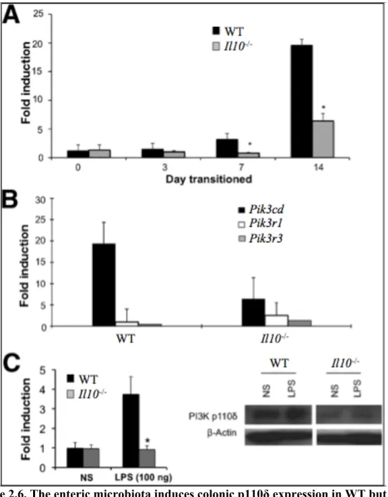
Methods: Colons and macrophages from PI3K p110δKD mice were evaluated for colonic inflammation and innate immune dysfunction. Colonic p110δ mRNA expression was examined in IL-10-deficient (Il10-/-) and wild type (WT) germ free (GF) mice during transition to a conventional microbiota. To assess polygenic impact on colitis
development, p110δKD mice were backcrossed to Il10-/- mice.
Results: A mild spontaneous colitis was demonstrated in p110δKD mice at 8 weeks with inflammation increasing with age. An inflammatory mucosal and systemic cytokine profile was characterized by expression of IL-12/23. In p110δKD macrophages,
augmented toll-like receptor signaling and defective bactericidal activity were observed. Consistent with an important homeostatic role for p110δ, WT mice raised in a GF
environment markedly upregulated colonic p110δ expression with the introduction of the enteric microbiota, however colitis-prone Il10-/- mice do not. Moreover, PI3K p110δKD mice crossed to Il10-/- mice developed severe colitis at an early age.
2.3 Introduction
The pathogenesis of the human inflammatory bowel diseases (IBD) Crohn’s disease (CD) and ulcerative colitis (UC) is complex, with abnormal immune responses in genetically susceptible individuals eliciting uncontrolled intestinal inflammation (Xavier and Podolsky, 2007). Genetic variants that confer CD susceptibility highlight the
importance of innate immune interactions with the enteric microbiota in controlling inflammation (Xavier and Podolsky, 2007). Commensal and pathogenic bacteria are recognized through conserved molecular microbial patterns by pattern-recognition receptors (PRRs), of which toll-like receptors (TLRs) form integral components (Abreu, 2010). Signaling through TLRs leads to the activation of NF-κB, culminating in the induction of inflammatory cytokines including IL-12/23 and TNF-α. This inflammatory response is essential for the eradication of infectious microorganisms; however, excessive and prolonged activation can be detrimental to the host. Although mechanisms by which the host distinguishes commensal from pathogenic bacteria are not well defined, under normal conditions TLR signaling initiated by the enteric microbiota is protective (Rakoff-Nahoum et al., 2004).
Phosphoinositide 3-kinases (PI3Ks) have emerged as important regulators of TLR signaling (Fukao and Koyasu, 2003; Liew et al., 2005). Class IA PI3Ks are a family of