Effects of feeding a soft diet and subsequent rehabilitation
on the development of the masticatory function
A . F U J I S H I T A , Y . K O G A , D . U T S U M I , A . N A K A M U R A , T . Y O S H I M I &
N . Y O S H I D A
Department of Orthodontics and Dentofacial Orthopedics, Graduate school of Biomedical Sciences, Nagasaki Univer-sity, Nagasaki, JapanSUMMARY It has been suggested that feeding a soft diet could possibly inhibit normal development of
the masticatory function. However, the
consequences of such changes in the alimentary habits have yet to be fully clarified. Therefore, the aim of this study was to determine whether a soft diet prevents the development of masticatory
function and whether a critical period for
programming the masticatory system exists. To
examine these hypotheses, we used a
three-dimensional jaw-movement tracking device and jaw muscle electromyography (EMG) to analyse masticatory function changes in mice. Jcl:ICR mice were divided into three groups, with the normal group fed a hard diet, the hypofunctional group fed a soft diet, and the rehabilitation group first fed a soft diet that was then changed to a hard diet. Our results showed that the excursion and duration of late-closing phase (occlusal phase) of
the chewing cycle and EMG activity in the
masseter muscle were not only reduced in the hypofunctional but also in the rehabilitation group as compared with the normal group. These results suggest that optimisation of the chewing pattern
and acquisition of appropriate masticatory
function are impeded by feeding a soft diet during the animal’s growth period and that no catch-up effect of the masticatory function is observed when there is a prolonged period of time prior to changing the diet from soft to hard. In conclusion, masticatory function can only be fully developed through a learning process such as exposure to chewing various kinds of foods with different food textures.
KEYWORDS:mastication, growth and development, jaw relation record, electromyography, masseter muscle, critical period
Accepted for publication 3 October 2014
Introduction
Mastication is one of the most fundamental functions for the conservation of life, with many organs involved in the complex ingestive process (1–5). While sucking is an inborn reflex in mammals that enables a baby to intake milk, mastication is a postna-tally acquired function and goes through a series of developmental steps early in life (6, 7). During the weaning period, the main feeding system changes from sucking to chewing, and subsequently, there are extensive morphological changes and maturation of the nervous system. In addition, exposure to chewing various kinds of foods with different food textures is
considered important in helping children develop masticatory functions after weaning. On the contrary, prolonged feeding of a soft diet may potentially impede normal development of the masticatory func-tion, as sensory stimulation while feeding is particu-larly important during the critical period (8).
Although many animal studies have investigated the effects of a soft diet on the growth of the craniofa-cial skeleton (9–11) and masticatory muscles (12, 13), few studies have focused on the development of sto-matognathic function. Liu et al. (14) examined the functional aspects and reported that the electromyo-graphic (EMG) activity of the jaw muscles was affected by the consistency of the daily diet. However,
Journal of Oral Rehabilitation201542; 266--274
there have been no reports that analysed jaw-move-ment patterns due to the difficulty of performing such studies.
The aim of our current study was to test the hypotheses that soft diets can impede the acquisition of appropriate masticatory function, and there exists a critical period representing a phase in the lifespan during which the development of the neural mecha-nism of mastication is activated. To test these hypoth-eses, we recorded both the three-dimensional jaw-movement trajectories and the EMG activities of the jaw muscles. These findings were then used to evalu-ate the changes in the development of the masticatory function in three groups of mice fed a hard diet, a soft diet and a diet that was changed from soft to hard at 15 weeks of age to determine whether there was a catch-up effect of the masticatory function.
Materials and methods
The experimental protocol of this study was approved by the Animal Welfare Committee of Nagasaki Uni-versity based on the Animal Care Standards of this institution (No. 0309120322-2, 2008). Every possible effort was taken to minimise animal suffering.
Experimental animals
At the age of 3 weeks, 36 male Jcl:ICR mice* were randomly divided into three groups that included a normal group (n= 13) that was fed a hard diet (chow pellet only; CE-2*), a hypofunctional group (n=11) that was fed a soft diet that consisted of only finely powdered pellet (<002 mm in diameter), and a reha-bilitation group (n =12) in which the mice were first fed a soft diet (pellet powder) until 15 weeks of age, with the diet then changed to a hard diet (chow pel-let) that continued from 15 until 20 weeks of age to determine whether there was functional rehabilitation of their masticatory system. Mice were weighed every week during the entire experimental period.
Functional recordings
The three-dimensional jaw movements and EMGs of the bilateral masseter muscles and unilateral digastric
muscle were recorded at 20 weeks of age, while the animals were eating chow pellets (hard food) or pow-dered pellets mixed with water shaped into balls that were 3 mm in diameter (soft food), respectively. This methodology has been described in detail in previ-ously published articles (15–17).
Data analysis
Table 1 lists the parameters used to analyse the jaw movements. Mean values for each parameter were calculated from 10 chewing cycles for each animal. In a previous study, we determined that jaw closing from the occlusal view could be divided into two phases: early-closing and closing (17). The late-closing phase is considered the occlusal phase of the chewing cycle and the time when most of the food grinding and possibly some tooth contact takes place. Electromyography activities in the masseter and digas-tric muscles were analysed in terms of burst duration and area (integrated EMG). Spike 2 software†aided in the data analysis.
Statistical analysis
Significant differences between the three groups were determined by an analysis of variance (ANOVA), with a
pairedt-test used to examine the differences between the hard and soft food chewing.Pvalues<005 were considered statistically significant. All values are dis-played as meansstandard error of the mean.
Results
Body weight
There was no significant difference in the body weight among the three groups throughout the experimental period.
Jaw movements
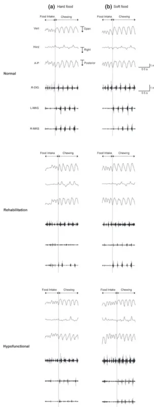
Figure 1 shows examples of jaw-movement trajecto-ries during the chewing of the hard or soft food in the normal, rehabilitation or hypofunctional group. The paths of three-dimensional jaw movements when
chewing hard or soft food were reconstructed in two dimensions by projecting on the sagittal, frontal and occlusal planes, respectively (Fig. 2).
Major differences in the paths of the jaw move-ments among the three groups were observed when chewing hard food. Although there was a significantly smaller excursion of the late-closing phase during hard food chewing in the hypofunctional and the rehabilitation groups compared to the normal group, no significant difference was observed when chewing soft food (Table 2). Comparison of the excursion of the late-closing phase between hard and soft food chewing demonstrated that there was a significantly larger excursion when chewing hard food versus soft food in the normal group (Table 3). No significant dif-ferences were observed in the hypofunctional and rehabilitation groups.
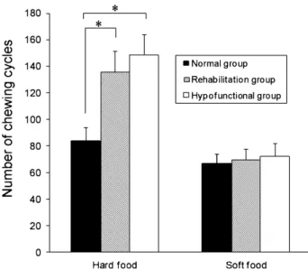
Similar results were observed for the chewing rhythm properties. For the total cycle duration, both the duration and excursion of the late-closing phase, they were significantly longer during the hard food chewing versus the soft food chewing in the normal group. However, there were no significant differences for these parameters in the hypofunctional and reha-bilitation groups when chewing either hard or soft food. A significantly larger number of chewing cycles in the masticatory sequence were observed in the hypofunctional and rehabilitation groups when chew-ing hard food, while there were no significant differ-ences among the three groups when chewing soft food (Fig. 3).
Jaw muscle activity
Figure 1 shows examples of the jaw muscle activity during hard or soft food chewing. The masticatory
sequence was divided into food intake and chewing periods based on the rhythm of the mandibular movement and the pattern of muscle activity. The area and the duration of the masseter activities were significantly larger and longer, respectively, in the normal group versus both the hypofunctional and the rehabilitation groups when chewing hard food (Table 2). On the other hand, no statistically signifi-cant differences were found for the activities of the digastric muscle among the three groups.
Comparison of the EMG activities for the different foods demonstrated that the area and the duration of the masseter burst were significantly larger and longer when chewing hard food versus soft food in the nor-mal group (Table 3). In contrast, no statistically signif-icant differences were observed in the masseter muscle activities between the hard and soft food chewing in the hypofunctional and the rehabilitation groups. There were also no significant differences for the area and the duration of the digastric muscle activities between hard and soft food chewing in any of the three groups.
Discussion
Influence of a soft diet on the development of masticatory function
To investigate the effect of feeding a soft diet on the development of masticatory function, we first com-pared the results obtained from a normal group fed a hard diet with those from a hypofunctional group fed a soft diet. Although the duration and excursion of the late-closing phase, which corresponds to the occlusal phase, were significantly longer when chew-ing hard food versus chewchew-ing soft food in the normal
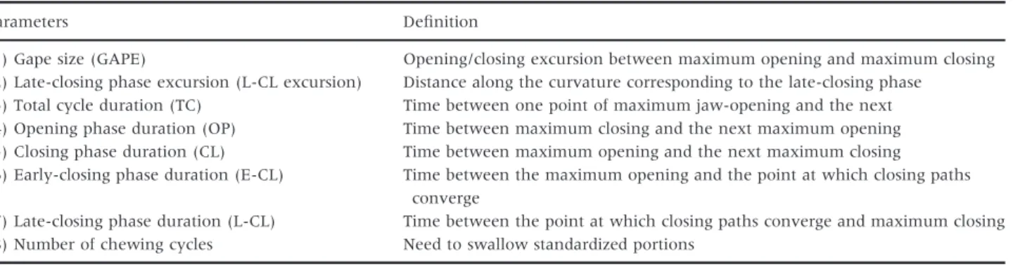
Table 1. Definitions of parameters used to analyse the jaw movements
Parameters Definition
(1) Gape size (GAPE) Opening/closing excursion between maximum opening and maximum closing
(2) Late-closing phase excursion (L-CL excursion) Distance along the curvature corresponding to the late-closing phase
(3) Total cycle duration (TC) Time between one point of maximum jaw-opening and the next
(4) Opening phase duration (OP) Time between maximum closing and the next maximum opening
(5) Closing phase duration (CL) Time between maximum opening and the next maximum closing
(6) Early-closing phase duration (E-CL) Time between the maximum opening and the point at which closing paths
converge
(7) Late-closing phase duration (L-CL) Time between the point at which closing paths converge and maximum closing
group, we found no significant differences for either parameter when foods of different hardness were examined in the hypofunctional group. These results indicate the capacity to perceive the food consistency is reduced in mice fed a soft diet. In our previous study, we found that the duration and excursion of the late-closing phase increased when chewing hard food as compared to chewing soft food in the normal mice (17). From this previous study, we concluded that mice fed a hard diet were capable of modifying their chewing pattern by increasing the excursion of the late-closing phase. As a result, these mice were able to extend the grinding path at the occlusal phase to increase the masticatory efficiency when chewing hard food versus when chewing soft food. We also found that mice raised on a soft diet were not able to regulate their chewing pattern in accordance with the food consistency. Furthermore, in the present study, we found that mice in the hypofunctional group did not show any significant difference in masseter activ-ity when chewing either hard or soft food, although we did observe an enhancing effect of the masseter activity when mice chewed hard food versus soft food in the normal group.
These results suggest that control of the masticatory force in response to changes in food hardness might be more difficult in mice that have been raised on a soft diet. Previous studies have shown that changes in food hardness are responsible for much of the variability in mastication and that sensory afferents such as muscle spindles and periodontal mechanoreceptors provide hardness-related feedback that modifies the mastica-tory central pattern generator (mCPG) output (18, 19). Thus, the decreased masticatory demands in our hypo-functional group may have resulted in an immaturity of the sensory receptors or an impaired sensory feed-back mechanism based on underdeveloped mCPG.
Effect of dietary change on the catch-up tendency of the masticatory function
To investigate the effect of daily diet changes from soft to hard diet on the subsequent catch-up of the masticatory function development and to verify whether a critical period exists for appropriate acquisi-tion of masticatory funcacquisi-tion during the growth period, we compared the results obtained from the rehabilita-tion group with those from the normal or the hypo-functional group.
(a) (b)
Fig. 1. Typical masticatory sequence. (a) chewing of hard food, (b) chewing of soft food. Upper three traces illustrate jaw move-ments in the vertical (Vert), horizontal (Horz) and antero-posterior (A-P) direction. The lower three traces show electromyographies (EMGs) of the right digastric muscles (R-DIG), left masseter ( L-MAS) and right masseter (R-MAS). The sequence was divided into food intake and chewing.
Fig. 2. Jaw-movement trajectories of the normal (upper), rehabilitation
(middle) and hypofunctional
(lower) groups in the sagittal (left), frontal (centre) and occlusal (right) planes when chewing hard or soft food. Tracings show 10 consecutive chewing cycles superimposed.
No statistically significant differences were observed in the masseter activity between the hard and soft food chewing in the rehabilitation as well as the hyp-ofunctional group. However, there was a significant difference observed in the normal group. Similarly, there were no statistically significant differences for the total cycle duration, or for the duration and excursion of the late-closing phase when chewing either hard or soft food in the rehabilitation as well as hypofunctional group, although there was a signifi-cant difference in the normal group. A previous study (20) that examined normally developed animals reported that the total cycle duration was longer
when chewing hard food versus soft food due to the lengthening of the duration of the occlusal phase. This indicates that when a harder food bolus is pres-ent between the grinding surfaces of the upper and lower teeth, the jaw movement becomes slower in the late-closing phase, which would extend the total cycle length. Therefore, the results in our current study indicate that mice in our rehabilitation group have greater difficulty in controlling the masticatory force or optimisation of the chewing patterns in accordance with the food consistency. Thus, these findings suggest that the lower sensory inputs due to a lack of experience with solid diet mastication during
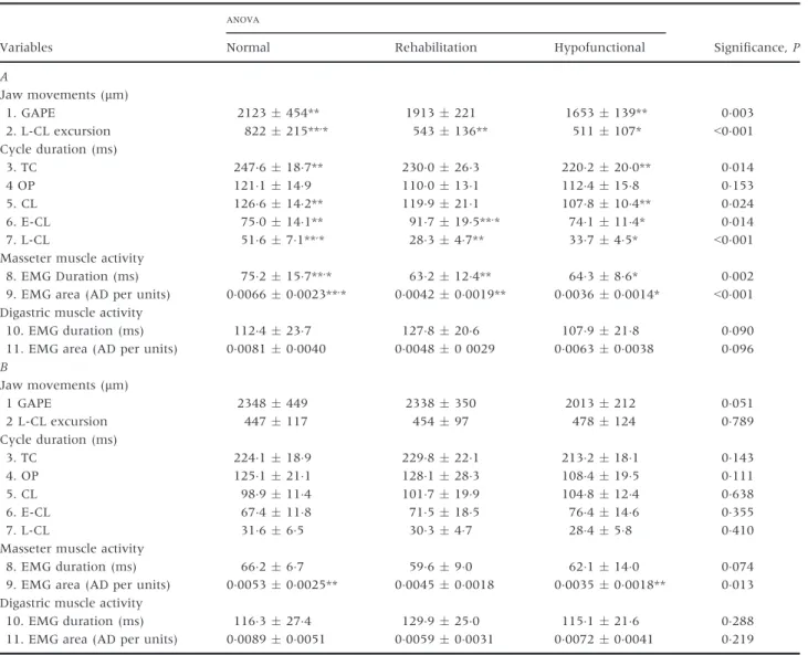
Table 2. Comparison of jaw movements, cycle durations and muscle activity between the experimental groups. (A) Hard food chew-ing. (B) Soft food chewchew-ing. Differences between the three experimental groups (normal, rehabilitation and hypofunctional) were investigated using an analysis of variance (ANOVA)
Variables
ANOVA
Significance,P
Normal Rehabilitation Hypofunctional
A Jaw movements (lm) 1. GAPE 2123454** 1913221 1653139** 0003 2. L-CL excursion 822215**,* 543136** 511107* <0001 Cycle duration (ms) 3. TC 2476187** 2300263 2202200** 0014 4 OP 1211149 1100131 1124158 0153 5. CL 1266142** 1199211 1078104** 0024 6. E-CL 750141** 917195**,* 741114* 0014 7. L-CL 51671**,* 28347** 33745* <0001
Masseter muscle activity
8. EMG Duration (ms) 752157**,* 632124** 64386* 0002
9. EMG area (AD per units) 0006600023**,* 0004200019** 0003600014* <0001
Digastric muscle activity
10. EMG duration (ms) 1124237 1278206 1079218 0090
11. EMG area (AD per units) 0008100040 000480 0029 0006300038 0096
B Jaw movements (lm) 1 GAPE 2348449 2338350 2013212 0051 2 L-CL excursion 447117 45497 478124 0789 Cycle duration (ms) 3. TC 2241189 2298221 2132181 0143 4. OP 1251211 1281283 1084195 0111 5. CL 989114 1017199 1048124 0638 6. E-CL 674118 715185 764146 0355 7. L-CL 31665 30347 28458 0410
Masseter muscle activity
8. EMG duration (ms) 66267 59690 621140 0074
9. EMG area (AD per units) 0005300025** 0004500018 0003500018** 0013
Digastric muscle activity
10. EMG duration (ms) 1163274 1299250 1151216 0288
11. EMG area (AD per units) 0008900051 0005900031 0007200041 0219
the growing period may have affected the develop-ment of the sensory afferents or mCPG in our hypo-functional and rehabilitation groups. This inadequate development of the masticatory function is also reflected by the increased number of chewing cycles in the rehabilitation group as well as in the hypofunc-tional group as compared with the normal group. It is entirely possible that a loss of masticatory efficiency will be compensated for by increasing the number of chewing cycles, and thus, animals with a lower masti-catory efficiency in our rehabilitation group will never be able to catch-up to the levels seen in the normal group. Moreover, our findings suggest that the matu-rity of the masticatory function will never reach nor-mal levels if the daily diet is changed from soft to hard after the optimum learning period for oral motor behaviour.
In both the rehabilitation and the hypofunctional groups, EMG activity in the masseter decreased as compared with the normal group. Kiliaridis and Shyu (21) found that the tetanic tension was weaker in rats raised on a soft versus a hard diet. These findings indicate that appropriate growth and functional matu-ration of the masticatory muscles may be impeded due to less muscle usage. Even after a rehabilitation of 5 weeks, a catch-up tendency in terms of the intensity of masseter muscle activity was not observed in these animals.
Previous study reported the correlation between food consistency and tooth wear (22). However, relationship between tooth wear and jaw movements is still contro-versial (23, 24). Therefore, further studies are needed to explore the effect of tooth wear on jaw movements and EMG activities in mice fed different diets.
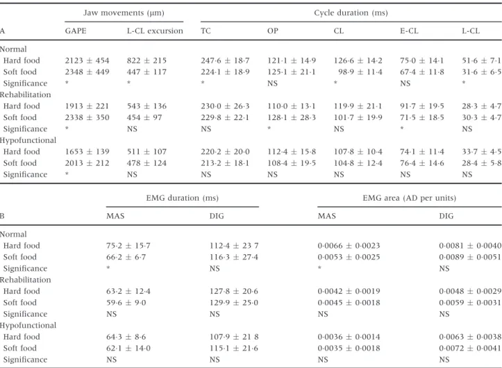
Table 3. Comparison of jaw movements, cycle durations and muscle activity between hard and soft food chewing in each of the three experimental groups. (A) Jaw movements and cycle durations. (B) Muscle activity. Values are presented as the standard error of the mean of each group. Individual differences were tested using a pairedt-test
A
Jaw movements (lm) Cycle duration (ms)
GAPE L-CL excursion TC OP CL E-CL L-CL
Normal Hard food 2123454 822215 2476187 1211149 1266142 750141 51671 Soft food 2348449 447117 2241189 1251211 989114 674118 31665 Significance * * * NS * NS * Rehabilitation Hard food 1913221 543136 2300263 1100131 1199211 917195 28347 Soft food 2338350 45497 2298221 1281283 1017199 715185 30347 Significance * NS NS * NS * NS Hypofunctional Hard food 1653139 511107 2202200 1124158 1078104 741114 33745 Soft food 2013212 478124 2132181 1084195 1048124 764146 28458 Significance * NS NS NS NS NS NS B
EMG duration (ms) EMG area (AD per units)
MAS DIG MAS DIG
Normal Hard food 752157 112423 7 0006600023 0008100040 Soft food 66267 1163274 0005300025 0008900051 Significance * NS * NS Rehabilitation Hard food 632124 1278206 0004200019 0004800029 Soft food 59690 1299250 0004500018 0005900031 Significance NS NS NS NS Hypofunctional Hard food 64386 107921 8 0003600014 0006300038 Soft food 621140 1151216 0003500018 0007200041 Significance NS NS NS NS
The conversion from sucking to mastication occurs regardless of whether mice are fed a hard or soft diet. However, mice raised on a soft diet only acquire mas-ticatory function at a much lower functional level. As these mice were adapted to eating only softer foods, they were incapable of modulating their chewing pat-tern or masticatory force during hard food chewing even if the diet was changed from soft to hard at 15 weeks of age. Therefore, these results suggest that there might be a critical period for the development of masticatory function during the several weeks after weaning and that feeding a soft diet during this time may inhibit the acquisition of the higher level of motor performance. Masticatory function will only properly develop concomitantly with the maturation of the peripheral sensory receptors in the oro-facial area and relevant central nervous system. As it is also well known that periodontal mechanoreceptors and jaw muscle spindles provide positive feedback (25), the present study results suggest the sensory feedback mechanism may be impaired in mice raised on a soft diet.
In conclusion, reduced masticatory stimuli, namely a lack of chewing experience with a hard diet, can impede optimisation of the chewing pattern and the acquisition of a more complex masticatory function during the growing period. Furthermore, when there
is a delay in the onset of specific learning for chewing various kinds of food with different consistencies beyond the critical period, the masticatory system cannot develop to reach a higher level of function. Additional studies that investigate whether immatu-rity of the peripheral receptors or underdevelopment of mCPG can cause impairment of the appropriate development of masticatory function will need to be undertaken in the future.
Acknowledgments
This study was supported in part by grants (No. 24792296 and 24659913) from the Ministry of Educa-tion, Culture, Sports, Science and Technology of Japan. None of the authors has any conflict of inter-ests to declare.
References
1. Dellow PG, Lund JP. Evidence for central timing of rhythmi-cal mastication. J Physiol. 1971;215:1–13.
2. Lavigne G, Kim JS, Valiquette C, Lund JP. Evidence that periodontal pressoreceptors provide positive feedback to jaw
closing muscles during mastication. J Neurophysiol.
1987;58:342–358.
3. Huang X, Zhang G, Herring SW. Effects of oral sensory affer-ents on mastication in the miniature pig. J Dent Res. 1993;72:980–986.
4. Liu ZJ, Masuda Y, Inoue T, Fuchihata H, Sumida A, Takada Ket al.Coordination of cortically induced rhythmic jaw and
tongue movements in the rabbit. J Neurophysiol.
1993;69:569–584.
5. Nakamura Y, Katakura N. Generation of masticatory rhythm in the brainstem. Neurosci Res. 1995;23:1–19.
6. Herring S. The ontogeny of mammalian mastication. Am Zool. 1985;25:339–349.
7. Langenbach GE, Weijs WA, Brugman P, van Eijden TM. A longitudinal electromyographic study of the postnatal matu-ration of mastication in the rabbit. Arch Oral Biol. 2001;46:811–820.
8. Denenberg VH. Effects of age and early experience upon conditioning in the G.57.BL/10 mouse. J Psychol. 1958;46: 211–226.
9. Kiliaridis S, Engstr€om C, Thilander B. The relationship between masticatory function and craniofacial morphology. I. A cephalometric longitudinal analysis in the growing rat fed a soft diet. Eur J Orthod. 1985;7:273–283.
10. Ito G, Mitani S, Kim JH. Effect of soft diets on craniofacial growth in mice. Anat Anz. 1988;165:151–166.
11. Kiliaridis S, Thilander B, Kjellberg H, Topouzelis N, Zafiria-dis A. Effect of low masticatory function on condylar growth: a morphometric study in the rat. Am J Orthod Dentofacial Orthop. 1999;116:121–125.
Fig. 3. Mean number of chewing cycles (s.d.) required for grinding a certain amount of hard or soft food in the three groups. Differences between the three groups were investigated using anANOVA.*denotes statistically significant pair-wise differ-ences (post hoccomparisons).
12. Maeda N, Kawasaki T, Osawa K, Yamamoto Y, Sumida H, Masuda Tet al.Effects of long-term intake of a fine-grained diet on the mouse masseter muscle. Acta Anat (Basel). 1987;128:326–333.
13. Langenbach G, van de Pavert S, Savalle W, Korfage H, van Eijden T. Influence of food consistency on the rabbit masse-ter muscle fibres. Eur J Oral Sci. 2003;111:81–84.
14. Liu ZJ, Ikeda K, Harada S, Kasahara Y, Ito G. Functional properties of jaw and tongue muscles in rats fed a liquid diet after being weaned. J Dent Res. 1998;77:366–376.
15. Koga Y, Yoshida N, Kobayashi K, Okayasu I, Yamada Y. Development of a three-dimensional jaw-tracking system implanted in the freely moving mouse. Med Eng Phys. 2001;23:201–206.
16. Okayasu I, Yamada Y, Kohno S, Yoshida N. New animal model for studying mastication in oral motor disorders. J Dent Res. 2003;82:318–321.
17. Utsumi D, Nakamura A, Matsuo K, Zeredo J, Koga Y, Yosh-ida N. Motor coordination of masseter and temporalis mus-cle during mastication in mice. J Stomat Occ Med. 2010;3:187–194.
18. Lund JP, Kolta A, Westberg KG, Scott G. Brainstem mecha-nisms underlying feeding behaviors. Curr Opin Neurobiol. 1998;8:718–724.
19. Lund JP, Kolta A. Generation of the central masticatory pat-tern and its modification by sensory feedback. Dysphagia. 2006;21:167–174.
20. Lund JP. Mastication and its control by the brain stem. Crit Rev Oral Biol Med. 1991;2:33–64.
21. Kiliaridis S, Shyu BC. Isometric muscle tension generated by masseter stimulation after prolonged alteration of the consistency of the diet fed to growing rats. Arch Oral Biol. 1988;33:467–472.
22. Teaford MF, Oyen OJ. Differences in the rate of molar wear between monkeys raised on different diets. J Dent Res. 1989;68:1513–1518.
23. Russell MD. The masticatory cycle in relation to occlusal wear and its treatment. J Dent. 1982;10:69–77.
24. Ekfeldt A, Karlsson S. Changes of masticatory movement characteristics after prosthodontic rehabilitation of individu-als with extensive tooth wear. Int J Prosthodont. 1996;9: 539–546.
25. Morimoto T, Inoue T, Masuda Y, Nagashima T. Sensory components facilitating jaw-closing muscle activities in the rabbit. Exp Brain Res. 1989;76:424–440.
Correspondence: Yoshiyuki Koga, Department of Orthodontics and Dentofacial Orthopedics, Graduate school of Biomedical Sciences, Nagasaki University, 1-7-1 Sakamoto, Nagasaki 852-8501, Japan. E-mail: [email protected]