Veterinary
Science
An immunohistochemical study of chromogranin A and Sp-1
immunoreactive cells in the gastrointestinal tract of ovariectomized rats
Sae-kwang Ku1, Hyeung-sik Lee2,*, Jae-hyun Lee31Pharmacology & Toxicology Lab., Central Research Laboratories, Dong-Wha Pharm. Ind. Co. Anyang 430-017, Korea 2Department of Herbal Biotechnology, Daegu Haany University , Daegu 712-715, Korea
3Department of Histology, College of Veterinary Medicine, Kyungpook National University, Daegu 702-701, Korea
The changes on the regional distributions and frequencies of two types of chromogranin, chromogranin A (CGA) and bovine Sp-1 chromogranin (BCG)-immunoreactive (IR) cells in gastrointestinal (GI) tract of osteoporotic Sprague-Dawley rat induced by ovariectomy were studied by immunohistochemical methods. The experimental animals were divided into two groups, one is non-ovariectomized group (Sham) and the other is ovariectomized group (OVX). Samples were collected from each part of GI tract at 10th week after ovariectomy or sham operation. CGA-IR cells were restricted to the stomach regions with various frequencies regardless of ovariectomy except for the fundus of OVX in which no cells were detected. In addition, BCG-IR cells were also restricted to the pylorus and duodenum regardless of ovariectomy. A significantly decrease of CGA IR cells was detected in OVX compared to that of Sham in both fundus and pylorus, and BCG-IR cells were also significantly decreased in the duodenum (p< 0.05). However, in the pylorus, BCG-IR cells in OVX showed similar frequency compared to that of Sham. In conclusion, the abnormality in density of chromogranin, a generally used GI endocrine cell marker, detected in this study may contribute to the development of GI symptoms in osteoporosis such as impairments of calcium and some lipids, frequently encountered in patients with postmenopausal osteoporosis.
Key words: ovariectomy, osteoporosis, chromogranin,
endo-crine, immunohistochemistry
Introduction
Osteoporosis is caused by an imbalance between bone resorption and bone formation, which results in bone loss and fractures after mineral flux. The frequency of fractures
significantly increases in osteoporosis, and hip fractures in senile patients are a very serious problem because it often limits the patients’ quality of life. The postmenopausal osteoporosis model using ovariectomized rat is useful for evaluation of osteoporetic drugs, because several parameters clearly decrease by the ovariectomy within 4 weeks after operation [33]. In addition, the ovariectomized rat bone loss model is suitable for studying problems that are relevant to postmenopausal bone loss, because ovariectomy that induced bone loss in the rat and postmenopausal bone loss share many similar characteristics including decreased intestinal absorption of calcium [12].
Gastrointestinal (GI) endocrine cells dispersed in the epithelia and gastric glands of the digestive tract synthesized various kinds of gastrointestinal hormones and played an important role in the physiological functions of the alimentary tract [1]. Until now, the endocrine cells are regarded as the anatomical units responsible for the production of gut hormones, and a change in their density would reflect the change in the capacity of producing these hormones [6]. Chromogranin (CG) belongs to a family of large anionic proteins, the members of which are known to be present in the secretory granules of a broad spectrum of amine and peptide-producing cells of adrenal medulla and gastrointestinal endocrine system, as well as in some neurons of the peptidergic and catecholaminergic nervous system of several mammals [9,26]. CGs have been found to occur in large variety of endocrine organs and cells outside the adrenal medulla, and they have been claimed as common “markers” of all neuroendocrine cells [3,8]. The appearance, regional distribution and relative frequency in GI tract of normal rat species are well recognized [2,10]. In addition, the changes of distribution and frequency of CG-immunoreactive (IR) cells in some diseases are also well demonstrated especially in some cancer status [14], colonic inertia [34], chronic constipation [16], familial amyloidotic polyneuropathy [7,24], antral atrophic gastritis [15] and some inflammatory bowel disease [5]. In addition, these GI CG-IR cells are also changed after vagotomized [29] and with ageing in human
*Corresponding author
Tel: +82-53-819-1436, Fax: +82-53-819-1574 E-mail: [email protected]
Sp-1 chromogranin; BCG)-generally used endocrine markers in the immunohistochemistry. In this study, each part of GI tract is sampled at 10th week after ovariectomy or sham-operation.
Materials and Methods
Experimental animals
Twenty Sprague-Dawley (SD) female rat (6-wk old upon receipt; Charles River, Japan) were used after acclimatization for 7 days. Animals were allocated 5 per polycarbonate cage in a temperature (20-25oC) and humidity (30-35%) controlled
room. Light: dark cycle was 12 hr: 12 hr and feed (Samyang, Korea) and water were supplied free to access. Half rats were ovariectomized group (OVX) and remainders were sham-operated group (Sham).
Bilateral ovariectomy
All rats were anesthetized with Ketamine hydrochloride (60 mg/2 ml/kg) and Xylazine hydrochloride (2.5 mg/2 ml/ kg) combination and subjected to operation. Bilateral ovariectomy was performed by removing both ovaries in the abdominal cavity, and sham operation (ovary identification) was performed in case of sham.
Tissue preparation and staining
After phlebotomy, each region of GI tract, fundus, pylorus, duodenum, jejunum, ileum, cecum, colon and rectum was collected from all experimental animals at 10th week after ovariectomy and/or sham-operation after 18hrs fasting to GI empty. Collected samples fixed in Bouin’s solution, then embedded in paraffin, sectioned (3~4µm) and
the sections were lightly counterstained with Mayer’s hematoxylin and the IR cells were observed under light microscope.
The specificity of each immunohistochemical reaction was determined as recommended by Sternberger [31], including the replacement of specific antiserum by the same antiserum, which had been preincubated with its corresponding antigen.
Quantity analyses
The frequency of IR cells was calculated using automated image analysis (Soft Image System, Germany) under microscope (Carl Zeiss, Germany) in the uniform area of GI mucosa among 1000 parenchymal cells. IR cell numbers were calculated as cell numbers/1000 parenchymal cells.
Statistical analysis
Results are expressed as the mean ± SD. Mann-Whitney U-Wilcoxon Rank Sum W test (M-W test) was used to analyze the significance of data with SPSS for Windows (Release 6.1.3; SPSS, USA) and a p-value of less than 0.05
was considered a significant difference.
Results
In this study, CGA-IR cells were restricted to the stomach regions with various frequencies regardless of ovariectomy except for the fundus of OVX in which no cells were detected. In addition, BCG-IR cells were also restricted to the pylorus and duodenum regardless of ovariectomy. Most of these IR cells in the mucosa of GI tract were generally spherical or spindle in shape (open type cell) while cells showing round in shape (close type cell) were also found in Table 1. Antisera used in this study
Antisera raised* Code Source Dilution
BCG† 805398 Dia Sorin, Stillwater, Minnesota, USA 1 : 1,000
CGA† A0430 DAKO Corp., Carpinteria, CA, USA 1 : 100
basal regions of mucosa. According to the location of the GI tract, different regional distributions and frequencies of CGA and BCG-IR cells were observed. CGA- and BCG-IR cells were mainly dispersed in the basal portions of gastric and intestinal mucosa rather than surface epithelia regions regardless of ovariectomy. CGA-IR cells were more numerously detected in the pylorus compared to that of pylorus in both sham (Fig. 1a~c) and OVX (Fig. 2a and b). In addition, more numerous BCG-IR cells were detected in the pylorus compared to that of duodenum in both sham (Fig. 3a~d) and OVX (Fig. 4a~d). However, no CGA-IR cells were demonstrated in the duodenum, jejunum, ileum, cecum, colon and rectum in both sham and OVX. No BCG-IR cells were detected in the fundus, jejunum, ileum, cecum, colon and rectum in this study.
Quantity of CGA-IR cells: Among 1,000 parenchymal
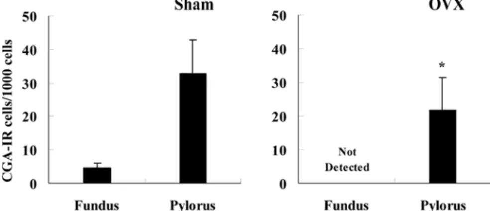
cells, CGA-IR cells in sham were detected in the fundus and pylorus with 4.50 ± 1.58 and 32.70 ± 10.13 cells/1,000 parenchymal cells, respectively. In OVX, CGA-IR cells
were detected with 21.60 ± 9.72 cells/1000 parenchymal cells in the pylorus only. In the fundus and pylorus, CGA-IR cells showed significant (p< 0.01 or p< 0.05) decrease in
OVX compared to that of sham (Fig. 5).
Quantity of BCG-IR cells: Among 1,000 parenchymal
cells, CGA-IR cells in sham were detected in the pylorus and duodenum with 2.10 ± 1.20 and 3.20 ± 1.40 cells/1,000 parenchymal cells, respectively. In OVX, BCG-IR cells were detected with 2.30 ± 1.25 and 2.00 ± 0.82 cells/1,000 parenchymal cells in the pylorus and duodenum, respectively. In the duodenum, BCG-IR cells showed significant (p<
0.05) decrease in OVX compared to that of sham but similar values are detected between sham and OVX (Fig 6).
Discussion
It is generally accepted that osteoporosis is metabolic and hormonal disorder that is clearly related to estrogen [25,27] and also osteoporetic patients and/or animals show quite different feeding habits [32]. The GI endocrine cells were generally divided into two types, one was round to spherical shaped close type cells which were located in the stomach regions, and the other was spherical to spindle shaped open type cells which were situated in the intestinal regions. In addition, the endocrine cells are regarded as the anatomical units responsible for the production of gut hormones, and a change in their density would reflect the change in the capacity of producing these hormones [6]. CGs have been found to occur in large variety of endocrine organs and cells outside the adrenal medulla, and they have been claimed as Fig. 1. CGA-IR cells in the fundus (a, b) and pylorus (c) of sham;
Most of CGA-IR cells were dispersed in the basal mucosa. PAP method, a and c: ×150; b: ×300.
Fig. 2. CGA-IR cells in the pylorus (a, b) of OVX; Most of
CGA-IR cells were dispersed in the basal mucosa. PAP method,
common “markers” of all neuroendocrine cells [3,8]. Although, the distributional patterns of these CG-IR cells in the GI tract of Rodentia were seldom, Hawkins et al. [11] reported that CGA-IR cells were demonstrated throughout the whole GI tract of 7 species of laboratory animals including mouse. However, it is also reported that single use of CG is not suitable as a marker of endocrine cells in some mice species because the relative frequencies of CG-IR cells were not detected or lower than that of other types of IR cells in case of some regions [17,20]. In addition, the appearance of CG-IR cells was also changed by using antisera in rat [10]. In this study, CGA-IR cells were restricted to the fundus and pylorus of sham and to the pylorus of OVX, and BCG-IR cells were also restricted to
the pylorus and duodenum of both groups.
In the present study, the changes of the CGA- and BCG-IR cells in the GI tract after ovariectomy were observed by immunohistochemical technique, the PAP method. Although, the frequency detected in this study was lower than that of previous study [2,10,11], CGA- and BCG-IR cells significantly (p< 0.01 or p< 0.05) decreased in detected regions of GI
tract as results of ovariectomy under same conditions except for duodenum in which similar values were detected between sham and OVX. These results are well corresponded to that of silver techniques-other endocrine markers in ovariectomized osteoporetic rats [18,19]. It was generally accepted that the changes of CG-IR cells were clearly related to digestive status of animals. In colonic inertia patients, a significantly Fig. 3. BCG-IR cells in the pylorus (a, b) and duodenum (c, d) of
sham; Most of BCG-IR cells were dispersed in the basal mucosa of the pylorus and/or crypts of the duodenum. PAP method, a and c: ×150; b and d: ×300.
Fig. 4. BCG-IR cells in the pylorus (a, b) and duodenum (c, d) of
OVX; Most of BCG-IR cells were dispersed in the basal mucosa of the pylorus and/or crypts of the duodenum. , PAP method, a and c: ×150; b and d: ×300.
Fig 5. Number of CGA-IR cells in the fundus and pylorus, and their changes after ovariectomy (OVX).
increase of CG-IR cells were demonstrated in colonic mucosa [34] and they also increased in the rectum of patients with chronic constipation [16], tumor [14], ulcerative colitis [5] and Crohn’s disease [5]. In addition, a significantly decrease of CG-IR cells were also observed in the abomasum of vagotomized calf [29], patients with familial amyloidotic polyneuropathy [7,24] and antral atrophic gastritis [15]. It has been postulated that the changes in the GI endocrine cells are a selective process to meet the new demands exerted by the dramatic decrease in intestinal absorption [4] and osteoporetic patients and/or experimental animals shows impairment of absorption of calcium ion [13,23] and increase of absorption of cholesterol and other lipids [21]. Therefore, the decrease of GI CG-IR cells may be responsible for the malabsorption of calcium and lipids that occur in patients with postmenopausal osteoporosis and these decreases of endocrine cells are also detected with aging especially to cells that release the hormone regulating GI motility [22].
In conclusion, ovariectomy induced severe quantitative changes of GI CG-IR cell density, and the abnormality in density of GI endocrine cells may contribute to the development of gastrointestinal symptoms in osteoporosis such as impairments of calcium and some lipids, frequently encountered in patients with postmenopausal osteoporosis. However, the target or individual changes of GI endocrine cells are not clear. Therefore, elucidation of the changes of individual GI endocrine cells using immunohistochemistry will provide mechanisms for understanding GI disorder that occurs in various diseases. Further detailed studies with immunohistochemical will be needed.
References
1.Bell FR. The relevance of the new knowledge of gastrointestinal hormones to veterinary science. Vet Sci Commun 2, 1979, 305-314.
2.Bjorkqvist M, Dornonville de la Cour C, Zhao CM, Gagnemo-Persson R, Hakanson R, Norlen P. Role of gastrin in the development of gastric mucosa, ECL cells and A-like cells in newborn and young rats. Regul Pept 2002,
108, 73-82.
3.Cohn DV, Elting JJ, Frick M, Elde R. Selective localization of the parathyroid secretory protein I/adrenal medulla chromogranin A protein familiy in a wide variety of endocrine cells of the rat. Endocrinology 1984, 144, 1963-1974.
4.El-Salhy M. The nature and implication of intestinal endocrine cell changes in celiac disease. Histol Histopathol 1998, 13, 1069-1075.
5.El-Salhy M, Danielsson A, Stenling R, Grimelius L.
Colonic endocrine cells in inflammatory bowel disease. J Intern Med 1997, 242, 413-419.
6.El-Salhy M, Sitohy B. Abnormal gastrointestinal endocrine cells in patients with diabetes type I: relationship to gastric emptying and myoelectrical activity. Scand J Gastroenterol 2001, 36, 1162-1169.
7.El-Salhy M, Suhr O. Endocrine cells in rectal biopsy specimens from patients with familial amyloidotic polyneuropathy. Scand J Gastroenterol 1996, 31, 68-73.
8.Fujita T, Kanno T, Kobayashi S. The paraneuron. pp. 1-286, Springer, New York, 1988.
9.Grube D, Yoshie S. Immunohistochemistry of chromogranin A and B, and secretogranin II in the canine endocrine pancreas. Arch Histol Cytol 1989, 52, 287-298.
10.Hashimoto Y, Ohki H, Sato F, Yanaihara N, Iwanaga T.
Immunohistochemical demonstration of chromogranin A in endocrine organs of the rat and horse by use of region-specific antibodies. Jpn J Vet Res 2001, 49, 13-17.
11.Hawkins KL, Lloyd RV, Toy KA. Immunohistochemical localization of Chromogranin A in normal tissues from laboratory animals. Vet Pathol 1989, 26, 488-498.
12.Kalu DN. The ovariectomized rat model of postmenopausal bone loss. Bone Miner Res 1991, 15, 175-191.
13.Kalu DN, Chen, C. Ovariectomized murine model of postmenopausal calcium malabsorption. J Bone Miner Res 1999, 14, 593-601.
14.Kimura N, Funakoshi A, Aunis D, Tateishi K, Miura W, Nagura H. Immunohistochemical localization of chromostatin and pancreastatin, chromogranin A-derived bioactive peptides, in normal and neoplastic neuroendocrine tissues. Endocr Pathol 1995, 6, 35-43.
15.Kitani M, Asada Y, Sakata J, Kitamura K, Sumiyoshi A, Fig 6. Number of BCG-IR cells in the pylorus and duodenum, and their changes after ovariectomy (OVX).
20.Ku SK, Lee HS, Lee JH. An immunohistochemical study of the gastrointestinal endocrine cells in the ddY mice. J Vet Sci
2004, 5, 87-95.
21.Loest HB, Noh SK, Koo SI. Green tea extract inhibits the lymphatic absorption of cholesterol and alpha-tocopherol in ovariectomized rats. J Nutr 2002, 132, 1282-1288.
22.Lucini C, De Girolamo P, Coppola L, Paino G, Castaldo L. Postnatal development of intestinal endocrine cell populations in the water buffalo. J Anat 1999, 195, 439-446.
23.Mitamura R, Hara H, Aoyama Y, Chiji H. Supplemental feeding of difructose anhydride III restores calcium absorption impaired by ovariectomy in rats. J Nutr 2002,
132, 3387-3393.
24.Nyhlin N, Anan I, El-Salhy M, Ando Y, Suhr OB.
Endocrine cells in the upper gastrointestinal tract in relation to gastrointestinal dysfunction in patients with familial amyloidotic polyneuropathy. Amyloid 1999, 6, 192-198. 25.O’Toole K, Fenoglio-Preiser C, Pushparaj N. Endocrine
30.Solcia E, Capella C, Vassallo G, Buffa R. Endocrine cells of the gastric mucosa. Int Rev Cytol 1975, 42, 223-286. 31.Sternberger LA. The unlabeled antibody
peroxidase-antiperoxidase (PAP) method. In: Sternberger LA. (ed), Immunocytochemistry, pp. 104-169, John Wiley & Sons, New York, 1979.
32.Thomas T. Leptin: a potential mediator for protective effects of fat mass on bone tissue. Joint Bone Spine 2003, 70, 18-21. 33.Yamaguchi K, Yada M, Tsuji T, Kuramoto M, Uemura D.
Suppressive effect of norzoanthamine hydrochloride on experimental osteoporosis in ovariectomized mice. Biol Pharm Bull 1999, 22, 920-924.
34.Zhao RH, Baig KM, Wexner SD, Woodhouse S, Singh JJ, Weiss EG, Nogueras JJ. Abnormality of peptide YY and pancreatic polypeptide immunoreactive cells in colonic mucosa of patients with colonic inertia. Dig Dis Sci 2004, 49, 1786-1790.