Imaging characterization
of non-hypersecreting adrenal masses:
comparison between MR and radionuclide techniques
S. MAUREA1, C. CARACÒ2, M. KLAIN1, C. MAINOLFI1, M. SALVATORE1
Aim.In patients with non-hypersecreting adrenal mass-es, tumor characterization is clinically relevant to estab-lish the appropriate treatment planning. The aim of this study was to comparatively characterize such adrenal lesions using MR and radionuclide techniques.
Methods.Thirty patients with non-hypersecreting uni-lateral adrenal tumors underwent both MR and adren-al scintigraphy. MR was performed using SE T1- (pre- and post-gadolinium DTPA) and T2-weighted images as well as in- and out-phase chemical-shift imaging (CSI). MR qualitative and quantitative (signal intensity ratios) eval-uation was performed. Radionuclide studies consisted of iodine-131 nor-cholesterol (n=20), iodine-131 MIBG (n=15) and fluorine-18 FDG PET (n=11) scans. Histology (n=16), biopsy (n=3) or clinical-imaging follow-up (n=11) demomstrated 13 adenomas, 3 cysts, 2 myelolipomas, 4 pheochromocytomas (pheos), 4 carcinomas, 1 sarco-ma and 3 metastases. Comparative isarco-maging analysis was focused on adenomas, pheos and malignant tumors.
Results.Qualitative MR evaluation showed: signal T2-hyperintensity in 46% of adenomas and in 100% of phe-os and malignant tumours, no gadolinium enhance-ment in 92% of adenomas and definite signal intensity loss on CSI in 100% of such tumour lesions, gadolin-ium enhancement in 100% of pheos and in 63% of malig-nancies and no absolute change of signal intensity on CSI in 100% of both pheos and malignancies. Quantitative MR analysis demonstrated: significantly higher signal T2-hyperintensity of pheos compared to adenomas and malignancies as well as significantly higher
enhance-1Division of Radiology and Nuclear Medicine,
Department of Biomorphological and Functional Sciences, “Federico II” University of Naples, Naples, Italy,
2Institute of Biostructures and Bioimages,
National Council for Researches (CNR), Naples, Italy
ment after gadolinium in pheos compared to adenomas and malignancies (p<0.03). Radionuclide studies showed significantly increased nor-cholesterol uptake only in adenomas (n=13), significant MIBG accumulation only in pheos (n=4) and FDG activity only in malignant adren-al lesions (n=8). Conclusion.MR techniques may pro-vide some presumptive criteria to characterize non-hypersecreting adrenal masses, such as no gadolinium enhancement and definite signal intensity loss on CSI in adenomas or quantitatively measured T2-hyperinten-sity and gadolinium enhancement in pheos. On the oth-er hand, radionuclide modalities offoth-er more specific findings in this setting since nor-cholesterol and MIBG scans are respectively able to reveal benign tumours such as adenoma and pheochromocytoma, while FDG imaging allows identification of malignant adrenal lesions. Adrenal scintigraphy is recommended in those patients, when MR images are uncertain or inconclu-sive.
KEY WORDS: Glands - Neoplasms - Magnetic resonance imag-ing - Radiopharmaceutics.
T
he high resolution of anatomic imaging tech-niques such as computed tomography (CT) and magnetic resonance (MR), used in patients with sus-pected abdominal diseases, frequently results in detection of unexpected adrenal masses.1, 2In this set-This paper has been part of the scientific program, as an oralpresen-tation, of the VI National Meeting of the Italian Medicine (AIMN), 15-19 November 2002, Genova, Italy.
Received April 25, 2003. Accepted for publication ???????????.
Address reprint requests to: S. Maurea, Via Raffaele De Cesare 7, 80132 Napoli, Italy. E-mail: [email protected]
ting, the main clinical question consists of differen-tial diagnosis between benign and malignant adren-al lesions in order to select appropriate treatment.2
As initial diagnostic approach, clinical and laborato-ry assessment of cortical and medullalaborato-ry adrenal func-tion allow the identificafunc-tion of hypersecreting adren-al lesions and, hence, characterization of these tumours.3 A tumour mass, however, may not cause
adrenal hyperfunction since it may be non-hyper-secreting or secretes non-active products. In such conditions, lesion typing remains uncertain. CT and MR accurately provide anatomic details of adrenal tumours as well as presumptive criteria for diagno-sis.4 In particular, adrenal adenomas may be
sug-gested by CT on the basis of low attenuation coeffi-cient on un-enhanced images and/or early as well as rapid washout on enhanced scans.5Similarly, MR is
able to characterize adenomas using chemical-shift sequence or pheochromocytomas showing clearly increased signal intensity on T2-weighted images and significant enhancement after gadolinium admin-istration.6
In patients with non-hypersecreting adrenal mass-es, radionuclide adrenal imaging performed with specifically labelled radiopharmaceuticals which tar-get elements of adrenal function provide specific metabolic information for diagnosis, thus, comple-menting morphological imaging modalities.7, 8 In
particular, a diagnostic role of molecular imaging has been identified in the management of patients with non-hypersecreting adrenal masses.9 In this
regard, several radiotracers which display unique biological behaviour may be used in nuclear medi-cine for adrenal lesion evaluation. These include tracers labelled with single photon emitters 7, 10such
as radio-iodine labelled nor-cholesterol and meta-iodobenzylguanidine (MIBG) and gallium-67 cit-rate.11 Alternatively there are agents labelled with
positron emitters such as hydroxy-ephedrine,12
deoxy-glucose 13-15and metomidate.16Finally,
radio-labeled somatostatin analogs have been proposed to identify somatostatin receptors in malignant adren-al tumours.17
In this study, we compared the results of MR imag-ing techniques and those of radionuclide studies in patients with non-hypersecreting adrenal masses in order to evaluate imaging criteria for accurately per-forming non-invasive preoperative tumour character-ization and, hence, to differentiate benign from malig-nant lesions.
Materials and methods
Patient population
Thirty patients (10 male and 20 female, mean age 51±13 years) with non-hypersecreting unilateral adren-al masses (tumour size=4.6±3.0 cm) detected on ultra-sound and/or CT studies underwent both MR imaging and adrenal scintigraphy using appropriate radiophar-maceuticals. The selection criteria for patient enroll-ment consisted of the presence of non-hypersecreting adrenal tumours discovered in subjects who had diag-nostic evaluation of non-adrenal disorders: abdominal pain (n=7), abdominal trauma (n=3), biliary tract stone disease (n=3), renal cysts (n=4), anuria (n=3) or dur-ing the stagdur-ing (n=5) or follow-up (n=5) for malignant tumours. No patient showed signs and/or symptoms of adrenal hyper-secretion. A total of 46 adrenal radio-nuclide studies was analyzed consisting of iodine-131 nor-cholesterol scintigraphy (n=20), iodine-iodine-131 MIBG imaging (n=15) and fluorine-18 deoxyglucose (FDG) positron emission tomography (PET) (n=11). Histology (n=16), biopsy (n=3) or 2-years clinical-imaging follow-up (n=11) were used as standard of ref-erence demonstrating a total of 22 benign adrenal lesions, of which 13 were adenomas, 3 were cysts, 2 were myelolipomas and 4 were pheochromocytomas (pheos), as well as a total of 8 malignant tumours, of which 4 were carcinomas, 1 was a sarcoma and 3 had metastases. Informed consent, as required by the Institutional Clinical Research Sub-panel on Human Studies at our Institute, was obtained in all patients.
Laboratory analysis
In all patients, the evaluation of adrenal function consisted of screening tests for excess of mineral-corticoid, glucomineral-corticoid, androgen and catechola-mine secretion. In particular, measurement of plas-ma aldosterone levels and renin activity in clinostat-ic as well as orthostatclinostat-ic posture was performed. Plasma cortisol and corticotropin levels were meas-ured at 8:00 a.m. and 11:00 p.m. and a 24-hour urine assay for free cortisol was performed. Measurements of serum dehydroepiandrosterone sulfate, 17-hydrox-yprogesterone, androstenedione, testosterone and electrolyte levels were included. An overnight low-dose dexamethasone (DS) suppression test (1 mg orally at 11:00 p.m. and measurement of serum cor-tisol level at 8:00 a.m. the following morning) was also performed. For the evaluation of medullary
adrenal function, plasma catecholamine levels, 24-hour urinary excretions of catecholamines and their metabolites, vanillylmandelic acid and metaneph-rine, were measured. Hormonal values were deter-mined by radioimmunoassay or immunoradiomet-ric assay methods using commercially available kits. Urinary catecholamine, vanillylmandelic acid and metanephrine levels were measured by high-perfor-mance liquid chromatography. Sodium and potas-sium levels were assessed by flame photometry with lithium as an internal standard.
Magnetic resonance
MR imaging studies were performed with a 1.5 Tesla superconducting magnet scanner (Magnetom, Siemens, Erlangen, Germany). A spin echo technique was used to obtain 5 mm contiguous 3-dimensional sections of the abdomen. T1-weighted images (TR/TE=600/15 ms) and T2-weighted images (TR/TE= 2000/15-90 ms) were obtained. T1-weighted images were also acquired after the intravenous administra-tion of gadolinium-DTPA (0.2 ml/kg of weight body, Magnevist, Schering). MR imaging studies were also integrated using chemical-shift (CS) sequence acquir-ing in-phase and out-of-phase images (TR/TE=100/6-4 ms).
Nor-cholesterol imaging
Before nor-cholesterol injection, thyroid iodine uptake was blocked with a saturated solution of potas-sium iodide (200 mg per day orally, starting the day before tracer administration and continuing for 8 days). Iodine-131 nor-cholesterol (37 MBq, CIS Bio International, Cedex, France) was injected intrave-nously. Adrenal scintigraphy was performed 5 (early) and 7 (delayed) days after tracer injection using a large field of view γ camera (Orbiter, Siemens, Erlangen, Germany) with a high-energy collimator and a 20% window centered at 364 Kev. Early and delayed posterior abdominal views were acquired. A mild laxative (bisacodyl) was given (10 mg) twice daily beginning 2 days before the 1stday of imaging
to reduce interfering colonic iodine-131 activity. When required, norcholesterol adrenal scintigraphy was also performed with a concomitant DS suppression test. For this, 1 mg of DS was administered 4 times daily for 7 days prior to nor-cholesterol and for 5 days after trac-er injection.
MIBG imaging
Before MIBG injection, thyroid iodine uptake was blocked with a saturated solution of potassium iodide (200 mg per day orally, starting before tracer admin-istration and continuing for 8 days). Iodine-131 MIBG (37 MBq, Amersham Sorin, Saluggia, Italy) was administered intravenously. Posterior abdominal spot views were obtained at 24, 48 and 72 hours after tracer injection using a large field of view γcamera (Orbiter, Siemens, Erlangen, Germany) with a high-energy collimator and a 20% window centered at 364 Kev.
Fluorine-18 FDG PET
PET imaging was performed using a whole-body PET EXACT 47 scanner (Siemens, Erlangen, Germany). Patients were studied in fasting conditions for at least 4-6 hours before FDG injection. Patients were posi-tioned on the PET gantry using a rectilinear scan com-puterized program localized on the superior abdo-men. Before injection of 18F FDG, abdomen
transmis-sion scan using a rod source of Ge-67 for the attenu-ation correction of the corresponding emission scans was performed for 20 minutes. Thereafter, patients were intravenously injected with 370 MBq of 18F FDG.
Abdomen emission imaging was acquired between 30 and 45 minutes after FDG administration. Images were reconstructed using filtered backprojection smoothed with a Hann filter with a cutoff frequency of 0.4 cycles/pixels by SUN Workstation System (Siemens, Erlangen, Germany) generating three-dimensional PET scans as axial, coronal and sagittal views.
Data analysis
Adrenal function, either cortical and medullary, was considered normal when the corresponding hormone values were in the normal range. The ana-tomic characteristics of adrenal tumours were assessed on MR images; in particular, tumour size was measured as maximal diameter in centimeters and the characteristics of lesion margins (regular or irregular) were assessed. Comparative evaluation of MR and radionuclide imaging findings was focused in adenomas, pheos and malignant adrenal lesions, both primary tumours and metastases; the remaining adrenal lesions, represented by cysts or myelolipo-ma, were not considered for the comparative
imag-ing analysis. The results of histology after surgery, adrenal biopsy or 2-years clinical-imaging follow-up were considered the standards of references; in particular, this latter criterion was used in 11 cases of non-hypersecreting cortical adenomas, ranging as maximal diameter between 2.0 and 3.0 cm, which were not surgically removed for the small tumor size.
MR images were evaluated using both qualitative and quantitative analysis. Signal intensity of adrenal lesions was qualitatively assessed on T2-weighted images in terms of hypo-, iso- or hyperintensity com-pared to that of liver tissue; furthermore, lesion enhancement on T1-weighted images after gadolin-ium administration (yes or no) as well as signal inten-sity loss on out-phase CS images (yes or no) were also
analyzed, as previously described.4, 6Signal intensity
of adrenal lesions was quantitatively assessed on T2-weighted images as well as on T1-T2-weighted images after gadolinium in terms of signal intensity ratios (SIRs) using region of interest analysis. SIRs consist-ed of ratios between absolute signal intensity of adrenal lesion and that of liver, fat and muscle tissues as well as that of image background. This compara-tive analysis was performed among adenomas, phe-os and malignant adrenal tumors using the Student’s “t” test for unpaired data. Data were expressed as mean±1SD. Probability values <0.05 were consid-ered significant.
Radionuclide studies were qualitatively evaluated independently without knowledge of clinical and pathologic findings. In particular, the presence of
TABLEI.—Qualitative MR and radionuclide imaging results of patient population.
# Lesion Site Size° MR T2* MR Gd^ MR CSI” nor-Chol MIBG FDG
1 Adenoma L 3.5 Hyperintense No Yes + — na
2 Adenoma L 3.0 Isointense No Yes + — na
3 Adenoma L 2.5 Isointense No Yes + — na
4 Adenoma R 6.0 Hyperintense Yes Yes + — na
5 Adenoma L 3.0 Hyperintense No Yes + na na
6 Adenoma R 2.5 Isointense No Yes + na na
7 Adenoma L 2.0 Hyperintense No Yes + na na
8 Adenoma L 2.5 Isointense No Yes + — na
9 Adenoma R 3.0 Hyperintense No Yes + na na
10 Adenoma L 3.0 Isointense No Yes + — —
11 Adenoma R 2.0 Hypointense No Yes + — —
12 Adenoma R 2.0 Hypointense No Yes + na na
13 Adenoma L 2.5 Hyperintense No Yes + na na
14 Pheochromocytoma L 5.0 Hyperintense Yes No - + na
15 Pheochromocytoma R 3.5 Hyperintense Yes No na + na
16 Pheochromocytoma L 2.5 Hyperintense Yes No na + na
17 Pheochromocytoma L 5.5 Hyperintense Yes No na + na
18 Carcinoma R 12.0 Hyperintense Yes No na — —
19 Carcinoma L 6.5 Hyperintense No No na na —
20 Carcinoma L 10.0 Hyperintense No No na — —
21 Carcinoma R 5.0 Hyperintense Yes No — na —
22 Sarcoma L 13.0 Hyperintense Yes No na na —
23 Lung metastasis L 2.0 Hyperintense No No — na —
24 Lung metastasis R 5.0 Hyperintense Yes No na na —
25 Melanoma metastasis R 4.5 Hyperintense Yes No na na —
26 Cyst L 5.5 Hyperintense No No na — na
27 Cyst L 5.0 Hyperintense No No — — na
28 Cyst L 5.0 Hyperintense No No — na na
29 Myelolipoma R 11.0 Hyperintense No Yes — na na
30 Myelolipoma R 4.0 Hyperintense No Yes — na —
°: maximal diameter in centimeters; *: lesion signal intensity compared to liver signal intensity; ^: enhancement after gadolinium administration; “: signal intensity loss; nor-Chol: nor-cholesterol; MIBG: metaiodobenzylguanidine; FDG: fluorine-deoxy-glucose; L: left; R: right; +: increased tracer uptake by adre-nal lesion (scan positive); —: no tracer uptake by adreadre-nal lesion (scan negative); na: not available.
abnormally increased uptake of nor-cholesterol, MIBG or FDG was analyzed in the adrenal regions when a tumour lesion was detected. The intensity of tumour uptake was qualitatively evaluated on a high-resolution display by 2 independent and experi-enced nuclear medicine physicians. In case of dis-agreement, final interpretation was determined by consensus reading. Adrenal activity was considered abnormal when tracer uptake was greater than initial blood pool and surrounding background activity and when no similar uptake was observed on the contralateral side.
Results
In all patients, laboratory evaluation of both corti-cal and medullary adrenal function, confirmed normal levels of the corresponding hormones. The results of qualitative MR imaging and radionuclide scans are illustrated in Table I.
MR qualitative analysis
In patients with adrenal adenomas, the evaluation of T2-weighted images demonstrated signal hyperin-tensity in 46% of cases; no significant lesion enhance-ment after gadolinium administration occurred in the majority (92%) of these tumors (Figure 1); finally,
sig-nal intensity loss on out-phase CS images was observed in all cases (100%), (Figure 2). In patients with pheos, the evaluation of T2-weighted images demonstrated signal hyperintensity in all cases (100%) and similarly significant lesion enhancement occurred after gadolinium administration in all cases (100%), (Figure 3); no CS signal intensity change from in-phase to out-in-phase imaging occurred in pheos. In patients with adrenal malignant tumors, the evaluation of T2-weighted images demonstrated signal hyperin-tensity in all cases (100%); significant lesion enhance-ment after gadolinium administration occurred in the majority (63%) of these tumors. Signal intensity loss on out-phase CS images in these tumours was not observed.
MR quantitative analysis
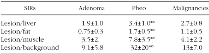
The results of quantitative evaluation of SIRs on T2-weighted MR images are reported in Table II; in particular, signal hyperintensity of pheos was signif-icantly higher compared to that of adenomas and malignant tumors, while no differences were observed between these latter adrenal lesions. The results of quantitative evaluation of SIRs on T1-weighted MR images performed after gadolinium administration are reported in Table III; in particular, signal intensity after gadolinium was significantly higher in pheos compared to that of adenomas and malignant tumors,
Figure 1.—A 42-year-old woman with a small (2 cm) right adrenal adenoma. A) Abdominal T1-weighted axial MR imaging shows a right adren-al mass with low signadren-al intensity. B) Abdominadren-al T1-weighted axiadren-al MR imaging after gadolinium administration shows no lesion enhance-ment.
while no differences were observed between differ-ent types of adrenal tumours.
Radionuclide imaging
The results of qualitative evaluation of nuclear stud-ies showed abnormally increased nor-cholesterol uptake in 100% of cases with adenomas (Figure 4) and, similarly, abnormal MIBG activity in 100% of pheos (Figure 5) as well as increased FDG uptake in 100% of malignant adrenal tumors (Figure 6); in par-ticular, no false positive or negative findings were observed in all series of radionuclide studies.
Discussion
The data on imaging of asymptomatic non-hyper-secreting adrenal masses has recently increased since the wide use of highly sensitive diagnostic techniques such as CT and MRI for the evaluation of the abdomen. In patients with these non-secreting tumours, the main role of imaging is not just to identify the presence of the tumour but to help characterise its type in a non-invasive way. This will help to ensure appropriate treatment. It is of particular importance to differentiate between benign and malignant lesions. CT and MR studies offer accurate anatomic details of any
adren-Figure 2.—A 37-year-old man with a small (2 cm) right adrenal adenoma. A) Abdominal T1 in-phase chemical-shift MR imaging shows lesion low signal intensity. B) Abdominal T1 out-phase chemical-shift MR imaging clearly shows loss of lesion signal intensity compared to in-phase acquisition.
Figure 3.—A 29-year-old man with a large (5.5 cm) left adrenal pheochromocytoma. A) Abdominal T1-weighted axial MR imaging shows a left adrenal mass with low signal intensity. B) Abdominal T2-weighted axial MR imaging shows a left adrenal mass with high signal inten-sity. C) Abdominal T1-weighted axial MR imaging after gadolinium administration shows significant, but inhomogeneous lesion enhancement.
al tumour and will give an initial analysis of lesion characterization.5, 6, 18, 19In particular, adrenal
adeno-mas may be suggested by CT on the basis of low attenuation coefficient on un-enhanced images and/or early as well as rapid washout on enhanced scans. Similarly, MR is able to characterize adenomas using dynamic-gadolinium or chemical-shift sequences.
Pheos also show specific MR features such as clearly increased signal intensity on T2-weighted images and significant enhancement after gadolinium administra-tion. Since gadolinium-enhanced MR imaging shows a considerable overlap in the characteristics of benign and malignant masses, its clinical applicability is lim-ited in distinguishing adenomatous from non-ade-nomatous adrenal lesions. Conversely, chemical-shift acquisition using the basis of fat content of a lesion can determine adenomas from malignant lesions. It is well known that adenomas contain a large amount of cytoplasmatic lipid in contrast to adrenal metastases which contain little or none. In the majority of aden-omas, the MR pattern seen consists of a reduction of signal intensity on out-of-phase scan compared with in-phase images, whereas in malignant lesions signal intensity remains unchanged. However, some aden-omas may contain insufficient lipid to result in loss of signal on out-of-phase scan as well as malignant pri-mary and secondary adrenal tumors may have lipid component showing loss of signal intensity on chem-ical-shift imaging and therefore this may reduce the sensitivity of this technique. Despite the effective-ness, this overlap in MRI appearances and tumour type mean that there remains a need for complemen-tary functional imaging.
Radionuclide modalities using specific tracers such as nor-cholesterol, MIBG and FDG may provide in vivo
tissue characterization of adrenal tumours being able
TABLEII.—Quantitative analysis of T2-weighted MR images.
SIRs Adenoma Pheo Malignancies
Lesion/liver 1.9±1.0 3.4±1.0*° 2.7±0.8
Lesion/fat 0.75±0.3 1.7±0.5*° 1.1±0.5
Lesion/muscle 3.5±2. 7.8±3.5*° 4.1±2.2
Lesion/background 9.1±5.8 32±20*° 13±7.0
SIRs: signal intensity ratios; Pheo: pheochromocytoma; p<0.001 vsadenoma; °p<0.03 vsmalignancies.
TABLEIII.—Quantitative analysis of T1-weighted gadolinium MR images.
SIRs Adenoma Pheo Malignancies
Lesion/liver 1.0±0.25 1.4±0.28*° 1.0±0.2
Lesion/fat 0.62±0.16 0.92±0.21*° 0.5±0.1
Lesion/muscle 2.0±0.5 2.6±0.35*° 1.8±0.6
Lesion/background 16±12 35±31*° 23±13
SIRs: signal intensity ratios; Pheo: pheochromocytoma; p<0.001 vsadenoma; °p<0.03 vsmalignancies.
Figure 4.—A 22-year-old woman with a 2.5 cm right adrenal adenoma. A) Abdominal T1-weighted axial MR imaging after gadolinium admin-istration shows a right adrenal mass. B) Abdominal posterior view of iodine-131 nor-cholesterol delayed imaging shows a round area of increased and exclusive tracer uptake in the right adrenal bed corresponding to the mass; no activity was observed in the contralateral side.
to differentiate between benign and malignant abnor-malities. Since these agents have no relation to each other and are taken up by individual parts of adren-als on the basis of entirely separate mechanism, they are able to differentiate different types of tumours.7, 12-15In particular, radiolabeled nor-cholesterol
scintigra-phy allows the characterization of functioning, but not hypersecretory, benign cortical adenomas; similar-ly, MIBG imaging has been demonstrated to be
use-ful to identify non-hypersecreting pheos 20, 21and
flu-oro-18 FDG using PET scanning has been shown to be able to recognize malignant adrenal tumours on the basis of increased glucose metabolism.13-15
In the present study, we compared the results of MR techniques and those of radionuclide studies in a group of patients with non-hypersecreting adrenal masses in order to evaluate imaging criteria for accu-rately performing tumour characterization. On the
Figure 5.—A 32-year-old man with a 3 cm right adrenal pheochromocytoma. A) Abdominal T1-weighted axial MR scan shows a round right adrenal mass. B) Abdominal posterior view of MIBG imaging shows a round area of intense and exclusive tracer uptake in the right adren-al bed; no activity was observed in the contradren-alateradren-al side.
Figure 6.—A 44-year-old woman with a 4.5 cm right adrenal metastasis by melanoma. A) Abdominal T1-weighted axial MR scan shows a large right adrenal mass. B) Uncorrected abdominal FDG PET scan shows intense tracer uptake by the right adrenal lesion; diffuse physiologic activ-ity is detectable in the liver and normal focal uptake is present in the upper pole of the right kidney as well as in the left kidney.
basis of our analysis, MR qualitative patterns to iden-tify adrenal adenomas consisted of no lesion enhance-ment after gadolinium or, mainly, signal intensity loss on out-phase CS imaging; this latter criterion is more appropriate since no gadolinium enhancement occurs also in other benign adrenal tumours.6, 18, 22Conversely,
the comparative evaluation of T1-T2 signal intensity changes demonstrated in our experience inhomoge-neous findings not allowing definite adenoma charac-terization since on T2 images 54% of these lesions were hypo- or iso-intense and the other 46% showed hyperintensity. For pheos, T2 signal hyperintensity and significant gadolinium enhancement were char-acteristicly occurring in all cases; likewise, in adren-al madren-alignancies signadren-al hyperintensity was found in all cases, while gadolinium enhancement occurred only in 63% of these tumours. These observations suggest that T2 signal hyperintensity is not accurate in differentiating adrenal masses, as previously reported by our group.22Since CS is not able to identify
phe-os or adrenal malignancies, other methodological approaches may be needed. For this purpose, the results of MR quantitative analysis showed that the degree of T2 signal hyperintensity and of lesion enhancement after gadolinium were significantly high-er in pheos compared to those of adenomas and malignant tumours but these latter lesions were not distinguished according to these quantitative criteria. These findings confirm the data of previous investiga-tions 4, 6, 7suggesting that, although the availability of
several technical methods, MR imaging provide only some presumptive criteria for tissue characterization in patients with non-hypersecreting adrenal masses. In particular, signal intensity loss on out-phase CS sequence seems to be the best marker for adenomas and the quantitative assessment of T2 or T1-gadolin-ium signal intensity allows to better characterize phe-os, while no specific MR criteria for adrenal malig-nancies are available.
In our series, the results of radionuclide studies were more homogeneous compared to those of MR imaging in terms of adrenal tumour characterization selectively showing nor-cholesterol uptake in adeno-mas, increased MIBG activity in pheo and abnormal FDG accumulation in, both primary and metastatic, adrenal malignancies. Although only a limited group of radionuclide scans were available, the 3 different radiotracers we used were able to identify different types of adrenal masses. In fact, nor-cholesterol uptake occurred in 100% of adenomas, MIBG concentration
was found in 100% of pheo, FDG accumulation was observed in 100% of malignancies, with no collec-tion of false negative or positive results. Therefore, according to our experience 10, 15, 23and that of others,7, 13, 14, 21radionuclide imaging using specific
radiocom-pounds offers specific non-invasive tissue character-ization in patients with non-hypersecreting adrenal masses. In this regard, the selection of the appropri-ate radioagent to be used depends on the clinical patient history but it may be limited if appropriate radiopharmaceuticals and nuclear equipment are not available. Because benign adenomas are the most common cause of non-hypersecreting adrenal tumors, labeled nor-cholesterol should be the 1st choice for
patients with no history of cancer disease. In case of a normal nor-cholesterol scan, MIBG should be used to confirm or rule out the presence of non-hyperse-creting pheo. If MIBG is also normal, FDG PET may be considered when the clinical suspicion of malignan-cy is high. Conversely, when neoplastic patients are evaluated, FDG PET should be initially performed followed, if normal, by nor-cholesterol and, in sequence, MIBG studies.
Nuclear studies are not routinely used in the diag-nostic protocols for managing patients with non-hypersecreting adrenal masses as well as are infre-quently considered for clinical decision making in such field.24, 25 Although combinations of anatomic
criteria by CT and MR are currently used to identify malignancy or benignancy without resorting to radio-nuclide imaging studies, on the basis of recent expe-riences a diagnostic role of molecular imaging has been proposed in this setting which may effect patient management.9 In fact, a radionuclide diagnosis of
non-hypersecreting adenoma by nor-cholesterol requests surgical treatment only if large tumour size occurs, otherwise clinical and imaging follow-up is appropriate; similarly, nuclear characterization of phe-ochromocytoma by MIBG allows to correctly plan surgery with adequate patient preparation, and, final-ly, early identification of adrenal malignancy by FDG PET may determine timely tumour resection with pos-sible favourable patient prognosis.
Conclusions
In conclusion, in patients with non-hypersecreting adrenal masses MR imaging may provide some pre-sumptive criteria to characterize tumour lesions; no
gadolinium enhancement and definite signal intensity loss on CSI suggest adenomas, while quantitatively measured T2-hyperintensity and/or gadolinium enhancement are able to identify pheos. Conversely, radionuclide techniques offer more specific findings since nor-cholesterol and MIBG uptake occur only in benign lesions such adenoma and pheos, respective-ly, and FDG accumulation detects adrenal malignancies. Therefore, multi-agents adrenal scintigraphy is strong-ly recommended in the diagnostic protocol of patients with non-hypersecreting adrenal masses, particularly when MR findings are uncertain and inconclusive.
References
1. Kloos RT, Gross MD, Francis IR, Korobkin M, Shapiro B. Incidentally discovered adrenal masses. Endocr Rev 1995;16:460-84.
2. Gross MD, Shapiro B. Clinical review 50. Clinical silent adrenal masses. J Clin Endocrinol Metab 1993;77:885-8.
3. Ross MS, Aron DC. Hormonal evaluation of the patient with an inci-dentally discovered adrenal mass. N Engl J Med 1990; 323: 1402-5.
4. Mayo-Smith WW, Boland GW, Noto RB, Lee M. State-of-the-art adrenal imaging. Radiographics 2001;21:995-1012.
5. Korobkin M, Brodeur FJ, Francis IR, Quint LE, Dunnik NR, Londy F. CT time-attenuation washout curves of adrenal adenomas and nonadenomas. Am J Roentgenol 1998;170:747-52.
6. Heinz-Peer G, Honigschnabl S, Schneider B, Niederle B, Kaserer K, Lechner G. Characterization of adrenal masses using MR imag-ing with histopathologic correlation. Am J Roentgenol 1999;173: 15-22.
7. Francis IR, Gross MD, Shapiro B, Korobkin M, Quint LE. Integrated imaging of adrenal disease. Radiology 1992;184:1-13.
8. Falke THM, Sandler MP. Classification of silent adrenal masses: time to get pratical. J Nucl Med 1994;35:1152-4.
9. Lawson MA. Role of molecular imaging in management of nonhy-persecreting adrenal masses. J Nucl Med 2001;42:893-4. 10. Maurea S, Klain M, Mainolfi C, Ziviello M, Salvatore M. The
diag-nostic role of radionuclide imaging in the evaluation of patients with non-hypersecreting adrenal masses. J Nucl Med 2001;42: 884-92.
11. Truong B, Jolles PR, Mullaney JM. Primary adrenal lymphoma: gallium scintigraphy and correlative imaging. J Nucl Med 1997;38:1770-1.
12. Shulkin BL, Wieland DM, Schwaiger M, Thompson NW, Francis IR, Haka MS et al.PET scanning with hydroxyephedrine: an approach to the localization of pheochromocytoma. J Nucl Med 1992;33: 1125-31.
13. Boland GW, Goldberg MA, Lee MJ, Mayo-Smith WW, Dixon J, McNicholas MM et al.Indeterminate adrenal mass in patients with cancer: evaluation at PET with 2-[F-18]-fluoro-2-deoxy-D-glucose. Radiology 1995;194:131-4.
14. Erasmus JJ, Patz EF, McAdams HP, Murray JG, Herndon J, Coleman RE et al.Evaluation of adrenal masses in patients with broncho-genic carcinoma using 18F fluorodeoxyglucose positron emission tomography. AJR Am J Roentgenol 1997;168:1357-60.
15. Maurea S, Mainolfi C, Bazzicalupo L, Panico MR, Imparato C, Alfano B et al.1999. Imaging of adrenal tumors using FDG PET: comparison of benign and malignant lesions. AJR Am J Roentgenol 1999;173:25-9.
16. Bergstrom M, Juhlin C, Bonasera TA, Sundin A, Rastad J, Akerstrom G et al.PET imaging of adrenal cortical tumors with the 11beta-hydroxylase tracer C-11 metomidate. J Nucl Med 2000;41:275-82. 17. Maurea S, Lastoria S, Caraco C, Klain M, Vanella P, Acampa W et al. The role of radiolabeled somatostatin analogs in adrenal imag-ing. Nucl Med Biol 1996;23:677-80.
18. Slapa RZ, Jakubowski W, Januszewicz A, Kasperlik-Zaluska AA, Dabrovska E, Fijuth J et al. Discriminatory power of MRI for dif-ferentiation of adrenal non-adenomas vs adenomas evaluated by means of ROC analysis: can biopsy be obviated? Eur Radiol 2000;10:95-104.
19. Caoili EM, Korobkin M, Francis IR, Cohan RH, Platt JF, Dunnick NR
et al.Adrenal masses: characterization with combined unenhanced and delayed enhanced CT. Radiology 2002;222:629-33.
20. Maurea S, Lastoria S, Cuocolo A, Celentano L, Salvatore M. The diagnosis of non-functioning pheochromocytoma: the role of I-123 metaiodobenzylguanidine imaging. Clin Nucl Med 1995;20:22-4. 21. Barzon L, Scaroni C, Sonino N, Fallo F, Gregianin M, Macri C et al.
Incidentally discovered adrenal tumors: endocrine and scinti-graphic correlates. J Clin Endocrinol Metab 1998;83:55-62. 22. Maurea S, Caracò C, Castelli L, Filice S, Alfano B, Ruffolo F et al.
La risonanza magnetica nello studio delle neoplasie surrenaliche: analisi qualitative e quantitative dell’intensità di segnale. Radiol Med 1998;95:199-207.
23. Maurea S, Klain M, Caracò C, Ziviello M, Salvatore M. Diagnostic accuracy of radionuclide imaging using iodine-131 nor-choleste-rol or MIBG in patients with hypersecreting or non-hypersecret-ing adrenal tumors. Nucl Med Comm 2002;23:951-60.
24. Young WF. Management approaches to adrenal incidentalomas: a view from Rochester, Minnesota. Endocrinol Metab Clin North Am 2000;29:159-85.
25. Schteingart DE. Management approaches to adrenal incidentalo-mas: a view from Ann Arbor, Michigan. Endocrinol Metab Clin North Am 2000;29:127-39.