The concept of a ‘target-rich, lead-poor’ pipeline in drug discovery, and widespread concern about the attrition rate of chemical compounds in (pre)clinical development, are together fuelling the search for better quality hits and chemical lead series. Researchers are rising to this challenge by devising new ways to identify chemical leads for specific protein targets and by using as starting points chemical structures that reflect the physical properties of successful oral drug molecules. A particular approach to lead identification for drug discovery involves the selection, screening and opti-mization of so-called fragments (also referred to as needles1, shapes2, binding elements3or seed templates4).
This review discusses fragment-based lead discovery, and focuses on the output of this new approach by collating published examples from 25 protein targets. These targets are primarily enzymes, and the screening techniques used include X-ray crystallography, nuclear magnetic resonance (NMR) spectroscopy,in vitro
bioassays and mass spectrometry.
The concepts that underpin the chemical frag-ments approach can be traced back to the pioneering work of Jencks5and Ariens6, who showed that
drug-like molecules can be regarded as the combination of two or more individual binding epitopes (or frag-ments). Screening for these binding epitopes using low-molecular-mass molecules is the essence of the fragments approach. The screened fragments are smaller (typically with Mr= 120–250 or 8–18 non-hydrogen
atoms), have less functionality and are correspondingly weaker than most hits from high-throughput screening (HTS), with typical binding affinities in the range mM–30 µM . Accordingly, biophysical screening meth-ods, including NMR7,8and X-ray crystallography9,10,
are particularly suitable, given the range of binding affinities that they can detect. In addition, these methods can afford significant structural understanding of the ligand–protein binding event, which is crucial in pri-oritizing fragment hits and rapidly developing them into leads. This process is illustrated in FIG. 1.
HTS is currently the established approach for hit identification within the pharmaceutical industry. Despite many successes from HTS, there is a need to find alternative approaches to lead discovery. Fragment-based approaches offer a number of attractive features compared with HTS. First, the number of compounds typically screened is in the range of only a hundred to a few thousand, because lower complexity compounds (fragments) have a higher probability of matching a target protein-binding site11. Second, a high proportion
of the atoms in a fragment hit are directly involved in the desired protein-binding interaction, so fragments can be described as efficient binders (that is, high binding energies per unit molecular mass); generally in HTS, larger and more potent compounds are identi-fied, but the compounds are less efficient binders. Third, when the binding interaction is structurally validated and understood, the subsequent chemical optimization
FRAGMENT-BASED LEAD DISCOVERY
David C. Rees, Miles Congreve, Christopher W. Murray and Robin Carr
Fragment-based lead discovery is gaining momentum in both large pharmaceutical companies and
biotechnology laboratories as a complementary approach to traditional screening. This is because
fragment-based approaches require significantly fewer compounds to be screened and
synthesized, and are showing a high success rate in generating chemical series with lead-like
properties. Compared with traditional screening hits, the starting fragments have considerably lower
molecular mass, and although the binding interactions of these fragments with a target protein are
weak, they are structurally understood through X-ray crystallography or NMR, and they exhibit high
‘ligand efficiency’. Here, we use examples from 25 different protein targets to describe chemical
strategies that exploit this structural knowledge to rapidly develop fragments into high-affinity leads.
Astex Technology, 436 Cambridge Science Park, Milton Road, Cambridge CB4 0QA, UK. Correspondence to D.C.R. e-mail: [email protected] doi:10.1038/nrd1467
RULE OF FIVE
Identifies several key properties that should be considered for compounds with oral delivery in mind. These properties are molecular mass <500 Da, cLogP <5, number of hydrogen-bond donors ≤5 and number of hydrogen-bond acceptors ≤10.
benefits from extensive design, synthesis of only a few compounds and a high success rate. And last, starting the chemical optimization stage with a low-molecular-mass fragment is likely to produce leads whose Mris still within the range desired for lead-likeness12–14. FIGURE 2 schematically illustrates how fragments and HTS hits compare as starting points for drug dis-covery (see also FIG. 3), and TABLE 1gives more detail on the differences between fragment-based lead discovery and HTS.
Drug-like, lead-like and fragment-like
At present, there are several guidelines for defining drug-like properties14,15, such as the ‘Lipinski RULE OF FIVE’16, which is used to maximize an oral drug candi-date’s probability of surviving development, and a more recent analysis based on the number of rotatable bonds, which indicated an upper limit of seven rotatable bonds in orally bioavailable drugs17.
Although these guidelines are useful for assessing the risk profile of an oral drugcandidate entering development, they are not necessarily as relevant for assessing the optimum properties of a lead. For example, studies of 450 pairs of commercial drugs and their corresponding leads indicated that, on average, leads had lower Mr, lower lipophilicity (cLogP), fewer aro-matic rings and fewer hydrogen-bond acceptors11.
A similar analysis concluded that libraries of com-pounds with Mr= 100–350 and cLogP = 1–3 are superior for finding leads compared with those com-prising drug-like compounds, with higher Mrand cLogP. The reason for this is that current lead-optimization practices routinely increase both Mr(on average by ~80) and lipophilicity (on average more than 1 Log unit) over those of initial leads. So, if the initial lead already possesses drug-like physical properties, then the optimization process is likely to result in drug can-didates with poorer drug-like properties. Overall, this suggests that ‘small is beautiful’ in quality hits and leads12,13.BOX 1describes the results from an analysis of
fragment hits against a range of targets, which imply that a ‘rule of three’ might be useful when constructing fragment libraries18.
The remainder of this review will present examples of fragment-based approaches in lead discovery, catego-rized into the following four types: fragment evolution; fragment linking; fragment self-assembly and fragment optimization (BOX 2).TABLES 2–6show the structures of the starting fragments and the optimized lead molecules from the 25 examples discussed.
Lead identification by fragment evolution
Identification of initial fragments using a direct binding technique is most useful if it is supported by some infor-mation about the binding mode of the fragment. With this type of information it is possible to develop hypotheses about how to build up larger and more complex molecules that target additional interactions in the active site of the protein. This ‘evolution’ leads to tighter-binding molecules, which can then be further optimized (FIG. 4; TABLE 2).
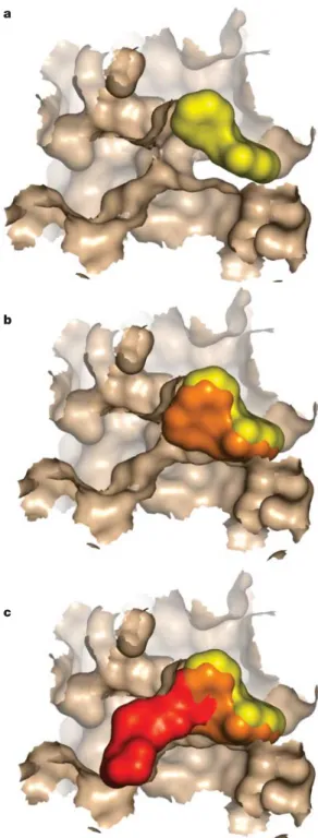
a
b
c
Figure 1 | Surface representation of fragment growth against p38 mitogen-activated protein kinase. a| The starting fragment is shown in yellow binding at the ATP-binding site (potency >1 mM, Mr = 200). b| A compound is displayed in orange (potency ~50 µM, Mr = 250) that was derived from
the first chemistry iteration on the starting fragment (also shown in yellow). c| A subsequent lead molecule is shown in red (potency ~ 300 nM, Mr = 425) superimposed on the earlier fragments. All binding modes are taken from crystal structures and in all cases the displayed protein surface is taken from the crystal structure of the red molecule in c. This was done to illustrate the evolution of the fragment in a clearer way, because there are changes in protein conformation in going from ato c53. The protein surface has been clipped to allow a clearer view of the inhibitors. The figure is based on data obtained during an Astex Technology programme.
at concentrations of 10–200 µΜunder conditions that favour disulphide exchange19. The cysteine-captured
ligands (not shown in the table) are readily identified by mass spectrometry and form stable complexes, even though in the absence of the tethering the ligands might bind very weakly. This approach was used to generate a very potent inhibitor ofthymidylate synthase. The affinity of the untethered ligand (~mM) was improved 3,000-fold by the synthesis of a small set of analogues guided by crystallographic structures of the tethered ligands.
Table 2, entry 3.Fragment chemistry was used to track the activity of inhibitors of the ATP-binding site ofp38 mitogen-activated protein (MAP) kinase2. On
the basis of a library of carefully selected drug-like scaffolds, and using NMR-based screening, a frag-ment with Kd= 1 mM was identified; by adding first one and then a second aromatic ring on to the central five-membered heterocyclic ring, activity reached Ki= 200 nM.
Table 2, entry 4.High-throughput X-ray crystallo-graphy screening of fragments using crystals of p38 MAP kinase led to the identification of a pyridyl indole derivative that has IC50 = 33 µΜ20. This compound was
one of a range of different hits identified by interpreta-tion of electron density in the active site following X-ray structure determination of fragment-soaked crystals. Fragment evolution based on the X-ray structure led to the lead illustrated (IC50 = 142 nM), and required the synthesis of only 70 compounds.
Table 2, entry 5.Fragment evolution against urokinase was initiated from the benzamidine analogue naph-thamidine, and resulted in a 1,000-fold increase in activity21. The optimization was driven by cycles of
structure-based design (SBD), and significant increases in selectivity were also observed over the starting frag-ments. The results are impressive because outside of the S1 specificity pocket in which the amidines bind, uro-kinase exhibits a relatively flat solvent-exposed active site compared with other serine proteases, such as thrombin and factor Xa.
In another example (not shown in TABLE 2), a sub-micromolar inhibitor against thrombin was discovered starting from aminobenzamidine (Ki= 34 µM). Using structure-based drug design, ten compounds were syn-thesized, and one compound showed a ~1,000-fold improvement in potency (Ki= 95 nM)22.
Table 2, entry 6. Structure-based screening was applied to block the binding of endogenous ligands to human adipocyte fatty-acid-binding protein FABP4 (REF. 23). Hits were initially located via an NMR screen and then subsequently crystallized in the protein. Limited structure-based optimization led to a low-molecular-mass 10 µM lead. Crystallization of this lead molecule showed that the binding mode for the initial fragment was retained in the more decorated molecule.
Table 2, entry 1.A method referred to as ‘needle screen-ing’ has been used to identify inhibitors that bind to the ATP-binding site of the bacterial enzyme DNA gyrase1.
Fourteen classes of needle hits (or fragments) were identified by in vitrobioassay and validated by biophysi-cal methods including NMR,SURFACE PLASMON RESONANCE (SPR) and X-ray crystallography. Subsequent three-dimensional-structure guided optimization using infor-mation obtained from the X-ray crystal structure of the ATP-binding pocket led to a compound that is reported to be >10,000-fold more active than the starting inda-zole fragment (maximal non-effective concentration (MNEC) in DNA gyrase inhibition 0.03 and >250 µg per ml, respectively). The authors suggest that needle screening provides chemical starting points that have no unnecessary structural elements, and therefore reduces the risk of toxicity or metabolic instability.
Table 2, entry 2.A native or engineered cysteine in a protein was allowed to react with a small library of disulphide-containing molecules (~1,200 compounds) SURFACE PLASMON RESONANCE
(SPR). A phenomenon which occurs when light is reflected off thin metal films to which target molecules are immobilized and addressed by ligands in a mobile phase. If binding occurs to the immobilized target then the local refractive index changes, which leads to the apparent rate constants for the association and dissociation phases of the reaction. The ratio of these values gives the apparent equilibrium constant (affinity).
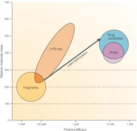
Relative molecular mass
Potency/efficacy 1 mM 100 µM 1 µM 10 nM 1 nM 0 100 200 300 400 500 600 700 Drugs Drug candidates Lead optim izatio n HTS hits Fragments
Figure 2 |Schematic comparison of the usual molecular mass and potency ranges of high-throughput screening hits with fragments as starting points for lead identification and drug discovery.The figure shows graphically a broad generalization of the range of molecular mass and potency for high-throughput screening (HTS) hits and fragments, superimposed on the typical requirements for leads, drug candidates and oral drugs using the same criteria. Fragment hits will have an Mrin the range 120–250 and low potency (mM–30 µM); to become useful leads, fragment potency will need to be increased, almost always with a resulting increase in Mr. HTS hits will have a much broader range of Mr(perhaps 250–600) and tend to be in the low-µM to high-nM potency range. Often, Mrwill need to be reduced and potency retained or increased to produce a quality lead series. Lead molecules themselves tend to be relatively potent for their size — for example, 100-nM potency for Mr = 350 (REF. 13). Oral small-molecule drug candidates tend to have
Mr<500 and be highly potent and efficacious, and launched oral drugs tend to have Mrsignificantly below 50014,15. There are, of course, many exceptions to the above historical generalizations.
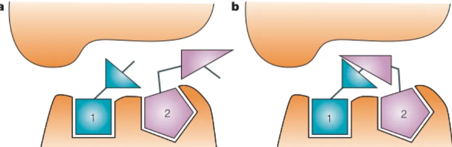
Lead identification by fragment linking
TABLES 3,4show examples in which two fragments have been identified that bind in separate binding sites that are close enough to each other to be chemically linked (FIG. 5). For this to be an efficient lead-identification approach, one needs to both identify the initial frag-ments and also have a process that allows the appropriate linking to be achieved in an efficient manner.
The potency increase achievable from optimally linking two fragments is often assumed to benefit from an approximate additivity effect, such that the free energy of binding of the joined molecule is approximately equal to the sum of the free energies of binding of the frag-ments (that is, two millimolar fragfrag-ments when joined together lead to a micromolar inhibitor)5. Such additivity
requires that the contribution from the linker is negligible and that the loss in rigid-body entropy on binding of all components to the enzyme is very small. Recently, an analysis of the experimental energetics associated with optimally linked fragments has suggested that the rigid-body entropy loss on protein binding constitutes a barrier of around three orders of magnitude to the binding affinity, and that this barrier is essentially independent of molecular mass25. The analysis implies that there should
be a super-additivity effect when two fragments are linked in an optimal fashion. Such super-additivity is observed for entries 2 and 7 in TABLE 3.
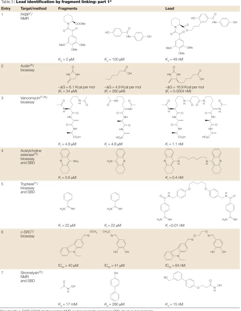
Table 3, entry 1.SAR by NMR7was used to identify a
potent 49 nM inhibitor of the FK506-binding protein (FKBP) binding domain by linking two weaker inhibitors (2 µM and 100 µM). NMR screening of a set of 1,000 fragments — including pipecolinic acid deriv-atives, a class of compounds known to bind to FKBP — identified the pipecolinic acid (Kd= 2 µM) and the diphenyl amide (Kd= 100 µM). Use of15N-13C-filtered
protein–ligand NUCLEAR OVERHAUSER EFFECT(NOE) data Table 2, entry 7.NMR screening (that is, structure–activity
relationships (SAR) by NMR7) was used to identify a
series of triazine-containing compounds that bind in the mM range to ErmAM methyl transferase24(for example, TABLE 2, entry 7, NMR Kd = 1 mM), an enzyme target for ameliorating antibiotic resistance. Optimization of this initial lead using parallel synthesis led to inhibitors in the low µΜrange (for example, table entry Ki= 7.5 µΜ). NMR and X-ray structures show that these non-nucleo-side compounds bind to the S -adenosylmethionine-binding site on the Erm protein and that there is scope for the incorporation of additional binding interactions.
NUCLEAR OVERHAUSER EFFECTS (NOEs). Changes in the intensity of NMR signals, which are caused by through-space dipole–dipole coupling. Upper distance constraints obtained from 1H–1H NOEs are used for
NMR structure determination of biological macromolecules.
Figure 3 |Schematic representation of a low-quality HTS hit.The high-throughput screening (HTS) hit is large and makes surface contact with the receptor without forming high-quality interactions in key pockets. The affinity is spread throughout the entire molecule and, in the absence of structural information, the medicinal chemist does not know which areas of the molecule to focus on during hit optimization. Experience shows that optimization of these kinds of hits is very difficult. This is in contrast to the schematic in FIG. 4, in which fragment 1 is much smaller, makes high-quality contacts with the receptor and has relatively weak affinity. It has been shown that such fragments can often be built up into attractive leads with the aid of structural information (for example, TABLE 2)
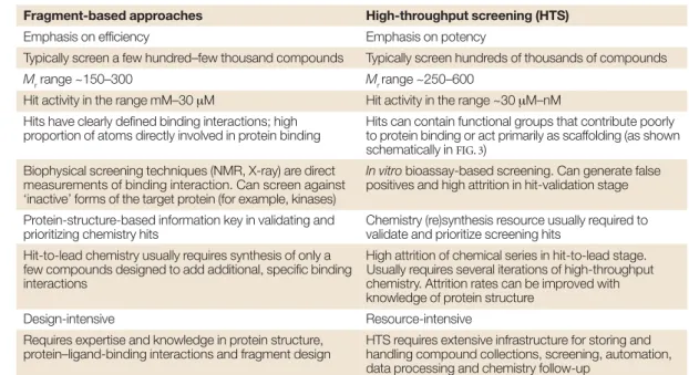
Table 1 | Comparison of fragment-based approaches and high-throughput screening
Fragment-based approaches High-throughput screening (HTS)
Emphasis on efficiency Emphasis on potency
Typically screen a few hundred–few thousand compounds Typically screen hundreds of thousands of compounds
Mrrange ~150–300 Mr range ~250–600
Hit activity in the range mM–30 µM Hit activity in the range ~30 µM–nM
Hits have clearly defined binding interactions; high Hits can contain functional groups that contribute poorly proportion of atoms directly involved in protein binding to protein binding or act primarily as scaffolding (as shown
schematically in FIG. 3)
Biophysical screening techniques (NMR, X-ray) are direct In vitrobioassay-based screening. Can generate false measurements of binding interaction. Can screen against positives and high attrition in hit-validation stage ‘inactive’ forms of the target protein (for example, kinases)
Protein-structure-based information key in validating and Chemistry (re)synthesis resource usually required to
prioritizing chemistry hits validate and prioritize screening hits
Hit-to-lead chemistry usually requires synthesis of only a High attrition of chemical series in hit-to-lead stage. few compounds designed to add additional, specific binding Usually requires several iterations of high-throughput
interactions chemistry. Attrition rates can be improved with
knowledge of protein structure
Design-intensive Resource-intensive
Requires expertise and knowledge in protein structure, HTS requires extensive infrastructure for storing and protein–ligand-binding interactions and fragment design handling compound collections, screening, automation,
data processing and chemistry follow-up
Table 3, entry 5.The quaternary structure of human mast cell tryptase, a trypsin-like serine protease30, has
been used to motivate the design of linked dimers of benzamidine31. An aspect of this approach was the key
observation that tryptase exists as a tetramer, and thereby provides the opportunity to couple ligands to form linked dimeric binders. The benzamidines illus-trated in TABLE 3were designed to bind into the S1 pock-ets of neighbouring tryptase monomers with a flexible linker region spanning the space between the two monomers, and the SAR of these inhibitors is supportive of this mechanism. It is interesting to note that APC-2059 (REF. 32), a tryptase inhibitor that has advanced to Phase II clinical trials, is also a dibasic inhibitor with an extended linking group (although this inhibitor was not derived directly from the fragment-linking approach). Table 3, entry 6.A fragment library of oximes was screened at high concentration against the non-receptor tyrosine kinase c-SRCto identify two weak hits (IC50= 40 µΜand 41 µΜ,respectively)3. The two respective
aldehydic precursors of the active oximes were then linked via a small library of di-hydroxylamine linkers to scope out and identify the optimum linking spacer group. This approach allowed the identification of a potent linked di-oxime (IC50 = 64 nM) without any structural knowledge of the target or binding sites. Table 3, entry 7.An elegant approach to lead identifica-tion has been developed that uses NMR screening of a secondary site in the presence of a small ligand that binds to a known active-site ‘hot spot’33. In this way, a
simple bi-phenyl analogue that binds to the P1′SITEof stromelysin with a Kd= 280 µΜwas identified in the presence of acetohydroxamic acid, which itself binds to the zinc ion in the catalytic site. Subsequently, an NMR structure of two fragments bound in the active site of the protein was solved. This allowed structure-guided design of a linked compound, which was found to be a very potent lead (Kd= 15 nM).
Table 4, entry 1.A technology termed extended teth-ering, related to that described in TABLE 2(entry 2), was used to identify a novel non-peptide inhibitor of allowed sufficient information on the binding site to be
obtained to guide the fragment linking.
Table 3, entry 2.Key questions concerning the efficiency of fragment linking have been addressed by detailed analysis of avidin binding by biotin analogues26.
Femtomolar inhibitors were fragmented into their con-stituent parts and their affinities were measured. The example illustrates the super-additivity in binding affinity that can be achieved when two fragments are joined together in a near-optimal fashion.
Table 3, entry 3.The binding properties ofVANCOMYCIN derivatives have been extensively studied with multi-valent acetyl-Lys-D-Ala-D-Ala molecules27,28. Here, only
the dimeric analogue is represented in the table. There is a large increase in affinity despite the introduction of additional rotatable bonds.
Table 3, entry 4.Starting from the crystal structure of an acridine analogue in acetylcholine esterase, it was dis-covered that a second molecule of the acridine analogue could be modelled in the active site29. Several dimeric
analogues with different linker lengths were synthesized, and an inhibitor that was 1,000-fold more potent than the original molecule was identified.
VANCOMYCIN
Vancomycin is an antibiotic that acts by binding to cell-wall precursors that terminate in the sequence D-Ala-D-Ala, thereby
inhibiting cell-wall synthesis. P1′SITE
The substrate residue that occurs immediately after the scissile amide bond in a protease. It is the key specificity site of matrix metalloproteases like stromelysin.
Box 1 |Rule of three
The properties of 40 fragmenthits identified against a range of targets using high-throughput X-ray crystallo-graphic screening technology has been examined18. The results indicated that on average fragment hits possessed properties consistent with a ‘rule of three’ in which:
•Mr<300
• Number of hydrogen-bond donors ≤3 • Number of hydrogen-bond acceptors ≤3 • cLogP = 3
In addition, it was noted that:
• The number of rotatable bonds was, on average,≤3 • Polar surface area was = 60 Å2
Box 2 |Summary of fragment-based approaches
Fragment evolution
An initial fragment is optimized by adding functionality to bind to adjacent regions of the active site (illustrated schematically in FIG. 4, with examples given in TABLE 2).
Fragment linking
Two (or more) fragments, which bind to proximal parts of the active site, are joined together to give a larger, higher-affinity-binding molecule (illustrated schematically in FIG. 5, with examples given in TABLES 3,4).
Fragment self-assembly
Fragments with complementary functional groups are allowed to react together in the presence of the protein target and the most potent larger molecule is detected (illustrated schematically in FIG. 6with examples given in TABLE 5). This includes approaches usually termed dynamic combinatorial chemistry.
Fragment optimization
Fragment approaches are used to optimize drug-like properties of a lead other than just binding affinity (illustrated schematically in FIG. 7, with examples given in TABLE 6).
these library fragments were coupled to known reversible cysteine-binding elements to generate potent reversible molecules.
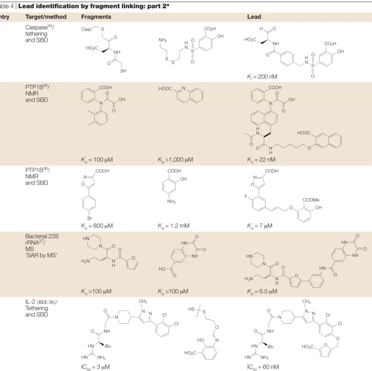
caspase-3, a cysteine protease34. Mass spectrometry was
used to identify the library members that had formed a disulphide bond to the tethered thiol. Subsequently,
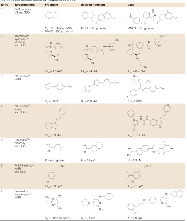
Table 2 | Lead identification by fragment evolution*
Entry Target/method Fragment Evolved fragment Lead
1 DNA gyrase1/
VS and SBD
Kd= 10 mM (by NMR) MNEC = 8 µg per ml MNEC = 30 ng per ml
MNEC >250 µg per ml 2 Thymidylate synthase19/ tethering and SBD IC50= 1.1 mM IC50= 24 µM IC50= 330 nM 3 p38 kinase2/ NMR Kd= 1 mM Kd= 200 µM Ki= 200 nM 4 p38 kinase20/ X-ray and SBD IC50= 33 µM IC50= 142 nM 5 Urokinase21/ bioassay and SBD Ki= not reported Ki= 5.9 µM Ki= 6.3 nM 6 FABP4 (REF. 23)/ NMR and SBD IC50= 590 µM IC50= 10 µM 7 Erm methyl transferase24/ NMR Kd= 1 mM (by NMR) Kd= 75 µM Ki= 7.5 µM
*See also FIG. 4. FABP, fatty-acid-binding protein; MNEC, maximal non-effective concentration; NMR, nuclear magnetic resonance; SBD, structure-based design; VS, virtual screening.
N H N N H N S O O N H N S O O O S O O N O OH S O O N O OH NH O CO2H CO2H H S O O N O NH NH O H CO2H CO2H CO2H N CO2H CO2H N S F CO2H CH3 N F N N N H N N H N H N H N O N N H2N HN H2N HN H2N HN HN O NH CO2H HO CO2H HO N N N NH2 NH2 CH3 S N N N NH NH2 N OH HN N N NH2 HN
overlay of the modelled tethered hits with a known crys-tal structure indicated a coupling of two of the fragments and allowed identification of sub-100 nM inhibitors.
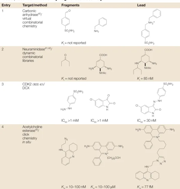
Lead identification by fragment self-assembly
The use of reactive fragments that are capable of self-assembly in the presence of a template molecule (such as a protein) (FIG. 6)is a large and growing field39.TABLE 5 cites some key examples in which two separate frag-ments are linked together to form a larger and more active inhibitor in the presence of the protein target itself. These examples are categorized here as ‘fragment self-assembly’ because the protein is used to self-select or to catalyse the synthesis of its own inhibitor without covalent attachment of the protein to the inhibitor. This definition forms the basis for distinguishing the examples in TABLE 6from those in TABLE 5.
Table 5, entry 1.In this prototype example, a mixture of imines was prepared from four amines and three alde-hydes under reversible conditions and then reduced to the corresponding amines40. High-performance liquid
chromatography was used to demonstrate the presence of all possible amines. When the same reaction is per-formed in the presence of carbonic anhydrase, the proportion of one amine is increased and this is pre-sumed to correspond to the strongest inhibitor. This is referred to as dynamic combinatorial chemistry39. Table 5, entry 2.Similar dynamic combinatorial chem-istry with imines has been used to identify a neur-aminidase inhibitor from diamine and ketone building blocks41,42. The amplification factor in the formation of
the secondary amine (table entry Ki = 85 nM), as determined by liquid chromatography–mass spec-trometry (LC–MS), was >30. The structure of this lead is closely related to the active component of osel-tamivir (Tamiflu; Roche), a marketed influenza neur-aminidase inhibitor.
Table 5, entry 3.A complementary approach has been reported in which inhibitors are formed from a dynamic combinatorial library in the presence of protein crystals and the binary complex is then observed directly by X-ray crystallography43. This has
been termed ‘dynamic combinatorial X-ray crystal-lography’ (DCX) and is illustrated by the identification of a previously reported inhibitor of cyclin-dependent kinase-2 (table entry IC50 = 30 nM) from a mixture of essentially inactive hydrazines and isatins as potential fragments for adjacent binding pockets within the ATP site.
Table 4, entries 2 and 3.Fragment linking has been applied to the extremely challenging target protein tyrosine phosphatase-1B35,36. Phosphatases
dephosphoryl-ate peptide substrdephosphoryl-ates by recognizing the doubly acidic phosphate group and key peptide residues; it is therefore not surprising that it is difficult to identify drug-like small-molecule leads against phosphatases. NMR screening of the catalytic phosphate-binding site, and a secondary phosphate-binding site, enabled the identifi-cation of a potent tool molecule (entry 2) and a useful hit (entry 3) by fragment linking.
Table 4, entry 4.Mass spectrometry has been used as the method for detecting the initial fragment hits for the 1061 region of bacterial 23S rRNA, a technique referred to as ‘SAR by MS’37. This subdomain of the ribonucleic
acid is part of the binding site for the antibiotic thiostrepton.In vitrobinding experiments showed that the two fragments bind to different sites on the RNA; subsequently, several fused compounds were synthesized that have markedly tighter binding. The authors reported that traditional HTS assays for this antibacterial target gave very low hit rates.
Table 4, entry 5.A potent small-molecule inhibitor of interleukin-2 (IL-2) was identified through the use of fragment tethering and fragment assembly38.
Ana-lysis of the X-ray structure of a known 3-µM inhibitor revealed that the protein is adaptive and able to undergo significant rearrangement, which creates small-molecule-binding sites. Ten individual cysteine mutants were designed to search the perimeter of the IL-2 binding ‘hot spot’. These mutants were then screened against a library of 7,000 disulphide-containing fragments. Analysis of the bound disulphide-tethered fragments indicated that these fragments could occupy a deep hydrophobic cavity within the adaptive region. An
a
1
b
1
Figure 4 |Fragment evolution. a| Fragment 1 binds to the receptor at one site. b| The lead molecule is evolved by building away from the starting fragment and making good contact with the upper surface and then by growing into a second pocket. For examples, see TABLE 2.
a 1 b 2 c 1 2
Figure 5 | Fragment linking. a| Fragment 1 binds to the receptor at one site. b| Fragment 2 binds to the receptor at an adjacent site.
Table 3 | Lead identification by fragment linking: part 1*
Entry Target/method Fragments Lead
1 FKBP7/
NMR
Kd= 2 µM Kd= 100 µM Kd= 49 nM
2 Avidin26/
bioassay
–∆G = 6.1 Kcal per mol –∆G = 4.9 Kcal per mol –∆G = 16.9 Kcal per mol
(Ki= 34 µM) (Ki= 260 µM) (Ki= 0.0004 nM) 3 Vancomycin27,28/ bioassay Ki= 4.8 µM Ki= 4.8 µM Ki= 1.1 nM 4 Acetylcholine esterase29/ bioassay and SBD Ki= 0.6 µM Ki= 0.4 nM 5 Tryptase31/ bioassay and SBD Ki= 22 µM Ki = 22 µM Ki<0.01 nM 6 c-SRC3/ bioassay IC50= 40 µM IC50= 41 µM IC50= 64 nM 7 Stromelysin33/ NMR and SBD Kd= 17 mM Kd= 280 µM Kd= 15 nM
*See also FIG. 5. FKBP, FK506-binding protein; NMR, nuclear magnetic resonance; SBD, structure-based design
MeO OMe OMe O N O COOMe OMe OMe MeO O N O O O HO O HN OH HO O HN OH NH HN O O OH HN NH O O OH O N H N H O HN O NH CO2H O O N H N H N H N H HN O NH NH NH CO2H HO2C O O O O O O NH O NH O NH O HN O HO2C NH2 N H2N N NH H2N H2N NH N H N H N N NH H2N O H N O O H N O NH H2N N N O O N HO OH N N OCH3 CH3O N HO OH N H OH O OH NC O N H OH O
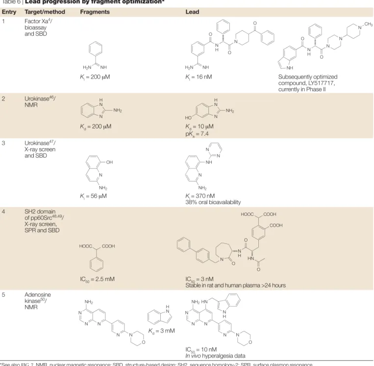
fragment. Subsequent optimization using a combination of medicinal chemistry and structure-based drug design led to the replacement of the benzamidine, a moiety that is often associated with poor oral bioavailability. Further lead optimization resulted in the discovery of the orally bioavailable candidate LY517717, and at a recent confer-ence presentation it was disclosed that this compound has successfully completed Phase I clinical trials, with clinical development ongoing. This example demon-strates that it is possible to generate an advanced drug candidate starting from a fragment with weak potency. Table 6, entry 2.One of the challenges in discovering useful urokinase inhibitors is to identify leads that do not have highly basic amidine or guanidine groups (pKa
>9). An NMR screen of more than 3,000 compounds led to the identification of 2-aminobenzimidazole as a weak (IC50 = 200 µΜ) but competitive inhibitor that binds to the same site on urokinase as the more tradi-tional inhibitors, but is less ionized at physiological pH (pKa= 7.5)46. Screening simple analogues of this hit led
to the 5-hydroxy derivative, IC50 = 10 µΜ(pKa = 7.4), and an X-ray co-complex structure was determined which could be used for subsequent optimization. Table 6, entry 3.The same target, urokinase, has been the subject of a fragment screen using X-ray crystallo-graphy47. Mixtures of weakly basic compounds were
assessed as urokinase ligands by crystal-soaking experi-ments. From a total of some 61 compounds, 8-hydroxy-2-amino quinoline was identified, and had Ki = 56 µΜ and pKa= 7.3. An attraction of the X-ray screening technique is that it generates structural information that can be used directly in subsequent lead optimization. In this case, the optimized compound had a 100-fold increase in enzyme activity (Ki = 370 nM), and, importantly, had 38% oral bioavailability.
Table 6, entry 4.Several drug discovery laboratories have targeted the sequence homology-2 (SH2) domain of the SRC protein family, and although nanomolar inhibitors have been identified, they are characterized by the presence of a phosphate group that is a liability in terms of its rapid hydrolysis and its contribution to poor cell penetration. An ‘SAR by X-ray’ approach has been developed and applied to find replacements for the phosphate group48. Small aromatic compounds were
screened as phenyl phosphate surrogates by performing crystal-soaking X-ray structure determination for ~20 compounds. Optimization led to a phenyl tricarboxylic acid replacement for the phenyl phosphate that retained activity (IC50 = 3 nM) and was stable in rat plasma for more than 24 hours (versus the phosphate analogue t1/2 = 0.6 hours). In a subsequent publication from the same laboratory49, an SPR (Biacore) assay was used as
a pre-screen before X-ray soaking of around 200 fragments to identify phosphotyrosine replacements. Table 6, entry 5.A strategy to break down an existing chemical lead into its fragments and scaffold, identify replacements for the fragments and then incorporate Table 5, entry 4.The building blocks are substantially
larger and higher affinity than many of the fragments described above, but the example is included to illus-trate the concept of self-assembly44,45. A femtomolar
inhibitor of acetylcholinesterase (Kd= 77 fM) has been identified from an array of building blocks containing an acetylene or azide group which undergo an irre-versible 1,3 dipolar cyclo-addition reaction as a result of binding to the enzyme. This is referred to as ‘click chemistry in situ’. The enzyme binds both fragments simultaneously, thereby positioning the azide and acetyl-ene groups sufficiently close to each other to allow them to react together much faster than would otherwise be the case. Structural knowledge of the binding interac-tions of acetylcholinesterase inhibitors facilitated the selection of these particular fragments.
Lead progression by fragment optimization TABLE 6shows examples in which the fragment approach has been used to optimize or modify properties other than just binding potency of a lead (FIG. 7).
Table 6, entry 1.This example illustrates the identification of a clinical candidate from a fragment-based discovery programme4. The known binder, benzamidine (200 µM),
was chosen as a seed template for the S1 POCKETof factor Xa. A structure-based virtual screening method was used to target proximal enzyme pockets and to drive three iter-ations of chemical synthesis. This resulted in the com-pound shown in TABLE 6, which has an IC50 = 16nM and is more than 10,000 times more potent than the initial
a
1 2
b
1 2
Figure 6 | Fragment self-assembly. a| Fragments 1 and 2 bind to receptor sites simultaneously with reacting groups positioned within conformational reach of each other, increasing the effective molarity of reacting groups. b| Lead molecule formed in the active site. For examples, see TABLE 5.
a
1
b
3
Figure 7 | Lead progression via fragment optimization. a| Existing lead molecule discovered by fragment-based approach. b| Lead molecule re-engineered to address optimization of a particular property (for example, selectivity, cell-based activity, oral activity or efficacy). For examples, see TABLE 6.
S1 POCKET
The pocket on a protease occupied by the substrate residue which immediately precedes the scissile amide bond. It is the key specificity pocket of trypsin-like serine proteases such as factor Xa, urokinase, tryptase and thrombin, in which lysine or arginine are the favoured substrate residues.
Summary and outlook
The fragments described in this review have Mr= 120–250 and binding affinity in the range mM–30 µΜ. The weak absolute potency of fragments belies their high efficiency as ligands, because fragments are extremely potent for their size51. Furthermore, crystal
structures of nanomolar leads that have been developed starting from fragments show very similar binding modes to those seen in the crystal structure of the iso-lated fragments, illustrating that fragments form very strong interactions with their proteins and provide good starting points for chemical optimization. the newly identified fragments back into the original
scaffold has been disclosed50. This offers the potential
to modify existing leads in terms of pharmacokinetic properties or side-effect profile. A known adenosine kinase inhibitor (Ki = 1.7 nM) was fragmented, and NMR screening was used to identify alternative groups for a meta-bromo phenyl side chain fragment. An example was the indole ring system (Kd = 3 mM), and when this was incorporated into the ATP-hinge-binding heterocycle scaffold, it gave a modified lead (IC50 = 10 nM) that retained potency in vitroand in vivo
in a hyperalgesia test.
Table 4 | Lead identification by fragment linking: part 2*
Entry Target/method Fragments Lead
1 Caspase34/ tethering and SBD Ki= 200 nM 2 PTP1B35/ NMR and SBD Kd= 100 µM Kd>1,000 µM Kd= 22 nM 3 PTP1B36/ NMR and SBD Kd= 800 µM Kd= 1.2 mM Kd= 7 µM 4 Bacterial 23S rRNA37/ MS ‘SAR by MS’ Kd>100 µM Kd>100 µM Kd= 6.5 µM 5 IL-2 (REF. 38)/ Tethering and SBD IC50= 3 µM IC50= 60 nM
*See also FIG. 5. Casp, caspase; IL, interleukin; MS, mass spectrometry; NMR, nuclear magnetic resonance; PTP, protein tyrosine phosphatase; SAR, structure–activity relationship; SBD, structure-based design.
O HO2C Casp S NH O SH N OH O O
COOH HOOC N COOH
HOOC N OH O O CO2H OH S H N S S NH2 O O CO2H HO2C OH H N O NH H O S O O H N N H O O O Br N O COOH COOH NH2 OH COOH COOMe N O F O OH HN N O N H O H2N O NH HN O HO O O HN N O N H O H2N O HN O NH HN O O N NH O O iBu HN NH2 CH3 HN N N Cl Cl N NH O O iBu HN NH2 CH3 HN N N Cl Cl O O HO2C HO HO2C N O S HS
activity is driven to the nanomolar affinity level. Some of the protein targets are ones that have been regarded as difficult to obtain attractive small-molecule leads against using conventional screening. Although fragment-based lead discovery is still too young for its output to be measured in terms of clinical impact, one example of a compound in Phase II is cited in which the initial chemical approach used fragments (see TABLE 6, entry 1).
The technique of fragment-based lead discovery can be seen as orthogonal to that of HTS; the screening techniques, affinity,Mr, size of compound libraries and chemical strategies for the subsequent ‘hit-to-lead’ stage are all different. It therefore represents an alternative and complementary strategy, and also presents interest-ing challenges, both organizationally and culturally, for companies that already have significant infrastructure for HTS. A simplistic summary is that the fragment approach emphasizes efficiency and design, whereas HTS emphasizes affinity and numbers.
Generally, fragments are identified using a biophysical screening method, most commonly NMR or protein crystallography, supported by a conventional enzyme bioassay. Consequently, information about the structure of the fragment–protein-binding interaction is gener-ated as part of the screening. This structural information means that it is possible to incorporate a large element of design in optimizing the fragment into a high-affinity lead, either by growing additional binding groups or joining two fragments together. As a result, fragments can be optimized into nanomolar leads via the synthesis of significantly fewer compounds than in traditional approaches.
The examples in this review are derived more or less equally in a three-way split from academic, biotechnology and established pharmaceutical laboratories, and cover 25 protein targets, the majority of which are enzymes. In all cases, the affinity of the starting fragments is increased by orders of magnitude, and in ~80% of the examples the
Table 5 | Lead identification by fragment self-asembly*
Entry Target/method Fragments Lead
1 Carbonic anhydrase40/ virtual combinatorial chemistry Ki = not reported 2 Neuraminidase41,42/ dynamic combinatorial libraries Ki= not reported Ki= 85 nM 3 CDK2 (REF. 43)/ DCX IC50>1 mM IC50>1 mM IC50= 30 nM 4 Acetylcholine esterase45/ click chemistry in situ Kd= 10–100 nM Kd= 10–100 µM Kd= 77 fM
*See also FIG. 6. CDK, cyclin-dependent kinase; DCX, dynamic combinatorial X-ray crystallography.
SO2NH2 O NH2 NH2+ SO2NH2 O COOH NHAc NH2 H2N COOH NHAc NH2 HN H2N NH SO2NH2 N H O O Cl N H O N NH SO2NH2 Cl HN N3 N N+ H2N NH2 (CH2)6CCH N+ H2N NH2 N N N HN N
that increasing Mrseems to correlate with attrition in clinical development, it is particularly attractive to start lead optimization with Mr<300. However, in the past, hits have often been selected on the basis of criteria asso-ciated with absolute potency or binding affinity, whereas the fragment approach emphasizes binding efficiency; that is, binding energy divided by Mr. Distinctive chemical strategies have been devised for building
low-Mrfragments into nanomolar leads. Fragment evolution involves growing out from an initial fragment, whereas in the fragment-linking strategy two separate fragments are linked together. In these respects, the fragment approach represents a new way of working for hit and lead identification in drug discovery projects.
Furthermore, screening for low-Mrfragment hits, as expected, has a substantially higher hit rate than HTS. This efficiency is carried into the chemical optimization stage, as knowledge about the protein–fragment-binding interaction allows higher affinity to be achieved with only a few compounds being synthesized.
Proponents of the fragments approach (see REF. 52 for a recent complementary review) argue that starting with a low-Mrcompound in which the entire molecule is needed for the target protein binding increases the chance of discovering molecules with the desired physicochemical properties of oral drugs (FIG. 2). In view of the widespread recognition that lead optimization is usually accompanied by increasing Mrby 100–150, and
Table 6 | Lead progression by fragment optimization*
Entry Target/method Fragments Lead
1 Factor Xa4/ bioassay and SBD Ki= 200 µM Ki= 16 nM Subsequently optimized compound, LY517717, currently in Phase II 2 Urokinase46/ NMR Kd= 200 µM Kd= 10 µM pKa= 7.4 3 Urokinase47/ X-ray screen and SBD Ki= 56 µM Ki= 370 nM 38% oral bioavailability 4 SH2 domain of pp60Src48,49/ X-ray screen, SPR and SBD IC50= 2.5 mM IC50= 3 nM
Stable in rat and human plasma >24 hours
5 Adenosine
kinase50/ NMR
Kd= 3 mM
IC50= 10 nM
In vivo hyperalgesia data
*See also FIG. 7. NMR, nuclear magnetic resonance; SBD, structure-based design; SH2, sequence homology-2; SPR, surface plasmon resonance.
NH H2N H2N NH N H N O O O N H N HO NH2 N NH2 H N N OH NH2 N NH NH2 N N COOH HOOC HOOC COOH COOH N H O HN O N O N N N NH2 N N O H N N N N NH2 N N O HN N H N N N CH3 N H NH O O
1. Boehm, H. J. et al. Novel inhibitors of DNA gyrase: 3D structure based biased needle screening, hit validation by biophysical methods, and 3D guided optimization. A promising alternative to random screening. J. Med. Chem.
43, 2664–2674 (2000).
An early description of a strategy to identify needles via virtual screening, in vitroscreening and validation by biophysical methods; also provides an example of fragment evolution applied to DNA gyrase. 2. Fejzo, J. et al. The SHAPES strategy: an NMR-based
approach for lead generation in drug discovery. Chem. Biol.
6, 755–769 (1999).
3. Maly, D. J., Choong, I. C. & Ellman, J. A. Combinatorial target-guided ligand assembly: identification of potent subtype-selective c-Src inhibitors. Proc. Natl Acad. Sci. USA97, 2419–2424 (2000).
4. Liebeschuetz, J. W. et al. PRO_SELECT: combining structure-based drug design and array-based chemistry for rapid lead discovery. 2. The development of a series of highly potent and selective factor Xa inhibitors. J. Med. Chem.45, 1221–1232 (2002).
5. Jencks, W. P. On the attribution and additivity of binding energies. Proc. Natl Acad. Sci. USA78, 4046–4050 (1981). 6. Farmer, P. S. & Ariens, E. J. Speculations on the design of
nonpeptidic peptidomimetics. Trends Pharmacol. Sci.3, 362–365 (1982).
7. Shuker, S. B., Hajduk, P. J., Meadows, R. P. & Fesik, S. W. Discovering high-affinity ligands for proteins: SAR by NMR.
Science274, 1531–1534 (1996).
Prototype description of the SAR by NMR technique and an early example of fragment linking to obtain nM ligands for the FK506-binding protein.
8. Pellecchia, M., Sem, D. S. & Wuthrich,K. NMR in drug discovery. Nature Rev. Drug Discov.1, 211–219 (2002). 9. Kuhn, P., Wilson, K., Patch, M. G. & Stevens, R. C. The
genesis of high-throughput structure-based drug discovery using protein crystallography. Curr. Opin. Chem. Biol.6, 704–710 (2002).
10. Blundell, T. L., Jhoti, H. & Abell, C. High-throughput crystallography for lead discovery in drug design. Nature Rev. Drug Discov.1, 45–54 (2002).
Review of the techniques required for high-throughput X-ray crystallography and discussion of this as a screening technique for lead discovery. 11. Hann, M. M., Leach, A. R. & Harper, G. Molecular
complexity and its impact on the probability of finding leads for drug discovery. J. Chem. Inf. Comput. Sci.41, 856–864 (2001).
12. Oprea, T. I., Davis, A. M., Teague, S. J. & Leeson, P. D. Is there a difference between leads and drugs? A historical perspective.J. Chem. Inf. Comput. Sci.41, 1308–1315 (2001).
13. Teague, S. J., Davis, A. M., Leeson, P. D. & Oprea, T. The design of leadlike combinatorial libraries. Angew. Chem. Int. Ed. Engl.38, 3743–3748 (1999).
14. Wenlock, M. C., Austin, R. P., Barton, P., Davis, A. M. & Leeson, P. D. A comparison of physiochemical property profiles of development and marketed oral drugs. J. Med. Chem.46, 1250–1256 (2003).
15. Vieth, M. et al. Characteristic physical properties and structural fragments of marketed oral drugs. J. Med. Chem.
47, 224–232 (2004).
16. Lipinski, C. A., Lombardo, F., Dominy, B. W. & Feeney, P. J. Experimental and computational approaches to estimate solubility and permeability in drug discovery and development settings. Adv. Drug Deliv. Rev.46, 3–26 (2001). 17. Veber, D. F. et al. Molecular properties that influence the oral
bioavailability of drug candidates. J. Med. Chem.45, 2615–2623 (2002).
18. Congreve, M., Carr, R., Murray, C. & Jhoti, H. A ‘rule of three’ for fragment-based lead discovery? Drug Discov. Today8, 876–877 (2003).
19. Erlanson, D. A. et al. Site-directed ligand discovery. Proc. Natl Acad. Sci. USA97, 9367–9372 (2000). 20. Frederickson, M., Gill, A. L., Padova, A. &
Congreve, M. S. Preparation of indoles as p38 MAP kinase inhibitors. (Astex Technology Limited, UK. UK Patent WO 03/087087 (2003).
21. Wendt, M. D. et al. Identification of novel binding interactions in the development of potent, selective 2-naphthamidine inhibitors of urokinase: synthesis, structural analysis, and sar of N-phenyl amide 6-substitution. J. Med. Chem.47, 303–324 (2004).
22. Bohm, H. J., Banner, D. W. & Weber, L. Combinatorial docking and combinatorial chemistry: design of potent non-peptide thrombin inhibitors. J. Comput. Aided Mol. Des.13, 51–56 (1999).
23. van Dongen, M. J. P. et al. Structure-based screening as applied to human FABP4: a highly efficient alternative to HTS for hit generation. J. Am. Chem. Soc.124, 11874–11880 (2002).
24. Hajduk, P. J. et al. Novel inhibitors of Erm methyltransferases from NMR and parallel synthesis. J. Med. Chem.42, 3852–3859 (1999).
25. Murray, C. W. & Verdonk, M. L. The consequences of translational and rotational entropy lost by small molecules on binding to proteins. J. Comput. Aided Mol. Des.16, 741–753 (2002).
26. Green, N. M. Avidin. Adv. Protein Chem.29, 85–133 (1975). 27. Rao, J. & Whitesides, G. M. Tight binding of a dimeric
derivative of vancomycin with dimeric L-Lys-D-Ala-D-Ala.
J. Am. Chem. Soc.119, 10286–10290 (1997). 28. Rao, J., Lahiri, J., Weis, R. M. & Whitesides, G. M. Design,
synthesis, and characterization of a high-affinity trivalent system derived from vancomycin and L-Lys-D-Ala-D-Ala.
J. Am. Chem. Soc.122, 2698–2710 (2000).
29. Pang, Y. P., Quiram, P., Jelacic, T., Hong, F. & Brimijoin, S. Highly potent, selective, and low cost
bis-tetrahydroaminacrine inhibitors of acetylcholinesterase. Steps toward novel drugs for treating Alzheimer’s disease.
J. Biol. Chem.271, 23646–23649 (1996).
30. Pereira, P. J. et al. Human β-tryptase is a ring-like tetramer with active sites facing a central pore. Nature392, 306–311 (1998).
31. Burgess, L. E. et al. Potent selective nonpeptidic inhibitors of human lung tryptase. Proc. Natl Acad. Sci. USA96, 8348–8352 (1999).
32. Rice, K. D. et al. Dibasic inhibitors of human mast cell tryptase. Part 2: structure–activity relationships and requirements for potent activity. Bioorg. Med. Chem. Lett.
10, 2361–2366 (2000).
33. Hajduk, P. J. et al. Discovery of potent nonpeptide inhibitors of stromelysin using SAR by NMR. J. Am. Chem. Soc.119, 5818–5827 (1997).
34. Erlanson, D. A. et al.In situassembly of enzyme inhibitors using extended tethering. Nature Biotechnol.21, 308–314 (2003).
35. Szczepankiewicz, B. G. et al. Discovery of a potent, selective protein tyrosine phosphatase 1B inhibitor using a linked-fragment strategy. J. Am. Chem. Soc.125, 4087–4096 (2003).
36. Liu, G. et al. Fragment screening and assembly: a highly efficient approach to a selective and cell active protein tyrosine phosphatase 1B inhibitor. J. Med. Chem.46, 4232–4235 (2003).
37. Swayze, E. E. et al. SAR by MS: a ligand based technique for drug lead discovery against structured RNA targets.
J. Med. Chem.45, 3816–3819 (2002).
38. Braisted, A. C. et al. Discovery of a potent small molecule IL-2 inhibitor through fragment assembly. J. Am. Chem. Soc.125, 3714–3715 (2003).
39. Ramstrom, O. & Lehn, J. M. Drug discovery by dynamic combinatorial libraries. Nature Rev. Drug Discov.1, 26–36 (2002).
40. Huc, I. & Lehn, J. M. Virtual combinatorial libraries: dynamic generation of molecular and supramolecular diversity by self-assembly. Proc. Natl Acad. Sci. USA94, 2106–2110 (1997).
41. Hochgurtel, M. et al. Ketones as building blocks for dynamic combinatorial libraries: highly active neuraminidase inhibitors generated via selection pressure of the biological target.
J. Med. Chem.46, 356–358 (2003). 42. Hochgurtel, M. et al. Target-induced formation of
neuraminidase inhibitors from in vitrovirtual combinatorial libraries. Proc. Natl Acad. Sci. USA99, 3382–3387 (2002). 43. Congreve, M. S. et al. Detection of ligands from a dynamic
combinatorial library by X-ray crystallography. Angew. Chem. Int. Ed. Engl.42, 4479–4482 (2003). 44. Bourne, Y. et al. Freeze-frame inhibitor captures
acetylcholinesterase in a unique conformation. Proc. Natl Acad. Sci. USA101, 1449–1454 (2004).
45. Lewis, W. G. et al. Click chemistry in situ:
acetylcholinesterase as a reaction vessel for the selective assembly of a femtomolar inhibitor from an array of building blocks. Angew. Chem. Int. Ed. Engl.41, 1053–1057 (2002). 46. Hajduk, P. J. et al. Identification of novel inhibitors of
urokinase via NMR-based screening. J. Med. Chem.43, 3862–3866 (2000).
47. Nienaber, V. L. et al. Discovering novel ligands for macromolecules using X-ray crystallographic screening.
Nature Biotechnol.18, 1105–1108 (2000). An early example of X-ray crystallography-driven screening applied to the discovery of urokinase inhibitors by fragment optimization. 48. Lesuisse, D. et al. SAR and X-ray. A new approach
combining fragment-based screening and rational drug design: application to the discovery of nanomolar inhibitors of Src SH2. J. Med. Chem.45, 2379–2387 (2002). 49. Lange, G. et al. Requirements for specific binding of low
affinity inhibitor fragments to the SH2 domain of (pp60)Src are identical to those for high affinity binding of full length inhibitors. J. Med. Chem.46, 5184–5195 (2003). 50. Hajduk, P. J. et al. Design of adenosine kinase inhibitors
from the NMR-based screening of fragments. J. Med. Chem.43, 4781–4786 (2000).
51. Hopkins, A. L, Groom, C. R & Alex, A. Ligand efficiency: a useful metric for lead selection. Drug Discov. Today9, 430–431 (2004).
This short article describes a simple but useful way to quantify ‘ligand efficiency’ (the contribution of each non-hydrogen atom in a lead to the overall binding affinity).
52. Erlanson, D. A., McDowell, R. S. & O’Brien, T. Fragment-based drug discovery. J. Med. Chem. 47, 3463–3482 (2004).
53. Teague, S. J. Implications of protein flexibility for drug discovery. Nature Rev. Drug Discov.2, 527–541 (2003).
Acknowledgements
H. Jhoti for scientific input, R. Taylor for figure 1 and H. Sore for proof reading.
Competing interests statement
The authors declare competing financial interests: see Web version for details.
Online links
DATABASES
The following terms in this article are linked online to: EntrezGene:
Caspase-3 | c-SRC | cyclin-dependent kinase-2 | p38 MAP kinase | protein tyrosine phosphatase-1B | tryptase | thymidylate kinase | urokinase