in Recurrent Hepatitis C Infection after Liver
Transplantation
Ziv Ben-Ari,
1Anat R. Tambur,
2Orit Pappo,
3Jaqueline Sulkes,
4Vera Pravica,
5Ian Hutchinson,
5Tirza Klein,
6Ran Tur-Kaspa,
1and Eytan Mor
7Background.Recurrent hepatitis C virus (HCV) infection is particularly aggressive in the post liver transplantation setting, with rapid progression of liver fibrosis. Platelet-derived growth factor (PDGF) is reportedly involved in the pathogenesis of liver fibrosis. The aim of this study was to evaluate the possible contribution of molecular variants of the PDGF-B gene to recurrent HCV infection after liver transplantation.
Methods.DNA was extracted from peripheral blood mononuclear cells of 40 patients who underwent liver transplan-tation for chronic HCV infection and genotyped for polymorphisms in PDGF-B at positions⫹1135 (A to C) and⫹286 (A to G). Intrahepatic PDGF-B expression was detected by immunohistochemistry and assessed semiquantitatively. Forty-seven healthy individuals served as controls.
Results.Recurrent HCV infection occurred in 34 patients (85%) after a median interval of 10.5 months (range 1.5–60.0). A statistically significant difference was observed in the distribution of the PDGF-B gene polymorphism at position⫹1135, but not
⫹286 between patients and controls (P⫽0.05). The A/A genotype occurred at a highly significantly increased rate in patients with recurrent HCV infection than in those without (64.7% vs. 16.67%,P⫽0.0001), and in patients with severe than in those with nonsevere recurrence (100% vs. 53.85%,P⫽0.05). The expression level of intrahepatic PDGF-B was found to be highly correlated with the fibrosis stage (P⬍0.0001). Further analysis yielded a highly statistically significant relationship between the PDGF-B gene polymorphism at position⫹1135 and clinical parameters of disease severity.
Conclusions.PDGF-B gene polymorphism appears to be associated with severe recurrent HCV infection after liver transplantation. PDGF-B may play an essential role in the development and progression of hepatic fibrosis. These findings, if confirmed, may have important therapeutic implications.
Keywords:PDGF, Gene, Polymorphism, Recurrent HCV, Liver transplantation. (Transplantation2006;81: 392–397)
H
epatitis C virus (HCV)-related liver failure is the leading indication for orthotopic liver transplantation (OLT) worldwide. After liver transplantation, recurrence of HCV infection occurs in virtually all patients. The majority show HCV-induced allograft hepatitis after follow-up of at least 5 years (1). The natural history of the disease is characterized by progression to cirrhosis in 6% to 23% of patients at a median of 3 to 4 years after transplantation; the estimated cumulative probability of developing HCV-related graft cirrhosis at 5years is 30% (1– 4). The development of cirrhosis is associated with reduced graft and patient survival (2), and once cirrhosis is established, the 1-year actuarial risk for decompensation is 42% (5).
HCV-related disease progression is particularly aggres-sive in the posttransplantation setting. Fibrosis progresses significantly faster than in immunocompetent patients (6), suggesting that the time to the development of cirrhosis is shorter—approximately 9 to 12 years (6). The rate of progres-sion is affected by a range of virus-, host-, and environment-related variables (7), namely: donor and recipient age, sex, and histocompatibility; year of transplantation; pretrans-plantation HCV RNA levels; viral genotype and quasispecies; use of immunosuppressive agents; and histological findings on the first liver biopsy.
Platelet-derived growth factor (PDGF), a cationic gly-coprotein of 24 kDa, is reportedly involved in the pathogen-esis of liver fibrosis (8 –11). PDGF is released by endothelial cells, macrophages, fibroblasts, and vascular smooth muscle cells as a homo- or heterodimer composed of PDGF-A or PDGF-B chains. It exists as an AA, BB, or AB isoform, co-valently linked by disulfide bonds (12). It has been implicated in the fibroproliferative response in various chronic inflam-matory disorders (13). The human PDGF-B gene has been localized to chromosome 22 (q12.3– q13.1) and spans 12 kb of V-cis-related sequences interrupted by four intervening sequences (14). Novel single nucleotide polymorphisms were noted in the promoters, 5=-UTRs and introns of PDGF-B. Substitutions observed were G¡A at position ⫹286
(5=-UTR, accession number AF169594) and A¡C at position 1Liver Institute and Department of Medicine D, Rabin Medical Center,
Bei-linson Campus, Petah Tiqva and Sackler Faculty of Medicine, Tel Aviv University, Tel Aviv, Israel.
2Department of Histocompatibility and Immunogenetics, Rush Medical Center, Chicago, IL.
3Department of Histopathology, Rabin Medical Center, Beilinson Campus, Petah Tiqva and Sackler Faculty of Medicine, Tel Aviv University, Tel Aviv, Israel. 4Epidemiology Unit, Rabin Medical Center, Beilinson Campus, Petah Tiqva
and Sackler Faculty of Medicine, Tel Aviv University, Tel Aviv, Israel. 5Immunology Research Group, University of Manchester, Manchester,
United Kingdom.
6Tissue Typing Laboratory, Rabin Medical Center, Beilinson Campus, Petah Tiqva and Sackler Faculty of Medicine, Tel Aviv University, Tel Aviv, Israel. 7Department of Organ Transplantation, Rabin Medical Center, Beilinson Campus, Petah Tiqva and Sackler Faculty of Medicine, Tel Aviv Univer-sity, Tel Aviv, Israel.
Address correspondence to: Ziv Ben Ari, M.D., The Liver Institute and De-partment of Medicine D Rabin Medical Center Beilinson Campus Petah Tiqva, 49100, Israel.
E-mail: gbenari@bezeqint.net.
Received 10 December 2004. Revision requested 28 December 2004. Accepted 19 May 2005.
The publisher apologizes for the delay of the publication of this article. Copyright © 2006 by Lippincott Williams & Wilkins
ISSN 0041-1337/06/8103-392
DOI: 10.1097/01.tp.0000173645.89064.c7
⫹1135 (first intron, accession number AF169595 (newly dis-covered by Ian Hutchinson and Vera Pravica, co-authors). There is no linkage disequilibrium between the two polymor-phisms. In the liver, PDGF is released by inflammatory cells and promotes the proliferation of hepatic stellate cells (HSCs), the main source of extracellular matrix proteins (15). During hepatic fibrogenesis, PDGF is overexpressed (16), and HSCs undergo a process of activation, developing a myofi-broblast-like phenotype (17) associated with increased pro-liferation and collagen synthesis (18). PDGF-B is the best characterized chemotactic factor for HSCs (19 –21), and is recognized as their most potent mitogen.
The aim of the present study was to evaluate the possi-ble association of molecular variants in the gene encoding PDGF-B with the risk of developing recurrent HCV infection after liver transplantation and with its severity.
PATIENTS AND METHODS Patients
The initial sample included all 46 patients who under-went OLT for chronic HCV infection at Rabin Medical Cen-ter between 1992 and 2002. Six were laCen-ter excluded because of concomitant alcoholic liver disease (n⫽2), a history of hepa-titis B (n⫽2), or missing data (n⫽2). All participants had more than 1 year of follow-up (mean, 49.3⫾28.4 months). The diagnosis of recurrent HCV infection was based on the presence of viremia by quantitative polymerase chain reac-tion (PCR) assay, increased serum transaminase levels, and histologic findings at diagnosis of lobular hepatitis in associ-ation with hepatocyte necrosis and midzonal macrovesicular steatosis. The immunosuppressive regimen included cyclo-sporine, azathioprine, or mycophenolate mofetil (CellCept), in addition to corticosteroids in 17 patients and tacrolimus and corticosteroids in 23 patients. Episodes of acute, histo-logically proven cellular rejection were treated with three consecutive boluses of intravenous Solu-Medrol, 1.0 g/d, and steroid-resistant episodes were treated with OKT3.
Patient files were reviewed for demographic character-istics, pretransplant HCV RNA load and genotype, immuno-suppressive regimen, rejection episodes, interval to recur-rence, and clinical progression at follow-up based on levels of serum bilirubin, and alanine aminotransferase (ALT), pres-ence of hepatic decompensation, retransplantation, and death due to recurrent HCV infection.
Severe recurrence was defined as stage 3– 4 fibrosis, or the presence of recurrent fibrosing cholestasis or graft failure (22).
The control group included 47 healthy (HCV Ab-neg-ative, HCV RNA-negative) volunteers (25 male, 22 female; mean age, 50.3⫾9.8 years) matched for sex and age (HCV Ab negative, HCV RNA negative) to the study group.
Genetic Polymorphism Assessment
The genetic profile of PDGF-B was analyzed in all pa-tients.
DNA Extraction
Genomic DNA was isolated by proteinase K digestion of fresh peripheral blood mononuclear cells, followed by phe-nol extraction and ethaphe-nol precipitation. DNA samples were
quantified and subjected to specific PCR reactions as de-scribed.
PDGF-B Gene Polymorphism
DNA was amplified by amplification refractory muta-tion system-polymerase chain reacmuta-tion (ARMS-based PCR) (23) in a 10:1 reaction containing 200 M deoxyribonucleoside (dNTPs); 1.5 mM magnesium chloride; 8.5% sucrose (w/v); 0.25 units Taq polymerase; 5 M specific control primers and 1 M internal control primers. PDGF-B⫹286 primers were de-signed (by Pravica V & Hutchinson I) to detect an A to G polymorphism: 5=-AAGGCCGGAACAGCTGAAA-3= and GGTCCGTCTGCCCGCCC/T; PCR product size was 323 bp. PDGF-B⫹1135 primers were designed to detect an A to C polymorphism: 5=-TGTTCTCGGGTTCCCAAAGG-3= and ATTCATTACCTTCGCCCCCC/A; PCR product size was 263 bp. The internal control primers amplified the human growth hormone gene: sense primer 5= -GCCTTCCCAAC-CATTCCCTTA-3=and antisense primer 5= -TCACGGATT-TCTGTTGTGTTTC-3⬘; PCR product size was 429 bp. PCR included 1 cycle of 95°C⫻1 min; 10 cycles of 95°C⫻15 sec-onds, 69°C⫻50 seconds, 72°C⫻40 seconds; and 20 cycles of 95°C⫻20 seconds, 59°C⫻50 seconds, 72°C⫻50 seconds.
Method validation was performed using alternative techniques: restriction fragment length polymorphism.
Histological Assessment
One pathologist (O.P.) blindly reviewed all hepatic specimens of the patients for overall necroinflammatory ac-tivity (grade 0 to 12) and fibrosis (stage 0 to 4) according to Knodell’s score. Intercurrent disease processes, such as acute cellular rejection, cytomegalovirus infection, biliary obstruc-tion, and ischemia were ruled out by serologic, immunohis-tochemical, radiological and endoscopic studies.
PDGF-B Immunohistochemistry Staining and Scoring
Immunohistology was performed on deparaffinized liver biopsy sections.
Briefly, sections (4l) of formalin-fixed, paraffin-em-bedded tissues were immunostained with mouse antihuman PDGF-B (U 376-UC, BioGenex, San Ramon, CA) diluted 1:100. Antigen retrieval was performed before application of the primary antibody. The tissue sections were placed in a bath with citrate buffer pH⫽6.0 and microwaved in a pres-sure cooker on high power (900 –1000 W) for 13 min until the pressure cooker was fully pressurized. Thereafter, the micro-wave level was reduced to 40% for another 5 min. Endoge-nous peroxidase reactions were blocked using Dako’s block-ing kit (Dako Co., Carpinterie, CA). Sections were then incubated with the primary antibody for 45 min followed with the Dako LSAB⫹kit peroxidase, which consists of la-beled streptavidin biotin reagents. Reactive sites were re-vealed by incubation with DAB (3.3-diaminobenzidine) (Dako). Sections were counterstained with hematoxylin. Pos-itive controls were sections of squamous carcinoma and neg-ative controls were the liver biopsies run concurrently with-out the primary antibody.
The intrahepatic PDGF-B expression was evaluated semiquantitatively according to the number of cells that stained positively (24).
Virological Assays
HCV RNA was tested by nested reverse-transcription polymerase chain reaction assay (RT-PCR) (Cobas Amplicor HCV Monitor Test, Roche Diagnostic Systems, Branchburg, NJ). Analytical sensitivity of the assay was 600 IU/ml. HCV genotypes were determined by a line-probe hybridization as-say (INNO-LiPa Innogenetics, Ghent, Belgium) directed to the 5=untranslated regions of the different HCV genotypes.
Statistical Analysis
Pearson and Spearman correlation coefficients and the significance for it (p) were calculated between the variables.
Chi-squared test or Fisher exact test were used, as ap-propriate, to analyze statistically significant relationships be-tween categorical variables (i.e., study vs. control group, re-currence, mutations⫹286 and⫹1135), and chi-squared test for equal proportions was used to analyze the distribution of each type of mutation between the study and control groups. Studentttest was used to analyze statistically significant dif-ferences in mean continuous parameters between the groups. To analyze statistically significant differences in mean contin-uous parameters between more than two groups of categori-cal variables, analysis of variance was used with Duncan mul-tiple comparison option. Due to the small sample size in some subgroups, a non-parametric Kruskal-Wallis test was also done. Multivariate stepwise logistic regression models were fitted to the data in order to predict mortality, disease recur-rence or disease severity. A p value less than or equal to 0.05 was considered statistically significant.
RESULTS
The baseline characteristics of the patients in this study are presented in Table 1: mean age was 52.7⫾9.6 years; 52.5% were male. HCV genotypes were available for 30 patients: 26 (86.6%) were genotype 1, 3 were genotype 2a and one was
genotype 3a. Mean pretransplant HCV RNA load was 414,954.8⫾880,991.4 IU/ml. The immunosuppressive regi-men was based on cyclosporine in 35% of the patients and on tacrolimus in 65%. The rejection rate was 45% (in 18 pa-tients). Of these, 12 patients developed mild rejection, five moderate, and only one patient severe (treated with OKT3).
Recurrent HCV infection developed in 34 patients (85%) after a median interval of 10.5 months (range 1.5– 60.0 months); in 8 patients, the recurrent infection appeared early (⬍12 months) and was severe. Mean serum ALT level at re-currence was 139.7⫾121.0 U/L, and mean serum bilirubin, 1.8⫾2.0 mg/dl; mean necroinflammatory score was 5.3⫾2.4, and mean fibrosis score, 1.7⫾1.1. Six of the 34 patients (17.6%) showed histologically proven cirrhosis during the follow-up period. The mean duration of follow-up was 49.8⫾28.4 months. The remaining six patients did not have histological recurrence of HCV. In all of them, liver biopsy was done during follow-up because of an increase in serum ALT level, which was found to be due to other causes and ultimately resolved.
Phenotypic Expression
The genotype and allele frequencies were determined in patients and controls (Table 2).
A statistically significant difference was observed in the distribution of the PDGF-B gene polymorphism at position
⫹1135 between the patients who underwent liver transplan-tation for HCV and the control group (P⫽0.05). However, no such statistically significant difference was observed at posi-tion⫹286.
Table 3 summarizes the phenotypic expression de-duced from the gene polymorphism in PDGF-B in the pa-tients with and without HCV infection after liver transplan-tation and in the patients with severe and non-severe recurrent disease. The allelic polymorphism of PDGF-B at position⫹1135 examined the presence of an A or C
nucleo-TABLE 1. Baseline Characteristics of Patients (n⫽40) (mean⫾SD)
Age (years) (mean) 52.7⫾9.6
Sex (F/M) 47.5%/52.5% Immunosuppression (cyclo/tacrolimus) 35%/65% Rejection episodes (n) 18 (45%)* Recurrent HCV (n) 34 (85%)
Alanine aminotransferase (U/L)** 139.7⫾121.0
Bilirubin (mg/dl)** (mean) 1.8⫾2.0
Necroinflammatory score** 5.3⫾2.4
Fibrosis score** 1.7⫾1.1
Cirrhosis (n) 6 (17.6%)
Viral genotype 1 (n) 26 (86.6%)***
Viral load (IU/ml) 414,954.8⫾880,991.4
Time to recurrence (months) (median) 10.5 (range, 1.5–60.0) Severe recurrence (n) 8 (23.5%) Follow-up (months) (mean) 49.8⫾28.4
* Of these, 12 patients developed mild rejection, 5 moderate, and 1 se-vere.
** At diagnosis of recurrent HCV after transplantation. *** HCV genotypes were available for 30 patients.
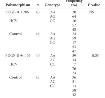
TABLE 2. Frequency of PDGF-B polymorphism
Polymorphism n Genotype Frequency (%) Pvalue PDGF-B⫹286 40 AA 20 NS AG 64 HCV GG 16 52 48 Control 46 AA 24 AG 59 GG 17 53 47 PDGF-B⫹1135 40 AA 59 0.05 AC 34 HCV CC 7 76 24 Control 43 AA 36 AC 51 CC 13 58 42
tide in this position, which translated directly into three phe-notypic expressions. A highly statistically significant differ-ence was observed between the patients who developed recurrent HCV infection after liver transplantation and those who did not (P⫽0.0001). The majority of patients (64.71%) with recurrent HCV infection exhibited the A/A genotype, 32.35% exhibited the A/C genotype, and 2.94% the C/C ge-notype; by contrast, 16.67% of the patients who did not de-velop HCV recurrence exhibited the A/A genotype, 16.67% the A/C genotype, and 66.66% the C/C genotype (Fig. 1).
A highly statistically significant difference was also ob-served among the patients with recurrence, by severity of dis-ease (P⫽0.05). All patients with severe recurrence had the A/A genotype. Of the patients with non-severe recurrence, 53.85% exhibited the A/A genotype, 42.31% the A/C geno-type, and 3.84% the C/C genotype (Fig. 1).
The allelic polymorphism of PDGF-B at position⫹286 examined the presence of an A or G nucleotide in this posi-tion, which translated directly into three phenotypic expres-sions. No statistically significant difference was observed be-tween the patients who developed a recurrent HCV infection and those who did not (Table 3). Within the group of patients with a recurrence, there was no statistical difference between those with severe and non-severe recurrent disease (Table 3).
Intrahepatic PDGF-B Immunohistochemistry
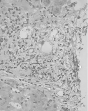
There was a statistically significant difference between the PDGF-B gene polymorphism at position⫹1135 and the intrahepatic immunohistochemistry for PDGF-B (P⫽0.027). PDGF-B immunoreactivity was found in mesenchymal cells of portal areas and fibrous septa localized diffusely in the cytoplasm compartment. A highly statistically significant cor-relation was found between the intrahepatic immunohisto-chemistry for PDGF-B and the fibrosis score (P⬍0.0001). The number of positive cells increased with progression of fibrosis (Figs. 2 and 3) (Table 4). No statistically significant correlation was found between PDGF-B immunohistochem-istry and the necroinflammatory score. A statistically signifi-cant correlation was found between PDGF-B immunohisto-chemistry and the serum bilirubin level (P⫽0.003), serum ALT (P⫽0.0002), severity of recurrence (P⫽0.0008), death due to HCV (P⫽0.0004).
Relationship between the PDGF-B Gene Polymorphism and Clinical Parameters
On univariate analysis, highly statistically significant correlation was noted between the PDGF-B gene
polymor-TABLE 3. PDGF-B gene polymorphism in patients after liver transplantation for HCV infection (n⫽40)
Posttransplantation HCV recurrence PDGF-B gene polymorphism ⴙ1135 (%) ⴙ286 (%) AA AC CC AA AG GG No recurrence (n⫽6) 1 (16.67) 1 (16.67) 4 (66.66) 3 (50.0) 3 (50.0) 0 Recurrence (n⫽34) 22 (64.71) 11 (32.35) 1 (2.94) 5 (14.7) 23 (67.65) 6 (17.65) Pvalue 0.0001 NS Severe recurrence (n⫽8) 8 (100) 0 0 1 (12.5) 6 (75.0) 1 (12.5) Nonsevere recurrence (n⫽26) 14 (53.85) 11 (42.31) 1 (3.84) 4 (15.38) 17 (65.38) 5 (19.23) Pvalue 0.05 NS
FIGURE 1. PDGF-B gene polymorphism (⫹1135) and re-current HCV infection.
FIGURE 2. Immunohistochemical detection of PDGF-B in liver biopsy in a patient with recurrent HCV infection and a fibrosis stage 0. No PDGF-B immunoreactivity was found.
phism at position ⫹1135 and recurrent HCV infection (P⫽0.001), serum ALT level (P⫽0.007), and serum bilirubin level (P⫽0.04) at recurrence, necroinflammatory score (P⫽0.03), fibrosis score (P⫽0.001), cirrhosis (P⫽0.003), se-vere recurrence (P⫽0.022), and death due to recurrent HCV infection (8 patients) (P⫽0.06) (Table 5). Factors found to have no association were immunosuppressive regimen, rejec-tion episodes, viral load, and viral genotype before transplan-tation, time to recurrence and retransplantation. Although multivariate analysis was performed, no statistical predictive factors were entered into the model due to sample size restric-tions (small number of deaths or recurrence events).
No statistically significant correlation was noted be-tween the PDGF-B gene polymorphism at position⫹286 and
the studied clinical parameters (Table 5), except for the necroinflammatory score (P⫽0.04).
DISCUSSION
Variables associated with the rapid disease progression of HCV reinfection after transplantation are under intensive investigation (7). To the best of our knowledge, this is the first study to examine the role of molecular variants of the gene encoding PDGF-B in this process.
We focused on two PDGF-B gene polymorphisms, at positions⫹1135 (A to C) and⫹286 (A to G). A statistically significant difference was noted in the distribution of the PDGF-B polymorphism at position⫹1135 between the study and control groups (P⫽0.05). No such relationship was found for position⫹286. Further analysis yielded significant relationships of the polymorphism at position ⫹1135 with both risk of recurrence and its severity. Specifically, the A/A genotype occurred at a significantly higher rate in patients with recurrence than in those without (64.7% vs. 16.67%,
P⫽0.0001), and in patients with severe recurrence than in those with non-severe recurrence (100% vs. 53.85%,
P⫽0.05). In addition, the polymorphism at position⫹1135 was associated with higher clinical parameters of disease at recurrence (serum ALT, bilirubin, fibrosis score, cirrhosis, and severity of infection). Due to the small number of events (death or recurrence), the model could not be fitted into a multivariate context. By contrast, the PDGF gene polymor-phism at position⫹286 did not correlate with recurrent HCV infection (p⫽NS).
These findings might help to shed light on the mecha-nisms underlying the accelerated course of HCV infection in liver transplant recipients compared to immune-competent individuals (6) both before and after the development of cir-rhosis (5). The posttransplantation period is known to be characterized by a faster rate of fibrogenesis until graft cirrho-sis develops and a greater risk of subsequent decompensation
FIGURE 3. Immunohistochemical detection of PDGF-B in liver biopsy in a patient with recurrent HCV infection and a fibrosis stage 3. The number of positive cells increased with the progression of fibrosis.
TABLE 4. PDGF-B expression levels in liver samples with different stages of fibrosisa
Stages of fibrosis n Expression levels (meanⴞSD)
F0 1b 3.6⫾1.1 F1 11 5.3⫾3.2 F2 14 9.2⫾3.4 F3 2 13.4⫾5.1 F4 6 19.6⫾5.9 aP⬍0.0001.
bn⫽number of cases in each fibrosis stage of patients with recurrent
HCV infection.
TABLE 5. Relationship between PDGF-B gene polymorphism and clinical parameters
Parameter Pvalue ⴙ1135 ⴙ286 Sex NS NS Age NS NS Immunosuppression (cyclosporine/tacrolimus) NS NS Rejection episodes NS NS Recurrence 0.001 NS Alanine aminotransferase 0.007 NS Bilirubin 0.04 NS Necroinflammatory score 0.03 0.04 Fibrosis score 0.001 NS Immunohistochemistry 0.027 NS Cirrhosis 0.003 NS Viral genotype NS NS Viral load NS NS Time to recurrence NS NS Severity 0.022 NS
Death due to hepatitis C virus 0.06 NS
than before transplantation (3,6, 25–27). Nevertheless al-though most patients develop recurrent HCV within 5 years, some maintain minimal to moderate liver damage whereas others advance rapidly to end-stage disease and graft failure (1–3,6,28). One of the key mediators of progressive liver disease are HSCs (15,18), they are considered to be the main source of extracellular matrix protein in the liver (19 –21). The present study was prompted by evidence indicating that the PDGF-B is one of the most potent mitogenic factor for HSCs (19 –21). PDGF is overexpressed during active hepatic fibrogenesis and may be involved in the transformation of HSCs to myofibroblast-like cells in vivo (16). These findings were supported by the study of Pinzani et al. (8), who re-ported markedly increased PDGF-A and -B chain mRNA ex-pression in cirrhotic livers, indicating the functional involve-ment of PDGF/PDGF-R (receptor) in liver fibrogenesis. Malizia et al. (9) also noted a high expression of PDGF-A and -B in mononuclear and proliferating ductal cells in livers with diseases of various etiologies. Indeed, we have found a statis-tically significant relationship between PDGF-B gene poly-morphism at position⫹1135 and the intrahepatic immuno-histochemistry for PDGF-B, and a highly statistically significant correlation between the immunohistochemistry for PDGF-B and the fibrosis stage. Therefore, PDGF-B may play an essential role in the development and progression of hepatic fibrosis in recurrent HCV infection after liver trans-plantation. The management of PDGF activity by antagonists or by soluble PDGF-B receptor that inhibit PDGF signaling and PDGF-induced proliferation in culture of HSC (29) might prevent aggressive liver fibrosis and improve prognosis in patients with recurrent HCV infection after liver trans-plantation.
Our data emphasize the potential importance of PDGF-B gene polymorphism in recurrent severe HCV infec-tion and the role of PDGF-B in the development and progres-sion of hepatic fibrosis. Further analysis in a larger cohort of patients is needed to confirm our results. These findings, if confirmed, may ultimately help clinicians therapy in selected patients at risk to design preemptive preventive
REFERENCES
1. Gane E, Portmann B, Naoumov N, et al. Long-term outcome of hepa-titis C infection after liver transplantation.N Engl J Med1996; 334: 815–820.
2. Prieto M, Berenguer M, Rayon M, et al. High incidence of allograft cirrhosis in hepatitis C virus genotype 1b infection following transplan-tation: relationship with rejection episodes.Hepatology1999; 29: 250– 256.
3. Testa G, Crippin JS, Netto GJ, et al. Liver transplantation for hepatitis C: recurrence and disease progression in 300 patients.Liver Transpl
2000; 6: 553–561.
4. Sanchez-Fueyos A, Resttepo J-C, Quinto I, et al. Impact of the recur-rence of hepatitis C after liver transplantation on the long term viability of the graft.Transplantation2002; 73: 56–63.
5. Berenguer M, Prieto M, Rayon JM, et al. Natural history of clinically compensated HCV-related graft cirrhosis following liver transplanta-tion.Hepatology2000; 32: 852–858.
6. Berenguer M, Ferrell L, Watson J, et al. HCV-related fibrosis
progres-sion following liver transplantation: increase in recent years.J Hepatol
2000; 32: 673–684.
7. Everson GT. Impact of immunosuppressive therapy on recurrence of hepatitis C.Liver Transpl2002; 8: s19–s27.
8. Pinzani M, Milani S, Herbst H, et al. Expression of platelet-derived growth factor and its receptors in normal human liver and during ac-tive hepatic fibrogenesis.Am J Pathol1996; 148: 785–800.
9. Malizia G, Brunt EM, Peters MG, et al. Growth factors and procollagen type 1 gene expression in human liver disease.Gastroenterology1995; 108: 145–156.
10. Grappone C, Pinzani M, Parola M, et al. Expression of platelet-derived growth factor in newly formed cholangiocytes during experimental biliary fibrosis in rats.J Hepatol1999; 31: 100–109.
11. Faiz Kabir Uddin Ahmed A, Ohtani H, Nio M, et al. In situ expression of fibrogenic growth factors and their receptors in biliary atresia: com-parison between early and late stages.J Pathol2000; 192: 73–80. 12. Ross R, Raines EW, Bowen-Pope DF. The biology of platelet-derived
growth factor.Cell1986; 46: 155–169.
13. Ross R, Masuda J, Raines EW, et al. Localization of PDGF-B protein in macrophages in all phases of atherogenesis.Science1990; 248: 1009–1012. 14. Collins T, Ginsburg D, Boss JM, et al. Cultured human endothelial cells express platelet-derived growth factor B chain: cDNA cloning and structural analysis.Nature1985; 316: 748–750.
15. Gressner AM, Bachem MG. Cellular sources of noncollagenous matrix proteins: role of fat storing cells in fibrogenesis.Semin Liver Dis1990; 10: 30–46.
16. Friedman SL, Arthur MJ. Activation of cultured rat hepatic lipocytes by Kupffer cell conditioned medium. Direct enhancement of matrix syn-thesis and stimulation of cell proliferation via induction of platelet-derived growth factor receptors.J Clin Invest1989; 84: 1780–1785. 17. Friedman SL. Molecular regulation of hepatic fibrosis, an integrated
cellular response to tissue injury.J Biol Chem2000; 275: 2247–2250. 18. Milani S, Herbst H, Schuppan D, et al. Cellular localization of type 1, 3
and 4 procollagen gene transcripts in normal and fibrotic human liver.
Am J Pathol1990; 137: 59–70.
19. Carloni V, Romanelli RG, Pinzani M, et al. Focal adhesion kinase and phospholipase C gamma involvement in adhesion and migration of human hepatic stellate cells.Gastroenterology1997; 112: 522–531. 20. Ikeda K, Wakahara T, Wang YQ, et al. In vitro migratory potential of
rat quiescent hepatic stellate cells and its augmentation by cell activa-tion.Hepatology1999; 29: 1760–1767.
21. Marra F, Gentilini A, Pinzani M, et al. Phosphatidylinositol 3-kinase is required for platelet-derived growth factor’s actions on hepatic stellate cells.Gastroenterology1997; 112: 1406–1409.
22. Gaglio PJ, Malireddy S, Levitt BS, et al. Increased risk of cholestatic hepatitis C in recipients of grafts from living versus cadaveric liver donors.Liver Transplantation2003; 9: 1028–1035.
23. Perrey C, Turner SJ, Pravica V, et al. ARMS-PCR technologies to de-termine IL-10, TNF-alfa, TNF-beta and TGF-beta 1 gene polymor-phisms.Transplant Immunol1999; 7: 127–128.
24. Lou S-M, Li Y-M, Wang K-M, et al. Expression of platelet-derived growth factor-BB in liver tissues of patients with chronic hepatitis B.
World J Gastroenterol2004; 10: 385–388.
25. Poynard T, Bedossa P, Opolon P. Natural history of liver fibrosis pro-gression in patients with chronic hepatitis C.Lancet1997; 349: 825– 832.
26. Fattovitch G, Giustina G, Degos F, et al. Morbidity and mortality in compensated cirrhosis type C: a retrospective follow-up study of 384 patients.Gastroenterology1997; 112: 463–472.
27. Hu K-Q, Tong MJ. The long-term outcomes of patients with compen-sated hepatitis C related cirrhosis and history of parenteral exposure in the United States.Hepatology1999; 29: 1311–1316.
28. Feray C, Gigou M, Samuel D, et al. The course of hepatitis C virus infection after liver transplantation.Hepatology1994; 20: 1137–1143. 29. Borkham-Kamphorst E, Stoll D, Gressner AM, Weiskirchen R.
Inhib-itory effect of soluble PDGF-beta receptor in culture-activated hepatic stellate cells.Biochem Biophys Res Commun2004; 317: 451–462.