REVIEW ARTICLE
Interleukin-6 role in head and neck
squamous cell carcinoma progression
Moaz M. Choudhary
a, Thomas J. France, Theodoros N. Teknos,
Pawan Kumar
*
Department of Otolaryngology-Head and Neck Surgery and Comprehensive Cancer Center, The Ohio State University, Columbus, OH 43210, USA
Received 9 May 2016; accepted 12 May 2016
Available online 20 July 2016
KEYWORDS Head and neck squamous cell carcinoma; Interleukin-6; JAK; STAT3; MAPK; PI3K/Akt; Nanog
Abstract Interleukin-6 (IL-6) is a pleiotropic cytokine which plays an important role in a num-ber of cellular processes including proliferation, survival, differentiation, migration and inva-sion. IL-6 mediates its downstream effects by activating a number of signaling cascades including JAK/STAT, PI3K/AKT and MAPK pathways. In addition to its effects on tumor cells, IL-6 also regulates tumor progression and tumor metastasis by modulating tumor angiogenesis and tumor lymphangiogenesis. A number of studies have shown that IL-6 levels are markedly upregulated in cancer patients. We and others have shown that high IL-6 expression indepen-dently predicts tumor recurrence, tumor metastasis and poor survival in head and neck cancer patients. Therefore targeting IL-6 signaling is a potential therapeutic strategy for the treat-ment of head and neck squamous cell carcinoma (HNSCC). In this review, we discuss the cur-rent understanding of the role of IL-6 in HNSCC progression and potential therapeutic strategies to target IL-6 signaling for the treatment of head and neck cancer patients. Copyrightª2016 Chinese Medical Association. Production and hosting by Elsevier B.V. on behalf of KeAi Communications Co., Ltd. This is an open access article under the CC BY-NC-ND license (http://creativecommons.org/licenses/by-nc-nd/4.0/).
* Corresponding author. 420 W. 12th Avenue, Room 464, Columbus, OH 43210, USA. Tel.:þ1 6146854325. E-mail address:Pawan.Kumar@osumc.edu(P. Kumar).
Peer review under responsibility of Chinese Medical Association.
Production and Hosting by Elsevier on behalf of KeAi
a Current address: Department of Internal Medicine, Rutgers New Jersey Medical School, Newark, NJ 07103, USA.
http://dx.doi.org/10.1016/j.wjorl.2016.05.002
2095-8811/Copyrightª2016 Chinese Medical Association. Production and hosting by Elsevier B.V. on behalf of KeAi Communications Co., Ltd. This is an open access article under the CC BY-NC-ND license (http://creativecommons.org/licenses/by-nc-nd/4.0/).
Available online atwww.sciencedirect.com
ScienceDirect
Introduction
Head and neck squamous cell carcinoma (HNSCC) remains a major health care problem worldwide, comprising almost 50% of all malignancies in some developing nations.1e3 Although advancements in the anti-cancer treatments including surgery, radiation and chemotherapy have increased the local control of HNSCC, the overall survival rates have not improved significantly over the last three decades.3,4Five year survival rates for patients with early stage localized head and neck cancers are more than 80% but drops to 40% when the disease has spread to the neck nodes, and to below 20% for patients with distant meta-static disease.4 Acquisition of chemoresistance and meta-static phenotype are the major causes of treatment failure and mortality in these patients.1,5It is therefore imperative that we gain a better understanding of the molecular mechanisms that contribute to the aggressive tumor phenotype in order to develop novel and effective strate-gies for the treatment of head and neck cancer patients.
Interleukin-6 (IL-6) is one of the key molecules that has been widely studied and implicated in poor clinical out-comes in HNSCC patients.6e10 IL-6 was initially identified and cloned as B-cell stimulatory factor-2.11e13At the same time a number of other molecules (IFN-b2, plasmacytoma growth factor and hepatocyte-stimulating factor) were independently cloned and found to be identical to IL-6.14e16Accumulating evidence has shown that IL-6 plays an important role in a number of biological process including immune regulation, hematopoiesis, inflammation and oncogenesis.17e19IL-6 is produced by a wide variety of cell types including immune cells (macrophages, dendritic cells and B-cells), endothelial cells and tumor cells.20e23We and others have shown that IL-6 levels are markedly elevated in the blood samples from cancer patients including HNSCC and independently predict tumor recurrence, poor survival and tumor metastasis.6e8A number of mechanistic studies have corroborated these clinical observations. Using our
in vitroandin vivohead and neck cancer models, we were able to demonstrate that IL-6 is a potent inducer of epithelial to mesenchymal transition (EMT) in head and neck cancer cell lines thereby promoting regional (lymph node) and distant (lung) metastasis.20 Similarly, Lederle et al24demonstrated that IL-6 promotes malignant growth of squamous cell carcinoma by regulating a complex cyto-kine and protease network. Recent studies have also high-lighted the role of IL-6 in the acquisition of chemoresistance and stem cell phenotype in cancer cells.25e28 We hereby present a review of recent studies that demonstrate the role of IL-6 in head and neck cancer progression.
Clinical significance of IL-6 in HNSCC
The association of IL-6 with clinical parameters (clinico-pathological factors) and oncological outcomes in HNSCC has been largely studied over the past two decades. Several studies have shown elevated levels of IL-6 in HNSCC.6,8,26,29 In a study of 65 untreated HNSCC patients and 20 healthy volunteers, Mojtahedi et al29found that serum levels of IL-6 and IL-18 were significantly increased in HNSCC patients
compared to healthy individuals, however only the differ-ence of IL-6 levels was found to be statistically significant. In addition, they showed that IL-6 concentration increased as tumor stage progressed and a significant difference was observed between stage IV vs stage I/II/III disease. These results suggest the activation of the Th2 arm of the immune response in HNSCC patients. However, Lathers et al30found elevated levels of IL-2 and GM-CSF in addition to IL-4, IL-6 and IL-10, thereby suggesting that HNSCC patients might have incomplete Th2 skewing. This theory of incomplete Th2 immune switch was further supported by the work of Sparano et al31where they examined blood samples from 58 patients of histologically proven HNSCC and showed that there were significantly higher levels of IL-6 and IL-10 as compared to IL-12. In addition, they showed that T3 and T4 patients had a positive relationship between tumor size and serum IL-6 levels. Similarly, in a caseecontrol study of 90 HNSCC patients and 39 controls, Riedel et al8showed higher levels of IL-6 in serum of HNSCC compared to healthy controls. They also showed a statistically significant cor-relation between serum IL-6 concentrations and with higher tumor stage and positive lymph nodes. In a prospective study of 85 patients with primary HNSCC, Tartour et al32 showed a significant association between higher lymph node (N) classification and elevated serum IL-6 levels.
In addition to serum levels of IL-6, tumor IL-6 expression both at mRNA and protein levels are also directly correlated with higher tumor stage and positive lymph nodes.9,33Wang et al33examined IL-6R and IL-6 mRNA expression in 86 oral squamous cell carcinoma tumor specimens and showed significantly higher IL-6R and IL-6 mRNA expression in tumor samples as compared to normal mucosa. IL-6 and IL-6R mRNA levels were also associated with larger tumors and more advanced histological grade. We have recently examined IL-6 expression in HNSCC by immunohistochem-istry and our results show a direct correlation between IL-6 expression and tumor stage, tumor recurrence, perineural invasion, extracapsular spread and inversely associated with HPV status.9
Considering the direct correlation between elevated IL-6 levels and high risk clinicopathological features in HNSCC, it does not come as a surprise that increased IL-6 levels are also associated with poor oncological outcomes in HNSCC and are reflective of a high burden of disease. This concept is further strengthened by the reduction of IL-6 levels after treatment in HNSCC patients. Several reports have studied IL-6’s association with oncological outcomes in HNSCC. Allen et al34studied numerous cytokines (IL-6, IL-8, growth-related oncogene-1 [GRO-1], VEGF and hepatocyte growth factors) longitudinally in a small prospective study of 30 patients with advanced oropharyngeal HNSCC receiving chemoradiation treatment by measuring serum levels at baseline and every 3 months. They showed a significant decrease in disease specific survival with longitudinal in-crease in levels of IL-6, suggesting that IL-6 could be a biomarker of treatment response and survival. Similarly, De Schutter et al35 did a retrospective study of 34 patient samples showing that pre-treatment IL-6 serum level is an independent predictor of local control, disease free sur-vival and overall sursur-vival. These concepts where further strengthened by our large prospective, longitudinal cohort study of 444 patients, where we have shown that serum IL-6
level was an independent predictor of recurrence and poor prognosis.
Elevated IL-6 levels are not only linked to tumor pro-gression but have also shown to mediate chemo and radio-resistance in HNSCC. Jinno et al26have shown that the high IL-6 expressing group showed significantly poor tumor response to the preoperative chemoradiotherapy as compared to the negative or low IL-6 expressing group. Similarly, Argiris et al36 showed that IL-6 levels were inversely associated with tumor response to induction chemotherapy in HNSCC patients with stages IIIeIVB receiving cisplatin doclataxel and Cetuximab. Recently, Stanam et al37 showed that upregulated IL-6 expression contributes to erlotinib resistance in HNSCC. Additionally, De Schutter et al35report a potential link between IL-6 and radio-resistance in HNSCC. High IL-6 levels in HNSCC pa-tients have also been found to be associated with health behaviors. We have recently shown that smoking (current and former) and decreased sleep was directly correlated with higher IL-6 levels.38We also showed that smoking was negatively associated with response to treatment and was an independent predictor of recurrence and poor prognosis. In addition, higher IL-6 levels were associated with muco-sitis and weight loss in HNSCC patients.
IL-6 effects on head and neck cancer
progression and metastasis
IL-6 is one of the cytokines that is commonly overexpressed in most cancer types including HNSCC.6,7,39e43IL-6 is a part of the cytokine family that shares a common glycoprotein 130 receptor (gp130; also known as CD130).44,45 Other members of IL-6 family include IL-11, leukemia inhibitory factor (LIF), ciliar neurotrophic factor (CTNF), oncostatin M (OSM), cardiotrophin-1 (CT-1) and cardiotrophin like cyto-kine (CLC).46,47IL-6 mediates its signaling by binding to an 80 kDa type-1 cell surface cytokinea-receptor subunit (IL-6R; also known as CD126)48 and a universally expressed 130 kDa gp130 to form an IL-6/IL-6R/gp130 complex that is clustered into a dimer structure (Fig. 1).49Once IL-6/IL-6R/ gp130 complex is formed, it recruits multiple signaling partners to mediate the IL-6’s wide range of cellular func-tions. One of the widely studied IL-6-mediated signaling pathway is the Jak family and STAT family of transcription factors.46 Dimerization of IL-6/IL-6R/gp130 leads to the activation of Jak family of kinases (Jak1, Jak2 and Tyk2) and recruitment of STAT proteins (STAT1, STAT3 and STAT5).50 STAT3 is one main STAT family member that is extensively studied in cancer.51Once STAT3 is activated, it forms a dimer in which the SH2 domain of one phospho-STAT3 binds to the phosphorylated Tyr705 of another STAT3 molecule and vice versa.52 The STAT3 dimers then translocate to the nucleus to regulate the expression a number of genes. The activation of JAK/STAT3 signaling by IL-6 is tightly regulated by SOCS (suppressor of cytokine signaling) and PIAS (protein inhibitor of activated STAT).53 In addition to the activation of JAK/STAT3 pathway, IL-6 also mediates its signaling by activating PI3K/AKT, RAS/ MAPK and Wnt signaling pathways.20,54,55
Although gp130 is universally expressed in human cells, IL-6R (gp126) expression is highly restricted to a few types of
cells.56Recent studies have shown that IL-6 is still able to mediate its signaling in the cells that lack IL-6R expression through an alternate signaling mechanism known as ‘trans-signaling’.57,58In the ‘classical signaling’ pathway, IL-6 binds to membrane-bound IL-6R, whereas in ‘trans-signaling’ circulating IL-6 binds to the soluble form of IL-6R (sIL-6R) and then this complex binds to membrane-bound gp130 and mediates the downstream signaling (Fig. 1). The significance of trans-signaling was highlighted by the presence of the biologically active form of sIL-6R in the blood samples from cancer patients.59,60The importance of the sIL-6R is high-lighted by the finding that 70% of the sIL-6R forms complexes with IL-6 in the blood and then binds directly to the membrane-bound gp130.61In addition, sIL-6R can function as a carrier protein for IL-6, thereby markedly prolonging the plasma half-life and signaling of IL-6.62
A number of studies have shown that IL-6 mediates persistent activation of the STAT3 pathway and up regula-tion of downstream target genes in head and neck cancers leading to increased tumor cell proliferation, migration, survival, invasion, epithelial to mesenchymal transition (EMT), cancer stem cell expansion, and chemo-resistance.20,37,63 In addition to IL-6, STAT3 can also be activated by EGFR that is often overexpressed in head and neck cancer cells.64 However, recent studies have shown that the persistent activation of STAT3 in HNSCC is pre-dominantly mediated by IL-6 and not by the EGFR pathway.65,66 Sriuranpong et al65 examined the interplay between STAT3, IL-6 and EGFR pathways using a panel of HNSCC cell lines. They showed that STAT3 was active in most of the cell lines, although only 3 out 10 HNSCC cell lines were moderately to strongly positive for activated EGFR. Even in EGFR-positive cell lines, STAT3 activation was not dependent on EGFR activation, as activated STAT3 persisted after treatment with AG1478 (EGFR inhibitor). In contrast, immunodepletion of IL-6 or blocking of gp130 (the signaling unit of the IL-6R complex) abolished STAT3 phos-phorylation, thereby confirming IL-6-dependent and EGFR-independent STAT3 activation. In a similar study, Squarize et al66 showed an increased activity of IL-6 promoter in HNSCC cells, which was dependent on the presence of an intact NFkappaB site. Higher IL-6 expression in turn pro-moted STAT3 activation in the EGFR-independent manner. The IL-6/STAT3 pathway has also been shown to promote cell survival in HNSCC cells by modulating the expression of serpin B3/B4, known as squamous cell carcinomas antigens 1 and 2.67A study by Ahmed et al67showed that targeting of IL-6/STAT3 signaling by a novel antibody against gp130 significantly inhibited STAT3 activation leading to prompt disappearance of cysteine proteases of serpin B3/B4 mRNAs and markedly enhancing tumor cell apoptosis. Recent studies have also shown that IL-6 modulates HNSCC tumorigenesis through epigenetic gene silencing via altering the CpG promoter methylation and repressing a number of tumor suppressor genes including CHFR, GATA5 and PAX6.68
Epithelial-mesenchymal transition (EMT) is a key process in tumor metastatic cascade that is characterized by the loss of cellecell junctions and cell polarity, resulting in the acquisition of migratory and invasive properties.69e71 We have recently shown that IL-6 promotes EMT in head and neck cancer cells by repressing E-cadherin expression via
the JAK/STAT3 signaling pathway.20 STAT3 knockdown in tumor cells or normal keratinocytes significantly decreased IL-6-mediated cell scattering, cell motility and reversal of EMT phenotype. Furthermore, tumor cells overexpressing IL-6 showed marked increase in lymph node and lung metastasis in a SCID mouse xenograft model. IL-6 has also been shown to modulate tumor cell motility by a number of downstream mediators. In our study, we show that IL-6/ STAT3 signaling modulate cell motility by regulating Focal Adhesion Kinase (FAK) activation.20Whereas Su et al63have shown that IL-6 enhances head and neck tumor cell motility by stabilizing Twist via the activation of casein kinase 2. Similarly, IL-6 has been shown to enhance head and neck cancer cell invasiveness by enhancing the expressions of MMP-1 and MMP-9.72e74 In addition, IL-6 also promotes tumor growth, metastasis and chemoresistance by enhancing tumor stem cell phenotype.25,28 We have recently shown that RhoC modulates cancer stem cell phenotype in head and neck cancer cells by regulating the expression of key stem cell transcription factors (nanog, oct3/4 and sox2) via the IL-6/STAT3 signaling pathway.25 IL-6/STAT3 signaling cascade was also shown to enhance cancer stem cell survival in HNSCC.28 Blocking of IL-6 signaling by tocilizumab (humanized anti-IL-6R antibody) significantly decreased cancer stem cell population and
markedly enhanced the anti-tumor effects of conventional chemotherapy.75
IL-6/STAT3 signaling as a potential target to
treat HNSCC patients
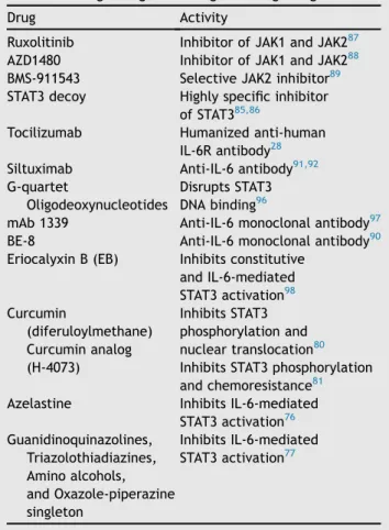
A number of clinical studies have shown that IL-6 levels are directly associated with poor overall survival, higher tumor stage, tumor recurrence and metastasis in a number of cancer types including HNSCC.6e8,26,36,72 Therefore, tar-geting IL-6 signaling is a potential therapeutic strategy for the treatment of patients with HNSCC. Because IL-6 has been shown to use multiple downstream molecules to mediate its biological function, it comes as no surprise that molecules with diverse modes of action have been tried as therapeutics to block IL-6 signaling (Table 1). Johnson et al76 employed high-content imaging (HCS) assays to screen 1726 compounds from the Library of Pharmacolog-ically Active Compounds and identified 51 inhibitors of IL-6-induced pSTAT3 activation. However, only three of these inhibitors selectively inhibited STAT3 as compared with STAT1. They subsequently confirmed Azelastine, an H1 receptor antagonist, as a selective inhibitor of IL-6-induced pSTAT3 activation that also reduced the growth
Fig. 1 IL-6 signaling pathways. IL-6 can mediate its intracellular signal by using both membrane-bound IL-6 receptor (IL-6 classical signaling) or soluble IL-6 receptor (IL-6 trans-signaling). In the classical IL-6 signaling, IL-6 binds to membrane-bound IL-6R (non-signaling receptor) and gp130 (signaling arm of IL-6 receptor) to form a complex. A complete functional complex consists of dimers of IL-6, IL-6R and gp130. IL-6 trans-signaling occurs in cells that lacks IL-6R. In this pathway, IL-6 first binds to soluble IL-6R (sIL-6R) to form a complex and then this complex binds to gp130 at the cell surface to mediate the intracellular signaling. A soluble form of IL-6 is released from cell surfaces by proteolytic cleavage (ADAM 17) or splicing of IL-6R mRNA. In both types of signaling, JAKs binds to Box1 and 2 of gp130 which leads to activation of JAKs. STATs also binds to gp130 but are phosphorylated by JAKs. IL-6 also activate PI3K/Akt pathway via JAKs. Both SOCS and SHP2 bind to gp130 via tyrosine 759. SOCS negatively regulate IL-6/STAT3 signaling by inhibiting both JAKs and STATs. SHP2 plays an important role in relating IL-6-mediated MAPK signaling.
of HNSCC cell lines. In the subsequent study, the same group screened a library of 94,491 compounds from the Molecular Library Screening Center Network (MLSCN) for the ability to inhibit IL-6-induced pSTAT3 activation.77Two hundred and three concentration-dependent inhibitors of IL-6-induced pSTAT3 activation were identified from this library and finally four chemical series (Guanidinoquina-zolines, Triazolothiadiazines, Amino alcohols, and Oxazole-piperazine singleton) progressed to lead optimi-zation stage.
Recent studies have demonstrated that curcumin, the constituent of the spice turmeric, in addition to its anti-inflammatory function also show potent anti-proliferative property in several tumor types. Curcumin has been shown to mediate its anti-tumor effects by inhibiting a number of signaling pathways including IL-6/STAT3 pathway.78,79 However, use of curcumin has been limited due to its poor bio-absorption.80 In order to overcome the poor bio-absorption of curcumin, we have recently developed a novel class of curcumin analogs, based on diarylidenylpiperidones (DAP), by incorporating a piperidone link to the beta-diketone structure and flu-oro substitutions on the phenyl groups.81This compound (H-4073) showed >5 fold higher cellular uptake in head and neck cancer cell lines as compared to curcumin and inhibited cell proliferation in a dose dependent manner. H-4073 mediated its anti-tumor effects by inhibiting JAK/ STAT3, FAK, Akt and VEGF signaling pathways that play important roles in cell proliferation, migration, survival and angiogenesis. In addition, pre-treatment of cisplatin-resistant HNSCC cell lines with H-4073 significantly reversed chemoresistance. In the SCID mouse xenograft model, H-4073 significantly enhanced the anti-tumor and anti-angiogenesis effects of cisplatin, with no added systemic toxicity. Another plant based natural compound ()-epigallocatechingallate (EGCG) has been shown to inhibit IL-6-mediated STAT3 phosphorylation.82e84 Lin et al82 showed that EGCG induces Fas/CD95-mediated apoptosis in head and neck squamous cell carcinoma cells by inhibiting constitutive and IL-6-induced JAK/ STAT3 signaling. EGCG was also able to block EGFR signaling and enhanced the anti-tumor effects of 5-flurouracil based chemotherapy.83 Recently, a chemi-cally modified cyclic STAT3 decoy oligonucleotide with improved serum and thermal stability was designed and tested in a nude mouse model. STAT3 decoy demon-strated a significant decrease in tumor volume compared with the control groups (mutant cyclic STAT3 decoy or saline) in conjunction with down modulation of STAT3 target gene expression without any significant side ef-fects.85 This STAT3 decoy oligonucleotide was further tested in phase 0 clinical trials.86Intravenous injection of the cyclic STAT3 decoy inhibited tumor growth and downregulated STAT3 target genes in the tumors.
Although targeting of the IL-6/STAT3 pathway in the head and neck cancer seems very promising, it has been challenging so far to target STAT3 using small molecule inhibitors in humans. What appears more promising is the use of JAK receptor antagonists. IL-6 phosphorylates STAT3 through Janus kinases (JAK) 1 & 2. Ruxolitinib is the first FDA approved drug that targets JAK1 and JAK2 kinases for myelofibrosis.87 Other drugs including AZD1480, a
preclinical JAK1/2 inhibitor, suppressed IL-6-induced STAT3 phosphorylation and showed subsequent human tumor growth suppression.88 Additionally, BMS-911543, a JAK2 selective inhibitor, showed similar suppression of tumor growth.89These drugs have not been tested in HNSCC yet. However, their potential benefits by blocking IL-6/STAT3 signaling do warrant HNSCC studies.
Another strategy to target IL-6/STAT3 signaling is to block IL-6 binding to IL-6R through neutralizing antibodies. Antibodies targeting IL-6 prevent the association of the ligand with its receptor, leading to prevention of STAT3 activation. Most of the studies using anti-IL-6 antibodies so far have been carried out using murine or humanized monoclonal antibodies.90e92 BE-8, a murine anti-IL-6 antibody, was developed by Diaclone and showed limited anti-tumor response.90It was shown that BE-8 could not block the daily IL-6 production>8 mg.90In addition BE-8 had a short half-life (3e4 days) and there is a natural drawback of murine antibodies inducing host anti-mouse antibody response.93 In contrast Siltuximab is a chimeric humanized monoclonal antibody that was developed by Centocor and has a much longer half-life (2 weeks).94 Siltuximab is currently in a phase II clinical trials for treatment in prostate cancer and multiple myeloma.91,95 Recently, tocilizumab, a humanized anti-IL-6R antibody (developed by Hoffmann-La Roche and Chugai), was tested in head and neck cancer model and was shown to disrupt primary human tumor initiation mediated by cancer stem cells.28
Table 1 Drugs designed to target IL-6 signaling.
Drug Activity
Ruxolitinib Inhibitor of JAK1 and JAK287 AZD1480 Inhibitor of JAK1 and JAK288 BMS-911543 Selective JAK2 inhibitor89
STAT3 decoy Highly specific inhibitor of STAT385,86
Tocilizumab Humanized anti-human IL-6R antibody28
Siltuximab Anti-IL-6 antibody91,92 G-quartet
Oligodeoxynucleotides
Disrupts STAT3 DNA binding96
mAb 1339 Anti-IL-6 monoclonal antibody97 BE-8 Anti-IL-6 monoclonal antibody90
Eriocalyxin B (EB) Inhibits constitutive and IL-6-mediated STAT3 activation98 Curcumin (diferuloylmethane) Curcumin analog (H-4073) Inhibits STAT3 phosphorylation and nuclear translocation80
Inhibits STAT3 phosphorylation and chemoresistance81 Azelastine Inhibits IL-6-mediated
STAT3 activation76 Guanidinoquinazolines, Triazolothiadiazines, Amino alcohols, and Oxazole-piperazine singleton Inhibits IL-6-mediated STAT3 activation77
Concluding remarks
In this review, we examined the role of IL-6 as a prognostic marker for patients with HNSCC, the importance of IL-6 in HNSCC tumor proliferation and metastasis, and finally the therapeutic potential of targeting the IL-6/STAT3 pathway. There is extensive data available to associate IL-6 levels in patients with clinicopathological parameters and oncolog-ical outcomes in HNSCC. Although IL-6 receptor is not expressed universally in different cell types, IL-6 can still mediate its intracellular signaling in these cells through trans-signaling mechanisms. As a result, IL-6/STAT3 signaling pathway is examined as an important therapeu-tic target. There has been extensive progress made in designing and testing novel drugs that target IL-6 signaling (Table 1), but we still need continued efforts to develop novel drugs which could effectively block this signaling pathway without significant adverse effects.
Funding program
NIH/NCI-CA178649 (P. Kumar) and The Ohio State University Comprehensive Cancer Center.
References
1.Leemans CR, Braakhuis BJ, Brakenhoff RH. The molecular biology of head and neck cancer. Nat Rev Cancer. 2011;11: 9e22.
2.Mehanna H, Paleri V, West CM, Nutting C. Head and neck cancerepart 1: epidemiology, presentation, and prevention. BMJ. 2010;341:c4684.
3.Siegel R, Naishadham D, Jemal A. Cancer statistics, 2012.CA Cancer J Clin. 2012;62:10e29.
4.Kalavrezos N, Bhandari R. Current trends and future perspec-tives in the surgical management of oral cancer.Oral Oncol. 2010;46:429e432.
5.Worden FP, Kumar B, Lee JS, et al. Chemoselection as a strategy for organ preservation in advanced oropharynx can-cer: response and survival positively associated with HPV16 copy number.J Clin Oncol. 2008;26:3138e3146.
6.Duffy SA, Taylor JM, Terrell JE, et al. Interleukin-6 predicts recurrence and survival among head and neck cancer patients. Cancer. 2008;113:750e757.
7.Chen Z, Malhotra PS, Thomas GR, et al. Expression of proin-flammatory and proangiogenic cytokines in patients with head and neck cancer.Clin Cancer Res. 1999;5:1369e1379. 8.Riedel F, Zaiss I, Herzog D, Go¨tte K, Naim R, Ho¨rmann K. Serum
levels of interleukin-6 in patients with primary head and neck squamous cell carcinoma.Anticancer Res. 2005;25:2761e2765. 9.Kumar B, Brown NV, Swanson BJ, et al. High expression of myoferlin is associated with poor outcome in oropharyngeal squamous cell carcinoma patients and is inversely associated with HPV-status.Oncotarget. 2016;7:18665e18677.
10. Moskowitz HS, Gooding WE, Thomas SM, et al. Serum biomarker modulation following molecular targeting of epidermal growth factor and cyclooxygenase pathways: a pilot randomized trial in head and neck cancer.Oral Oncol. 2012;48:1136e1145. 11. Hirano T, Yasukawa K, Harada H, et al. Complementary DNA for
a novel human interleukin (BSF-2) that induces B lymphocytes to produce immunoglobulin.Nature. 1986;324:73e76. 12. Yoshizaki K, Nakagawa T, Fukunaga K, Tseng LT, Yamamura Y,
Kishimoto T. Isolation and characterization of B cell
differentiation factor (BCDF) secreted from a human B lym-phoblastoid cell line.J Immunol. 1984;132:2948e2954. 13.Yasukawa K, Hirano T, Watanabe Y, et al. Structure and
expression of human B cell stimulatory factor-2 (BSF-2/IL-6) gene.EMBO J. 1987;6:2939e2945.
14.Gauldie J, Richards C, Harnish D, Lansdorp P, Baumann H. Interferon beta 2/B-cell stimulatory factor type 2 shares identity with monocyte-derived hepatocyte-stimulating factor and regulates the major acute phase protein response in liver cells.Proc Natl Acad Sci USA. 1987;84:7251e7255.
15.Van Snick J, Cayphas S, Szikora JP, et al. cDNA cloning of murine interleukin-HP1: homology with human interleukin 6. Eur J Immunol. 1988;18:193e197.
16.Woloski BM, Fuller GM. Identification and partial character-ization of hepatocyte-stimulating factor from leukemia cell lines: comparison with interleukin 1.Proc Natl Acad Sci USA. 1985;82:1443e1447.
17.Bethin KE, Vogt SK, Muglia LJ. Interleukin-6 is an essential, corticotropin-releasing hormone-independent stimulator of the adrenal axis during immune system activation.Proc Natl Acad Sci USA. 2000;97:9317e9322.
18.Kopf M, Baumann H, Freer G, et al. Impaired immune and acute-phase responses in interleukin-6-deficient mice.Nature. 1994;368:339e342.
19.Stahl EA, Raychaudhuri S, Remmers EF, et al. Genome-wide association study meta-analysis identifies seven new rheuma-toid arthritis risk loci.Nat Genet. 2010;42:508e514.
20.Yadav A, Kumar B, Datta J, Teknos TN, Kumar P. IL-6 promotes head and neck tumor metastasis by inducing epithelial-mesenchymal transition via the JAK-STAT3-SNAIL signaling pathway.Mol Cancer Res. 2011;9:1658e1667.
21.Sironi M, Breviario F, Proserpio P, et al. IL-1 stimulates IL-6 production in endothelial cells.J Immunol. 1989;142:549e553. 22.Watson JM, Sensintaffar JL, Berek JS, Martı´nez-Maza O. Constitutive production of interleukin 6 by ovarian cancer cell lines and by primary ovarian tumor cultures.Cancer Res. 1990; 50:6959e6965.
23.Andersson U, Sander B, Andersson J, Mo¨ller G. Concomitant production of different lymphokines in activated T cells.Eur J Immunol. 1988;18:2081e2084.
24.Lederle W, Depner S, Schnur S, et al. IL-6 promotes malignant growth of skin SCCs by regulating a network of autocrine and paracrine cytokines.Int J Cancer. 2011;128:2803e2814. 25.Islam M, Sharma S, Teknos TN. RhoC regulates cancer stem
cells in head and neck squamous cell carcinoma by over-expressing IL-6 and phosphorylation of STAT3.PLoS One. 2014; 9:e88527.
26.Jinno T, Kawano S, Maruse Y, et al. Increased expression of interleukin-6 predicts poor response to chemoradiotherapy and unfavorable prognosis in oral squamous cell carcinoma.Oncol Rep. 2015;33:2161e2168.
27.Liu CC, Lin JH, Hsu TW, et al. IL-6 enriched lung cancer stem-like cell population by inhibition of cell cycle regulators via DNMT1 upregulation.Int J Cancer. 2015;136:547e559. 28.Krishnamurthy S, Warner KA, Dong Z, et al. Endothelial
interleukin-6 defines the tumorigenic potential of primary human cancer stem cells.Stem Cells. 2014;32:2845e2857. 29.Mojtahedi Z, Khademi B, Hashemi SB, et al. Serum
interleukine-6 concentration, but not interleukine-18, is asso-ciated with head and neck squamous cell carcinoma progres-sion.Pathol Oncol Res. 2011;17:7e10.
30.Lathers DM, Achille NJ, Young MR. Incomplete Th2 skewing of cytokines in plasma of patients with squamous cell carcinoma of the head and neck.Hum Immunol. 2003;64:1160e1166. 31.Sparano A, Lathers DM, Achille N, Petruzzelli GJ, Young MR.
Modulation of Th1 and Th2 cytokine profiles and their associ-ation with advanced head and neck squamous cell carcinoma. Otolaryngol Head Neck Surg. 2004;131:573e576.
32.Tartour E, Deneux L, Mosseri V, et al. Soluble interleukin-2 receptor serum level as a predictor of locoregional control and survival for patients with head and neck carcinoma: results of a multivariate prospective study. Cancer. 1997;79: 1401e1408.
33.Wang YF, Chang SY, Tai SK, Li WY, Wang LS. Clinical significance of interleukin-6 and interleukin-6 receptor expressions in oral squamous cell carcinoma.Head Neck. 2002;24:850e858. 34.Allen C, Duffy S, Teknos T, et al. Nuclear factor-kappaB-related
serum factors as longitudinal biomarkers of response and sur-vival in advanced oropharyngeal carcinoma.Clin Cancer Res. 2007;13:3182e3190.
35.De Schutter H, Landuyt W, Verbeken E, Goethals L, Hermans R, Nuyts S. The prognostic value of the hypoxia markers CA IX and GLUT 1 and the cytokines VEGF and IL 6 in head and neck squamous cell carcinoma treated by radiotherapy þ/ chemotherapy.BMC Cancer. 2005;5:42.
36.Argiris A, Lee SC, Feinstein T, et al. Serum biomarkers as po-tential predictors of antitumor activity of cetuximab-containing therapy for locally advanced head and neck can-cer.Oral Oncol. 2011;47:961e966.
37.Stanam A, Love-Homan L, Joseph TS, Espinosa-Cotton M, Simons AL. Upregulated interleukin-6 expression contributes to erlotinib resistance in head and neck squamous cell carcinoma. Mol Oncol. 2015;9:1371e1383.
38.Duffy SA, Teknos T, Taylor JM, et al. Health behaviors predict higher interleukin-6 levels among patients newly diagnosed with head and neck squamous cell carcinoma. Cancer Epi-demiol Biomarkers Prev. 2013;22:374e381.
39.Chang CH, Hsiao CF, Yeh YM, et al. Circulating interleukin-6 level is a prognostic marker for survival in advanced nonsmall cell lung cancer patients treated with chemotherapy. Int J Cancer. 2013;132:1977e1985.
40.Okada S, Okusaka T, Ishii H, et al. Elevated serum interleukin-6 levels in patients with pancreatic cancer. Jpn J Clin Oncol. 1998;28:12e15.
41.Wong VW, Yu J, Cheng AS, et al. High serum interleukin-6 level predicts future hepatocellular carcinoma development in pa-tients with chronic hepatitis B. Int J Cancer. 2009;124: 2766e2770.
42.Nakashima J, Tachibana M, Horiguchi Y, et al. Serum inter-leukin 6 as a prognostic factor in patients with prostate cancer. Clin Cancer Res. 2000;6:2702e2706.
43.Zhang GJ, Adachi I. Serum interleukin-6 levels correlate to tumor progression and prognosis in metastatic breast carci-noma.Anticancer Res. 1999;19:1427e1432.
44.Hibi M, Murakami M, Saito M, Hirano T, Taga T, Kishimoto T. Molecular cloning and expression of an IL-6 signal transducer, gp130.Cell. 1990;63:1149e1157.
45.Taga T, Hibi M, Hirata Y, et al. Interleukin-6 triggers the as-sociation of its receptor with a possible signal transducer, gp130.Cell. 1989;58:573e581.
46.Heinrich PC, Behrmann I, Haan S, Hermanns HM, Mu ¨ller-Newen G, Schaper F. Principles of interleukin (IL)-6-type cytokine signalling and its regulation. Biochem J. 2003;374: 1e20.
47.Heinrich PC, Behrmann I, Mu¨ller-Newen G, Schaper F, Graeve L. Interleukin-6-type cytokine signalling through the gp130/Jak/STAT pathway.Biochem J. 1998;334:297e314. 48.Yamasaki K, Taga T, Hirata Y, et al. Cloning and expression of
the human interleukin-6 (BSF-2/IFN beta 2) receptor.Science. 1988;241:825e828.
49.Skiniotis G, Boulanger MJ, Garcia KC, Walz T. Signaling con-formations of the tall cytokine receptor gp130 when in com-plex with IL-6 and IL-6 receptor.Nat Struct Mol Biol. 2005;12: 545e551.
50.Hunter CA, Jones SA. IL-6 as a keystone cytokine in health and disease.Nat Immunol. 2015;16:448e457.
51. Yu H, Lee H, Herrmann A, Buettner R, Jove R. Revisiting STAT3 signalling in cancer: new and unexpected biological functions. Nat Rev Cancer. 2014;14:736e746.
52. Levy DE, Lee CK. What does Stat3 do?J Clin Invest. 2002;109: 1143e1148.
53. Kershaw NJ, Murphy JM, Lucet IS, Nicola NA, Babon JJ. Regu-lation of Janus kinases by SOCS proteins.Biochem Soc Trans. 2013;41:1042e1047.
54. Kim MS, Lee WS, Jeong J, Kim SJ, Jin W. Induction of meta-static potential by TrkB via activation of IL6/JAK2/STAT3 and PI3K/AKT signaling in breast cancer. Oncotarget. 2015;6: 40158e40171.
55. Linnskog R, Jo¨nsson G, Axelsson L, Prasad CP, Andersson T. Interleukin-6 drives melanoma cell motility through p38a -MAPK-dependent up-regulation of WNT5A expression. Mol Oncol. 2014;8:1365e1378.
56. Wolf J, Rose-John S, Garbers C. Interleukin-6 and its receptors: a highly regulated and dynamic system. Cytokine. 2014;70: 11e20.
57. Rose-John S. IL-6 trans-signaling via the soluble IL-6 receptor: importance for the pro-inflammatory activities of IL-6.Int J Biol Sci. 2012;8:1237e1247.
58. Rose-John S, Scheller J, Elson G, Jones SA. Interleukin-6 biology is coordinated by membrane-bound and soluble re-ceptors: role in inflammation and cancer.J Leukoc Biol. 2006; 80:227e236.
59. Jurczyszyn A, Czepiel J, Biesiada G, et al. HGF, sIL-6R and TGF-b1 play a significant role in the progression of multiple myeloma.J Cancer. 2014;5:518e524.
60. Won HS, Kim YA, Lee JS, et al. Soluble interleukin-6 receptor is a prognostic marker for relapse-free survival in estrogen receptor-positive breast cancer. Cancer Invest. 2013;31: 516e521.
61. Gaillard JP, Mani JC, Liautard J, Klein B, Brochier J. Inter-leukin-6 receptor signaling. I. gp80 and gp130 receptor inter-action in the absence of interleukin-6. Eur Cytokine Netw. 1999;10:43e48.
62. Peters M, Jacobs S, Ehlers M, et al. The function of the soluble interleukin 6 (IL-6) receptor in vivo: sensitization of human soluble IL-6 receptor transgenic mice towards IL-6 and pro-longation of the plasma half-life of IL-6.J Exp Med. 1996;183: 1399e1406.
63. Su YW, Xie TX, Sano D, Myers JN. IL-6 stabilizes Twist and en-hances tumor cell motility in head and neck cancer cells through activation of casein kinase 2.PLoS One. 2011;6:e19412. 64. Rubin Grandis J, Zeng Q, Drenning SD. Epidermal growth factor
receptor-mediated stat3 signaling blocks apoptosis in head and neck cancer.Laryngoscope. 2000;110:868e874.
65. Sriuranpong V, Park JI, Amornphimoltham P, Patel V, Nelkin BD, Gutkind JS. Epidermal growth factor receptor-independent constitutive activation of STAT3 in head and neck squamous cell carcinoma is mediated by the autocrine/paracrine stimu-lation of the interleukin 6/gp130 cytokine system.Cancer Res. 2003;63:2948e2956.
66. Squarize CH, Castilho RM, Sriuranpong V, Pinto Jr DS, Gutkind JS. Molecular cross-talk between the NFkappaB and STAT3 signaling pathways in head and neck squamous cell carcinoma.Neoplasia. 2006;8:733e746.
67. Ahmed ST, Darnell Jr JE. Serpin B3/B4, activated by STAT3, promote survival of squamous carcinoma cells.Biochem Bio-phys Res Commun. 2009;378:821e825.
68. Gasche JA, Hoffmann J, Boland CR, Goel A. Interleukin-6 promotes tumorigenesis by altering DNA methylation in oral cancer cells.Int J Cancer. 2011;129:1053e1063.
69. Kalluri R. EMT: when epithelial cells decide to become mesenchymal-like cells.J Clin Invest. 2009;119:1417e1419. 70. Lee MY, Chou CY, Tang MJ, Shen MR. Epithelial-mesenchymal
progression, epidermal growth factor receptor overexpression, and snail up-regulation.Clin Cancer Res. 2008;14:4743e4750. 71. Mandal M, Myers JN, Lippman SM, et al. Epithelial to mesen-chymal transition in head and neck squamous carcinoma: as-sociation of Src activation with E-cadherin down-regulation, vimentin expression, and aggressive tumor features.Cancer. 2008;112:2088e2100.
72. Kanazawa T, Nishino H, Hasegawa M, et al. Interleukin-6 directly influences proliferation and invasion potential of head and neck cancer cells.Eur Arch Otorhinolaryngol. 2007;264: 815e821.
73. Nishino H, Miyata M, Kitamura K. The effect of interleukin-6 on enhancing the invasiveness of head and neck cancer cells in vitro.Eur Arch Otorhinolaryngol. 1998;255:468e472. 74. Sundelin K, Roberg K, Gre´nman R, Ha˚kansson L. Effects of
cy-tokines on matrix metalloproteinase expression in oral squa-mous cell carcinoma in vitro. Acta Otolaryngol. 2005;125: 765e773.
75. Mochizuki D, Adams A, Warner KA, et al. Anti-tumor effect of inhibition of IL-6 signaling in mucoepidermoid carcinoma. Oncotarget. 2015;6:22822e22835.
76. Johnston PA, Sen M, Hua Y, et al. High-content pSTAT3/1 im-aging assays to screen for selective inhibitors of STAT3 pathway activation in head and neck cancer cell lines.Assay Drug Dev Technol. 2014;12:55e79.
77. Johnston PA, Sen M, Hua Y, et al. HCS campaign to identify selective inhibitors of IL-6-induced STAT3 pathway activation in head and neck cancer cell lines.Assay Drug Dev Technol. 2015;13:356e376.
78. Goel A, Kunnumakkara AB, Aggarwal BB. Curcumin as “Cur-ecumin”: from kitchen to clinic.Biochem Pharmacol. 2008;75: 787e809.
79. Adams BK, Cai J, Armstrong J, et al. EF24, a novel synthetic curcumin analog, induces apoptosis in cancer cells via a redox-dependent mechanism.Anticancer Drugs. 2005;16:263e275. 80. Anand P, Kunnumakkara AB, Newman RA, Aggarwal BB.
Bioavailability of curcumin: problems and promises. Mol Pharm. 2007;4:807e818.
81. Kumar B, Yadav A, Hideg K, Kuppusamy P, Teknos TN, Kumar P. A novel curcumin analog (H-4073) enhances the therapeutic efficacy of cisplatin treatment in head and neck cancer.PLoS One. 2014;9:e93208.
82. Lin HY, Hou SC, Chen SC, et al. ()-Epigallocatechin gallate induces Fas/CD95-mediated apoptosis through inhibiting constitutive and IL-6-induced JAK/STAT3 signaling in head and neck squamous cell carcinoma cells.J Agric Food Chem. 2012; 60:2480e2489.
83. Masuda M, Suzui M, Weinstein IB. Effects of epigallocatechin-3-gallate on growth, epidermal growth factor receptor signaling pathways, gene expression, and chemosensitivity in human head and neck squamous cell carcinoma cell lines.Clin Cancer Res. 2001;7:4220e4229.
84. Chang CM, Chang PY, Tu MG, et al. Epigallocatechin gallate sensitizes CAL-27 human oral squamous cell carcinoma cells to the anti-metastatic effects of gefitinib (Iressa) via synergistic suppression of epidermal growth factor receptor and matrix metalloproteinase-2.Oncol Rep. 2012;28:1799e1807.
85.Sen M, Paul K, Freilino ML, et al. Systemic administration of a cyclic signal transducer and activator of transcription 3 (STAT3) decoy oligonucleotide inhibits tumor growth without inducing toxicological effects.Mol Med. 2014;20:46e56.
86.Sen M, Thomas SM, Kim S, et al. First-in-human trial of a STAT3 decoy oligonucleotide in head and neck tumors: implications for cancer therapy.Cancer Discov. 2012;2:694e705.
87.Vaddi K, Sarlis NJ, Gupta V. Ruxolitinib, an oral JAK1 and JAK2 inhibitor, in myelofibrosis. Expert Opin Pharmacother. 2012; 13:2397e2407.
88.Hedvat M, Huszar D, Herrmann A, et al. The JAK2 inhibitor AZD1480 potently blocks Stat3 signaling and oncogenesis in solid tumors.Cancer Cell. 2009;16:487e497.
89.Purandare AV, McDevitt TM, Wan H, et al. Characterization of BMS-911543, a functionally selective small-molecule inhibitor of JAK2.Leukemia. 2012;26:280e288.
90.Trikha M, Corringham R, Klein B, Rossi JF. Targeted anti-interleukin-6 monoclonal antibody therapy for cancer: a re-view of the rationale and clinical evidence.Clin Cancer Res. 2003;9:4653e4665.
91.Fizazi K, De Bono JS, Flechon A, et al. Randomised phase II study of siltuximab (CNTO 328), an anti-IL-6 monoclonal antibody, in combination with mitoxantrone/prednisone versus mitoxantrone/prednisone alone in metastatic castration-resistant prostate cancer.Eur J Cancer. 2012;48: 85e93.
92.Puchalski T, Prabhakar U, Jiao Q, Berns B, Davis HM. Pharma-cokinetic and pharmacodynamic modeling of an anti-interleukin-6 chimeric monoclonal antibody (siltuximab) in patients with metastatic renal cell carcinoma.Clin Cancer Res. 2010;16:1652e1661.
93.Beck JT, Hsu SM, Wijdenes J, et al. Brief report: alleviation of systemic manifestations of Castleman’s disease by monoclonal anti-interleukin-6 antibody.N Engl J Med. 1994;330:602e605. 94.van Zaanen HC, Lokhorst HM, Aarden LA, et al. Chimaeric anti-interleukin 6 monoclonal antibodies in the treatment of advanced multiple myeloma: a phase I dose-escalating study. Br J Haematol. 1998;102:783e790.
95.Orlowski RZ, Gercheva L, Williams C, et al. A phase 2, ran-domized, double-blind, placebo-controlled study of siltuximab (anti-IL-6 mAb) and bortezomib versus bortezomib alone in patients with relapsed or refractory multiple myeloma.Am J Hematol. 2015;90:42e49.
96.Jing N, Zhu Q, Yuan P, Li Y, Mao L, Tweardy DJ. Targeting signal transducer and activator of transcription 3 with G-quartet ol-igonucleotides: a potential novel therapy for head and neck cancer.Mol Cancer Ther. 2006;5:279e286.
97.Fulciniti M, Hideshima T, Vermot-Desroches C, et al. A high-affinity fully human anti-IL-6 mAb, 1339, for the treatment of multiple myeloma.Clin Cancer Res. 2009;15:7144e7152. 98.Yu X, He L, Cao P, Yu Q. Eriocalyxin B inhibits STAT3 signaling
by covalently targeting STAT3 and blocking phosphorylation and activation of STAT3.PLoS One. 2015;10:e0128406.