DEVELOPMENT OF NON-STEROIDAL AROMATASE
INHIBITOR-BASED PROTOCOL FOR THE CONTROL OF
OVARIAN FUNCTION USING A BOVINE MODEL
A Thesis submitted to the College of Graduate Studies and Research in Partial Fulfilment of the Requirements
for the Degree of Doctor of Philosophy
in the Department of Veterinary Biomedical Sciences University of Saskatchewan
Saskatoon
By
Maria Jimena Yapura
i
PERMISSION TO USE POSTGRADUATE THESIS
In presenting this thesis in partial fulfillment of the requirements for a Postgraduate degree from the University of Saskatchewan, I agree that the Libraries of this University may take it freely available for inspection. I further agree that permission for copying of this thesis in any manner, in whole or in part, for scholarly purposes may be granted by the professor or professors who supervised my thesis work or, in their absence, by the Head of the Department or the Dean of the College in which my thesis work was done. It is understood that any copying or publication or use of this thesis or parts thereof for financial gain shall not be allowed without my written permission. It is also understood that due recognition shall be given to me and to the University of Saskatchewan in any scholarly use which may be made of any material in my thesis.
Request for permission to copy or to make other use of material in this thesis in whole or part should be addressed to:
Head of Department of Veterinary Biomedical Sciences Western College of Veterinary Medicine
52 Campus Drive University of Saskatchewan
Saskatoon, Saskatchewan S7N 5B4 Canada
ii
ABSTRACT
Five studies were designed to characterize the effects of a non-steroidal aromatase inhibitor, letrozole, on ovarian function in cattle. The general hypothesis was that non-steroidal aromatase inhibitors have potential as a steroid-free option for the control of ovarian function for the purposes of fixed-time artificial insemination and embryo production. The specific objectives were to determine the effect of route and vehicle, type of aromatase inhibitor, and duration of aromatase inhibitor treatment (short vs prolonged) on ovarian follicles in cattle, and to test the efficacy of an aromatase inhibitor-based protocol to synchronize ovulation in cattle. In the first experiment, heifers were treated with letrozole intravenously (n=10) or intramuscularly (n=10) or allocated in iv and im control groups (n=5/group). During the second experiment, heifers were
divided randomly into two groups (n=15/group) and an intravaginal device containing 1 g of letrozole or a blank device (control) was inserted. The third experiment was designed with the
goal of formulating and testing an intravaginal device that provides biologically active circulating concentrations of an aromatase inhibitor for a minimum of 4 days. The biological significance of the pharmacokinetic differences between the letrozole intravaginal devices resulting from the third study was evaluated during the fourth study. A final study was designed
to determine the effect of stage of the estrous cycle on the proportion of animals that ovulated and the synchrony of ovulation of heifers treated with an aromatase inhibitor-based ovulation-synchronization protocol and to determine subsequent pregnancy outcomes. In all the studies, the effects of aromatase inhibitor on ovarian function were assessed by transrectal ultrasound examination of the ovaries, and blood samples were collected for hormone concentration determination. Results demonstrated that route of administration, or more precisely, the nature of
iii
the vehicle used for the administration of letrozole (intravenous, intramuscular depot, short release intravaginal or prolonged release intravaginal) has an impact on the effects of letrozole on hormonal profiles and ovarian dynamics. The intramuscular route appeared to provide a prolonged release of letrozole from the injection site which had a marked effect on estradiol production, dominant follicle lifespan, and CL form and function. Letrozole treatment during the
ovulatory follicle wave by means of a gel-based intravaginal releasing device during the second study resulted in more rapidly growing dominant follicles and larger ovulatory follicles, delayed ovulation (by 24 h) of a single follicle and formation of a CL that secreted higher levels of progesterone. A wax-based vehicle allowed for a steady and continuous delivery of the active compound over the treatment period. During the third study, the addition of a letrozole-containing gel coating increased the rate of initial absorption and hastened the increase on plasma concentrations of the active ingredient, while the letrozole-containing wax-based vehicle prolonged drug-delivery from the intravaginal device. When tested in vivo during the fourth study, we confirmed that letrozole-impregnated intravaginal devices formulated with a wax base plus a gel coat vehicle was most suitable for the application of a letrozole-based protocol for the synchronization of ovulation in cattle, since it effectively delivered elevated concentrations of letrozole, and reduced estradiol production resulting in increased follicular growth and lifespan, without adversely affecting progesterone production. The application of a letrozole-impregnated
intravaginal device for 4 days, combined with PGF treatment at device removal and GnRH 24 h post-device removal increased the percentage of ovulations and synchrony of ovulation in cattle, regardless the stage of the estrous cycle at initiation of treatment. As observed in previous studies, the effects observed could be associated with an increase in circulating LH
iv
concentrations. However, the effects of treatment on gonadotropin concentrations are
inconclusive, possibly due to inadequate sampling frequency. The impact of letrozole treatment of oocyte fertility remains unknown. The results of the five experiments support our general hypothesis that non-steroidal aromatase inhibitors have potential as a steroid-free option for the control of ovarian function in cattle. However, further research is needed in order to elucidate the effects of letrozole treatment during the proestrous on oocyte competence and fertility of the resulting ovulations in cattle.
v
ACKNOWLEDGEMENTS
I want to express my gratitude to my supervisor, Dr. Gregg P. Adams, for his guidance and support throughout my program. I consider myself fortunate to have been admitted as his student even when I could not speak a word of English. He believed in me and always encouraged me to go beyond my own expectations. I appreciate his understanding and his endless patience. Thanks for inspiring in me the love for teaching. I also extend my appreciation to my committee members Dr. Jaswant Singh, and Dr. Roger Pierson for their inputs, critiques, and insightful comments. I will be forever thankful to Dr. Reuben Mapletoft for his helpful disposition and genuine interest in my program. I would not have had the chance to pursue this wonderful career without Dr Mapletoft’s support.
I take this opportunity to express my most heartfelt gratitude to the Mapletoft family, for their embracement and love. They truly made me feel part of their family and I will always cherish beautiful memories of the time we shared together.
I am immensely thankful to my dear friends Maria Fernanda, Rodrigo, Andrea, Alvaro, Paula, Marcos, Rodolfo, Sergio, Orleigh, Mahsa, and Sunita for their help, the laughs, the fun memories and their support. You will always be in my hearth and I wish you all a happy life, filled with success and accomplishments.
There are many people that have, directly or indirectly, participated in the completion of this thesis. I specially thank my colleagues and members of the Repro Lab as well as all our summer students for their assistantship at the farm; without their help this work could not have been completed. The interactions with the professors in the department, my fellow students, colleagues and officemates were stimulating and rewarding. I also take this opportunity to
vi
acknowledge funding from NSERC Industrial Partnership Scholarship, Bioniche Life Science Inc., and University of Saskatchewan Devolved Scholarship.
I am thankful to my husband, Dr Luca Panizzi, for his support, his unconditional love and for always believing in me. Thanks for choosing me, for being my partner in life, for caring so much and for making me feel at home in any place in the world where life may take us.
Finally, I want to thank all my family members in Argentina and Italy for standing by my side during every important moment in my life. Special thanks to my sisters, and my parents for their never-ending support during this journey. I thank them for their love and encouragement, for constantly reminding me of my roots and of every sacrifice we made to get to where we are today. Thank for always waiting for me to come back home. I want them to remember that I am always dreaming of coming back home.
vii
DEDICATION
I would like to dedicate this dissertation to my sisters Eugenia, Laura and Emilia; for being my inspiration and bedrock, and for their immense love. You are my source of strength to overcome any adversity. You have become beautiful, strong and fearless women; I could not be more proud of you all. I specially thank Eugenia and Laura for giving me the blessing and honour of being an aunt and godmother of my beloved Ignacio y Jimena. To them I dedicate this work as well, in a hopeless attempt that it will make up for all the birthdays, Christmas and important event of their lives that, very regrettably, I have missed. You are and always will be in my prayers. I love you all.
Me gustaría dedicar esta disertación a mis hermanas Eugenia, Laura y Emilia; por ser mi inspiración y mis cimientos, y por su inmenso amor. Ustedes son mi fuente de fortaleza para superar cualquier adversidad. Se han convertido en mujeres hermosas, fuertes y valientes; no podría estar más orgullosa de todas ustedes. Especialmente le quiero agradecer a Eugenia y Laura por regalarme la bendición y el honor de ser tía y madrina de mis amados Ignacio y Jimena. A ellos también les dedico este trabajo, en un intento desesperanzado de compensar por todos los cumpleaños, navidades y momentos importantes de sus vidas que muy dolorosamente me he perdido. Están y siempre estarán en mis oraciones. Los amo.
viii
TABLE OF CONTENTS
PERMISSION TO USE POSTGRADUATE THESIS ...i
ABSTRACT ... ii
ACKNOWLEDGEMENTS ... v
DEDICATION ... vii
TABLE OF CONTENTS ... viii
LIST OF TABLES ... xvi
LIST OF FIGURES ... xviii
LIST OF ABBREVIATIONS ... xxiv
CHAPTER 1: GENERAL INTRODUCTION ... 1
CHAPTER 2: LITERATURE REVIEW ... 5
2.1 Reproductive Physiology in Cattle ... 5
2.1.1 Hypothalamus-Pituitary-Ovarian Axis ... 5
2.1.2 Ovarian follicular wave development in cattle ... 8
2.1.3 Gonadotropins and the control of follicular growth ... 9
2.2 Control of the Estrous Cycle ... 12
ix
2.2.2 Estradiol and progesterone-based protocols ... 15
2.3 Decline in Fertility and its Impact on Reproductive Efficiency ... 16
2.3.1 Proposed mechanisms for the decline in fertility ... 17
2.3.2 Methods to improve fertility post-AI and embryo transfer ... 19
2.4 Estrogens and Other Sex Steroid Hormones ... 20
2.4.1 Steroidogenic pathway for the synthesis of sex steroid hormones ... 20
2.4.2 Source and actions of estrogens ... 21
2.4.3 Regulation of steroidogenesis in bovine ovary ... 25
2.4.4 Local effects of estrogens in the ovary... 26
2.4.5 Metabolism and elimination of estrogens ... 27
2.5 Aromatase Enzyme ... 28
2.5.1 Classification ... 28
2.5.2 CYP19 gene: Structure and regulation ... 28
2.6 Estrogen Receptors ... 30
2.6.1 Classification and structure ... 30
2.6.2 Mechanism of action ... 31
2.6.3 Distribution of estrogen receptors ... 32
2.7 Use of Estrogens in Food Producing Animals: Regulatory Limitations ... 33
x
2.8.1 Classification ... 35
2.9 Letrozole ... 36
2.9.1 Pharmacokinetic parameters of letrozole ... 37
2.9.2 The use of letrozole in cancer therapy ... 38
2.9.3 Letrozole in the treatment of infertility in women ... 39
2.9.4 Letrozole toxicity ... 40
2.9.5 Use of letrozole in cattle... 41
2.10 The Bovine Model for the Study of Human Reproduction ... 42
CHAPTER 3: GENERAL HYPOTHESIS ... 44
CHAPTER 4: GENERAL OBJECTIVES ... 44
CHAPTER 5: EFFECT OF VEHICLE AND ROUTE OF ADMINISTRATION
OF A NON-STEROIDAL AROMATASE INHIBITOR ON OVARIAN
FUNCTION IN A BOVINE MODEL ...45
5.1. Abstract ... 46
5.2. Introduction ... 47
5.3. Materials and Methods ... 49
5.3.1. Cattle ... 49
xi
5.3.3. Ovarian ultrasonography ... 50
5.3.4. Collection of blood samples ... 51
5.3.5. Hormone assays... 51 5.3.6. Letrozole concentrations ... 53 5.3.7. Statistical analyses... 55 5.4. Results ... 55 5.5. Discussion ... 65 5.6. Acknowledgements: ... 68
CHAPTER 6: NON-STEROIDAL AROMATASE INHIBITOR TREATMENT
WITH AN INTRAVAGINAL DEVICE AND ITS EFFECT ON
PRE-OVULATORY OVARIAN FOLLICLES IN A BOVINE MODEL ...69
6.1. Abstract ... 70
6.2. Background ... 71
6.3. Methods ... 73
6.3.1. Cattle ... 73
6.3.2. Treatments and examinations ... 73
6.3.3. Ovarian ultrasonography ... 74
6.3.4. Collection of blood samples ... 74
xii 6.3.6. Letrozole concentrations ... 76 6.3.7. Statistical analyses... 78 6.4. Results ... 78 6.5. Discussion ... 84 6.6. Acknowledgements: ... 87
CHAPTER
7:
FORMULATION
AND
PHARMACOKINETIC
CHARACTERISTICS OF AN INTRAVAGINAL DEVICE FOR AROMATASE
INHIBITOR DELIVERY IN CATTLE ...88
7.1. Abstract:... 89
7.2. Introduction ... 89
7.3. Materials and Methods ... 91
7.3.1. In vitro testing of different inhibitors of estradiol production ... 92
7.3.2. In vitro diffusion chamber studies ... 93
7.3.3. Preparation of letrozole-impregnated intravaginal devices ... 94
7.3.4. In vivo testing of letrozole intravaginal devices ... 95
7.3.5. Measurement of plasma letrozole concentration ... 96
7.3.6. Statistical analyses... 98
7.4. Results ... 99
xiii
7.4.2. Diffusion chamber study results:... 99
7.4.3. In vivo testing of letrozole intravaginal devices: ... 102
7.5. Discussion:... 105
7.6. Acknowledgement: ... 108
CHAPTER 8: EFFECT OF AROMATASE INHIBITOR INTRAVAGINAL
DEVICES ON OVARIAN FUNCTION IN CATTLE ...109
8.1 Abstract ... 110
8.2 Introduction ... 110
8.3 Materials and Methods ... 113
8.3.1 Cattle ... 113
8.3.2 Treatments and examinations ... 113
8.3.3 Ovarian ultrasonography ... 114
8.3.4 Collection of blood samples ... 114
8.3.5 Hormone assays... 115
8.3.6 Statistical analyses... 116
8.4 Results ... 116
8.5 Discussion ... 119
xiv
CHAPTER 9: SYNCHRONIZATION OF OVULATION IN CATTLE WITH AN
NON-STEROIDAL AROMATASE INHIBITOR-BASED PROTOCOL: A PILOT
STUDY ...122
9.1. Abstract ... 123
9.2 Introduction ... 124
9.3 Materials and Methods ... 127
9.3.1 Experiment 1 ... 127
9.3.1.1 Cattle ... 127
9.3.1.2 Treatments and examinations ... 127
9.3.1.3 Collection of blood samples ... 129
9.3.1.4 Hormone assays... 129
9.3.2 Experiment 2 - Artificial insemination pilot study... 130
9.3.3 Statistical analyses... 131
9.4 Results ... 131
9.4.1 Experiment 1 ... 131
9.4.2 Experiment 2 - Artificial insemination trial ... 136
9.5 Discussion ... 136
9.6 Acknowledgements ... 143
xv
10.1 Effects of non-steroidal aromatase inhibitor in estradiol concentration ... 145
10.2 Effects of non-steroidal aromatase inhibitor treatment on follicular dynamics… ... 148
10.3 Effects of non-steroidal aromatase inhibitor treatment in CL function ... 150
10.4 Effect of non-steroidal aromatase inhibitor treatment on gonadotropin secretion... 153
10.5 Effect of non-steroidal aromatase inhibitor treatment on fertility in cattle ... 155
10.6 Pharmacokinetics of letrozole in cattle ... 159
10.7 Summary ... 162
CHAPTER 11: GENERAL CONCLUSIONS ... 164
CHAPTER 12: FUTURE STUDIES ... 169
xvi
LIST OF TABLES
Table 5. 1. Follicle dynamics (mean ± SEM) in heifers treated intravenously (iv) or intramuscularly with letrozole (1mg/kg of body weight) or placebo (control). Data are presented as mean ± SEM. ... 56
Table 5. 2. Pharmacokinetics of letrozole after intravenous administration and AUClast after
intravenous and intramuscular administration of 1mg/kg in cattle. ... 65
Table 6. 1. Effects of a letrozole-containing intravaginal device on ovarian function in heifers (mean±SEM). ... 79
Table 6. 2. Pharmacokinetics of a letrozole-containing intravaginal device in heifers. ... 83
Table 7. 1. Treatment groups tested for estradiol inhibitory capability using an in vitro bovine granulosa cell culture. ... 93
Table 7. 2. Precision and accuracy data of calculated concentrations of calibration samples for letrozole in bovine plasma (n=6). ... 97
Table 7. 3. Precision and accuracy of the LCMS/MS method for determining letrozole concentrations in plasma samples ... 98
Table 7. 4. Cmax and tmax (mean±SD) in cattle after treatment with different
letrozole-containing intravaginal devices: Wax (DOPE) + gel coat (n=2), Wax + gel coat (n=4) and Wax (n=4). ... 104
xvii
Table 7. 5. Blood plasma letrozole content (AUClast) in cattle after treatment with different
letrozole-containing intravaginal devices: Wax (DOPE) + gel coat (n=2), Wax + gel coat (n=4) and Wax (n=4). ... 105
Table 8. 1. Effects of letrozole-containing intravaginal devices on ovarian function in heifers (mean±SEM). ... 116
Table 9. 1. Effects of a letrozole-containing intravaginal device in combination with PGF and GnRH on ovarian function in heifers (mean±SEM). ... 132
Table 9. 2. Effect of addition of letrozole to a PGF-GnRH-protocol on ovulation in heifers. ... 133 Table 9. 3. Distribution of ovulation in heifers given a letrozole-releasing intravaginal device for 4 days beginning on different days of the estrous cycle, followed by a luteolytic dose of prostaglandin and an ovulation-inducing dose of GnRH. ... 133
xviii
LIST OF FIGURES
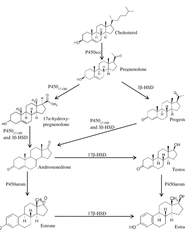
Figure 2. 1. Synthesis of estrogens in ruminants. Five main enzymes (P450 cholesterol side-chain cleavage enzyme (P450scc), 3β-hydroxysteroid dehydrogenase (3β-HSD), cytochrome P450 17α-hydroxylase (P45017-OH), 17β-hydroxysteroid dehydrogenase (17β-HSD), and P450
aromatase (P450arom)) are involved in the enzymatic pathway leading to the synthesis of estrogens from cholesterol. ... 23
Figure 2. 2. Chemical structure of steroidal aromatase inhibitors Exemestane and Formestane: its chemical structure resembles that of androstenedione (see Figure 2.1). ... 35
Figure 2. 3. Chemical structure of non-steroidal aromatase inhibitors letrozole and anastrozole: their chemical structures contain a triazole group that selectively binds to the heme group of the aromatase enzyme. ... 36
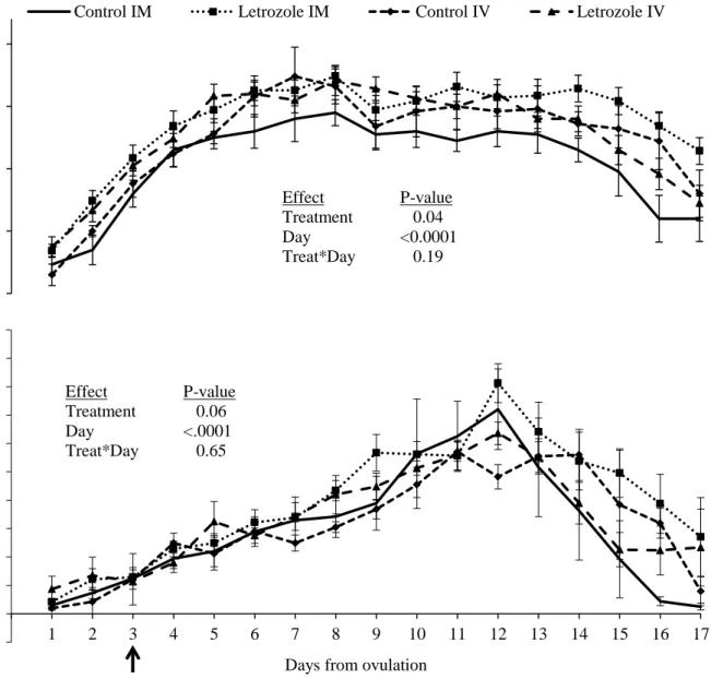
Figure 5. 1. Dominant follicle diameter profile (mean ± SEM) in heifers treated with letrozole intramuscularly (im, n=10) or intravenously (iv, n=10), and their respective placebo-treated controls (n=5 per group). In the bottom panel, im and iv routes were combined within letrozole and control groups. The arrow indicates the day of treatment. ... 57
Figure 5. 2. Corpus luteum diameter and plasma progesterone concentration (mean±SEM) in heifers treated with letrozole intramuscularly (im, n=10) or intravenously (iv, n=10), and their respective placebo-treated controls (n=5 per group). The arrow indicates the day of treatment.. 58
Figure 5. 3. Corpus luteum diameter and plasma progesterone concentration (mean ± SEM) in heifers treated with letrozole (intravenous and intramuscular routes combined, n=20) or a placebo (intravenous and intramuscular routes combined, n=10). (*) indicates P≤0.05 between
xix
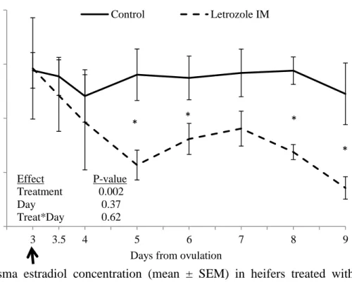
groups for that specific time point (analyzed by LSD). The arrow indicates the day of treatment. ... 59 Figure 5. 4. Plasma estradiol concentration (mean ± SEM) in heifers treated with letrozole intramuscularly (im, n=10) compared to placebo-treated controls (iv and im combined; n=10). (*) indicates P≤0.05 between groups for that specific time point (analyzed by LSD test). The arrow indicates the day of treatment. ... 60
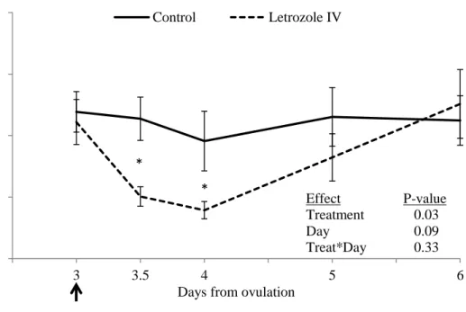
Figure 5. 5. Plasma estradiol concentration (mean ± SEM) in heifers treated with letrozole intravenously (iv, n=10) compared to placebo-treated controls (iv and im combined; n=10). (*) indicates P≤0.05 between groups for that specific time point (analyzed by LSD test). The arrow indicates the day of treatment. ... 61
Figure 5. 6. Plasma FSH concentration (mean ± sem) in heifers treated with letrozole (intravenous and intramuscular routes combined; n=20) compared to pacebo-treated controls (intravenous and intramuscular routes combined; n=10). Data were centralized to the day of post-treatment wave emergence. (*) indicates P≤0.05 between groups for that specific time point (analyzed by LSD test)... 62
Figure 5. 7. Plasma LH concentration (mean ± SEM) in heifers treated with letrozole intravenously (n=10) and intramuscularly (n10) compared to placebo-treated controls (intravenous and intramuscular routes combined; n=10). Data were centralized to the day of post-treatment wave emergence. ... 63 Figure 5. 8. Plasma letrozole concentration (mean ± SEM) during the first 12 h (A) and 8 d (B) in heifers after a single administration of 1 mg/kg of body weight intravenously (iv, n=4) or intramuscularly (im, n=4). ... 64
xx
Figure 6. 1. Dominant follicle diameter (mean±SEM) in heifers treated with a blank (control, n=15) or a letrozole-containing intravaginal device (letrozole, n=15). Devices were inserted on Day 1, indicated by the arrow (Day 0 = wave emergence). * On indicated days, values differed between groups (P≤0.05). ... 79
Figure 6. 2. Corpus luteum diameter (mean±SEM) following post-treatment ovulation in heifers treated with a blank (control, n=15) or a letrozole-containing intravaginal device (letrozole, n=15). ... 80
Figure 6. 3. Plasma progesterone concentrations (mean±SEM) following post-treatment ovulation in heifers treated with a blank (control, n=15) or a letrozole-containing intravaginal device (letrozole, n=15). * On indicated days, values differed between groups (P≤0.05). ... 80
Figure 6. 4. Plasma estradiol concentrations (mean ±SEM) in heifers treated with a blank (control, n=15) or a letrozole-containing intravaginal device (letrozole, n=15). Devices were given on Day 1, indicated by the arrow (Day 0 = wave emergence) of the ovulatory wave. * On indicated days, values differed between groups (P≤0.05). ... 81
Figure 6. 5. Plasma FSH concentrations (mean ±SEM) in heifers treated with a blank (control, n=15) or a letrozole-containing intravaginal device (letrozole, n=15). Devices were given on Day 1 (Day 0 = wave emergence) of the ovulatory wave. ... 82
Figure 6. 6. Plasma LH concentrations (mean ±SEM) in heifers treated with a blank (control, n=15) or a letrozole-containing intravaginal device (letrozole, n=15). Devices were given on Day 1, indicated by the arrow (Day 0 = wave emergence) of the ovulatory wave. ... 82
xxi
Figure 6. 7. Plasma letrozole concentration (mean±SEM) in heifers (n=4) given an intravaginal letrozole-releasing device for 6 days. * Between indicated time points, values differed (P≤0.05). ... 83
Figure 7. 1. Effect of three different inhibitors of estradiol production on estradiol secretion by bovine granulosa cells in culture. Cells were cultured in vitro for 6 days under non-luteinising conditions without treatment (negative control), or treatment with FSH alone (positive control) or with letrozole (A), anastrozole (B) or fenbendazole (C) at 1/10 x standard, standard or 10 x standard doses Data are presented as the mean ± SEM estradiol concentrations in three independent replicate cultures for each inhibitor. ab Values with no common superscript are different (P<0.05). ... 100
Figure 7. 2. Letrozole concentrations in saline during diffusion chamber trial for 24 hours. Letrozole was prepared in a liposome- or a wax-based vehicle and its diffusion through bovine vaginal mucosa was tested in diffusion chambers using phosphate buffered saline as perfusion buffer. Data from three diffusion chambers per formulation are presented as mean ± SEM. .... 101
Figure 7. 3. Letrozole concentrations in saline during diffusion chamber trial for 12 hours. Letrozole was prepared in a gel vehicle and its diffusion through bovine vaginal mucosa was tested in diffusion chambers using phosphate buffered saline as perfusion buffer. Data from two diffusion chambers per formulation are presented as mean ± SD. ... 102
Figure 7. 4. Letrozole concentrations in plasma (mean±SEM) during the first 12 h following treatment with a letrozole-containing intravaginal device in heifers. Letrozole devices were
xxii
prepared in three formulations: Wax (DOPE) + gel coat (n=2), Wax + gel coat (n=4), Wax only (n=4). a b c On indicated days, values differed among groups (P≤0.05). ... 103
Figure 7. 5. Plasma letrozole concentrations in heifers (mean ± SEM) over 12 days following treatment with a letrozole-containing intravaginal device. Letrozole devices were prepared in three formulations: Wax (DOPE) + gel coat (n=2), Wax + gel coat (n=4), Wax only (n=4). ab On indicated days, values differed among groups (P≤0.05). ... 104
Figure 8. 1. Dominant follicle diameter profiles (mean±SEM) in heifers treated with a blank intravaginal device (control, n=4), or a letrozole-containing device with wax + gel coat (n=4) or wax only (n=4). Devices were inserted on Day 3. abc On indicated days, values with no common subscripts are different (P≤0.05). ... 117
Figure 8. 2. Plasma estradiol concentrations (mean±SEM) in heifers treated with a blank intravaginal device (control, n=4), or a letrozole-containing device with wax + gel coat (n=4) or wax only (n=4). Devices were inserted on Day 3. ... 118
Figure 8. 3. Corpus luteum diameter profiles (mean±SEM) in heifers treated with a blank intravaginal device (control, n=4), or a letrozole-containing device with wax + gel coat (n=4) or wax only (n=4). Devices were inserted on Day 3. ... 118
Figure 8. 4. Plasma progesterone concentrations (mean±SEM) in heifers treated with a blank intravaginal device (control, n=4), or a letrozole-containing device with wax + gel coat (n=4) or wax only (n=4). Devices were inserted on Day 3. ... 119
xxiii
Figure 9. 1. Synchronization treatment schedule for Experiment 1. Heifers (48) were treated im with PGF followed by GnRH 24 h later to synchronize ovulation. Ultrasound examinations (U/S) were done daily to detect ovulation,(Day 0) and heifers were given an intravaginal device containing 3 g of letrozole for 4 days starting on Days 0, 4, 8, 12, or 16. At the time of device removal, heifers were given PGF followed by GnRH 24 h later. Ultrasound examinations were performed daily, from two days before device insertion to 9 days after the post-treatment ovulation. ... 128
Figure 9. 2. Plasma estradiol concentration (mean ± SEM) at letrozole device removal in heifers treated with a 4-day regimen of letrozole intravaginally compared to untreated controls. Devices were placed on Days 0 (n=10), 4 (n = 10), 8 (n = 8), 12 (n=11) or 16 (n=9; Day 0 = ovulation). Control samples were obtained from heifers in the Group 16 at Days 4, 8, 12 and 16, prior to treatment with letrozole on Day 16. Hence, Day 16 group lacked of a control group at device removal. ... 134
Figure 9. 3. Corpus luteum diameter profiles (mean ± SEM) in heifers after treatment with letrozole-releasing intravaginal device for 4 days followed by PGF at device removal and GnRH 24 h later. Devices were placed on Days 0 (n=10), 4 (n = 10), 8 (n = 8), 12 (n=11) or 16 (n=9; Day 0 = ovulation). ... 135
Figure 9. 4. Plasma progesterone (P4) profiles (mean ± SEM) in heifers after treatment with letrozole-releasing intravaginal device for 4 days followed by PGF at device removal and GnRH 24 h later. Devices were placed on Days 0 (n=10), 4 (n = 9), 8 (n = 8), 12 (n=11) or 16 (n=10; Day 0 = ovulation). ... 135
xxiv
LIST OF ABBREVIATIONS
µg micrograms 3β-HSD 3β-hydroxysteroid dehydrogenase 17β-HSD 17β-hydroxysteroid dehydrogenase A adenine AI artificial inseminationCIDR controlled internal drug release
CL corpus luteum
DES diethylstilbestrol
EB estradiol benzoate
EC estradiol cypionate
E-17β estradiol-17 beta
ELISA enzyme-linked immunosorbent assay FDA USA Food and Drug Administration FSH follicle stimulating hormone
FSHr follicle stimulating hormone receptor FTAI fixed-time artificial insemination
g gram
G guanine
GH growth hormone
GnRH gonadotropin releasing hormone
xxv
im intramuscular
iv intravenous
kg kilogram
LCMS/MS Liquid chromatography tandem mass spectrometry
LH luteinizing hormone
LHr luteinizing hormone receptor
mg milligram
mL millilitre
mm millimetre
MOET multiple ovulation and embryo transfer mRNA messenger ribonucleic acid
NADPH reduced nicotinamide adenine dinucleotide phosphate
ng nanogram
pg picogram
PGF prostaglandin F2α
P450arom P450 aromatase enzyme
P450scc P450 side-chain cleavage enzyme
P45017-OH P450 17α-hydroxylase
StAR steroidogenic acute regulatory protein
T thymine
1
CHAPTER 1: GENERAL INTRODUCTION
Reproductive efficiency is the single most important factor affecting profitability in the cattle industry. The impact of methods for controlling the estrous cycle in cattle may be illustrated by estimates of the use of artificial insemination (AI) and embryo transfer (ET) in cattle. A conservative estimate of the worldwide use of AI is 83 million cows per year – estimated to represent about 20% of the breedable cattle population (25% in North America) [1]. Worldwide, 53% of cows artificially inseminated are of dairy breeds and 39% are of beef breeds, but in Canada, the gap is much wider: 94% in dairy and 6% in beef. Regarding ET, just over 120,000 donor cows are collected each year worldwide and 800,000 embryos are transferred [2]. In Canada, those number do not exceed 13,500 and 55,000, respectively [3].
Estrogen-based protocols as a treatment for controlling and synchronizing ovulation in cattle, have revolutionized breeding practices and enabled beef producers to make use of AI as never before because labour and resources could now be pre-scheduled (e.g., fixed-time artificial insemination; FTAI). These protocols also allowed for the wider application of superovulation and embryo transfer due to their effectiveness to synchronize follicular wave emergence [4-8]. Steroid-induced wave synchronization is associated with regression of the dominant follicle followed by a surge in circulating FSH and subsequent emergence of a new follicular wave at a consistent interval post-treatment. Steroid-induced regression of the dominant follicle is a result of a systemic alteration in feedback of estradiol and progesterone on pituitary release of LH and FSH [9]. Estradiol suppresses FSH release [10] and has been reported to decrease LH pulse amplitude in sheep [11] and cattle [12]. Progesterone has been reported to decrease LH pulse frequency and suppress maximal diameter of the dominant follicle in a dose-dependent manner
2
in cattle [13-18]. Once the suppressive effects of estradiol are removed, FSH surges resulting in the emergence of a new wave of follicular development approximately 4 days after estradiol/progesterone treatment, regardless of the stage of development of the dominant follicle at the time of treatment [4, 5, 19].
However, increasing consumer sensitivity to the possible deleterious effects of estrogens in food and in the environment [20] has led to new regulations about the use of estrogenic products in livestock. The European Union has already banned the use of estrogenic products in food producing animals [21-24]. In United States [25] and Canada [26], estrogens cannot be used for synchronization of estrus except by prescription and custom-compounding. In 2007, New Zealand and Australia banned use of estrogens in lactating dairy animals [24]. The ban of the use of estrogens in livestock and lack of commercially availability of estrogenic preparations negatively impacts the implementation of reproductive biotechnologies in cattle production systems, limiting potential reproductive efficiency and genetic improvement provided by the use of AI or MOET [24]. In this context, the development of alternative methods for controlling ovarian function in cattle, with efficacy and predictability comparable with that of estrogen plus progesterone treatments [5], and with no toxic or harmful effects on human and animal health is needed.
Another important factor that negatively impacts reproductive efficiency, mainly in dairy industry, is the decline in fertility observed in high producing cows. Over a period of 15-20 years, the rate of decline in fertility has been 0.5% per annum in the USA [27]; in UK herds, pregnancy at first insemination has gone from 56% to 40%, approaching a decline rate of 1% per annum [28]. The dramatic decline in fertility has been associated with a reduction in expression
3
of heat, failure to ovulate, and poor luteal function. Regarding luteal function, cows that failed to carry a pregnancy had lower levels of progesterone at days 14 and 21 post-breeding compared to those that remained pregnant [29]. Several research groups have concluded that embryos developing under higher levels of progesterone early in the luteal phase grow more rapidly and were more likely to prevent prostaglandin F2α (PGF) secretion by the uterus; i.e. prevent luteolysis and achieve maternal recognition of pregnancy [30-32]. The development of a treatment to improve early luteal function and therefore support the development of a larger embryo will be of extreme importance not only for AI in dairy industry but also for the success of ET in both beef and dairy herds.
Aromatase inhibitors prevent the body from producing its own estrogens; thus, they could potentially be applied to the management of estrogen-dependent physiologic functions such as reproduction. Letrozole (Femara, Novartis) is a non-steroidal aromatase inhibitor that inactivates the aromatase enzyme by reversibly binding to the heme group of the P450 subunit of the enzyme. Letrozole is used as an adjuvant treatment for hormone-responsive breast cancer in post-menopausal women [33] and has been used as a fertility therapy for women undergoing assisted reproduction because of its putative effect on FSH secretion through removal of the negative feedback of estradiol [34]. A 5-day regimen of letrozole (2.5 mg/day from 3 to 7 days after the beginning of menses) has been used for ovarian stimulation in women [35], and larger or increasing doses of letrozole have been used to induce ovarian superstimulation [36, 37]. It has also been applied to lower the cost of superstimulatory treatments by reducing the dose of FSH required [38].
4
We conducted two studies to assess the effects of aromatase inhibitors on ovarian function in cattle. Contrary to the above mentioned hypothesis proposed to explain the effect of aromatase inhibitors in women [34], letrozole treatment of cattle did not induce follicular atresia or hasten emergence of a new follicular wave, whether given as a single intravenous dose on Day 3 post-ovulation [39] or in a 3-day regimen from Days 1-3, 3-5 or 5-7 post-post-ovulation [40]. Rather, letrozole treatment increased mean plasma LH concentrations resulting in a prolonged period of dominance of the extant DF and delayed emergence of the next follicular wave. In addition, the 3-day letrozole treatment was associated with greater corpus luteum (CL) diameters [40].
Studies done in cattle in our laboratory established a solid foundation for the development of letrozole-based synchronization and fertility treatments:
• Short or prolonged letrozole treatment extends the lifespan of the dominant follicle • Letrozole treatment can be used to delay follicle wave emergence
• Letrozole treatment induces the formation of a larger dominant follicle
• Letrozole treatment is luteotrophic (larger CL that secretes more progesterone) The set of studies presented in this dissertation aim to provide convincing data that aromatase inhibitors can be applied as a safe and effective method to control the estrous cycle and improve fertility in cattle.
5
CHAPTER 2: LITERATURE REVIEW
2.1Reproductive Physiology in Cattle
2.1.1 Hypothalamus-Pituitary-Ovarian Axis
The hypothalamus-pituitary-ovarian (HPO) axis is comprised by the anatomic and functional relationships existing among the hypothalamus, pituitary gland, and ovaries, thus allowing these endocrine glands to act as a single entity. The hormones involved in the control of this axis include gonadotropin-releasing hormone (GnRH, produced by the hypothalamus), luteinizing hormone and follicle-stimulating hormone (LH and FSH, respectively; produced by the anterior pituitary), and estradiol, inhibin and progesterone (synthetized in the ovaries). However, numerous other factors and hormones have been linked to the control of the HPO axis in mammals. Furthermore, the neuroendocrine nature of the HPO axis integrates external factors such as stress, nutrition, and photoperiod, in the control of the reproductive physiology [41].
The hypothalamus is located in the ventral brain and comprises groups of cell bodies known as hypothalamic nuclei (namely, the paraventricular nucleus, the surge center and the tonic or pulsatile center) [41, 42]. The neurons located in the surge and tonic centers secrete GnRH, while the neurons of the paraventricular nucleus synthetize oxytocin. Anatomic arrangements are of special importance for the communication between the hypothalamus and pituitary gland. GnRH-producing neurons release their secretion through long axons located in vicinity to the pituitary stalk [43]. Within the pituitary stalk there is highly complex capillary network, the hypothalamo-hypophyseal portal system, which allows GnRH to reach the anterior portion of the pituitary, the adenohypophysis. It is believed that the main function of this capillary system is to
6
prevent the very small amount of GnRH produced by the hypothalamus from getting diluted in the general circulation [42]. Once GnRH binds to its receptors on the gonadotroph cells, LH and FSH are produced and secreted into the blood stream in order to reach their target organ, the ovaries [43].
The gonadotropins (FSH and LH) play an important role in communicating with the ovaries. Gonadotropins activate the ovaries to produce different steroid hormones such as estrogen, androgens and progesterone which are linked to the regulation the estrous cycle. Positive and negative feedback loops are involved in the communication between the ovaries and the hypothalamus and pituitary [10, 44-46]. Estrogen receptors (ER) are present in the hypothalamus, pituitary and in the ovaries of many species, including cattle [47-50]. Hence, alteration in estradiol concentration could have direct and independent effects on all the endocrine glands that comprise the HOP axis. Details of the interactions between gonadotropins and ovarian sex steroids in cattle will be discussed in later sections of this literature review.
Non-steroidal ovarian products have also been related to the control of gonadotropin secretions [51, 52]. Inhibin is a protein produced by the granulosa cell of growing follicles under the influence of FSH. It comprises α and β sub-units and it is known to suppress FSH secretion by acting at hypothalamic and pituitary levels [43, 53-55]. The combination of two β sub-units results in a related protein, known as activin, which counteracts the effect of inhibin by stimulating FSH release [43]. Other factors present in the follicular fluid include follistatin, oxytocin, insulin-like growth factor (IGF), epidermal growth factor (EGF) and plasminogen activator (reviewed in [56]).
7
Kisspeptins (Kps) are a family of peptide hormones which bind to protein G-coupled receptor 54 (GPR54) and integrates nutritional and hormonal information which is critical to metabolism and regulation of reproduction [57]. All Kps are the product of the KISS-1 gene and contain, at the C-terminal region, a common deca-peptide sequence (Kp-10) that confers to them biological activity [57, 58]. Although Kp receptors (GPR54) have been described in the hypothalamus, pituitary, ovaries and placenta, its major role has been postulated as being the neuroendocrine regulator of GnRH release [58, 59]. Kisspeptin and hypothalamic GPR54 have been associated with the onset of pulsatile GnRH secretion observed at puberty and in seasonal breeders, like sheep [57-59].
Kisspeptin-secreting neurons contain ER and other sex steroid receptors and are located in close vicinity to GnRH-secreting cells in the hypothalamus [59, 60]. Two sub-populations of Kp-secreting cells have been identified: one is located in the arcuate nucleus (ARC) and the other is located in the anteroventral periventricular area (AVPV) [59, 60]. This differentiation of these two sub-populations of cells is relevant since the regulatory effects of estrogens are nucleus-specific. Thus, estrogens inhibit the expression of Kp at the ARC, causing a negative-feedback effect on gonadotropin secretion. As expected, reducing circulating E2 concentrations resulted in higher Kp mRNA levels and increased GnRH output [58]. In contrast, estrogens enhance Kiss1
expression at the AVPV in rodents mediated by ERα, which suggests that this population of neurons may be involved in the positive-feedback actions of estradiol to generate the preovulatory surge of gonadotropins [57, 59]. In cattle, administration of 100 pmole/kg of Kp during diestrus, proestrus and estrus resulted in increased circulating LH concentrations during diestrous and proestrus, but not during estrous [61].
8
It is important to highlight that, although estrogen plays a key role in the regulation of GnRH pulsatile secretion pattern, GnRH-releasing neurons seem to lack ER thus making the Kp/GPR54 system pivotal for the normal development of the reproductive functions [59].
2.1.2 Ovarian follicular wave development in cattle
In cattle, ovarian follicles develop in waves [62, 63]. A wave of follicular development begins with the synchronous recruitment of a mean of 24 (range of 8 to 41) small follicles (3 to 4 mm in diameter), an event that is referred to as wave emergence [64, 65]. Additionally, the growth pattern of these follicles at ≤3 mm in diameter has also been describes as occurring in a wave-like manner [66, 67]. In monovular species such as cattle, a single follicle is selected from this cohort of recruited follicles to continue growing (i.e. dominant follicle), while the other follicles in the group undergo atresia (i.e. subordinate follicles) [63, 68-70]. This “selection” process is confirmed by the deviation in diameter between the now dominant follicle and the largest subordinate follicle, which occurs when the dominant follicle is on average 8.5 mm in diameter and about 2.5 days after wave emergence [64]. If the dominant follicle is growing during the early luteal phase, this follicle becomes anovulatory and regresses. Three different phases of follicular growth have been described during anovulatory waves: 1) a growing phase, during which the dominant follicle grows actively; 2) a static phase, during which follicular diameter does not change significantly; and 3) a regressing phase, during which dominant follicle diameter begins to decrease [71]. However, if the dominant follicle is actively growing or in early static phase when regression of the corpus luteum begins, it will become the ovulatory follicle and as such it has the capability of triggering a gonadotropin surge that will lead to ovulation (reviewed in [72]).
9
Two and three of these waves of follicular development have been most commonly reported during an estrous cycle in cattle [70, 71, 73, 74]. In animals with two or three waves of follicular development, the first wave can be identified, on average, the day of ovulation. The second follicular wave emerges about day 10 and day 9 after ovulation in two-wave and three-wave cycles, respectively [66, 71]. In animals with three follicular waves per cycle, the last and ovulatory wave is detected in average on day 16 post-ovulation [75]. Estrous cycles composed of two waves of follicular development are consistently shorter than 3-wave cycles (19 to 20 days vs 22 to 23 days, respectively) [75].
2.1.3 Gonadotropins and the control of follicular growth
Follicle-stimulating hormone and LH play crucial roles in the regulation of ovarian follicular dynamics. Emergence of a follicular wave and the selection of a dominant follicle have been temporally associated with an increase and decrease, respectively, in FSH levels [76]. A transient increase in plasma LH concentration has been reported in relation to the time of follicular deviation defined as the measured point when a significant difference in diameter between the dominant follicle and the largest subordinate follicle is first identified [77]. Additionally, the peak in LH concentrations that encompasses ovulation has been well documented [78, 79].
As mentioned above, each wave of follicular development is preceded by a surge in FSH concentration [76]. Inhibiting the increase in FSH concentration by administration of a proteinaceous fraction of follicular fluid was followed by a delay in follicular wave emergence. Moreover, promotion of a surge of FSH by ablation of the extant DF resulted in the emergence of a new wave of follicular development within 2 days [76]. These results also provided evidence that intrafollicular factors were, at least in part, responsible for the changes in plasma FSH
10
concentrations related to follicular dynamics. Further clarification of this concept revealed that increasing plasma concentrations of intrafollicular components (e.g., estradiol, inhibins, IGF, among others), mainly synthesised by the dominant follicle, have a suppressing effect on circulating FSH concentrations and that this decrease in FSH levels was crucial for the process of dominant follicle selection and to ensure monovulation [45, 53, 68, 80].
A FSH-follicle coupling system involving all the follicles in the recruited wave is responsible for the initial drop in FSH concentrations observed after wave emergence. When the dominant follicle reaches a critical stage of development for selection (8.5 mm [81]), it acquires the capability to further suppress FSH concentrations to levels that are not compatible with continuous development of the subordinate follicles [10]. However, a shift from FSH to LH-dependency by the dominant follicle (by acquisition of LH receptors in the thecal and granulosa cells [77]) allows it to continue growing even under very low levels of plasma FSH [53]. A study of the effects of LH suppression by administration of exogenous progesterone before, during and after follicular deviation demonstrated that LH was not indispensable for the initiation of deviation. There was no difference in follicle diameters between progesterone-treated and control animals before and during deviation. However, the dominant follicle was smaller and grew at a slower rate when LH secretion was suppressed after deviation was initiated in the progesterone– treated group [14, 77].
The presence of a healthy dominant follicle at the end of the luteal phase leads to ovulation. The drop on progesterone concentration that takes place during luteolysis induces an increase in LH pulsatility that further feeds the growth of the extant dominant follicle [13]. Additionally, high estradiol production by the dominant follicle exerts positive feedback at hypothalamic level
11
to increase LH pulsatility. This loop of positive stimulation by high estradiol and low progesterone on GnRH producing cells by estradiol leads to the pre-ovulatory surge of LH
needed for final oocyte maturation and release [57, 58, 82]. The molecular mechanisms involved in this process have been discussed in previous sections in this review.
The active dominant follicle produces factors that inhibit plasma FSH secretion thus preventing the emergence of a new wave of follicular development during the dominance period [51, 53, 83]. At the end of the static phase (when the dominant follicle starts to regress) or after ovulation (when the source of the follicular components disappears), the negative feedback effect of follicular products (mainly estradiol) on gonadotropin secretion is removed. This results in a surge of FSH secretion followed by emergence of a new follicular wave [15, 46].
In summary, follicle wave emergence is preceded by an increase in plasma FSH concentrations. Follicular selection is related to a decline in circulating FSH concentrations caused mainly by the production of estradiol by the cohort, and in particular by the dominant follicle under the influence of LH. Low FSH concentrations maintained by the dominant follicle ensure that no further waves of follicular development occur during the period of dominance. When LH pulse frequency is low, the dominant follicle losses dominance and regresses, estradiol production declines, FSH surges and a new wave of follicular development is stimulated. However, if the dominant follicle is still viable at the end of the luteal phase, LH pulse frequency increases and stimulates a greater production of estradiol by the dominant follicle that will, in turn, elicit a preovulatory peak of LH and FSH secretion, and ovulation. The disappearance of the dominant follicle after ovulation removes the negative feedback effect of estradiol on FSH
12
secretion and a post-ovulatory surge of FSH followed by the emergence of a wave of follicular development is observed [84].
2.2Control of the Estrous Cycle
Based on our knowledge of reproductive physiology gained in the last three or four decades, several strategies and protocols to manipulate phenomena related to the estrous cycle (i.e., wave emergence, selection, luteolysis and ovulation) have been developed. The ideal method of control of the estrous cycle is one that is simple, effective and safe. For the purpose of this review, the rationale and implementation of protocols based on prostaglandin F2α (PGF) and
GnRH, and the use of estrogens alone or in combination with progesterone will be briefly discussed.
2.2.1 Prostaglandin and GnRH-based protocols for estrus synchronization
The identification of PGF as the luteolysin responsible for the regression of the CL in cattle provided a new means for controlling the length of the luteal phase and ovulation (reviewed in [85]). Several protocols applying different doses and intervals between doses of prostaglandins have since been designed [86, 87]. One of the main limitations of synchronizing ovulation by a single PGF treatment is that the state of maturity of the dominant follicle at the time of PGF determines the interval to estrus and ovulation. This interval has been described to be, on average, of 3 to 3.4 days. However, if the dominant follicle present at the time of PGF treatment is in the late static or regressing phase, the dominant follicle of the subsequent follicular wave will have to grow in order to reach preovulatory size; in this case the interval from PGF treatment to ovulation may take as much as 6 days [86, 87]. Further, newly formed CL during the
13
first 3 to 4 days after ovulation are refractory to the luteolytic effects of PGF; responsiveness increases as the CL develops [86]. These important sources of variability in interval from treatment to ovulation limit the use of prostaglandin-based protocols for fixed-time artificial insemination (FTAI). An alternative approach which uses two doses of PGF 11 to 14 days apart is widely accepted and used on many dairy and beef farms. The rationale for this approach is that approximately 67% of the animals (those with a CL ≥5 day-old or those experiencing natural luteal regression) would respond to the first PGF injection by undergoing luteolysis and ovulating. When the second injection of PGF is administered 11 to 14 days later, it is expected that 100% of the animals would have functional and PGF-responsive CLs [69]. However, the use of luteolytic doses of prostaglandin still relies on estrus detection efficiency to provide acceptable outcomes. Consequently, the rate of submission of animals for AI after detected estrus may limit the effectiveness of this protocol. Herd heat detection rates are very variable and have been reported to be ≤ 50% (range from about 30% to 65%) [88-91] in high producing dairy farms and between 53% and 73% in commercial beef farms [90, 92].
The use of fixed-time AI (FTAI) can overcome the negative impact of low estrus detection efficiency. FTAI eliminates the need for estrus detection but requires synchronous growth and ovulation of a viable dominant follicle in order to be successful. Pregnancy rates obtained with FTAI are usually comparable to those obtained after AI with high estrus detection rates, because all animals are inseminated regardless of whether or not they showed estrus [93]. Gonadotropin releasing hormone is commonly used to induce the pituitary release of gonadotropins (LH and FSH) which will induce ovulation and/or luteinisation of the dominant follicle [94, 95]. Emergence of a new wave of follicular development is expected approximately 2 days after
14
GnRH treatment [94], although its synchrony is highly dependent on the occurrence of ovulation after GnRH [95, 96]. The use of GnRH is usually combined with a luteolytic dose of PGF 7 days later: GnRH is intended to synchronize wave emergence while PGF synchronizes luteolysis. An additional dose of GnRH is used about 48 hours after PGF to induce an LH surge and further synchronize ovulation [93, 97]. Finally, animals are inseminated 16 to 20 hours after the second GnRH dose. This protocol is known as Ovsynch. Several studies in which Ovsynch protocols were used had pregnancy rates similar to those obtained using the two doses of PGF 14 days apart with high estrous detection management in lactating dairy cows (38.9% versus 37.8%, respectively) [93, 94]. However, after the application of Ovsynch to heifers, pregnancy rates were lower than in controls treated with PGF and inseminated after estrus detection (35.1% versus 74.4%, respectively). Poor ovulatory response to the first dose of GnRH and consequently poor synchronization of wave emergence were identified as the causes of the lack of success of the Ovsynch protocols in heifers [96, 98]. Ovulation was induced in 54% of dairy heifers after GnRH treatment [94], and it was reported that 27% to 44% of beef heifers treated with a single GnRH dose failed to ovulate [95, 96].
Alterations to the original Ovsynch protocol were introduced in order to improve the ovulatory response and synchronization. Pre-synchronization of follicular wave emergence using a double PGF treatment 14 days apart followed 12 days later by an Ovsynch protocol (Presynch), or using two consecutives Ovsynch protocol 7 days apart (Double-Ovsynch) improved the ovulatory response to first GnRH and therefore more heifers and cows responded to the second GnRH with synchronous ovulation [99, 100]. Additional modifications include the addition of a progestin device between first GnRH and PGF (to prevent early ovulation, mostly in heifers
15
[101]) and alterations on the timing of the second GnRH and AI (Cosynch [102]). Specifics of the application and effectiveness of these protocols are beyond the scope of the present thesis and have been reviewed elsewhere [103].
2.2.2 Estradiol and progesterone-based protocols
The interval from PGF-induced luteolysis to ovulation and the variability in the ovulatory response among animals to GnRH treatment depends on the status of the ovarian follicle at the time of treatment. Thus, a method that controls ovarian follicle recruitment would provide the advantage of knowing the stage of follicular development when the ovulatory or luteolytic treatment is given and would improve the ovulatory response and its synchrony.
Combinations of progesterone and estradiol have been used to hormonally induce a new wave of follicular development in a predictable interval of time [5, 12]. This method of synchronizing wave emergence is based on the negative feedback effects that estradiol has on FSH secretion during the luteal phase [104] or under the influence of an exogenous source of progesterone [4, 5, 19, 105]. Additionally, exogenous progesterone suppresses LH secretion leading to the regression of the extant dominant follicle. Reduced secretion of FSH and LH from the pituitary gland terminates the growth of both FSH- and LH-dependent follicles. Reduced circulating estradiol concentration (most likely due to metabolism and clearance) results in a FSH surge and in emergence of a new wave of follicular development about 4 days after estradiol and progesterone treatment [4, 106, 107]. This protocol includes, at the time of estradiol and progesterone treatment, the insertion of a progesterone releasing device for 7 to 9 days and a dose of PGF at the time of progesterone device removal to ensure luteal regression. Animals are inseminated 55 to 60 hours after progesterone withdrawal [107]. Additionally, different forms of
16
estradiol have been applied in these progestin-based protocols. It has been reported that the use of short-acting preparations such as E-17β or estradiol benzoate (EB) results in more synchronous wave emergence and ovulation than long-acting esters such as estradiol cypionate [8, 108, 109]. When estradiol is added to increase the synchrony of ovulation, it is recommended that it should be administered 24 hours after progesterone device withdrawal and AI should be performed 30-36 hours later [107].
A synchronized ovulatory response is essential for the efficient use of time, resources, labour and the application of reproductive management techniques such as FTAI [5, 109] and multiple ovulation and embryo transfer (MOET) [105, 106, 110]. The outcome of a superstimulation treatment is strongly influenced by the stage of follicular development at initiation of treatment. Optimal ovarian responses were obtained when superstimulatory treatments were initiated about the time of follicular wave emergence (day – 1 or 0 of the follicular wave) [5, 109]. Since progesterone plus estradiol treatments result in precise synchronization of wave emergence (on average 4.3 days after treatment), superstimulatory treatments are initiated on day 4 of the protocol, without the need for estrus detection and the 8 to 12-day waiting period for a natural follicular wave to emerge [4, 111].
2.3Decline in Fertility and its Impact on Reproductive Efficiency
Fertility can be defined as the ability of the animal to conceive and maintain pregnancy if served at the appropriate time in relation to ovulation [112]. This definition implies that fertility can be negatively impacted at several different steps in the pathway leading to obtaining a healthy offspring: altered ovarian function and cyclicity, poor estrous behaviour or detection, failure to ovulate, and embryonic or fetal loss. Strategies to control or modify estrous, ovulation, and
17
ovarian dynamics have been discussed in previous sections of this review. In this section we will briefly discuss aspects related to CL dysfunction and pregnancy failure.
2.3.1 Proposed mechanisms for the decline in fertility
It is now evident that fertility has declined with rising milk yields [113, 114]. In the USA the rate of decline in fertility was 0.5% per annum, over a period of 15-20 years,[27]; in UK herds, pregnancy at first insemination has gone from 56% to 40% between 1975-1982 and 1995-1998, approaching a decline rate of 1% per annum [28]. However, there is recent evidence that the historical decline in fertility has reached a nadir and has now begun to improve, at least in the United States [115].
This dramatic decline in fertility has been associated with a reduction in expression of estrus, failure of ovulation, and poor luteal function [114]. Strategies to control estrus, time to ovulation and even eliminate heat detection have already been discussed.
After ovulation, the CL is formed and secretes progesterone. In a non-bred female, and under the influence of estradiol of follicular origin, the endometrium will start secreting PGF about 16 days post-ovulation, which will induce the luteolytic process. However, if the animal has been bred and a conceptus in present in the uterus by day 16 post-ovulation, the embryo signals its presence and prevents the uterine secretion of PGF and luteolysis. Hence, the period between days 14 and 21 of the bovine estrous cycle are critical for the establishment of pregnancy [116].
From the process of pregnancy recognition and luteolysis, several steps can be identified in which a malfunction could lead to pregnancy failure. In cattle, the natural process of luteolysis is driven by estradiol [116]. Estradiol secreted by the growing dominant follicle of each follicular
18
wave in the cycle primes the uterine endometrium and induces the expression of oxytocin receptors [117]. Oxytocin, also of follicular origin, acts on the endometrial receptors stimulating the synthesis and secretion of PGF. It has been shown that administration of exogenous estradiol hastens the process of luteolysis in cattle [117].
Luteolysis is a default process in cattle, meaning that it has to be actively blocked in order to sustain a pregnancy. In cattle, the embryo is responsible for signaling its own presence. Embryonic secretion of interferon-τ prevents PGF secretion, although the precise mechanism of its action is still unknown. It has been proposed that interferon-τ blocks the effect of estradiol on the endometrium, hence preventing the acquisition of oxytocin receptors. An embryo that fails to secrete interferon-τ, or that secretes insufficient amounts of the protein, will fail to signal its presence and the process of luteolysis will take place [116].
Progesterone concentrations are high during the luteal phase of the estrous cycle, which is necessary to prepare the uterus for embryonic development. In order to block luteolysis and maintain high levels of progesterone, the embryonic trophoblast has to elongate and occupy the whole uterine lumen, meaning that larger embryos are more likely to succeed in preventing luteolysis. Progesterone levels have been associated with embryonic growth and elongation rate and interferon-τ secretion capabilities [116, 118, 119]. However, progesterone does not seem to act directly on the embryo in order to influence its growth. Rather, the effects that progesterone has on uterine secretion, from which the embryo feeds, are responsible for the increased trophoblastic elongation observed in cows supplemented with progesterone during the first week post-breeding [118, 120]. It has been shown that cows that failed to carry a pregnancy had lower levels of progesterone between days 14 and 21 post-breeding compared to those that remained
19
pregnant [29]. In dairy cows, low circulating progesterone concentrations have been associated with increases in metabolic rate and liver clearance of steroid hormones due to high dry matter intake [120] and thus, reduced embryonic survival.
2.3.2 Methods to improve fertility post-AI and embryo transfer
Several strategies to improve pregnancy rates post-breeding or embryo transfer have been tested. The main goals of such strategies can be summarized as follows: (1) to increase the antiluteolytic signal from the conceptus (by increasing progesterone concentration post-breeding or embryo transfer), and (2) to decrease the effect of estradiol (from the dominant follicle) on endometrial secretion of PGF [114, 116].
Several research groups have concluded that embryos developing under higher levels of progesterone early in the luteal phase grow more rapidly (i.e. trophoblast expansion) and were more likely to prevent prostaglandin F2α (PGF) secretion by the uterus; i.e. prevent luteolysis and achieve maternal recognition of pregnancy [30-32, 118, 119]. Higher levels of progesterone have been achieved by inducing the ovulation of a larger dominant follicle (which results of a larger CL) e.g. stimulating follicle growth with eCG, by treating with luteotrophic hormones (such as hCG, and GnRH) to induce accessories CLs and stimulate CL growth and progesterone secretion [120, 121], or by supplementation with exogenous progesterone (in most cases by the use of intravaginal devices) [30, 116, 118, 122].
Estradiol of follicular origin has been identified as the stimulus driving the expression of oxytocin receptors in the endometrium and, therefore, triggering the mechanism of luteolysis. It has been shown that reduction of circulating estradiol concentrations by ablation of the dominant follicle delays the occurrence of luteolysis, while estradiol supplementation hastens the initiation
20
of the luteolytic process [117]. In an attempt to reduce estradiol circulating concentrations during the critical period of maternal recognition of pregnancy (days 15 to 21 post-ovulation), researchers have treated cows with GnRH in order to induce ovulation and remove the estrogenic activity of a dominant follicle. It was concluded that increasing the proportion of cows with 3 waves of follicular development reduces the odds of having a highly estrogenic dominant follicle during such period over 2-wave animals, and therefore improves pregnancy rates [116, 117]. 2.4Estrogens and Other Sex Steroid Hormones
Steroid hormones are cholesterol-derivatives commonly classified, based on their physiological behaviour, in five groups: glucocorticoids, mineralocorticoids, progestins, androgens, and estrogens. The last three groups, also referred to as sex steroid hormones, are associated with the mechanisms that govern the reproductive physiology of mammals.
2.4.1 Steroidogenic pathway for the synthesis of sex steroid hormones
Estrogens are the final product of a complex biosynthetic pathway that, in ruminants, involves five main enzymes: the P450 cholesterol side-chain cleavage enzyme (P450scc), 3β-hydroxysteroid dehydrogenase (3β-HSD), cytochrome P450 17α-hydroxylase (P45017-OH),
17β-hydroxysteroid dehydrogenase (17β-HSD), and P450 aromatase (P450arom) [123, 124].
The rate limiting step in the biosynthesis of sex steroid hormones appears to be the incorporation of cholesterol into the mitochondria [125]. Free cholesterol is highly hydrophobic, and although it can cross the cellular membrane by diffusion, this process is extremely slow. Therefore, the incorporation of cholesterol inside the mitochondria depends on an active (energy