Introduction
Embryonic stem (ES) cells are pluripotent cells derived from the inner cell mass of the blastocyst stage embryo that can differentiate in vivo and in vitro into all cell lineages of the adult animal. The ability of these cells to adopt multiple fates makes ES cells prime candidates for use in stem cell therapies. However, unmodified ES cells cannot be used in regenerative therapies because they form teratomas after transplantation. In successful transplantation studies, ES cells are predifferentiated into the desired cell types, which may then integrate into the transplanted tissue (Brustle et al., 1999; Chen and Mok, 1995; Dinsmore et al., 1996; Liu et al., 2000). Hence, an understanding of how ES cells differentiate in vitro into neural cells is essential for developing and refining transplantation strategies for the nervous system, as well as for understanding the molecular mechanisms underlying neurogenesis.
A commonly used technique for differentiating ES cells into the neural lineage involves treatment of the cells with all-trans retinoic acid (RA) (Bain et al., 1995). However, the mechanisms by which ES cells are committed to the neural lineage using this technique are poorly understood. RA-mediated neural differentiation requires growth of ES cells as
embryoid bodies (EBs) for 4 days prior to the RA treatment (Bain et al., 1995), indicating that certain as yet undefined events must occur within the EB to make the cells responsive to the neurogenic effects of RA. Other techniques for promoting neural differentiation of ES cells include inhibition of BMP signaling (Gratsch and O’Shea, 2002), growth at low density (Tropepe et al., 2001; Ying et al., 2003) and co-culture with PA6 cells (Kawasaki et al., 2000). However, these protocols all require culture at very low densities and cell-cell/matrix interactions for neuronal differentiation, and the mechanisms underlying neuronal lineage commitment induced by these techniques remain unclear. Furthermore, the need to culture the cells at low density to achieve neuronal differentiation limits the number of cells that could potentially be obtained for transplantation strategies.
One candidate pathway for neural lineage commitment by ES cells is the Wnt/β-catenin pathway that has been shown to be a cell-cell (and/or cell-matrix) contact-regulated inducer of neurogenesis (Patapoutian and Reichardt, 2000). β-catenin exists in three cellular pools: membrane bound, cytoplasmic and nuclear. Membrane-bound β-catenin is associated with cadherin/adherens junctions and functions to bridge E-cadherin to the cytoskeleton (Aberle et al., 1996; Gumbiner
Culture of embryonic stem (ES) cells at high density inhibits both β-catenin signaling and neural differentiation. ES cell density does not influence β-catenin expression, but a greater proportion of β-catenin is targeted for degradation in density cultures. Moreover, in high-density cultures, β-catenin is preferentially localized to the membrane further reducing β-catenin signaling. Increasing
β-catenin signaling by treatment with Wnt3a-conditioned medium, by overexpression of β-catenin, or by overexpression of a dominant-negative form of E-cadherin promotes neurogenesis. Furthermore, β-catenin signaling is sufficient to induce neurogenesis in high-density cultures even in the absence of retinoic acid (RA), although RA potentiates the effects of β-catenin. By contrast, RA does not induce neurogenesis in high-density cultures in the absence of β-catenin signaling. Truncation of the armadillo domain of β-catenin, but not the C terminus or the N terminus, eliminates its proneural effects. The proneural effects of β-catenin reflect enhanced lineage commitment
rather than proliferation of neural progenitor cells. Neurons induced by β-catenin overexpression either alone or in association with RA express the caudal neuronal marker Hoxc4. However, RA treatment inhibits the β -catenin-mediated generation of tyrosine hydroxylase-positive neurons, suggesting that not all of the effects of RA are dependent upon β-catenin signaling. These observations suggest that β-catenin signaling promotes neural lineage commitment by ES cells, and that β-catenin signaling may be a necessary co-factor for RA-mediated neuronal differentiation. Further, enhancement of β -catenin signaling with RA treatment significantly increases the numbers of neurons generated from ES cells, thus suggesting a method for obtaining large numbers of neural species for possible use in for ES cell transplantation.
Key words: Embryonic stem cells, β-Catenin, Neurogenesis, Retinoic acid, Tyrosine hydroxylase, Cell density
Summary
β
-Catenin signaling is required for neural differentiation of
embryonic stem cells
José Javier Otero*, Weimin Fu, Lixin Kan, Adolfo E. Cuadra and John A. Kessler*
Department of Neurology, Northwestern University Feinberg School of Medicine, 303 East Chicago Avenue, Chicago, IL 60611, USA
*Authors for correspondence (e-mail: j-otero@northwestern.edu and jakessler@northwestern.edu)
Accepted 13 April 2004
Development 131, 3545-3557
and McCrea, 1993). In the cytoplasm, β-catenin turnover is regulated by a stepwise phosphorylation on its N terminus. Initially, β-catenin is phosphorylated on Ser45 by a ‘priming’ kinase (Liu et al., 2002). Phosphorylation at Ser45 primes β-catenin for phosphorylation at Ser33/37;Thr41 by Gsk3β (Kang et al., 2002). Phospho-Ser33/37;Thr41-β-catenin is then targeted for ubiquitin-directed proteolysis (Salic et al., 2000a), and increased phosphorylation by Gsk3βdecreases the nuclear pool of β-catenin (Lucas et al., 2001). During Wnt signaling, Gsk3β is inhibited, leading to the accumulation of unphosphorylated β-catenin in the cytoplasm and its translocation to the nucleus, where it interacts with members of the TCF/LEF family of transcription factors. The interaction of β-catenin with TCF/LEF transcription factors causes both inhibition of repression and activation of transcription. However, both Wnt and β-catenin can signal through other independent pathways (Korswagen, 2002; Kuhl, 2002; Pandur et al., 2002a; Pandur et al., 2002b; Tada et al., 2002; Yamanaka et al., 2002).
This study addresses the role of β-catenin signaling in neural lineage commitment by ES cells. We find that induction of differentiation at high density inhibits both β-catenin signaling and neural differentiation. However, overexpression of β-catenin is sufficient to induce neuronal lineage commitment even in the absence of RA or EB formation, and, unlike RA, can induce neuronal differentiation in high-density cultures.
Materials and methods
Cell culture
The WW6 ES cell (Ioffe et al., 1995) line was obtained from Dr Winfred Edelman (Albert Einstein College of Medicine). ES cells were cultured without feeder cells on gelatin-coated tissue culture flasks, in media described previously (Bain et al., 1995) with Lif (ESGRO, Chemicon). Cells were routinely observed for SSEA-1 (Fut4 – Mouse Genome Informatics) and Oct4 staining by immunofluorescence, and were never kept in continued cell culture for more than 15 passages. For EB inductions, dissociated ES cells were counted by Trypan Blue exclusion and seeded at indicated densities into 10 cm petri dishes (Falcon) in induction media (4–/4+ protocol) without feeder cells (Bain et al., 1995). Induction media was identical to the cell culture media except that Lif and β-mercaptoethanol was not added. On day 4, 5×10⁻⁷ M all-trans RA
(Sigma) was added. On the eighth day of the induction, EBs were dissociated by incubation with trypsin for 5-10 minutes and plated onto PDL/laminin-coated glass coverslips, in Neurobasal Media supplemented with B27 and L-glutamine (Life Technologies). As serum and B27 supplement contain retinoids that could be converted to active RA by the cells (Brewer et al., 1993), Knock Out Serum Replacement induction media was used [serum in induction media was replaced with 15% Knock Out Serum Replacement (Life Technologies)] for retinoid starvation studies. Knock Out Serum Relpacement (KOSR) is a serum supplement designed to culture undifferentiated ES cells in the presence of Lif. For the retinoid starvation studies, cells were plated on PDL/laminin-coated coverslips in media composed of 50% Knock Out DMEM/50% neurobasal media, supplemented with N2 supplement and 7.5% KOSR. Cells were fixed and stained 3-4 days after plating on the PDL/ laminin-coated coverslips. In addition to EB-mediated neuronal differentiation, neuronal induction protocols were performed as described previously (Gratsch and O’Shea, 2002; Ying et al., 2003). In the EB-independent protocol, cells were seeded onto gelatinized coverslips at 0.5-1.5×10⁴cells/cm2in DMEM/F12 with N2 and B27
supplements. In order to test density-dependent regulation of this
EB-independent induction protocol, ES cells were plated at 1×10⁶
cells/cm2in DMEM/F12 with N2 and B27 supplements, and fixed for
immunostaining 8-10 days after plating.
BrdU incorporation assay
To test for differences in proliferation in undifferentiated ES cells stably transfected with either empty vector or β-catenin ∆N, cells were grown on gelatin-coated coverslips in ES cell culture media plus Lif and pulsed for 3 hours with BrdU. To test for differences in proliferation between ES cells stably transfected with either empty vector or β-catenin ∆N during neural differentiation, cells were induced at 104cells/cm2without Lif, as described previously (Ying et
al., 2003). Cells were pulsed for 3 hours with BrdU at day 6 post-Lif withdrawal. BrdU-labeled cells were detected by staining with anti-BrdU antibodies (Chemicon).
Generation of stable cell lines
The effects of β-catenin on lineage commitment by ES cells were examined by transfecting the cells with various constructs of the β-catenin molecule and H2kd-cadherin [dominant-negative E-cadherin, a kind gift of Dr Fiona Watt (Zhu and Watt, 1996)]. H2kd-E-cadherin is a chimeric protein composed of the extracellular domain of the murine MHC class I antigen H2kd fused to the cytoplasmic and transmembrane domains of E-cadherin (16 amino acids of the extracellular domain of E-cadherin are also present). H2kd-E-cadherin has been shown to decrease endogenous levels of E-H2kd-E-cadherin, increase protein levels of β-catenin and increase β-catenin signaling (Vizirianakis et al., 2002; Zhu and Watt, 1996). As ES cells undergo several rounds of mitosis during the process of differentiation in vitro, stably transfected cell lines were established to avoid possible effects due to dilution of transiently transfected cells and/or preferential proliferation of nontransfected cells. The genes of interest were placed in the pcDNA3.1 expression plasmid (Invitrogen) under the control of a CMV promoter. Constitutively active promoters are widely used to study in vitro differentiation of ES cells (Chung et al., 2002; Gratsch and O’Shea, 2002). The truncation mutations were as follows: β-catenin total, full-length protein; β-β-catenin ∆N, first 128 amino acids truncated; β-catenin ∆C, last 113 amino acids truncated; and β-catenin ∆Armadillo, armadillo repeats deleted (Fig. 4). Cells were transfected by electroporation. Selection media containing 250 µg/ml geneticin (Life Technologies) was added 2-3 days post-electroporation. Colonies of geneticin-resistant ES cells were picked and expanded. Genomic DNA was extracted by proteinase K digestion and analyzed for insertion of plasmid DNA by PCR. PCR-positive cells were expanded in selection media, whereas differentiation experiments were performed in media without geneticin. Stable cell lines were routinely tested by PCR of genomic DNA to ensure continued integration of the construct (data not shown). None of the cell lines were observed to alter expression of SSEA-1 and Oct4, or to alter the undifferentiated morphology (data not shown). All experiments were repeated in independently derived clones to control for possible positional effects.
Noggin treatment
Noggin-Fc chimera (R&D Systems, ED50=0.1-1 µg/ml) was used to
inhibit BMP function at concentrations from 50 ng/ml to 2 µg/ml in the induction media. The higher concentration is sufficient to block the effects of 100 ng/ml of BMP added exogenously to cultures of neural stem cells (data not shown). The ES cells were seeded at high density (106cells/ml) in induction media and treated with noggin-Fc
from day 0 to day 8. EBs were plated onto PDL/laminin-coated coverslips in Neurobasal plus B27 supplement.
Wnt treatment
Conditioned media was used instead of induction media for the 4–/6+ protocol, with ES cells seeded at high density (106cells/ml). EBs
were dissociated and plated on PDL/laminin-coated coverslips in Neurobasal plus B27 supplement.
Western blotting
Cell lysates were isolated by using Cell Lysis Buffer (Roche Molecular Biochemicals) and protein concentration was quantified by using the BCA Protein Assay Kit (Pierce). Antibodies used were anti-β-catenin (Santa Cruz), anti-N-terminal-phospho anti-β-catenin (phospho-Ser33/37/Thr41 and phospho-Ser45/Thr41 sites were blotted, Cell Signaling), and anti-actin (Santa Cruz). Secondary antibodies conjugated to horse radish peroxidase were purchased from Santa Cruz. Detection of signal was performed by the Luminol Chemiluminescence Kit (Roche Molecular Biochemicals).
Immunocytochemistry
Cells plated on coverslips (Carolina Science and Math) were fixed in freshly prepared 4% paraformaldehyde and permeabilized with 0.2% Triton X-100. Primary antibodies used were as follows: anti-β-tubulin 3 (Sigma), anti-GABA (Sigma), anti-GFAP (Sigma), anti-Hoxc4 (BABCo), anti-neurofilament 200 (Sigma), anti-nestin (Chemicon), anti-NeuN (Chemicon), anti-Oct4 (Chemicon), anti-tyrosine hydroxylase (Sigma) and anti-SSEA-1 (Developmental Studies Hybridoma Bank). Appropriate secondary antibodies were purchased from Southern Biotechnology Associates. Coverslips were mounted onto glass slides (Fisher) with Antifade Kit (Molecular Probes). Quantification of percentage of cells immunoreactive for specific antigens was determined by capturing images from random fields. Hoechst-staining nuclei and cells positive for the markers indicated (e.g. nestin, β-tubulin 3, BrdU; numerator=stain, denominator= Hoechst-positive nuclei) were counted.
Confocal microscopy
Day 2 EBs were fixed, permeabilized, and stained with anti-β-catenin (Santa Cruz) antibody in aggregate form in 15 ml conical centrifuge tubes. Cells were then resuspended in Antifade reagent (Molecular Probes) and placed on glass slides. For dissociated cultures, cells were plated at 106 cells/cm2 on gelatin-coated coverslips. Analysis was
performed on a Zeiss LSM 510 Laser Scanning Confocal Microscope. Fluorescence intensity over cross-sections of the cells was analyzed by Metamorph software (v6.0).
Pitx2 RT-PCR
Total RNA was extracted using trizol (Invitrogen) and first strand cDNA synthesis was performed using the Thermoscropt rt-PCR kit (Invitrogen). Pitx2 forward primer, ACG GAT CCA TGA ACT GCA TGA AAG GCC CGC TG; Pitx2 reverse primer, TTT CTA GAT CAC ACC GGC CGG TCG ACT GC; actin forward primer, GTG AAA AGA TGA CCC AGA TC; actin reverse primer, TCA TGG ATG CCA CAG GAT TC. PCR was performed for 25 cycles.
TOPFLASH assay
In order to assay β-catenin signaling, the TCF/LEF-TOPflash construct [a kind gift of H. Clevers (van de Wetering et al., 1997), from hereon referred to as TCF/LEF-luciferase] was used. This promoter has multiple TCF/LEF consensus sites driving luciferase transcription. Total luminescence from the lysates was normalized by the activity of TK-renilla luciferase to control for the difference in transfection efficiencies between the experimental samples. Cell lysates were then used to measure luciferase activity. Luciferase assays were performed in EBs by transiently transfecting undifferentiated ES cells with TCF/LEF luciferase and TK-renilla luciferase (Fugene 6, Roche Molecular Biochemicals). After transient transfection, the cells were induced by EB formation at either high or low densities and incubated overnight. For the Wnt-conditioned media experiment, HEK293 cells were transfected with
TCF/LEF-TOPFLASH and treated with control conditioned, Wnt3a-conditioned media for 3 days, or co-transfected with H2kd-E-cadherin with TCF/LEF-luc. For β-catenin experiments, HEK293 cells were co-transfected with 5 ng TK-renilla luciferase, 0.3 µg TCF/LEF-Luciferase with or without different β-catenin expression constructs (0.1 µg transfected for low dosage and 0.3 µg transfected for high dosage) (Fig. 4). β-catenin constructs used were pcDNA3.1 (CMV-β-cat) or the retroviral pCLE (β-cat, β-cat∆N, β-cat∆C, β-cat∆NC and β-cat∆Arm). The promoter in the pCLE plasmid is known to be weaker than the CMV promoter in pcDNA3.1. Activity was detected by using a Berthold Luminomitor. The y axis in all TOPFLASH assays is defined as Relative Luciferase units, which we define as total luciferase activity from the tested promoter divided by TK-renilla luciferase activity so as to normalize for variability in transfection efficiency.
Electrophysiology
Cells were examined for voltage gated-channel function using the whole-cell patch clamp technique. All recordings were performed at room temperature using a Leica DM-IRB inverted microscope. Electrodes were fashioned from borosillicate glass, G150-F4 (Warner Instruments, Hampden, CT), pulled from Sutter instruments P97 horizontal pipette puller (Novato, CA) and fire polished with Narishigue MF-83 microforge (Setayaga-Ku, Japan). Pipettes had a resistance of 4-8 mΩwhen filled with pipette solution containing 105 mM KCl, 5 mM K-EGTA, 0.5 mM MgCl2, 5 mM Mg-ATP, 5 mM
Na-phosphocreatinine, 5 mM K-phophoenopyruvate, with osmolarity set to 255 milliosmoles and pH adjusted pH 7.3 with KOH. Cells were perfused in bath solution containing 115 mM NaCl, 5 mM HEPES, 5 mM KCl, 2 mM CaCl2, 0.5 mM MgCl2, 5 mM glucose, with
osmolarity set to 260 milliosmoles and pH adjusted to 7.4 with NaOH. Recordings were obtained with Axon, Axopatch 200B amplifier onto a PC using Axon pClamp 9.0 Acquisition Software (Union City, CA). Recordings were sampled at 5 kHz using an eight pole lowpass bessel filter set at 2 kHz. Capacitative currents were compensated using built-in analog circuits with series resistance error corrected to a mbuilt-inimum of 70%. For voltage gated-channel experiments, cells were maintained at –80 mV holding potential and depolarized stepwise for 100 ms, from –60 to +50 mV in 10 mV increments applied at 0.1 Hz.
Results
Culture at high density inhibits β-catenin signaling
The Wnt/β-catenin signaling cascade is a crucial contact-regulated process that is involved in neurogenesis (Baker et al., 1999; Cho and Dressler, 1998; Dorsky et al., 1998). We found that when ES cells were induced to differentiate by EB formation at high density (106cells/ml), a greater proportion
of β-catenin was phosphorylated on its N terminus, although the total amount of β-catenin remained unchanged (Fig. 1A). Phosphorylation of β-catenin at residues Ser33/37;Thr41 targets it for ubiquitin-directed proteolysis (Brantjes et al., 2002) and inhibits nuclear β-catenin (Lucas et al., 2001), therefore suggesting that β-catenin signaling is more active when ES cells are differentiated at low density.
As the subcellular localization of β-catenin is regulated by cell density in some cell types (Dietrich et al., 2002), the localization of β-catenin was also compared between low- and high-density cultures. Day 2 EBs seeded from either low (105
cells/ml) or high (106 cells/ml) densities were fixed and stained
it was diffusely localized throughout the cell (Fig. 1B). To analyze the distribution of β-catenin objectively and quantitatively, optical sections were analyzed for fluorescence intensity (see Materials and methods). Graphing the fluorescence intensity over a cross-section of high-density EBs revealed many peaks in fluorescence intensity at the cell membrane, and valleys in other cellular compartments (Fig. 1C,C′). By contrast, low-density EBs showed a very diffuse/nonlocalized pattern of staining, indicating that β-catenin was not localized to any specific subcellular compartment (Fig. 1B,B′). Differences between the peaks and the valleys of fluorescence intensity in the low-density and high-density seeded cultures were plotted graphically (Fig. 1D) and analyzed. The peak-valley differences in high-density seeded EBs were significantly larger than in the low-density seeded EBs, substantiating the visual observation of increased membrane localization of β-catenin in high-density cultures (Fig. 1D).
Culture at high density reduces TCF/LEF dependent transcription
To determine whether the differences in phosphorylation and localization of β-catenin between high and low density cultures resulted in differences in TCF/LEF-dependent transcription, undifferentiated ES cells were transiently transfected with a
TCF/LEF-luciferase plasmid (see Materials and methods) and then seeded for EB formation at either high or low densities. A dual luciferase reporting system with sufficient sensitivity and reproducibility to detect possible decreases in baseline TCF/LEF transcription was used. In fact, seeding EBs at high density resulted in a 2-fold repression of baseline luciferase activity when compared with luciferase activity in low-density cultures (Fig. 1E). In order to confirm this result, we examined the expression of Pitx2, a homeobox transcription factor involved in gabaergic neuron differentiation that is downstream of Wnt/β-catenin (Chazaud et al., 1999; Kioussi et al., 2002; Martin et al., 2002; Westmoreland et al., 2001). RT-PCR analysis revealed that levels of Pitx2 mRNA were significantly higher in the low-density cultures compared with the high-density cultures (Fig. 1F), supporting the conclusion that β-catenin signaling is more active in low-density cultures.
Neural differentiation of ES cells is inhibited at high density
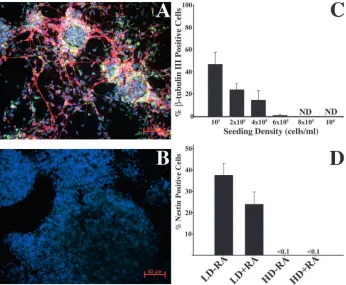
[image:4.612.241.560.74.445.2]To define the effects of cell density on neuronal lineage commitment, ES cells dissociated from monolayer cultures were induced by the 4–/4+ protocol (see Materials and methods) at various densities. Cells were then plated onto PDL/laminin-coated coverslips. Four days after plating the cells were stained with antibodies to β-tubulin 3 (red) and Fig. 1. Phosphorylation of β-catenin is inhibited
in low-density cultures. (A) Western blot analyses of total cell lysates extracted on days 2 (d2) and 4 (d4) after ES cells were seeded for EB formation at low (LD, 105cells/ml) and high densities (HD,
106cells/ml). Density did not alter the levels of
total β-catenin protein. However, the levels of phospho-β-catenin were significantly reduced in the low-density cultures when compared with the same time point in the high-density cultures using both antibodies (lower two blots). ES cells were seeded for EB formation at either low densities (B) or high densities (C) and stained with anti-β-catenin antibody. Fluorescence intensity over a random cross-section of an EB demonstrated diffuse staining in low-density EBs (B′), whereas high-density EBs resulted in peaks and valleys in fluorescent intensities (C′). (D) The difference in fluorescence intensity (*P<0.05, **P<0.01) between the peaks and valleys was quantified and plotted graphically. HD EBs had a higher average difference between the peaks and the valleys, demonstrating that β-catenin staining is more localized (B′,C′,D, y axis shows fluorescence intensity). (E) Undifferentiated ES cells were transiently transfected with an artificial TCF/LEF promoter and then seeded at high and low densities. Total luciferase activity was then assayed at day 1 post-seeding. High-density EBs repressed basal levels of TCF/LEF activity 2-fold (y axis shows relative luciferase units). (F) ES cells were differentiated by EB formation at low (105cells/ml) and high (106cells/ml) densities.
nestin (green), and counterstained with Hoechst dye (blue) (Fig. 2A-D). Plating at higher densities resulted in formation of EBs that were larger than those formed at low plating densities, and contact between EBs was increased in the high-density cultures. The maximal percentage of neurons was obtained when the cells were seeded at 105cells/ml (Fig. 2A).
As the seeding density was increased, the percentage of β-tubulin 3-positive cells in the cultures decreased in a linear fashion (Fig. 2A-D). Furthermore, as density increased, the number of nestin-positive cells similarly decreased, indicating that elevated density inhibited both neural and neuronal differentiation (Fig. 2D). At low density without RA treatment, there is a significant amount of neural differentiation, as demonstrated by nestin immunoreactivity. However, RA potentiates neural differentiation at low density by increasing the percentage of cells that are committed to the neural lineage (β-tubulin 3 + nestin-positive cells were 36% without RA and 73% with RA). If the RA-treated cultures are maintained for more than three days post-plating, virtually all of the nestin-positive cells observed go on to differentiate into GFAP-positive cells. As BMP signaling has been reported to inhibit neuronal differentiation of ES cells (Gratsch and O’Shea, 2002; Kawasaki et al., 2000; Tropepe et al., 2001) high-density RA-treated cultures were also RA-treated with a BMP antagonist, Noggin-Fc, to determine whether BMP signaling mediated the inhibitory effects of high density. Treatment of the high-density cultures with 50 ng/ml Noggin-Fc did not result in any detectable neuronal differentiation, and treatment with a saturating dose (2 µg/ml) resulted in minimal (<0.1% of cells) neuronal differentiation. Furthermore, treatment of low-density cultures (105 cells/ml) with media preconditioned by
high-density cultures (106 cells/ml) did not significantly inhibit
neuronal differentiation (data not shown), suggesting that the
inhibitory effect was not mediated solely by soluble factors. Finally, using two different EB-independent protocols (Gratsch and O’Shea, 2002; Ying et al., 2003), neuronal differentiation was also found to be inhibited by culture at high density (Fig. 7).
Stimulation of endogenous β-catenin overcomes density-dependent inhibition of neural
differentiation in ES cells
Stimulation of endogenous β-catenin signaling was performed by treatment with Wnt3a-conditioned media or by generation of stably transfected ES cells expressing H2kd-E-cadherin, a dominant-negative form of E-cadherin that has been shown previously to increase cellular levels of β-catenin (Vizirianakis et al., 2002; Zhu and Watt, 1996). The activity of these reagents in the stimulation of β-catenin signaling was assayed both by co-transfecting HEK293 cells with the TCF/LEF-luciferase plasmid with H2kd-E-cadherin and by treating TCF/LEF-luciferase-transfected HEK-293 cells with Wnt3a-conditioned media (Fig. 3A). H2kd-E-cadherin increased TCF/LEF-luciferase activity 2-fold, whereas treatment with Wnt3a-conditioned media resulted in more than 3-fold activation. To test whether Wnt3a-conditioned media could activate β-catenin in ES cells, we seeded ES cells for EB formation at high density in either control-conditioned media or Wnt3a-conditioned media and total cell lysates were analyzed by western blotting (Fig. 3B). Culture with Wnt3a-conditioned media resulted in a decrease in the levels of P-Ser33/37;Thr41 β-catenin (Fig. 3B, lower blot). In addition, H2kd-E-cadherin-expressing ES cells had higher levels of β-catenin protein relative to controls, as described previously (Vizirianakis et al., 2002; Zhu and Watt, 1996).
[image:5.612.221.567.71.356.2]To determine whether increasing endogenous β-catenin Fig. 2. Neural differentiation of ES cells is
regulated by cell density. ES cells were induced to differentiate by the 4–/4+ protocol at increasing seeding densities. After the induction, cells were plated onto PDL/laminin-coated coverslips and fixed 3-4 days post-plating. Coverslips were stained with anti-β-tubulin 3 (red) and anti-nestin (green), and counterstained with Hoechst dye (blue) to visualize nuclei. (A) 105
cells/ml seeding density; (B) 106 cells/ml
seeding density; (C) quantification of neuronal differentiation at different seeding densities (ND, none detected; error bars are s.d.). (D) Quantification of nestin-positive cells at low (105cells/ml) and high (106
signaling can overcome the inhibitory effects on differentiation of culture at high density, ES cells treated with Wnt3a-conditioned media or those expressing H2kd-E-cadherin were induced at high density by the 4–/4+ protocol (Fig. 4C-G). Conditioned media from untransfected cells was used as a control. Cultures treated with Wnt3a-conditioned media
contained many β-tubulin 3-positive (Fig. 3D) and nestin-positive cells. By contrast, virtually no cells immunoreactive for β-tubulin 3 (Fig. 3E), GFAP or nestin were detected in cultures treated with control media. Similarly, overexpression of H2Kd-E-cadherin in ES cells also promoted neuronal differentiation in high-density cultures (Fig. 3F), whereas cells transfected with an empty vector did not (Fig. 3E). Taken together, these observations suggest that increased β-catenin signaling in RA-treated ES cells can largely prevent the density-dependent inhibition of neural differentiation.
[image:6.612.51.557.66.273.2]As both treatment with Wnt3a-conditioned media or overexpression of H2Kd-E-cadherin could activate several different signaling pathways, we directly tested whether increasing β-catenin signaling alone can overcome the inhibitory effects of increased cell density by stably Fig. 3. Stimulation of endogenous β-catenin signaling overcomes the inhibitory effects of high density on RA-treated cultures. Stimulation of endogenous signaling was mediated by treatment with Wnt3a-conditioned media and by overexpression of H2kd-E-cadherin. (A) HEK293 cells were transfected with TCF/LEF-luciferase and tested for luciferase activity after treatment with Wnt3a-conditioned media or after
co-transfection of H2kd-E-cadherin with TCF/LEF-luc. H2kd-E-cadherin was able to increase luciferase activity, although Wnt3a treatment had a greater effect. (B) Changes in β-catenin degradation were analyzed in ES cells after Wnt3a treatment. ES cells were induced by EB formation in high density and were either treated with control-conditioned media or with Wnt3a-conditioned media. Total cell lysates were extracted on day 2 of the differentiation (2 days Wnt3a treatment) and analyzed by western blotting (B). Treatment with Wnt3a-conditioned media reduced the amount of phospho-β-catenin while not significantly altering the levels of total β-catenin. Wnt3a treatment resulted in a decrease in the levels of phopshorylated β-catenin signaling. ES cells were induced by EB formation at high density in either control-conditioned media (C) or Wnt3a-conditioned media (D), and differentiated by the 4–/4+ protocol. Wnt3a treatment resulted in many β-tubulin 3-positive cells (D), whereas control media did not (C). Cells were stably transfected with an empty vector (E), H2kd-E-cadherin (F) or full-length β-catenin (G), and induced by EB formation at high density using the 4–/4+ protocol. Overexpression of H2kd-E-cadherin and β-catenin resulted in neuronal differentiation in high-density cultures, but transfection with empty vector did not. (C-G) All cells were fixed 3-4 days post-plating, and stained with anti-β-tubulin 3 antibodies (green) and counterstained with Hoechst (blue).
Fig. 4. Modulation of β-catenin signaling regulates TCF/LEF
[image:6.612.71.263.533.738.2]transfecting ES cells with β-catenin. Control cells were transfected with a vector containing only the antibiotic resistance gene. Lif was then withdrawn and the β-catenin overproducing cells cultured at high density in the 4–/4+ EB protocol (Fig. 3G). Overexpression of β-catenin resulted in differentiation into β-tubulin 3-positive (Fig. 3G) and nestin-positive (data not shown) cells whereas virtually no such cells were present in the control cultures (Fig. 3E). This directly demonstrates that stimulation of β-catenin pathways can prevent the density-dependent inhibition of neural and neuronal differentiation in RA-treated cultures.
β-catenin induced neuronal differentiation of ES cells requires the armadillo domain and does not require RA treatment
To determine whether β-catenin signaling can by itself induce neurogenesis in ES cells in the absence of RA, and to define the domains of the β-catenin molecule that might be involved, we constructed various full-length and truncated constructs of β-catenin (Fig. 4A). We first tested the ability of these constructs to stimulate an artificial TCF/LEF-TOPFLASH promoter (Fig. 4B). HEK293 cells were co-transfected with the different β-catenin constructs and with a TCF/LEF promoter driving a luciferase reporter gene. In agreement with other studies (Peifer et al., 1991; van de Wetering et al., 1997; Vleminckx et al., 1999), transfection with full-length β-catenin, as well as with an N-terminal truncation of β-catenin (β-catenin ∆N), increased transcription of the reporter gene. The activity of β-catenin ∆N was several orders of magnitude higher than the activity of the full-length β-catenin construct as expected. By contrast, transfection of constructs with a truncation of the C-terminal portion of the molecule (β-catenin ∆C), or with a truncation of the armadillo domains (β-catenin ∆Armadillo), did not transactivate the reporter gene in this
assay, suggesting that both the armadillo domain and the C terminus are necessary for the transactivation of this promoter in HEK293 cells. In addition, stimulation of the promoter was dependent on the dosage of available β-catenin, as luciferase activity increased when the amount of transfected β-catenin pCLE plasmid (insert controlled by a retroviral LTR promoter) was increased or when β-catenin-pcDNA3.1 (insert transcription controlled by a stronger CMV promoter) was used. These data demonstrate that TCF/LEF driven transcription can be increased either by inhibiting Gsk3β-targeted degradation or by artificially increasing levels of total β-catenin.
To directly test the effects of β-catenin on ES cell differentiation and to determine which domains of β-catenin were necessary for its function, we then stably transfected ES cells with these various constructs of β-catenin. As serum and B27 supplement contain retinoids that could be converted to active RA by the cells (Brewer et al., 1993), the induction protocol was slightly altered so that ES cells cultured in the absence of RA were induced in a medium that lacks serum and any retinoids (see Materials and methods). As mammals cannot synthesize retinoids de novo, retinoid starvation has been widely used as a simple way to study retinoid dependent processes (Collins and Mao, 1999). After withdrawal of Lif and the formation of EBs, the cells that overexpressed either β-catenin ∆N or β-catenin ∆C differentiated into β-tubulin 3-immunoreactive cells in the high-density cultures with or without RA at all seeding densities tested (Fig. 5I-P). However, RA treatment of β-catenin-transfected cultures potentiated the neurogenic effects of β-catenin (Fig. 6A). By contrast, ES cells transfected with empty vector or with the β-catenin ∆Armadillo failed to differentiate into neurons in the high-density cultures, although they were still able to differentiate into neurons in low-density cultures treated with RA (Fig. 5A-H). β-tubulin 3-Fig. 5. The armadillo domain mediates
neurogenesis promoted by β-catenin and RA acts synergistically to enhance β-catenin-mediated neurogenesis. Cells were seeded at high (106cells/ml, HD) or low densities
(105cells/ml, LD) and treated with or without
[image:7.612.231.569.68.338.2]immunoreactive cells induced by overexpression of β-catenin with or without RA treatment were also found to be immunoreactive for NeuN (Neuna60 – Mouse Genome Informatics; Fig. 8E-H), neurofilament 200 (data not shown), Map2 (Fig. 8M-P), and synaptophysin (Fig. 8Q-T). Cells from high-density cultures transfected with β-catenin were patch clamped and were found to possess voltage-gated ion channels, whereas cells from control cultures did not (Fig. 6B and data not shown). The neuronal morphology of these cells, their expression of β-tubulin 3, NeuN, neurofilament, Map2 and synaptophysin, and their expression of voltage-gated channels, substantiate the conclusion that these cells are neurons.
β-catenin induced neural and neuronal lineage commitment does not require EB formation
Recently it has been demonstrated that ES cells can differentiate into neuron-like cells without having to go through an EB step (Gratsch and O’Shea, 2002; Ying et al., 2003). However, both of these protocols require culture at very low densities to induce differentiation. When ES cells were plated at 106cells/cm2[a
concentration 100-fold greater than that used by Ying and colleagues (Ying et al., 2003)] in DMEM/F12 with N2 and B27 supplements, cells immunoreactive to nestin and β-tubulin 3 were not detectable in either untransfected cells (data not shown) or cells transfected with an empty vector (Fig. 7A). However, overexpression of β-catenin ∆N (Fig. 7B) overcame the density-dependent inhibition of neural differentiation (for quantification, see Fig. 7C). Interestingly, GFAP- and CNPase-immunoreactive cells were not detected in β-catenin ∆N transfected cells seeded at high density. These cultures were stained with anti-catenin antibodies and the localization of β-catenin staining was analyzed by optical sectioning in a confocal microscope (Fig. 7E,F). In control cells most staining was localized to the cell membrane, whereas in cells stably transfected with β-catenin ∆N there was a more diffuse pattern of staining. These studies demonstrate that the stimulatory effects of β-catenin on neuronal lineage commitment by ES cells do not require formation of EBs.
Increased neuronal differentiation by β-catenin is not mediated by increased proliferation
β-catenin is a known mitogen (Kikuchi, 1999; Megason and
McMahon, 2002), and it is therefore possible that the increased neuronal differentiation was due to increased proliferation of neural progenitor cells and/or increased re-entry of neural progenitors into the cell cycle. ES cells stably transfected with β-catenin ∆N or with the empty vector were therefore tested for BrdU incorporation in order to study the effects of β-catenin ∆N expression on the proliferation of ES cells. Undifferentiated ES cells grown with Lif (see Materials and methods) were pulsed for 3 hours with BrdU and stained with anti-BrdU antibodies (Fig. 7D, left graph). BrdU incorporation by cells stably transfected with β-catenin ∆N did not differ significantly from cells transfected with the empty vector, although there was a slight trend in that direction (P=0.11, by
t-test). This is not surprising as undifferentiated ES cells
cultured with Lif already have a very high rate of proliferation. More importantly, we investigated whether ES cells stably transfected with β-catenin ∆N increased proliferation during neural differentiation by plating the cells at low density (104
cells/cm2) on gelatin-coated tissue culture dishes in
DMEM/F12 with B27 and N2 supplements without Lif. This has been shown previously by Ying and colleagues (Ying et al., 2003) to result in neural differentiation, with nestin-immunoreactive cells appearing by day 5 post-Lif withdrawal. Cells stably transfected with β-catenin ∆N or with empty vector were differentiated in vitro with this protocol and pulsed for 3 hours with BrdU (Fig. 7D, right graph) on day 6 post-Lif withdrawal. Interestingly, the cells transfected with β-catenin ∆N were found to have a statistically significant decrease in BrdU incorporation (P<0.01, by t-test) and had a decrease in the number of cells but an increase in the percentage of tubulin 3-immunoreactive cells, suggesting that activation of β-catenin signaling resulted in increased exit from the cell cycle and differentiation.
Phenotypic analysis of neurons generated by
β-catenin overexpression
[image:8.612.61.556.62.234.2]To determine whether the phenotypes of neurons induced by β-catenin and RA are the same, profiles of gene expression were compared in neurons derived from ES cells that overexpress β-catenin and in neurons induced by the 4–/4+ protocol with untransfected cells cultured at low density. We first used Hox gene expression profiles to characterize the Fig. 6. RA acts synergistically with β-catenin to
induce neuronal differentiation in ES cells. (A) The percentage of β-tubulin
3-immunoreactive cells was quantified from high density inductions with or without RA treatment (see Materials and methods). β-catenin ∆N induced a greater proportion of neurons than β-catenin ∆C did. Treatment with RA, along with either β-catenin ∆C or ∆N, resulted in an increase in the number of neurons, suggesting that there is synergy between RA and β-catenin signaling (ND, none detected; error bars show s.d.). (B) Cells with neuronal morphology from these cultures were patch clamped to determine whether they expressed voltage-gated ion channels (see
phenotype of the cells (Fig. 8). Virtually all neurons expressed Hoxc4 after induction of neuronal lineage commitment by RA treatment (Fig. 8V), by overexpression of β-catenin (Fig. 8A,C), or by both (Fig. 8B,D). This suggests that the neurons generated in all conditions are caudal in character. Furthermore, GABAergic neurons were detected in the culture in all conditions (Fig. 8E-H). Some neurons (<5%) induced by overexpression of β-catenin were immunoreactive for tyrosine hydroxylase (TH), the rate-limiting enzyme in dopamine biosynthesis. However, treatment with RA suppressed the generation of TH-positive cells, suggesting that not all of the effects of RA are dependent on β-catenin (Fig. 8I-L).
Discussion
We have found that β-catenin signaling and neural differentiation of ES cells are inhibited by culture at high density. This observation is consistent with prior studies of neural/neuronal differentiation of ES cells that have all used relatively low densities irrespective of whether EB or
dissociated cell culture techniques were used (Gratsch and O’Shea, 2002; Ying et al., 2003). The need to culture the cells at low density to achieve neuronal differentiation limits the number of cells that could potentially be obtained for transplantation strategies and raises questions about the mechanisms mediating neuronal differentiation of the cells. Our studies suggest that β-catenin signaling promotes both neural and neuronal differentiation of ES cells, and that the effects of increased cell density are mediated at least in part by inhibition of β-catenin signaling.
Similar to observations with keratinocytes (Dietrich et al., 2002), culture of ES cells at high density promotes membrane localization of β-catenin with a consequent decrease in signaling. Furthermore, although the levels of total β-catenin were not influenced by cell density, high-density cultures had a higher proportion of N-terminally phosphorylated β-catenin (Ser33/37;Thr41), which targets the molecule for degradation (Salic et al., 2000b). The sequestration of β-catenin by membrane binding and the targeting for degradation reduces the nuclear pool of β-catenin thereby reducing signaling (Novak and Dedhar, 1999). The suppression of baseline TCF/LEF activity and decreased Pitx2 transcription in our high-density cultures supports the conclusion that β-catenin signaling is diminished in these cultures.
[image:9.612.51.327.67.507.2]Although there are extensive data indicating that Wnt/β-catenin signaling enhances neuronal differentiation in the
Fig. 7. Overexpression of β-catenin overcomes
density-dependent inhibition of neural differentiation in an EB-independent protocol. ES cells transfected with the constructs indicated were induced to differentiate into the neural lineage in an EB-independent protocol (see Materials and methods) at a density of 106cells/cm2. Cells were fixed and stained 8-10
developing embryo (Baker et al., 1999; Dorsky et al., 1998; McGrew et al., 1999; Megason and McMahon, 2002) the effects of β-catenin on ES cells could also reflect enhanced proliferation and/or reduced exit of progenitor cells from the cell cycle, or survival of neural species, as well as an instructive effect on neural lineage commitment. In fact, recent studies have demonstrated a role for β-catenin in maintaining the proliferative state of neural stem cells. Overexpression of constitutively active β-catenin in neural stem cells increased neurogenesis primarily by decreasing cell cycle exit of neural progenitors (Chenn and Walsh, 2002), and β-catenin expression in the developing spinal cord maintained neural progenitor cells in a proliferative state with decreased neuronal differentiation (Zechner et al., 2003). Furthermore, it has been suggested that stabilization of β-catenin results in maintenance of pluripotency in human embryonic stem cells (Sato et al., 2004) and inhibition of differentiation of murine ES cells (Aubert et al., 2002; Haegele et al., 2003; Kielman et al., 2002). By contrast, our study shows that β-catenin facilitates neural
and neuronal differentiation of ES cells while not increasing proliferation of neural progenitors, and that it is associated with enhanced exit from cell cycle. Overexpression of β-catenin in the pluripotent P19 cell line induces neuronal differentiation (Israsena et al., 2004), whereas pharmacological inhibition of Gsk3β facilitated neuronal differentiation in P19 cells (Ding et al., 2003). Other investigators have also shown that β-catenin signaling can result in increased differentiation without affecting proliferation (Jin et al., 2001). It is possible that β-catenin exerts an effect either on proliferation, on differentiation, or on both, depending on the context of other signaling cascades. For example, many other molecules such as Shh, Lif and FGF have been shown to be potent mitogens or potent differentiation signals (Bartlett et al., 1998; Zhu et al., 1999) depending upon the cellular context. In the Chenn and Walsh study, and in the Zechner study, β-catenin was overexpressed in a setting where they were also exposed to significant levels of Fgf2 present in the ventricular zone (Vaccarino et al., 1999). By contrast, in our studies serum was removed from the cultures after EB dissociation. In fact, in other studies we find that the effects of β-catenin signaling in cultured neural progenitor cells are modified from pro-differentiation to maintenance of the proliferative state by the presence of Fgf2 (Israsena et al., 2004).
[image:10.612.43.347.68.506.2]Finally, it should also be emphasized that we found that the stimulatory effects of β-catenin on neurogenesis reflect effects on pre-neural cells, as well as on neural progenitor cells, so that mitogenic effects on neural progenitors could not possibly
Fig. 8.β-catenin overexpression results in a less
restricted population of neurons compared with RA-derived neurons. The phenotype of neurons generated by β-catenin ∆N and ∆C overexpression in high-density EB cultures was compared with neurons generated by RA treatment of low-density EB cultures.
(A-D) Hoxc4, red; β-tubulin 3, green; (E-H) NeuN, green; GABA, red; (I-L) tyrosine hydroxylase, red; β-tubulin 3, green; Hoechst stain blue (white arrows indicate TH-positive neurons); (M-P) Map2, green; β-tubulin 3, red; (Q-T) synaptophysin, green; β-β-tubulin 3, red. (A-D) All neurons generated by either β-catenin overexpression or RA treatment were positive for Hoxc4, a homeobox gene specific for caudal neurons. (E-H) Many neurons were positive for the
neurotransmitter GABA and all neurons were positive for NeuN. (I-L) Some neurons induced by
overexpression of β-catenin were immunoreactive for tyrosine hydroxylase (TH), the rate-limiting enzyme in catecholamine biosynthesis. By contrast,
underlie all of its actions. Nestin immunoreactive neural progenitor cells did not develop in high-density cultures in the absence of β-catenin signaling, but they did develop in response to increased β-catenin signaling, indicating an effect on pre-neural cells. However, in low-density cultures, which develop nestin-immunoreactive cells in the absence of exogenous RA treatment, β-catenin signaling promotes commitment of these progenitors to the neuronal lineage, indicating an effect on the neural stem cells as well.
The domains of β-catenin mediating its effects on neurogenesis in ES cells and the signaling pathways involved are unclear. In the canonical Wnt pathway, β-catenin interacts with members of the TCF/LEF family of transcription factors leading to both relief of repression and activation of transcription. Numerous genetic and biochemical studies suggest that the C-terminal domain of β-catenin is the primary transactivation domain (Peifer et al., 1991; van de Wetering et al., 1997), although it has been reported that a second transactivation domain may be present in the N terminus (Hsu et al., 1998). We found that in HEK293 cells the transactivation of TCF/LEF genes requires both the armadillo domain of β-catenin and the C-terminal transactivation domain. By contrast, although the armadillo domain of β-catenin was required to induce neural lineage commitment by ES cells, the C-terminal transactivation domain was not. Yet, β-catenin ∆C was unable to enhance TCF/LEF driven transcription in an artificial promoter system. This raises the possibility that some of the effects of β-catenin on neurogenesis might not be mediated by the classical TCF/LEF pathway, consistent with prior observations that β-catenin can signal through other transcription factors (Easwaran et al., 1999; Kioussi et al., 2002). However, there are several possible alternative explanations for the results of the truncation experiments. First, in some cell types the armadillo domain partly activates TCF/LEF pathways (N. Israsena and J.A.K., unpublished), raising the possibility that this is the case with ES cells. Alternatively, overexpression of β-catenin ∆C may lead to displacement of endogenous β-catenin from adherins junctions to the nucleus. However, this seems unlikely as the replacement of endogenous membrane bound β-catenin with β-catenin ∆C would be expected to reduce C-terminal immunostaining on the cell membrane, and immunohistochemical studies did not show any change in the relative staining when a C-terminal β-catenin antibody or an N-terminal β-β-catenin antibody was used.
RA signaling was incapable of inducing neural differentiation in high-density cultures in the absence of β-catenin signaling. There is a precedent for such dependence on β-catenin signaling of RA-mediated differentiation. In addition, the effects of RA in inducing endoderm in the F9 teratocarcinoma cell line are absolutely dependent upon β-catenin signaling (Lui et al., 2002), and inhibition of axin, an auxiliary factor to Gsk3βthat promotes β-catenin degradation, inhibits RA-mediated differentiation of P19 cells (Lyu et al., 2003). Nevertheless RA treatment enhanced neural differentiation in ES cells overexpressing β-catenin, suggesting that there is a synergistic interaction between the two signaling pathways. There are numerous types of crosstalk between RA and β-catenin signaling (for a review, see Katoh, 2002). For example, RA increases β-catenin protein stability and affinity for adherins junctions in a breast
cancer cell line (Byers et al., 1996), and RA treatment results in co-immunoprecipitation of β-catenin with the retinoic acid receptor and an increase in β-catenin-RAR driven transcription (Easwaran et al., 1999). Interestingly, RA has been shown to upregulate the Wnt receptor frizzled (Katoh, 2002), although we did not find a change in the expression of frizzled by western blotting in EBs after RA treatment (data not shown). Moreover, some caudal homeobox genes contain response elements for both β-catenin and RA signaling (Lickert and Kemler, 2002). Interestingly, neurons generated by either β-catenin overexpression or RA treatment were caudal in nature as evidenced by expression of Hoxc4 (Fig. 8). In addition, we found no statistically significant difference in the proportion of gabaergic neurons in β-catenin transfected cells with or without RA treatment. Nevertheless RA treatment inhibited the generation of TH-positive neurons by β-catenin, suggesting that RA exerts at least some effects independent of β-catenin signaling. Although β-catenin signaling has been demonstrated in the caudal neural tube it has not been shown previously that β-catenin can induce neurogenesis in an RA-independent manner. Wnt and FGF signaling inhibited the expression of cyp26, a cytochrome P450 oxidase that degrades RA and which is in part responsible for the spatially restricted signaling of RA in the caudal neural tube (Kudoh et al., 2002). Furthermore, the expression of caudal neural genes by Wnt signaling was mediated through RA signaling. By contrast, our studies demonstrate that β-catenin signaling can lead to the development of caudal neurons in a RA-independent fashion. In addition to inhibiting β-catenin signaling, it is possible that high-density culture increases BMP signaling, a known inhibitor of neural differentiation in ES cells (Kawasaki et al., 2000; Tropepe et al., 2001). However treatment with noggin-Fc or low-density inductions using media conditioned by high density inductions did not inhibit neural differentiation. Interestingly, it has been shown that inhibition of BMP signaling in epithelial bud development results in the upregulation of Lef1 and an increase in β-catenin signaling (Jamora et al., 2003). We, however, were unable to find any difference in Lef1 protein levels between high and low density cultures (data not shown).
In summary, our observations indicate that β-catenin signaling enhances neural lineage commitment by ES cells. Furthermore, β-catenin signaling may be a necessary co-factor for RA-induced neural differentiation. Culture of ES cells at increased density inhibits neurogenesis mediated by all of the previously described protocols for inducing neurogenesis (RA, antagonism of BMP signaling, or treatment with stromal cell membranes), apparently by both sequestering β-catenin at the cell membrane and by increasing phosphorylation of β-catenin. However, enhanced β-catenin signaling can overcome the inhibitory effects of increased cell density. These observations illustrate the importance of β-catenin signaling in neural lineage commitment by ES cells, and the synergy between RA and β-catenin signaling indicates a method for obtaining large numbers of neural species for possible use in therapeutic strategies involving ES cell transplantation.
References
Aberle, H., Schwartz, H. and Kemler, R. (1996). Cadherin-catenin complex,
protein interactions and their implications for cadherin function. J. Cell
Biochem. 61, 514-523.
Aubert, J., Dunstan, H., Chambers, I. and Smith, A. (2002). Functional
gene screening in embryonic stem cells implicates Wnt antagonism in neural differentiation. Nat. Biotech. 20, 1240-1245.
Bain, G., Kitchens, D., Yao, M., Huettner, J. E. and Gottlieb, D. I. (1995).
Embryonic stem cells express neuronal properties in vitro. Dev. Biol. 168, 342-357.
Baker, J. C., Beddington, R. S. and Harland, R. M. (1999). Wnt signaling
in Xenopus embryos inhibits bmp4 expression and activates neural development. Genes Dev. 13, 3149-3159.
Bartlett, P. F., Brooker, G. J., Faux, C. H., Dutton, R., Murphy, M., Turnley, A. and Kilpatrick, T. J. (1998). Regulation of neural stem cell
differentiation in the forebrain. Immunol. Cell Biol. 76, 414-418.
Brantjes, H., van Barker, N. E. J. and Clevers, H. (2002). TCF, Lady Justice
casting the final verdict on the outcome of Wnt signalling. Biol. Chem. 383, 255-261.
Brewer, G. J., Torricelli, J. R., Evege, E. K. and Price, P. J. (1993).
Optimized survival of hippocampal neurons in B27-supplemented Neurobasal, a new serum-free medium combination. J. Neurosci. Res. 35, 567-576.
Brustle, O., Jones, K. N., Learish, R. D., Karram, K., Choudhary, K., Wiestler, O. D., Duncan, I. D. and McKay, R. D. (1999). Embryonic stem
cell-derived glial precursors, a source of myelinating transplants. Science
285, 754-756.
Byers, S., Pishvaian, M., Crockett, C., Peer, C., Tozeren, A., Sporn, M., Anzano, M. and Lechleider, R. (1996). Retinoids increase cell-cell
adhesion strength, beta-catenin protein stability, and localization to the cell membrane in a breast cancer cell line, a role for serine kinase activity.
Endocrinology 137, 3265-3273.
Chazaud, C., Chambon, P. and Dolle, P. (1999). Retinoic acid is required in
the mouse embryo for left-right asymmetry determination and heart morphogenesis. Development 126, 2589-2596.
Chen, U. and Mok, H. (1995). Development of mouse embryonic stem (ES)
cells, IV. Differentiation to mature T and B lymphocytes after implantation of embryoid bodies into nude mice. Dev. Immunol. 4, 79-84.
Chenn, A. and Walsh, C. A. (2002). Regulation of cerebral cortical size by
control of cell cycle exit in neural precursors. Science 297, 365-369.
Cho, E. A. and Dressler, G. R. (1998). TCF-4 binds beta-catenin and is
expressed in distinct regions of the embryonic brain and limbs. Mech. Dev.
77, 9-18.
Chung, S., Sonntag, K. C., Andersson, T., Bjorklund, L. M., Park, J. J., Kim, D. W., Kang, U. J., Isacson, O. and Kim, K. S. (2002). Genetic
engineering of mouse embryonic stem cells by Nurr1 enhances differentiation and maturation into dopaminergic neurons. Eur. J. Neurosci.
16, 1829-1838.
Collins, M. D. and Mao, G. E. (1999). Teratology of retinoids. Annu. Rev. Pharmacol. Toxicol. 39, 399-430.
Dietrich, C., Scherwat, J., Faust, D. and Oesch, F. (2002). Subcellular
localization of beta-catenin is regulated by cell density. Biochem. Biophys.
Res. Commun. 292, 195-199.
Ding, S., Wu, T. Y., Brinker, A., Peters, E. C., Hur, W., Gray, N. S. and Schultz, P. G. (2003). Synthetic small molecules that control stem cell fate. Proc. Natl. Acad. Sci. USA 100, 7632-7637.
Dinsmore, J., Ratliff, J., Deacon, T., Pakzaban, P., Jacoby, D., Galpern, W. and Isacson, O. (1996). Embryonic stem cells differentiated in vitro as a
novel source of cells for transplantation. Cell Transplant. 5, 131-143.
Dorsky, R. I., Moon, R. T. and Raible, D. W. (1998). Control of neural crest
cell fate by the Wnt signalling pathway. Nature 396, 370-373.
Easwaran, V., Pishvaian, M., Salimuddin, X. and Byers, S. (1999).
Cross-regulation of beta-catenin-LEF/TCF and retinoid signaling pathways. Curr.
Biol. 9, 1415-1418.
Gratsch, T. E. and O’Shea, K. S. (2002). Noggin and chordin have distinct
activities in promoting lineage commitment of mouse embryonic stem (ES) cells. Dev. Biol. 245, 83-94.
Gumbiner, B. M. and McCrea, P. D. (1993). Catenins as mediators of the
cytoplasmic functions of cadherins. J. Cell Sci. Suppl. 17, 155-158.
Haegele, L., Ingold, B., Naumann, H., Tabatabai, G., Ledermann, B. and Brandner, S. (2003). Wnt signalling inhibits neural differentiation of
embryonic stem cells by controlling bone morphogenetic protein expression.
Mol. Cell. Neurosci. 24, 696-708.
Hsu, S. C., Galceran, J. and Grosschedl, R. (1998). Modulation of
transcriptional regulation by LEF-1 in response to Wnt-1 signaling and association with beta-catenin. Mol. Cell. Biol. 18, 4807-4818.
Ioffe, E., Liu, Y., Bhaumik, M., Poirier, F., Factor, S. M. and Stanley, P.
(1995). WW6, an embryonic stem cell line with an inert genetic marker that can be traced in chimeras. Proc. Natl. Acad. Sci. USA 92, 7357-7361.
Israsena, N., Hu, M., Fu, W., Kan, L. and Kessler, J. A. (2004). The
presence of FGF2 signaling determines whether beta-catenin exerts effects on proliferation or neuronal differentiation of neural stem cells. Dev. Biol.
268, 220-231.
Jamora, C., DasGupta, R., Kocieniewski, P. and Fuchs, E. (2003). Links
between signal transduction, transcription and adhesion in epithelial bud development. Nature 422, 317-322.
Jin, E. J., Erickson, C. A., Takada, S. and Burrus, L. W. (2001). Wnt and
BMP signaling govern lineage segregation of melanocytes in the avian embryo. Dev. Biol. 233, 22-37.
Kang, D. E., Soriano, S., Xia, X., Eberhart, C. G., De Strooper, B., Zheng, H. and Koo, E. H. (2002). Presenilin couples the paired phosphorylation
of beta-catenin independent of axin, implications for beta-catenin activation in tumorigenesis. Cell 110, 751-762.
Katoh, M. (2002). Regulation of WNT signaling molecules by retinoic acid
during neuronal differentiation in NT2 cells, threshold model of WNT action. Int. J. Mol. Med. 10, 683-687.
Kawasaki, H., Mizuseki, K., Nishikawa, S., Kaneko, S., Kuwana, Y., Nakanishi, S., Nishikawa, S. I. and Sasai, Y. (2000). Induction of midbrain
dopaminergic neurons from ES cells by stromal cell-derived inducing activity. Neuron 28, 31-40.
Kielman, M. F., Rindapaa, M., Gaspar, C., van Poppel, N., Breukel, C., van Leeuwen, S., Taketo, M. M., Roberts, S., Smits, R. and Fodde, R.
(2002). Apc modulates embryonic stem-cell differentiation by controlling the dosage of beta-catenin signaling. Nat. Genet. 32, 594-605.
Kikuchi, A. (1999). Roles of Axin in the Wnt signalling pathway. Cell Signal 11, 777-788.
Kioussi, C., Briata, P., Baek, S. H., Rose, D. W., Hamblet, N. S., Herman, T., Ohgi, K. A., Lin, C., Gleiberman, A., Wang, J. et al. (2002).
Identification of a Wnt/Dvl/beta-catenin→Pitx2 pathway mediating cell-type-specific proliferation during development. Cell 111, 673-685.
Korswagen, H. C. (2002). Canonical and non-canonical Wnt signaling
pathways in Caenorhabditis elegans, variations on a common signaling theme. BioEssays 24, 801-810.
Kudoh, T., Wilson, S. W. and Dawid, I. B. (2002). Distinct roles for Fgf,
Wnt and retinoic acid in posteriorizing the neural ectoderm. Development
129, 4335-4346.
Kuhl, M. (2002). Non-canonical Wnt signaling in Xenopus, regulation of axis
formation and gastrulation. Semin. Cell Dev. Biol. 13, 243-249.
Lickert, H. and Kemler, R. (2002). Functional analysis of cis-regulatory
elements controlling initiation and maintenance of early Cdx1 gene expression in the mouse. Dev. Dyn. 225, 216-220.
Liu, C., Li, Y., Semenov, M., Han, C., Baeg, G. H., Tan, Y., Zhang, Z., Lin, X. and He, X. (2002). Control of beta-catenin phosphorylation/degradation
by a dual-kinase mechanism. Cell 108, 837-847.
Liu, S., Qu, Y., Stewart, T. J., Howard, M. J., Chakrabortty, S., Holekamp, T. F. and McDonald, J. W. (2000). Embryonic stem cells differentiate into
oligodendrocytes and myelinate in culture and after spinal cord transplantation. Proc. Natl. Acad. Sci. USA 97, 6126-6131.
Lucas, J. J., Hernandez, F., Gomez-Ramos, P., Moran, M. A., Hen, R. and Avila, J. (2001). Decreased nuclear beta-catenin, tau hyperphosphorylation
and neurodegeneration in GSK-3beta conditional transgenic mice. EMBO J.
20, 27-39.
Lui, T., Lee, Y. N., Malbon, C. C. and Wang, H. Y. (2002). Activation of
the beta-catenin/LEF-TCF pathway is obligate for formation of primitive endoderm by mouse F9 totipotent teratocarcinoma cells in response to retinoic acid. J. Biol. Chem. 277, 30887-30891.
Lyu, J., Costantini, F., Jho, E. H. and Joo, C. K. (2003). Ectopic expression
of axin blocks neuronal differentiation of embryonic carcinoma p19 cells.
J. Biol. Chem. 278, 13487-13495.
Martin, D. M., Skidmore, J. M., Fox, S. E., Gage, P. J. and Camper, S. A.
(2002). Pitx2 distinguishes subtypes of terminally differentiated neurons in the developing mouse neuroepithelium. Dev. Biol. 252, 84-99.
McGrew, L. L., Takemaru, K., Bates, R. and Moon, R. T. (1999). Direct
regulation of the Xenopus engrailed-2 promoter by the Wnt signaling pathway, and a molecular screen for Wnt-responsive genes, confirm a role for Wnt signaling during neural patterning in Xenopus. Mech. Dev. 87, 21-32.
Megason, S. G. and McMahon, A. P. (2002). A mitogen gradient of dorsal
Novak, A. and Dedhar, S. (1999). Signaling through beta-catenin and Lef/Tcf. Cell. Mol. Life Sci. 56, 523-537.
Pandur, P., Lasche, M., Eisenberg, L. M. and Kuhl, M. (2002a). Wnt-11
activation of a non-canonical Wnt signalling pathway is required for cardiogenesis. Nature 418, 636-641.
Pandur, P., Maurus, D. and Kuhl, M. (2002b). Increasingly complex, New
players enter the Wnt signaling network. BioEssays 24, 881-884.
Patapoutian, A. and Reichardt, L. F. (2000). Roles of Wnt proteins in neural
development and maintenance. Curr. Opin. Neurobiol. 10, 392-399.
Peifer, M., Rauskolb, C., Williams, M., Riggleman, B. and Wieschaus, E.
(1991). The segment polarity gene armadillo interacts with the wingless signaling pathway in both embryonic and adult pattern formation.
Development 111, 1029-1043.
Salic, A., Lee, E., Mayer, L. and Kirschner, M. W. (2000a). Control of
beta-catenin stability, reconstitution of the cytoplasmic steps of the wnt pathway in Xenopus egg extracts. Mol. Cell 5, 523-532.
Salic, A., Lee, E., Mayer, L. and Kirschner, M. W. (2000b). Control of
beta-catenin stability, reconstitution of the cytoplasmic steps of the wnt pathway in Xenopus egg extracts. Mol. Cell 5, 523-532.
Sato, N., Meijer, L., Skaltsounis, L., Greengard, P. and Brivanlou, A. H.
(2004). Maintenance of pluripotency in human and mouse embryonic stem cells through activation of Wnt signaling by a pharmacological GSK-3-specific inhibitor. Nature Med. 10, 55-63.
Tada, M., Concha, M. L. and Heisenberg, C. P. (2002). Non-canonical Wnt
signalling and regulation of gastrulation movements. Semin. Cell Dev. Biol.
13, 251-260.
Tropepe, V., Hitoshi, S., Sirard, C., Mak, T. W., Rossant, J. and van der Kooy, D. (2001). Direct neural fate specification from embryonic stem cells,
a primitive mammalian neural stem cell stage acquired through a default mechanism. Neuron 30, 65-78.
Vaccarino, F. M., Schwartz, M. L., Raballo, R., Nilsen, J., Rhee, J., Zhou, M., Doetschman, T., Coffin, J. D., Wyland, J. J. and Hung, Y. T. (1999).
Changes in cerebral cortex size are governed by fibroblast growth factor during embryogenesis. Nat. Neurosci. 2, 246-253.
van de Wetering, M., Cavallo, R., Dooijes, D., van Beest, M., van Es, J., Loureiro, J., Ypma, A., Hursh, D., Jones, T. and Bejsovec, A. (1997).
Armadillo coactivates transcription driven by the product of the Drosophila segment polarity gene dTCF. Cell 88, 789-799.
Vizirianakis, I. S., Chen, Y. Q., Kantak, S. S., Tsiftsoglou, A. S. and Kramer, R. H. (2002). Dominant-negative E-cadherin alters adhesion and
reverses contact inhibition of growth in breast carcinoma cells. Int. J. Oncol.
21, 135-144.
Vleminckx, K., Kemler, R. and Hecht, A. (1999). The C-terminal
transactivation domain of beta-catenin is necessary and sufficient for signaling by the LEF-1/beta-catenin complex in Xenopus laevis. Mech. Dev.
81, 65-74.
Westmoreland, J. J., McEwen, J., Moore, B. A., Jin, Y. and Condie, B. G.
(2001). Conserved function of Caenorhabditis elegans UNC-30 and mouse Pitx2 in controlling GABAergic neuron differentiation. J. Neurosci. 21, 6810-6819.
Yamanaka, H., Moriguchi, T., Masuyama, N., Kusakabe, M., Hanafusa, H., Takada, R., Takada, S. and Nishida, E. (2002). JNK functions in the
non-canonical Wnt pathway to regulate convergent extension movements in vertebrates. EMBO Rep. 3, 69-75.
Ying, Q. L., Stavridis, M., Griffiths, D., Li, M. and Smith, A. (2003).
Conversion of embryonic stem cells into neuroectodermal precursors in adherent monoculture. Nat. Biotechnol. 21, 183-186.
Zechner, D., Fujita, Y., Hulsken, J., Muller, T., Walther, I., Taketo, M. M., Crenshaw, E. B., 3rd, Birchmeier, W. and Birchmeier, C. (2003).
beta-Catenin signals regulate cell growth and the balance between progenitor cell expansion and differentiation in the nervous system. Dev. Biol. 258, 406-418.
Zhu, A. J. and Watt, F. M. (1996). Expression of a dominant negative
cadherin mutant inhibits proliferation and stimulates terminal differentiation of human epidermal keratinocytes. J. Cell Sci. 109, 3013-3023.
Zhu, G., Mehler, M. F., Zhao, J., Yu Yung, S. and Kessler, J. A. (1999).