D
E
V
E
LO
P
M
E
N
T
INTRODUCTION
In the zebrafish embryo, precursors of the ectoderm derive from the animal pole region, while precursors of the endoderm and mesoderm originate from partially overlapping territories near the equatorial region, or margin, of the embryo (Kimmel et al., 1990; Warga and Nusslein-Volhard, 1999). The endodermal progenitors arise from the first four rows of marginal cells, while mesodermal precursors arise from the entire marginal region (Kikuchi et al., 2004). Fate-mapping experiments have shown that when single cells located near the margin are labelled at the late blastula stage, their progeny frequently populate both germ layers (Warga and Nusslein-Volhard, 1999). Therefore, in the most vegetal rows of cells, mesodermal and endodermal fates are intermingled and both germ layers share common mesendodermal precursors. The molecular mechanisms that allow segregation of these two germ layers are poorly understood.
In zebrafish, as in other vertebrates, signalling by secreted TGF factors of the Nodal family is crucial for the formation of the mesoderm and endoderm (Schier, 2003). The nodal-related genes cyclopsand squintare expressed in the first two rows of cells and are potent inducers of endoderm and mesoderm when overexpressed
(Erter et al., 1998; Feldman et al., 1998; Gritsman et al., 2000; Peyrieras et al., 1998; Rebagliati et al., 1998; Sampath et al., 1998). cyclop;squintdouble mutants or mutants lacking the maternal and zygotic contribution of oep(MZoep), which encodes a Nodal co-receptor, lack all endoderm and have little or no mesoderm (Agathon et al., 2003; Gritsman et al., 1999; Zhang et al., 1998).
Several lines of evidence suggest that in addition to being required for endoderm and mesoderm formation, differential Nodal signalling may also be involved in the separation of these two germ layers. First, many studies have documented that high levels of Nodal signalling promote endoderm formation and expression of endodermal determination genes, while lower levels of Nodal signalling promote mesoderm formation and expression of mesodermal genes such as brachyury (Alexander et al., 1999; Alexander and Stainier, 1999; Clements et al., 1999; Erter et al., 1998; Faucourt et al., 2001; Gritsman et al., 2000; Henry et al., 1996; Jones et al., 1995; Piccolo et al., 1999; Rodaway et al., 1999; Sun et al., 1999; Yasuo and Lemaire, 1999). Second, overexpression of low doses of lefty1, which encodes a potent endogenous antagonist of Nodal, suppresses endoderm formation while higher doses also affect the mesoderm (Thisse et al., 2000; Thisse and Thisse, 1999). Similarly, in zygotic oep mutants that have reduced Nodal signalling, the endoderm is absent while the mesoderm is modestly affected (Schier et al., 1997; Strahle et al., 1997). By contrast, MZoep embryos resemble cyclops;squintdouble mutants and lack all endoderm and most mesoderm (Agathon et al., 2003; Gritsman et al., 1999; Zhang et al., 1998). Finally, it has been demonstrated that Squint acts as a morphogen capable of inducing different cell fates and that it does not require a relay mechanism (Chen and Schier, 2001; Le Good et al., 2005).
Zebrafish endoderm formation is regulated by combinatorial
Nodal, FGF and BMP signalling
Morgane Poulain1, Maximilian Fürthauer2,*, Bernard Thisse2, Christine Thisse2and Thierry Lepage3,†
In the zebrafish embryo, the mesoderm and endoderm originate from common precursors and segregate during gastrulation by mechanisms that are largely unknown. Understanding how the signalling pathways that regulate endoderm and mesoderm formation interact is crucial to understanding how the germ layers are established. Here, we have analysed how the FGF and BMP pathways interact with Nodal signalling during the process of endoderm formation. We found that activation of the FGF/ERK pathway disrupts endoderm formation in the embryo and antagonizes the ability of an activated form of Tar/Acvr1b to induce endoderm at the animal pole. By contrast, inhibition of FGF signalling increases the number of endodermal precursors and potentiates the ability of Tar*/Acvr1b to induce endoderm at the animal pole. Using a pharmacological inhibitor of the FGF receptor, we show that reducing FGF signalling partially rescues the deficit of endoderm precursors in bonmutant embryos. Furthermore, we found that overexpression of BMPs compromises endoderm formation, suggesting that formation of endoderm precursors is negatively regulated by BMPs on the ventral side. We show that simultaneous inhibition of the FGF/Ras and BMP pathways results in a dramatic increase in the number of endoderm precursors. Taken together, these data strongly suggest that BMP and FGF-ERK pathways cooperate to restrict the number of endodermal progenitors induced in response to Nodal signalling. Finally, we investigated the molecular basis for the FGF-MAPK-dependent repression of endoderm formation. We found that FGF/ERK signalling causes phosphorylation of Casanova/Sox32, an important regulator of endoderm determination, and provide evidence that this phosphorylation attenuates its ability to induce sox17. These results identify a molecular mechanism whereby FGF attenuates Nodal-induced endodermal transcription factors and highlight a potential mechanism whereby mesoderm and
endoderm fates could segregate from each other.
KEY WORDS: Endoderm, Zebrafish, FGF, BMP, MAP kinase, Mesoderm, Casanova, Bon, ERK Development 133, 2189-2200 (2006) doi:10.1242/dev.02387
1National Institute for Medical Research, Division of Developmental Biology, The
Ridgeway, Mill Hill, London NW7 1AA, UK. 2Institut de Génétique et Biologie
Moléculaire et Cellulaire, CNRS/INSERM/ULP, BP 163, 67404 Illkirch cedex, CU de Strasbourg, France. 3UMR 7009 CNRS, Université de Paris VI, Observatoire
Océanologique de Villefranche sur Mer, 06230 Villefranche-sur-Mer, France.
*Present address: Max Planck Institute of Molecular Cell Biology and Genetics, Pfotenhauerstrasse 108, D-01307 Germany
†Author for correspondence (e-mail: lepage@obs-vlfr.fr)
D
E
V
E
LO
P
M
E
N
T
The endodermal determination program initiated by Nodal signalling requires the maternally expressed Smad2 factor (Dick et al., 2000; Gaio et al., 1999) and eomesodermin (Bjornson et al., 2005) as well as several zygotic transcription factors such as the Mix-like homeobox proteins Bon; the product of the bonnie and clyde gene (bon)(Alexander et al., 1999; Trinh et al., 2003) and Mezzo (Poulain and Lepage, 2002). Mix-like proteins act in parallel with the zinc-finger-containing factor Gata5, which is encoded by the faust gene to induce the sox-related gene casanova/sox32 (Kikuchi et al., 2004; Reiter et al., 1999). casanova/sox32mutants do not express the endodermal marker sox17, and lack endodermal precursors and organs derived from the gut tube (Alexander et al., 1999; Alexander and Stainier, 1999; Aoki et al., 2002a; Dickmeis et al., 2001; Kikuchi et al., 2001; Kikuchi et al., 2004; Reiter et al., 1999; Reiter et al., 2001; Sakaguchi et al., 2001). In the absence of Casanova activity, cells that normally contribute to the endoderm change their fate and differentiate into mesodermal cells. Therefore, Casanova is a key transcription factor in the endoderm determination network and is required for the expression of sox17.
In addition to Nodal, the FGF and BMP signalling pathways have been shown to play crucial roles in formation and patterning of mesoderm and endoderm in vertebrates. Basic FGF was first identified as a mesoderm-inducing factor that promotes ventral mesoderm formation (Kimelman, 1991). Then, studies in Xenopus and zebrafish demonstrated that FGF is required for posterior mesoderm formation through the maintenance of the expression of Tbox transcription factors such as No Tail and Tbx-16 (Bottcher and Niehrs, 2005; Griffin et al., 1995a; Griffin and Kimelman, 2003; Schulte-Merker and Smith, 1995). Because overexpression of a dominant-negative FGF receptor blocks the dorsal mesoderm inducing activity of Activin, FGF ligands have been proposed to act as competence factors needed by presumptive mesodermal cells to respond to TGF-signals (Cornell et al., 1995; Zhao et al., 2003). In zebrafish, FGF signals may relay the action of TGFligands over long distances, allowing activation of the pan-mesodermal marker brachyuryin cells distant from the Nodal source (Reiter et al., 1999). Consistent with these observations, FGF is required downstream of Nodal signalling to induce the co-receptor Oep in cells distant from the source of Nodal, a mechanism that contributes to the amplification and propagation of Nodal signals (Mathieu et al., 2004). Thus, the FGF pathway might be involved in the initiation of, as well as in the maintenance of, mesodermal populations. Finally, studies in zebrafish have shown in addition to its effects on anteroposterior patterning of the mesoderm, FGF signalling controls patterning along the DV axis by repressing the expression of BMPs (Fürthauer et al., 2004). Although a large body of evidence supports the role of FGF in mesoderm formation and patterning, the implication of FGF signalling in formation of the endoderm has received considerably less attention and the results obtained are unclear. In Xenopus, studies with vegetal pole explants by Henry et al. showed that FGF is required for expression of the pancreatic marker pdx1but not for expression of the intestinal marker intestinal fatty acid binding protein (IFABP) (Henry et al., 1996). By contrast, Gamer and Wright (Gamer and Wright, 1995) found that bFGF is a potent inhibitor of pdx1expression in vegetal pole explants. A third study concluded that overexpression of eFGF inhibits expression of mixer, while inhibition of FGF signalling in animal caps induces the ectopic expression of the key endodermal regulator mixerand of the gene marker endodermin(Cha et al., 2004). These observations suggest that in Xenopus, FGF signalling may antagonize endoderm specification. However, nothing is known about the role of FGF signalling in endoderm specification in other vertebrates.
Similarly, the roles of BMPs in patterning of the mesoderm and ectoderm are well documented but only a few studies focused on the role of BMPs on endoderm formation. Sasai et al. (Sasai et al., 1996) reported that, in Xenopus, overexpression of nogginor chordin induces endoderm in animal caps. Furthermore, they showed that endoderm induction by chordinis strongly potentiated by inhibition of FGF signalling and counteracted by activation of FGF signalling. Taken together these observations suggest that both BMP and FGF signalling antagonize endoderm formation in Xenopus but the molecular mechanism responsible for this antagonism is not known and these observations have not been extended to other vertebrates. Here, we have attempted to unravel the interactions between the Nodal, FGF and BMP pathways during formation of the endoderm in zebrafish. We show that both the FGF/MAPK and the BMP pathways antagonize endoderm formation in response to Nodal signals. We have found that activation of the FGF/MAPK pathway by overexpression of FGF ligands or constitutively active versions of Ras or ERK caused a severe reduction in the number of endodermal precursors in the whole embryo and antagonized the ability of Tar/Acvr1b to induce endoderm at the animal pole. By contrast, the triple inhibition of Fgf8, Fgf17b and Fgf24 caused a strong increase in the number of endodermal precursors while inhibition of FGF/ERK signalling potentiated the ability of Tar*/Acvr1b to induce endoderm at the animal pole. Furthermore, we found that overexpression of BMPs also inhibits endoderm formation and that simultaneous inhibition of the FGF/Ras and BMP pathways causes formation of an excess of endoderm in the embryo. Furthermore, we provide evidence that FGF/ERK signalling results in phosphorylation of Casanova and that this phosphorylation attenuates its activity. These results suggest that the FGF and BMP pathways counteract the Nodal signalling pathway and limit formation of endoderm. Importantly, they identify Casanova as a key factor at the crossroads between signalling pathways and highlight a potential molecular mechanism that may help explain the separation of the mesoderm and endoderm.
MATERIALS AND METHODS
Zebrafish strains, embryo manipulation, inhibitor treatments Adult zebrafish were maintained at 28.5°C using standard procedures (Westerfield, 1994). Wild-type embryos were collected by natural spawning from the AB strain. Mutant embryos were obtained by inter-crossing heterozygous carrier fish identified by random crossing. We used the bonnie
and clydem425mutant allele (Kikuchi et al., 2000). Embryos were genotyped
following the procedure published by Kikuchi et al. (Kikuchi et al., 2000). To inhibit FGFR activity, embryos were treated with SU5402 (Mohammadi et al., 1997), at 15 M after 1000 cell stage at 28.5°C in the dark. This concentration gave the most consistent results but some variability was observed in the effects of the drug, depending of the batch of inhibitor, the cell line and the time of addition of the drug. SU5402 activity was monitored by its ability to inhibit MAPK activity and sprouty4 expression (see Fig. S1 in the supplementary material).
RNA and oligonucleotides microinjection
Constructs for microinjection of Fgf8 (Fürthauer et al., 1997), DN-Fgfr1 and
noggin1(Furthauer et al., 1999), DN-Ras and CA-Ras (Whitman and
D
E
V
E
LO
P
M
E
N
T
gene (Fischer et al., 2003), while the Fgf8 morpholino phenocopied the
acerebellarmutation, which disrupts the fgf8gene. When co-injected, these
oligos caused severe defects in the posterior region of the embryo, which is known to require FGF signalling. The sequence of the morpholinos used are: MO Fgf8, 5⬘-GAGTCTCATGTTTATAGCCTCAGTA-3⬘; MO Fgf24: 5⬘-GACGGCAGAACAGACATCTTGGTCA-3⬘; MO Fgf17b, 5⬘ -AGT-GTTCAATATCCAGGGCTCTCCT-3⬘. noggin1mRNA was injected at 25
g/ml.
Whole-mount in situ hybridisation
In situ hybridisation was performed following a protocol adapted from Thisse et al. (Thisse et al., 2004) with antisense RNA probes and staged embryos. In some cases, the lineage tracer (FLDX) was detected after in situ hybridisation using an anti-fluorescein antibody coupled to alkaline phosphatase and Fast Red as substrate. The probes used in this publication have been described previously: sox17(Alexander and Stainier, 1999),
sprouty4(Fürthauer et al., 2001), nkx2.5(Chen and Fishman, 1996) and
foxi1(Fürthauer et al., 2004). All probes were synthesized with the T7 RNA
polymerase after linearization by NotI.
To count the number of cells expressing sox17, the embryos were photographed under different angles so that the whole surface was covered. Four to six different pictures were taken by progressively rotating the embryo and landmarks were used to delimit nonoverlapping areas. Alternatively, flat preparations of the blastoderm were made after removing the yolk platelets (see Fig. S2 in the supplementary material).
Immunochemistry and western blot
Capped mRNA of FLAG-tagged casanova/sox32(100 ng/l) were injected into embryos at the two-cell stage. When the embryos reached 30% epiboly (5 hpf), the chorion was removed with pronase and the yolk was removed using an ‘eye hair knife’ in an agarose chamber in MBS 1⫻(Modified Barth Saline Buffer) supplemented with gentamycin (Peng, 1991; Sagerstrom et al., 1996). Fifteen blastoderms were pooled, allowed to recover for 20 minutes after dissection in MBS 1⫻and then lysed in SDS sample buffer. Proteins were fractioned by sodium dodecyl sulphate-polyacrylamide gel electrophoresis (SDS-PAGE) and electrophoretically transferred to PVDF membranes. The replicate was blocked for 1 hour in 1% BSA, BRBT (Tris-HCl 10 mM, NaCl 150 mM, EDTA 1 mM, pH 7.5, Tween 0.01%) and incubated overnight at 4°C with a monoclonal antibody [␣ -Casanova-Phospho at 1/100 or ␣-Flag M2 (Kodak) at 1/1000]. After washing in BRBT, the membrane was incubated with the secondary anti body at 1/10000 (anti-Rabbit HRP or anti-mouse HRP conjugated antibodies, Amersham). Bound antibodies were revealed by ECL western blotting detection reagent (Pierce). The ␣-Casanova-Phospho antibody was made in rabbit by Eurogentec and was purified by affinity chromatography using HPLC (BioRad system). Immunostaining was performed essentially as described (Shinya et al., 2001).
RNA extraction and reverse transcription-polymerase chain reaction
Total RNA from staged embryos was extracted by the method of Chomczynski (Chomczynski, 1987). For RT-PCR, cDNA synthesis and PCR were performed as described by Sagerström (Sagerström, 1996). Primers pairs for histone H4 were synthesized according to previous published reports. Other primer pairs used were derived from the ORF of each protein: Zsox-17 forward, 5⬘-ACGAGGTGGAGTTTGAGCAC; Zsox-17 reverse, 5⬘-GGCTGCTCTAAAAGCTGCTG (amplified fragment 465 bp); Casanova forward, 5⬘-CAGCATTCTGTCCAGCAGAG; Casanova reverse, 5⬘-CAAAATCAGCAGCAATCTGG (amplified fragment 480 bp). Cycling parameters were as follows: initial denaturation at 94°C for 1 minute, annealing at 55°C for 1 minute, extension at 72°C for 1 minute 30 seconds. Thirty cycles were used for ease of comparison. Each experiment was repeated three times using one or three whole embryos.
Site-directed mutagenesis and construction of expression plasmids
To make pCS2 cas-S47A construct, the AGC codon encoding serine in position 47 of pCS2 caswas mutated GCC (alanine) by splicing PCR using the following oligonucleotides: Casanova-S47A Fw, 5⬘
-TCGGGCCCAT-TAGCCCCGGTGTCTGTC-3⬘; Casanova-S47A Rev; 5⬘ -GACAGACA-CCGGGGCTAATGGGCCCGA-3⬘. The exchange was verified by sequencing. pCS2 cas-WT-Flagand pCS2 cas-S47A-Flag were constructed as follows. A ClaI-EcoRI fragment containing Casanova was PCR amplified with the following oligonucleotides (restriction sites sequences underlined and ATG in bold): Fw-Casanova-ClaI-ATG, 5⬘-CCCATCGATATG TA-TCTCGACCGGATG-3⬘; Casanova-EcoRI-Rev, 5⬘ -CTTGAATTCCCTTT-TTGCTGTGGTCCAA-3⬘and cloned into the pCS2-Flag Vector digested with EcoRI and ClaI.
RESULTS
FGF/ERK signalling represses endoderm formation To test the role of FGF signalling on endoderm formation, embryos were injected at the one- or two-cell stage with synthetic Fgf8 mRNA and analysed by in situ hybridization for the expression of sox17, as a read out of endoderm specification. Injection of fgf8 RNA drastically reduced the number of sox17-expressing cells. In the most severe cases, only a few residual sox17progenitors were present in the injected embryos (Fig. 1C). To test if Fgf8 overexpression interfered with Nodal expression, we examined the expression of lefty1, which is a target of Nodal signalling (Thisse and Thisse, 1999). Expression of lefty1was not affected in the embryos overexpressing Fgf8 (data not shown), indicating that the loss of endodermal precursors caused by Fgf8 was not a consequence of interfering with Nodal expression. Similarly, overexpression of an activated form of Ras (CA-RAS) (Whitman and Melton, 1992) or of an activated form of human ERK2* (Emrick et al., 2001) strongly reduced the number of sox17-expressing cells (Fig. 1B,D). As FGFs are expressed (Draper et al., 2003; Fürthauer et al., 2004) and MAPK activity is detected in the endomesodermal
[image:3.612.332.515.405.645.2]D
E
V
E
LO
P
M
E
N
T
territory during gastrulation (Fig. S1 in the supplementary material), FGFs are good candidates for endogenous factors that negatively regulate formation of the endodermal precursors. To test this hypothesis we attempted to block FGF signalling using morpholinos. Injection of morpholinos directed against either Fgf8 or Fgf17b or Fgf24 mRNA alone did not affect significantly the number of endoderm precursors. By contrast, the triple knockdown of Fgf8, Fgf17b and Fgf24 resulted in a large increase in the number of endodermal precursors that formed (Fig. 1E,F; see Table 3). Taken together, these results show that endogenous FGF signals restrict endoderm formation during gastrulation.
FGF/ERK signalling antagonizes the ability of Tar*/Acvr1b to induce endoderm at the animal pole
To document the repressive effect of FGF/MAPK signalling on endoderm formation and circumvent cell-migration dependent effects, we used the ability of Nodal signals to induce endoderm at the animal pole as a model to examine the consequences of activating or inhibiting the FGF pathway. We used a constitutively activated form of Tar*/Acvr1b, a type I receptor for the Nodal factors, to activate Nodal signalling (Aoki et al., 2002b; Peyrieras et al., 1998; Renucci et al., 1996). To block FGF signalling, we used a dominant-negative form of FGFR1 (DN-FGFR1) (Fürthauer et al., 2004). In agreement with the known role of FGF in mesoderm formation, injection of the dominant-negative form of FGFR1 potently inhibited endogenous mesodermal expression (data not shown). To block FGF signalling, we also used the pharmacological agent SU5402, a specific inhibitor of FGFR function (Mohammadi et al., 1997).
Injection of RNA encoding Tar*/Acvr1b at the animal pole induced ectopic expression of the endodermal marker sox17(55%, n=40, Fig. 2E) and of the mesodermal marker ZFIN CB187(50%, n=14, Fig. 2B) (see Thisse et al., 2001 at http://zfin.org). In agreement with previous studies (Mathieu et al., 2004), we found that inhibition of FGF signalling reduced the ability of Tar*/Acvr1b to induce expression of the mesodermal marker CB187(6.9%, n=28, Fig. 2C). By contrast, the frequency of sox17 induction by tar*/acvr1b was moderately enhanced (75.5%, n=49, Fig. 2F). Similarly, whereas injection of very low doses 1-2 pg of tar*/acvr1b caused the ectopic expression of sox17in about 32% of the embryos, treatment with SU5402 following the injection increased this percentage to 54% (Table 1). RT-PCR assays confirmed that the level of casanova/sox32and sox17transcripts in response to tar*/acvr1b overexpression was increased following treatment with SU5402 (Fig. 2G). By contrast, when mRNA encoding an activated Erk2* was co-injected with tar*/acvr1b, the number of embryos expressing sox17ectopically was decreased to 23% (Table 1) and the size of the clones showing ectopic expression of the marker was strongly reduced (Fig. 2H,I). Taken together, these results show that the FGF/ERK pathway negatively regulates the ability of ectodermal cells to be converted into endoderm in response to tar*/acvr1b overexpression.
Inhibition of FGF/MAPK signalling partially rescues the phenotype of bonnie and clyde
mutants
The gain-of-function experiments and the experiments based on treatments with inhibitors strongly suggest that FGF-ERK signals antagonize endoderm formation during normal development. This raised the possibility that inhibiting FGF-ERK signals could rescue mutants displaying a reduced number of endoderm precursors.
Indeed, we found that inhibiting FGF signalling partially rescues the deficit of endodermal precursors of bon mutants (Kikuchi et al., 2000), which have a reduced expression of casanova/sox32(Aoki et al., 2002a). Although the average number of sox17-expressing cells in homozygous bonmutants at 75% epiboly is around 11, this number increased more than threefold following application of SU5402 at 15 M at the blastula stage (Fig. 3A-C; Table 2) and endodermal precursors could be detected even on the ventral side of the embryos. In addition, treatment with SU5402 partially rescued the cardia bifida phenotype caused by the defect in endoderm formation in this mutant (Fig. 3D-F, Table 2). However, in most cases, the rescued hearts beat at a very low frequency and did not differentiate properly into an atrium and a ventricle but remain as an elongated tube. We found that a combination of morpholinos against Fig. 2. FGF/ERK activity antagonizes the ability of tar*/acvr1bto induce endoderm. Expression of CB187(A-C) and sox17(D-F) in embryos at 80% epiboly. (A,D) Wild type. (B,C,E,F) tar*/acvr1bRNA at 200 ng/l was injected alone or in combination with DN-FGFR1 RNA at 500 ng/l in one animal pole blastomere at the 64-cell stage embryo. Injection of tar*/acvr1bRNA alone is able to induce ectopic CB187(B) and sox17 (E) expression at the animal pole (arrows). Co-injection with DN-FGFR1 abolishes the ability of tar*/acvr1bto induce CB187(C), whereas DN-FGFR1 slightly increases the ability of tar*/acvr1bto induce
sox17(F). (G) RT-PCR analysis of three whole embryos injected with
tar*/acvr1bRNA at 25 ng/l. FGF signal inhibition by treatment with 15
M of SU5402 after the 1000-cell stage enhances casand sox17
[image:4.612.319.556.59.351.2]D
E
V
E
LO
P
M
E
N
T
Fgf8 and Fgf24 also rescued the cardia bifida of bonmutants, while causing only a modest increase in the number of endodermal precursors present at 80% epiboly (Table 2). Thus, inhibition of FGF signalling is able to compensate for the reduced activity of the molecular cascade leading to endoderm formation. We then attempted to rescue the lack of endoderm of embryos injected with antisense morpholino oligonucleotides directed against casanova/sox32(Dickmeis et al., 2001). Treatment with SU5402, did not rescue endoderm formation in these embryos (not shown). These results suggest that blocking the FGF-MAPK pathway cannot compensate for the absence of Casanova but can attenuate the endodermal deficiency of mutants with a reduced expression of casanova/sox32.
BMP and FGF signalling cooperate to restrict endoderm formation
Taken together, our results so far indicate that FGF signalling is involved in regulating endoderm formation. However, in the course of experiments in which we tried to interfere with MAPK signalling at the level of Ras, we found that overexpression of RNA encoding a dominant-negative form of Ras (DN-Ras) (Whitman and Melton, 1992) did not, result in an overall excess of endoderm (354/477, n=88/93) (compare Fig. 5A,E with 5C,G). The number of endodermal cells was slightly increased dorsally (Fig. 5C), but in fact, fewer sox17-expressing cells were present in lateral and ventral regions (Fig. 5G). This observation suggests that, in addition to FGF signalling, the extent of endoderm formation is also regulated by signals emanating from the ventral side of the embryo. As BMPs are known to be essential for the specification of ventral cell fates (Kishimoto et al., 1997; Schmid et al., 2000), we investigated whether their activity modulates endoderm formation. The gene encoding the transcription factor Foxi1 (Nissen et al., 2003) displays a BMP-dependent expression in the prospective epidermis and can therefore be considered as a molecular readout of ongoing BMP signalling. In the ventral region of wild-type embryos, the expression domains of foxi1and sox17are strikingly complementary (Fig. 4A-C). Moreover, when FGF signalling is blocked using DN-Ras, DN-FGFR1 or SU5402, the expression of the bmpgenes and their target foxi1expands dorsally (Fig. 4D-I) (Fürthauer et al., 2004). In these FGF-signalling inhibited embryos, sox17expression becomes restricted to the residual dorsal BMP-free zone that does not express foxi1, consistent with a potential role of BMPs in restricting endoderm formation. In support of this hypothesis, co-injection of the three BMP ligands at the ventral margin created a local gap in sox17 expression (97.3%, n=38, Fig. 4K). Injection of the same
[image:5.612.45.563.72.115.2]molecules at the one-cell stage produced a range of effects going from a loss of endoderm on the ventral side (Fig. 4L, n=14/53) to an almost complete loss of endoderm precursors all around the embryo (not shown; n=39/53). We therefore investigated whether BMP signalling is responsible for the inhibition of endoderm formation in the ventrolateral region of FGF-depleted embryos. In agreement with a previous study, we found that the number of endodermal progenitors was not increased in zebrafish embryos mutant for bmp2bor bmp7(Tiso et al., 2002) (data not shown). Similarly, inhibition of BMP signalling with noggincaused only a slight increase in the number of endodermal precursors that formed (Fig. 5B,F, n=84/84 and Table 3). However, we found that simultaneously inhibiting MAPK signalling and BMP signalling caused a massive excess of endodermal precursors to form all around the embryo (Fig. 5D-H, n=70/88, Table 3). By counting individual sox17-positive cells in embryos at 75% epiboly co-injected with nogginand DN-Ras we found that the number of Table 1. FGF/ERK activity antagonizes the ability of Tar*Acvr1b to induce endoderm at the animal pole
TAR* (1 ng/l) TAR* (1 ng/l) TAR* (200 ng/l)
RNA injected at the 16-cell stage TAR* (1 ng/l) + SU5402 + Erk2* TAR* (200 ng/l) + DN-FGFR1
Embryos showing ectopicsox17expression (%) 32 54 23 55 75.5
[image:5.612.57.561.152.224.2]Total number of embryos 152 85 83 40 49
Table 2. Inhibition of the FGF pathway increases the number of sox17-expressing cells and partially rescues heart morphogenesis in bonnie and clydemutants
bon–/–+ Fgf8
bon–/– bon–/–+ SU5402 and Fgf24 MOs
Average number of cells expressing sox17per embryo 11 (n=12) 37 (n=13) Not determined
at about 80% epiboly
Average number of sox17-expressing cells at about 22 (n=38) Not determined 28 (n=25)
90% epiboly
Percentage of embryos with cardia bifida at 30 hpf 19 (n=62) 9 (n=403) 2 (n=86)
Fig. 3. Inhibition of the FGF/MAPK pathway rescues the phenotype of bonmutants.(A-C) Dorsal views of embryos at 80% epiboly showing expression of sox17in wild-type embryos (A), bon
mutant embryos (B) and bonmutant embryos treated with SU5402 at 15 M after 1000-cell stage (C). (D-F) Partial rescue of heart
[image:5.612.318.559.439.629.2]D
E
V
E
LO
P
M
E
N
T
endodermal precursors was increased by 50% (652/437, n=15, Table 3).Furthermore, the quadruple inhibition of Fgf8, Fgf17b, Fgf24 using morpholinos and of BMP signalling with noggin mRNA resulted in an even higher number of endodermal precursors compared with wild type (1000/437, n=6). These data suggest that FGF signals acting all around the margin cooperate with BMP signals that act ventrally to limit the number of endoderm precursors during normal development.
Casanova protein is phosphorylated by MAPK in vivo
[image:6.612.53.299.60.315.2]To better understand the molecular basis of the repressive effect of FGF/MAPK signalling on endoderm formation, we looked for putative consensus ERK phosphorylation sites in the transcription factors that act downstream of Nodal signals. Among the transcription factors regulated by Nodal signalling, the Smads have been shown to stand at the intersection of TGF and MAPK signalling pathways. All the Smads contain a cluster of ERK consensus sites in the linker domain and MAP kinase-dependent phosphorylation of these sites has been shown to downregulate their activity (Kretzschmar et al., 1997; Pera et al., 2003) (reviewed by Massagué, 2003). We therefore examined whether the repressive effect of FGF/ERK on specification of endoderm precursors was mediated by phosphorylation of Smad2 or Smad3. To test this idea, we overexpressed Smad2 and Smad3 constructs with the serines of the four putative phosphorylation sites in the Smad linker domain mutated to alanine (Kretzschmar et al., 1999). These mutant versions of Smads were not able to rescue the loss of endodermal precursors following overexpression of Fgf8 (not shown). Moreover, we found that the expression of lefty1, which is regulated by Nodal signalling, is unchanged in Fgf8-injected embryos, indicating that Fgf8 does not inhibit directly the Smads. Taken together, these observations suggested that the repressive effect of FGF/ERK on endoderm formation occurred downstream or in parallel of the Smads. Therefore, we focused on Casanova/Sox32, which is an important factor in the endoderm determination cascade acting downstream of Smads. We noticed that Casanova contains a single ERK consensus phosphorylation site PLSP (Gonzalez et al., 1991), which is conserved in zebrafish sox17, while the mouse and human Sox17 proteins contain a related sequence SLSP (Fig. 6A). In order to test whether this putative site is phosphorylated in vivo, we generated antibodies against a 15 amino acids peptide spanning the phosphorylated serine (Fig. 6A). Antibodies recognizing the phosphorylated peptide were purified by affinity chromatography. We were not able to detect the endogenous Casanova protein using these antibodies, probably because of the very low level of expression of this gene. However, these antibodies recognized a single protein of 37 kDa, the predicted molecular weight of Casanova, in protein extracts from embryos overexpressing casanova/sox32(Fig. 6B). These antibodies were specific for the phosphorylated form of Casanova as they did not recognize a protein band following injection of cas-S47AmRNA, which contains a mutated phosphorylation site in which serine 47 is replaced with alanine (Fig. 6B). As expected, Fig. 4. BMP signalling restricts ventral endoderm formation.
sox17(in blue) and foxi1(green) expression at 70% epiboly in wild type (A-C) and in embryos injected with DN-RasRNA (300 ng/l) at the one-cell stage (D-I). Arrows in C,F,I indicate the boundary between the epidermis and endoderm. (J-L) sox17expression at 75% epiboly in wild-type embryo (J); in an embryo injected into one ventral
[image:6.612.50.358.574.739.2]blastomere at the 16-cell stage with a mixture of RNAs encoding Bmp2b, Bmp4 and Bmp7 (5 ng/l, 5 ng/l and 40 ng/l) (K); and in an embryo injected at the one- or two-cell stage with a mixture of Bmp2b, Bmp4 and Bmp7 RNAs (2.5 ng/l, 2.5 ng/l and 20 ng/l) (L). (A-F) Lateral views; (G-I,L) dorsal views; (J,K) ventral views.
Fig. 5. Double inhibition of the FGF and BMP pathways causes formation of an excess of endoderm precursors all around the embryo. (A-H) sox17expression at 80% epiboly in wild-type embryos (A,E) or embryos injected at the one-cell stage with nogginRNA at 25 ng/l (B,F), DN-Ras
RNA at 300 ng/l (C,G), or co-injected with noggin
D
E
V
E
LO
P
M
E
N
T
the level of exogenous phosphorylated Casanova was markedly increased following co-injection with ERK2* (Fig. 6C). Similarly, the phosphorylated form of Casanova was no longer detected when Casanova was co-expressed with zebrafish MKP3, which encodes a zebrafish MAPK phosphatase (Fig. 6D). Treatment with SU5402 resulted in a reduction of the level of phosphorylated Casanova protein in embryos injected with casmRNA (Fig. 6E). By contrast, the MAPK P38 inhibitor SB205580 did not have any effect on the level of phosphorylated Casanova protein (Fig. 6F). These results suggest that Casanova protein is a target of ERK in vivo and that the FGF pathway is largely responsible for the phosphorylation of Casanova protein.
Phosphorylation of Casanova/Sox32 attenuates its activity
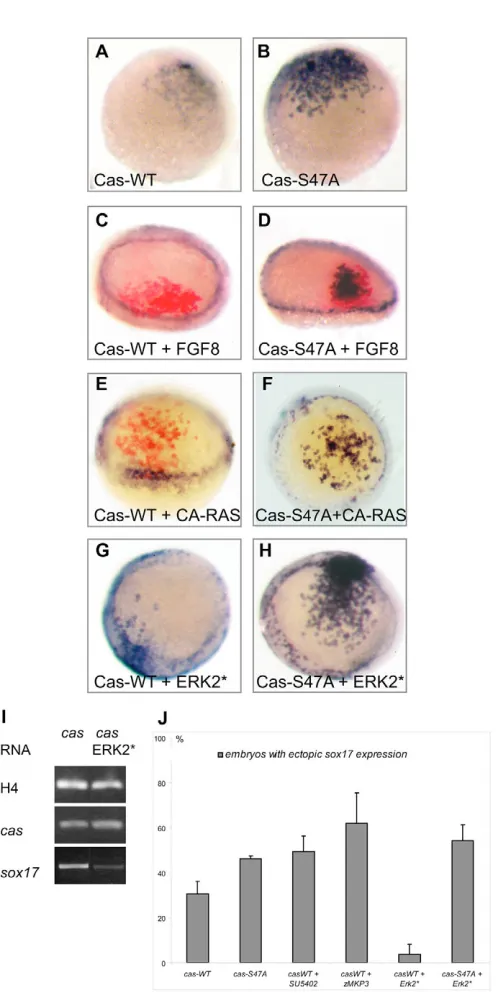
We next examined whether phosphorylation of Casanova modifies its activity. First, we compared the ability of low doses of cas-WTor cas-S47Ato induce ectopic sox17expression. When 20 pg of cas-WT RNAwas injected, it caused ectopic expression of sox17in 35% of the embryos (n=381, Fig. 7J, Table 4). When the same dose of mutated cas-S47A was injected, the percentage of embryos displaying sox17 was slightly increased to 47% (n=214). Furthermore, in situ hybridisation signals were consistently stronger in the cas-S47Athan in the cas-WT-injected embryos (Fig. 7A,B, see Fig. S3A-C in the supplementary material). These results suggest that the presence of the ERK target site has a negative effect on the ability of Casanova to induce sox17expression.
We then tested the effect of activating or inhibiting FGF-ERK signalling on the ability of ectopic wild-type or mutated Casanova to induce sox17expression at the animal pole. Injection of Fgf8 (Fig. 7C), a constitutively active Ras (Fig. 7E) or an activated version of ERK (Fig. 7G; see Fig. S3D,E in the supplementary material) strongly decreased the ability of Casanova to induce sox17 (Table 4). Importantly, activating the MAPK pathway with these reagents had only modest effects on the ability of the mutated form of Casanova to induce ectopic expression of sox17at the animal pole (Fig. 7D,F,H). This result indicates that the ERK consensus phosphorylation site is required for the attenuation of Casanova activity in response to FGF pathway activation. In agreement with this conclusion, inhibition of the FGF-ERK pathway by treatment with SU5402, or by co-injection with zebrafish MKP3, potentiated the ability of exogenous Casanova to induce sox17(Table 4, Fig. 7J). Taken together, these results suggest that FGF-ERK activity antagonises endoderm formation by acting at the level of transcription factors induced by Nodal signalling through direct phosphorylation of Casanova.
DISCUSSION
[image:7.612.48.563.72.126.2]We have investigated how the Nodal, FGF and BMP pathways interact during the process of endoderm formation in the zebrafish embryo. We found a double repressive effect of the FGF and BMP pathways on induction of endodermal precursors by Nodal signalling. When either of these pathways is Table 3. Double inhibition of the FGF and BMP pathways causes formation of an excess of endoderm precursors at 75% epiboly
Fgf8, Fgf8, Fgf17b and
RNA injected at the one-cell stage Wild type Noggin DN Ras DN Ras + Noggin Fgf17b and Fgf24 MOs Fgf24 MOs + Noggin
Average number of sox17-positive 437±47 498±89 354±64 652±87 802±63 1000±172
cells per embryo
[image:7.612.48.370.454.741.2]Total number of embryos 14 10 15 15 6 6
Fig. 6. Casanova protein is phosphorylated by MAPK in vivo. (A) Putative conserved MAPK phosphorylation sites (red box) in Casanova and in Sox17 from human, mouse and zebrafish. The blue line corresponds to the 15 amino acid peptide used to generate antibodies (‘␣
Casanova-Phospho’ antibody). (B-F) Western blot of protein extracts of embryos at 30% epiboly (15 embryos per lane). Embryos were injected with cas-flagRNA at 100 ng/l into one blastomere at the two-cell stage. Zebrafish mkp3
RNA (50 ng/l) (D) or SB203580 (200 M) (F) were co-injected with cas-flagRNA, while 15
M SU5402 was added to the medium after the injection (E). Overexpressed Casanova proteins are Flag tagged and detected by a mouse anti-Flag antibody. (B) The ‘␣Casanova-Phospho’ antibody recognised the product of ectopically expressed Casanova RNA (lane 2). Product of
D
E
V
E
LO
P
M
E
N
T
overactivated, the number of endodermal precursors formed is severely reduced. By contrast, when the FGF pathway is blocked, embryonic cells are more easily induced to form endoderm by activation of Nodal signalling. Moreover, we showed that inhibition of FGF signalling partially rescued the deficit of endoderm of bonnie and clydemutants, and that the simultaneous inhibition of both the FGF and BMP pathways results in an excess of endodermal precursors. Finally, we identified a molecular mechanism that explains the repressive action of FGF on endoderm formation. We show that FGF signals control the number of endodermal precursors formed via phosphorylation of the transcription factor Casanova.
FGF signalling and endoderm formation
Although a large number of studies have implicated FGF signalling in the control of mesoderm formation and patterning, only a few studies, mainly in Xenopus, have addressed the role of FGF signalling in formation of the endodermal germ layer. In Xenopus, numerous observations indicate a repressive role of FGF signalling on endoderm formation. Gamer and Wright (Gamer and Wright, 1995) found that bFGF is a potent inhibitor of the expression of the pancreatic marker pdx1. Bouwmeester et al. (Bouwmeester et al., 1996) found that co-injection of XFD, a dominant-negative version of Fgfr1, potentiates the ability of chordinto induce expression of endodermal markers in animal caps assays. Finally, a recent study showed that overexpression of eFGF inhibits expression of mixer and that inhibition of FGF signalling in animal caps induces the ectopic expression of mixerand endodermin(Cha et al., 2004). Taken together, these studies suggest that in XenopusFGF signalling antagonizes the molecular pathways that control endoderm specification.
To investigate whether FGF and Nodal signalling interact during endoderm formation in zebrafish, we ectopically expressed FGF signals. Overexpression of FGFs, as well as overexpression of activated versions of the FGF receptor, activated forms of Ras and the MAP kinase ERK, all strongly interfered with formation of the endoderm. Reciprocally, inhibition of FGF signalling with antisense morpholinos oligonucleotides caused a dramatic increase in the number of endodermal precursors. By contrast, interfering with the MAPK pathway using DN-Ras only increased the number of endoderm precursors present on the dorsal side as well as the number of endoderm precursors induced by ectopic activation of the Nodal pathway at the animal pole. Finally, we showed that treatments with SU5402 or injection of morpholinos against Fgf8 and Fgf24 partially rescued the deficit of endoderm precursors and the associated cardia bifida phenotype of the bonnie and clydemutant, which have a reduced expression of casanova/sox32. This finding is consistent with the results of David and Rosa (David and Rosa, 2001), who showed that grafted wild-type endodermal cells rescue thecardia bifida defects of casanova/sox32 mutants. Therefore, attenuation of FGF signalling partially compensates for the lack of a downstream effector of Nodal probably by increasing the activity of Casanova. This provides a further indication that in the intact embryo, endoderm formation in response to Nodal signalling is negatively regulated by FGF.
[image:8.612.53.301.51.553.2]Studies with zebrafish mutants and experiments with inhibitors had previously shown that FGF and Nodal signalling cooperate during mesoderm formation (Griffin and Kimelman, 2003; Griffin et al., 1995b; Mathieu et al., 2004). Our results show that during endoderm formation there is a strong antagonism between the FGF and the Nodal signalling pathways.
Fig. 7. Casanova activity is negatively regulated by the FGF-ERK signalling pathway. Expression of sox17(blue) in embryos at 30% epiboly (A,B) or 50% epiboly (C-H). In C-E, FLDX used as lineage marker was revealed by immunochemistry (red labelling). Embryos injected at the 16-cell stage with RNA encoding Casanova-WT (A) or Casanova-S47A (B), each at 10 ng/l (see also Fig. S2 in the
D
E
V
E
LO
P
M
E
N
T
Endoderm formation and BMP signalling
Previous studies had suggested that, in Xenopus, endoderm formation is regulated negatively by signals emanating from the ventral side. In particular, Sasai et al. reported that overexpression of nogginor chordininduces endoderm in animal caps and that this effect is strongly potentiated by inhibition of FGF signalling (Sasai et al., 1996). However, Tiso et al. found that, in zebrafish, the number of endoderm precursors is neither affected by overexpression of BMP nor reduced in the swirlmutant which has a disrupted bmp2 gene (Tiso et al., 2002). By contrast, we found that overexpression of a cocktail of bmp2, bmp4and bmp7potently affects the number of endoderm precursors and that overexpression of nogginincreases, although only slightly, the number of endodermal precursors. More strikingly, we found that while inhibition of the BMP signalling pathway with nogginmRNA results in only a modest increase in the number of endodermal precursors, when nogginand DN-Ras are co-injected, they cause formation of a massive excess of endodermal precursors. Finally, the largest number of endodermal precursors was observed following simultaneous inhibition of BMP signalling with noggin and of FGF signalling with morpholinos, further implicating BMP signals in restricting endoderm formation. Taken together, these experiments reveal a previously unknown role for BMP signals in repressing endoderm formation. Therefore, our results show that in addition to being positively regulated by signals from the TGFfamily, formation of the endoderm is negatively regulated by a combination of FGF and BMP signals (Fig. 8).
Repression of endoderm formation by FGF or BMP may not be mediated by phosphorylation or competition for Smad2/3
Phosphorylation of Smad in their inter-linker region has been implicated in modulating the response to Tgfsignals (Massagué, 2003; Pera et al., 2003). In Xenopusgastrulae, it has been shown that MAP kinase-dependent phosphorylation of Smad2 inhibits its translocation into the nucleus. As a consequence, animal blastomeres loose their competence to respond to Tgfand differentiate into ectoderm instead of mesoderm (Grimm and Gurdon, 2002). Consequences of Smad2 phosphorylation are not clear as different studies gave opposite results. One report has shown that in human cells, activation of Ras caused the exclusion of Smad2 and Smad3 from the nucleus (Kretzschmar et al., 1999). These data contrast with an earlier report in which Erk2-dependant phosphorylation of Smad2 correlated with an increase in the nuclear localisation and activity of Smad2 (de Caestecker et al., 1998). Our finding that a Nodal target gene such as lefty1is expressed normally following overexpression of Fgf8 suggests that, in zebrafish, the antagonism between endoderm formation and FGF/ERK signalling does not rely on an inhibitory phosphorylation of Smads. The inability of a Smad2 mutant than can not be phosphorylated by ERK to rescue endoderm formation in FGF overexpressing embryos reinforces this conclusion.
Although the inhibitory effect of FGF signalling on endoderm formation can be explained by an inhibitory phosphorylation of Casanova, the molecular mechanism responsible for the inhibitory action of BMP is not presently known. Both the Nodal signalling
[image:9.612.49.567.72.126.2]pathway and the BMP signalling pathway share a common downstream component, Smad4. It is therefore tempting to speculate that the inhibitory action of BMP on endoderm formation may be caused by a competition between Smad1/Smad5, which are Table 4. FGF/ERK signalling negatively regulates the activity of Casanova at the animal pole
Casanova-WT Casanova-WT Casanova-WT Casanova-S47A
RNA injected at the 16-cell stage Casanova-WT Casanova-S47A + SU5402 + Mkp3 + Erk2* + Erk2*
% Embryos showing ectopic sox17 30 46 49 64 3 54
expression
Total number of embryos 381 214 182 205 167 118
Fig. 8. Schematic representation of the repressive effects of the BMP and FGF pathways on endoderm formation. (A) Wild-type context: (a-c) schematic representation of signalling activities of Nodal, FGF and BMP signalling. This representation is speculative and based on the potential range of signals and the expression pattern and range of antagonists (Schier and Talbot, 2005). (B) Formation of the endoderm is negatively regulated by a combination of FGF and BMP signals. (C) DN-Ras overexpressing embryos: inhibition of MAPK signalling promotes endoderm formation on the dorsal side but causes a loss of endoderm precursors on the ventral side owing to increased expression of the BMPs. (D) Triple inhibition of FGF signalling with morpholinos or combined inhibition of Ras and BMP signalling with DN-Ras + noggin: simultaneous removal of the BMP and FGF dependent inhibitions promotes endoderm formation all around the embryo.
Aa Nodal signalling activity domain
Ab FGF signalling activity domain
Ac BMP signalling activity domain
B Wild-type conditions
C +DN-RAS
D MO FGFs or DN-RAS+Noggin
BMP inhibition by FGF
Endoderm induction by Nodal
Endoderm inhibition by BMP
Endoderm inhibition by FGF
Limit of endodermal domain
Endodermal domain
[image:9.612.315.563.213.588.2]D
E
V
E
LO
P
M
E
N
T
activated by BMP signalling, and Smad2/Smad3, which are activated by Nodal signalling for binding to Smad4 (Candia et al., 1997). However, we found that overexpression of Smad4 was not able to rescue the loss of sox17expression in BMP-overexpressing embryos (not shown), therefore, a competition at the level of Smad proteins does not seem to be the cause of the inhibitory action of BMPs on endoderm formation.
Casanova/Sox32 as a transcription factor at the crossroad of the FGF and Nodal signalling pathways
We have uncovered a potential molecular mechanism whereby FGF signalling acts to attenuate endoderm formation by identifying Casanova, a Nodal target gene, as a major target of this repression. First, we found that overexpressed Casanova is phosphorylated and that the level of phosphorylation is correlated with activation or inhibition of this pathway. Second, we showed that activation of FGF/ERK attenuates the ability of Casanova to induce sox17when ectopically expressed at the animal pole, while inhibition of FGF/ERK makes Casanova a more potent inducer of endoderm. Finally, we showed that although the ability of wild type Casanova to induce endoderm at the animal pole is antagonized by overexpression of FGF or activation of the ERK pathway, a mutated phosphorylation-insensitive form of Casanova is no longer subject to this inhibition. Therefore, these data support the hypothesis that Casanova is at the crossroads of the Nodal and the MAPK signalling pathways, and that the antagonism between these pathways is caused, at least in part, by phosphorylation of this transcription factor. Nevertheless, interactions between these pathways could occur at other levels. Fgf8 or CA-Ras overexpression decrease the expression of sox17and of casanova/sox32. This suggests that Casanova may not be the only target of this repressive mechanism. In addition to Casanova, MAPK consensus sites are present in factors acting at the level of, or downstream of, Casanova, such as Eomesodermin, Gata5 and Sox17. Future experiments should address whether Eomesodermin, Gata5 and Sox17 are also involved in the crosstalk between the MAPK and Nodal signalling pathways. Alternatively, FGF may additionally affect the expression of casanova/sox32 by compromising the activity of upstream regulators in the cascade. For example, the homeobox proteins Bon and Mezzo act upstream of Casanova and are both able to bind purified Smad2 (Germain et al., 2000) (M.P. and T.L., unpublished). Phosphorylation of Smad2/3 may affect their interaction with Mixer and Mezzo.
In zebrafish, the endodermal and mesodermal precursors originate from a common endomesodermal territory and both require Nodal signalling. The molecular mechanisms that allow these two cell fates to segregate during gastrulation are not well understood. It was previously known that factors promoting endoderm formation, such as Casanova or Mezzo repress mesoderm formation when overexpressed (Aoki et al., 2002a; Kikuchi et al., 2001; Poulain and Lepage, 2002). Our finding that endogenous FGF signals strongly antagonize endoderm formation by downregulating Casanova shows that a reciprocal negative interaction exists between factors that promote mesoderm formation and transcription factors required for endoderm formation. This mutual antagonism may therefore help to understand how mesodermal- or endodermal-specific gene regulatory networks are established in the precursors of these two germ layers, allowing different cell fates to be segregated.
In conclusion, we have shown that the FGF and BMP signals antagonize endoderm formation by Nodal factors. Furthermore, we have shown that Casanova is subject to an inhibitory
phosphorylation in response to FGF signalling and therefore stands at the intersection between the FGF and Nodal signalling pathways. This phosphorylation may represent a general mechanism whereby FGF attenuates Nodal-induced endodermal transcription factors, and therefore these results may help to understand how mesoderm and endoderm segregate from each other.
We thank F. Rosa for the tar*/acvr1b, nkx2.5and casanova/sox32plasmids, and for the casanova/sox32morpholino oligonucleotides. We thank N. G. Ahn for sending us ERK2* plasmid, J. Massagué for Smad2 and Smad3 constructs, and M. Whitman for CA-Ras and DN-Ras plasmids. We thank D. Meyer for the bonnie and clyde strain. We thank our colleagues at the Marine Station of Villefranche for help and support, and Lydia Besnardeau for excellent technical help. We thank Clare Hudson and Hitoyoshi Yasuo for fruitful discussions and careful reading of the manuscript. We particularly thank Jean-Phillipe Chambon for helping us with HPLC chromatography and western blotting. We thank Laurent Gilleta for taking care of the zebrafish. This work was supported by the CNRS, by the Asssociation pour la Recherche contre le Cancer, by the Ligue Nationale Contre le Cancer, by the Ministère de la Recherche and by the National Institute of Health. M.F. was supported by CNRS and HFSP.
Supplementary material
Supplementary material for this article is available at http://dev.biologists.org/cgi/content/full/133/11/2189/DC1
References
Agathon, A., Thisse, C. and Thisse, B.(2003). The molecular nature of the zebrafish tail organizer. Nature424, 448-452.
Alexander, J. and Stainier, D. Y.(1999). A molecular pathway leading to endoderm formation in zebrafish. Curr. Biol.9, 1147-1157.
Alexander, J., Rothenberg, M., Henry, G. L. and Stainier, D. Y.(1999). casanova plays an early and essential role in endoderm formation in zebrafish. Dev. Biol.215, 343-357.
Aoki, T. O., David, N. B., Minchiotti, G., Saint-Etienne, L., Dickmeis, T., Persico, G. M., Strahle, U., Mourrain, P. and Rosa, F. M.(2002a). Molecular integration of casanova in the Nodal signalling pathway controlling endoderm formation. Development129, 275-286.
Aoki, T. O., Mathieu, J., Saint-Etienne, L., Rebagliati, M. R., Peyrieras, N. and Rosa, F. M.(2002b). Regulation of nodal signalling and mesendoderm formation by TARAM-A, a TGFbeta-related type I receptor. Dev. Biol.241, 273-288.
Bjornson, C. R., Griffin, K. J., Farr, G. H., 3rd, Terashima, A., Himeda, C., Kikuchi, Y. and Kimelman, D.(2005). Eomesodermin is a localized maternal determinant required for endoderm induction in zebrafish. Dev. Cell9, 523-533. Bottcher, R. T. and Niehrs, C.(2005). Fibroblast growth factor signaling during
early vertebrate development. Endocr. Rev.26, 63-77.
Bouwmeester, T., Kim, S., Sasai, Y., Lu, B. and De Robertis, E. M.(1996). Cerberus is a head-inducing secreted factor expressed in the anterior endoderm of Spemann’s organizer. Nature382, 595-601.
Candia, A. F., Watabe, T., Hawley, S. H., Onichtchouk, D., Zhang, Y., Derynck, R., Niehrs, C. and Cho, K. W.(1997). Cellular interpretation of multiple TGF-beta signals: intracellular antagonism between activin/BVg1 and BMP-2/4 signaling mediated by Smads. Development124, 4467-4480. Cha, S. W., Hwang, Y. S., Chae, J. P., Lee, S. Y., Lee, H. S., Daar, I., Park, M. J.
and Kim, J.(2004). Inhibition of FGF signaling causes expansion of the endoderm in Xenopus. Biochem. Biophys. Res. Commun.315, 100-106. Chen, J. N. and Fishman, M. C.(1996). Zebrafish tinman homolog demarcates
the heart field and initiates myocardial differentiation. Development122, 3809-3816.
Chen, Y. and Schier, A. F.(2001). The zebrafish Nodal signal Squint functions as a morphogen. Nature411, 607-610.
Chomczynski, P. and Sacchi, N.(1987). Single-step method of RNA isolation by acid guanidinium thiocyanate-phenol-chloroform extraction. Anal. Biochem. 162, 156-159.
Clements, D., Friday, R. V. and Woodland, H. R.(1999). Mode of action of VegT in mesoderm and endoderm formation. Development126, 4903-4911. Cornell, R. A., Musci, T. J. and Kimelman, D.(1995). FGF is a prospective
competence factor for early activin-type signals in Xenopus mesoderm induction. Development121, 2429-2437.
David, N. B. and Rosa, F. M.(2001). Cell autonomous commitment to an endodermal fate and behaviour by activation of Nodal signalling. Development 128, 3937-3947.
D
E
V
E
LO
P
M
E
N
T
Cloning and characterization of zebrafish smad2, smad3 and smad4. Gene246, 69-80.
Dickmeis, T., Mourrain, P., Saint-Etienne, L., Fischer, N., Aanstad, P., Clark, M., Strahle, U. and Rosa, F.(2001). A crucial component of the endoderm formation pathway, CASANOVA, is encoded by a novel sox-related gene. Genes Dev.15, 1487-1492.
Draper, B. W., Stock, D. W. and Kimmel, C. B.(2003). Zebrafish fgf24 functions with fgf8 to promote posterior mesodermal development. Development130, 4639-4654.
Emrick, M. A., Hoofnagle, A. N., Miller, A. S., Ten Eyck, L. F. and Ahn, N. G. (2001). Constitutive activation of extracellular signal-regulated kinase 2 by synergistic point mutations. J. Biol. Chem.276, 46469-46479.
Erter, C. E., Solnica-Krezel, L. and Wright, C. V.(1998). Zebrafish nodal-related 2 encodes an early mesendodermal inducer signaling from the extraembryonic yolk syncytial layer. Dev. Biol.204, 361-372.
Faucourt, M., Houliston, E., Besnardeau, L., Kimelman, D. and Lepage, T. (2001). The pitx2 homeobox protein is required early for endoderm formation and nodal signaling. Dev. Biol.229, 287-306.
Feldman, B., Gates, M. A., Egan, E. S., Dougan, S. T., Rennebeck, G., Sirotkin, H. I., Schier, A. F. and Talbot, W. S.(1998). Zebrafish organizer development and germ-layer formation require nodal-related signals. Nature 395, 181-185.
Fischer, S., Draper, B. W. and Neumann, C. J.(2003). The zebrafish fgf24 mutant identifies an additional level of Fgf signaling involved in vertebrate forelimb initiation. Development130, 3515-3524.
Fujii, R., Yamashita, S., Hibi, M. and Hirano, T. (2000). Asymmetric p38 activation in zebrafish: its possible role in symmetric and synchronous cleavage. J. Cell Biol. 150, 1335-1348.
Fürthauer, M., Thisse, C. and Thisse, B.(1997). A role for FGF-8 in the dorsoventral patterning of the zebrafish gastrula. Development124, 4253-4264. Furthauer, M., Thisse, B. and Thisse, C.(1999). Three different noggin genes
antagonize the activity of bone morphogenetic proteins in the zebrafish embryo. Dev. Biol.214, 181-196.
Fürthauer, M., Reifers, F., Brand, M., Thisse, B. and Thisse, C.(2001). sprouty4 acts in vivo as a feedback-induced antagonist of FGF signaling in zebrafish. Development128, 2175-2186.
Fürthauer, M., Van Celst, J., Thisse, C. and Thisse, B.(2004). Fgf signalling controls the dorsoventral patterning of the zebrafish embryo. Development131, 2853-2864.
Gaio, U., Schweickert, A., Fischer, A., Garratt, A. N., Muller, T., Ozcelik, C., Lankes, W., Strehle, M., Britsch, S., Blum, M. et al.(1999). A role of the cryptic gene in the correct establishment of the left-right axis. Curr. Biol.9, 1339-1342.
Gamer, L. W. and Wright, C. V.(1995). Autonomous endodermal determination in Xenopus: regulation of expression of the pancreatic gene XlHbox 8. Dev. Biol. 171, 240-251.
Germain, S., Howell, M., Esslemont, G. M. and Hill, C. S.(2000).
Homeodomain and winged-helix transcription factors recruit activated Smads to distinct promoter elements via a common Smad interaction motif. Genes Dev. 14, 435-451.
Gonzalez, F. A., Raden, D. L. and Davis, R. J.(1991). Identification of substrate recognition determinants for human ERK1 and ERK2 protein kinases. J. Biol. Chem.266, 22159-22163.
Griffin, K. J. and Kimelman, D.(2003). Interplay between FGF, one-eyed pinhead, and T-box transcription factors during zebrafish posterior development. Dev. Biol.264, 456-466.
Griffin, K., Patient, R. and Holder, N.(1995a). Analysis of FGF function in normal and no tail zebrafish embryos reveals separate mechanisms for formation of the trunk and the tail. Development121, 2983-2994.
Griffin, K. J. P., Patient, R. K. and Holder, N. H. K.(1995b). Analysis of FGF function in normal and no tailzebrafish embryos reveals separate mechanisms for formation of the trunk and tail. Development121, 2983-2994.
Grimm, O. H. and Gurdon, J. B.(2002). Nuclear exclusion of Smad2 is a mechanism leading to loss of competence. Nat. Cell Biol. 4, 519-522. Gritsman, K., Zhang, J., Cheng, S., Heckscher, E., Talbot, W. S. and Schier, A.
F.(1999). The EGF-CFC protein one-eyed pinhead is essential for nodal signaling. Cell97, 121-132.
Gritsman, K., Talbot, W. S. and Schier, A. F.(2000). Nodal signaling patterns the organizer. Development127, 921-932.
Henry, G. L., Brivanlou, I. H., Kessler, D. S., Hemmati-Brivanlou, A. and Melton, D. A.(1996). TGF-beta signals and a pattern in Xenopus laevis endodermal development. Development122, 1007-1015.
Jones, C. M., Kuehn, M. R., Hogan, B. L. M., Smith, J. C. and Wright, C. V. E. (1995). Nodal-related signals induce axial mesoderm and dorsalize mesoderm during gastrulation. Development121, 3651-3662.
Kikuchi, Y., Trinh, L. A., Reiter, J. F., Alexander, J., Yelon, D. and Stainier, D. Y.(2000). The zebrafish bonnie and clyde gene encodes a Mix family homeodomain protein that regulates the generation of endodermal precursors. Genes Dev.14, 1279-1289.
Kikuchi, Y., Agathon, A., Alexander, J., Thisse, C., Waldron, S., Yelon, D.,
Thisse, B. and Stainier, D. Y.(2001). casanova encodes a novel Sox-related protein necessary and sufficient for early endoderm formation in zebrafish. Genes Dev.15, 1493-1505.
Kikuchi, Y., Verkade, H., Reiter, J. F., Kim, C. H., Chitnis, A. B., Kuroiwa, A. and Stainier, D. Y.(2004). Notch signaling can regulate endoderm formation in zebrafish. Dev. Dyn.229, 756-762.
Kimelman, D.(1991). The role of fibroblast growth factor in early Xenopus development. Ann. N. Y. Acad. Sci. 638, 275-282.
Kimmel, C. B., Warga, R. M. and Schilling, T. F.(1990). Origin and organization of the zebrafish fate map. Development108, 581-594.
Kishimoto, Y., Lee, K. H., Zon, L., Hammerschmidt, M. and Schulte-Merker, S.(1997). The molecular nature of zebrafish swirl: BMP2 function is essential during early dorsoventral patterning. Development124, 4457-4466. Kretzschmar, M., Doody, J. and Massague, J.(1997). Opposing BMP and EGF
signalling pathways converge on the TGF-beta family mediator Smad1. Nature 389, 618-622.
Kretzschmar, M., Doody, J., Timokhina, I. and Massague, J.(1999). A mechanism of repression of TGFbeta/Smad signaling by oncogenic Ras. Genes Dev.13, 804-816.
Le Good, J. A., Joubin, K., Giraldez, A. J., Ben-Haim, N., Beck, S., Chen, Y., Schier, A. F. and Constam, D. B.(2005). Nodal stability determines signaling range. Curr. Biol.15, 31-36.
Massagué, J.(2003). Integration of Smad and MAPK pathways: a link and a linker revisited. Genes Dev.17, 2993-2997.
Mathieu, J., Griffin, K., Herbomel, P., Dickmeis, T., Strahle, U., Kimelman, D., Rosa, F. M. and Peyrieras, N.(2004). Nodal and Fgf pathways interact through a positive regulatory loop and synergize to maintain mesodermal cell
populations. Development131, 629-641.
Mohammadi, M., McMahon, G., Sun, L., Tang, C., Hirth, P., Yeh, B. K., Hubbard, S. R. and Schlessinger, J.(1997). Structures of the tyrosine kinase domain of fibroblast growth factor receptor in complex with inhibitors. Science 276, 955-960.
Nissen, R. M., Yan, J., Amsterdam, A., Hopkins, N. and Burgess, S. M.(2003). Zebrafish foxi one modulates cellular responses to Fgf signaling required for the integrity of ear and jaw patterning. Development130, 2543-2554.
Peng, H. B.(1991). Xenopus laevis: practical uses in cell and molecular biology. In Methods in Cell Biology. Vol. 36 (ed. B. K. Kay and H. B. Peng), pp. 657-662. Santa Barbara: Harcourt Brace Jovanovich.
Pera, E. M., Ikeda, A., Eivers, E. and De Robertis, E. M.(2003). Integration of IGF, FGF, and anti-BMP signals via Smad1 phosphorylation in neural induction. Genes Dev.17, 3023-3028.
Peyrieras, N., Strahle, U. and Rosa, F.(1998). Conversion of zebrafish blastomeres to an endodermal fate by TGF-beta-related signaling. Curr. Biol.8, 783-786.
Piccolo, S., Agius, E., Leyns, L., Bhattacharyya, S., Grunz, H., Bouwmeester, T. and De Robertis, E. M.(1999). The head inducer Cerberus is a multifunctional antagonist of Nodal, BMP and Wnt signals. Nature397, 707-710.
Poulain, M. and Lepage, T.(2002). Mezzo, a paired-like homeobox protein is an immediate target of Nodal signalling and regulates endoderm specification in zebrafish. Development129, 4901-4914.
Rebagliati, M. R., Toyama, R., Haffter, P. and Dawid, I. B.(1998). cyclops encodes a nodal-related factor involved in midline signaling. Proc. Natl. Acad. Sci. USA95, 9932-9937.
Reiter, J. F., Alexander, J., Rodaway, A., Yelon, D., Patient, R., Holder, N. and Stainier, D. Y.(1999). Gata5 is required for the development of the heart and endoderm in zebrafish. Genes Dev.13, 2983-2995.
Reiter, J. F., Kikuchi, Y. and Stainier, D. Y.(2001). Multiple roles for Gata5 in zebrafish endoderm formation. Development128, 125-135.
Renucci, A., Lemarchandel, V. and Rosa, F.(1996). An activated form of type I serine/threonine kinase receptor TARAM-A reveals a specific signalling pathway involved in fish head organiser formation. Development122, 3735-3743.
Rodaway, A., Takeda, H., Koshida, S., Broadbent, J., Price, B., Smith, J. C., Patient, R. and Holder, N.(1999). Induction of the mesendoderm in the zebrafish germ ring by yolk cell- derived TGF-beta family signals and discrimination of mesoderm and endoderm by FGF. Development126, 3067-3078.
Sägerstrom, C. G., Grinbalt, Y. and Sive, H.(1996). Anteroposterior patterning in the zebrafish, Danio rerio: an explant assay reveals inductive and suppressive cell interactions. Development122, 1873-1883.
Sakaguchi, T., Kuroiwa, A. and Takeda, H.(2001). A novel sox gene, 226D7, acts downstream of Nodal signaling to specify endoderm precursors in zebrafish. Mech. Dev.107, 25-38.
Sampath, K., Rubinstein, A. L., Cheng, A. M., Liang, J. O., Fekany, K., Solnica-Krezel, L., Korzh, V., Halpern, M. E. and Wright, C. V.(1998). Induction of the zebrafish ventral brain and floorplate requires cyclops/nodal signalling. Nature395, 185-189.
D
E
V
E
LO
P
M
E
N
T
Schier, A. F.(2003). Nodal signaling in vertebrate development. Annu. Rev. Cell Dev. Biol.19, 589-621.
Schier, A. F. and Talbot, W. S.(2005). Molecular genetics of axis formation in Zebrafish. Annu. Rev. Genet. 39, 561-613.
Schier, A. F., Neuhauss, S. C., Helde, K. A., Talbot, W. S. and Driever, W. (1997). The one-eyed pinhead gene functions in mesoderm and endoderm formation in zebrafish and interacts with no tail. Development124, 327-342. Schmid, B., Furthauer, M., Connors, S. A., Trout, J., Thisse, B., Thisse, C. and
Mullins, M. C.(2000). Equivalent genetic roles for bmp7/snailhouse and bmp2b/swirl in dorsoventral pattern formation. Development127, 957-967. Schulte-Merker, S. and Smith, J. C.(1995). Mesoderm formation in response to
Brachyuryrequires FGF signalling. Curr. Biol. 5, 62-67.
Shinya, M., Koshida, S., Sawada, A., Kuroiwa, A. and Takeda, H.(2001). Fgf signalling through MAPK cascade is required for development of the subpallial telencephalon in zebrafish embryos. Development128, 4153-4164. Strahle, U., Jesuthasan, S., Blader, P., Garcia-Villalba, P., Hatta, K. and
Ingham, P. W.(1997). one-eyed pinhead is required for development of the ventral midline of the zebrafish (Danio rerio) neural tube. Genes Funct.1, 131-148.
Sun, B. I., Bush, S. M., Collins-Racie, L. A., LaVallie, E. R., DiBlasio-Smith, E. A., Wolfman, N. M., McCoy, J. M. and Sive, H. L.(1999). derriere: a TGF-beta family member required for posterior development in Xenopus. Development 126, 1467-1482.
Thisse, B., Wright, C. V. and Thisse, C.(2000). Activin- and Nodal-related factors control antero-posterior patterning of the zebrafish embryo. Nature403, 425-428.
Thisse, B., Heyer, V., Lux, A., Alunni, V., Degrave, A., Seiliez, I., Kirchner, J.,
Parkhill, J. P. and Thisse, C.(2004). Spatial and temporal expression of the zebrafish genome by large-scale in situ hybridization screening. Methods Cell Biol.77, 505-519.
Thisse, C. and Thisse, B.(1999). Antivin, a novel and divergent member of the TGFbeta superfamily, negatively regulates mesoderm induction. Development 126, 229-240.
Tiso, N., Filippi, A., Pauls, S., Bortolussi, M. and Argenton, F.(2002). BMP signalling regulates anteroposterior endoderm patterning in zebrafish. Mech. Dev.118, 29-37.
Trinh, L. A., Meyer, D. and Stainier, D. Y.(2003). The Mix family homeodomain gene bonnie and clyde functions with other components of the Nodal signaling pathway to regulate neural patterning in zebrafish. Development130, 4989-4998.
Warga, R. M. and Nusslein-Volhard, C.(1999). Origin and development of the zebrafish endoderm. Development126, 827-838.
Westerfield, M.(1994). The Zebrafish Book. Eugene OR: Institute of Neurosciences, University of Oregon.
Whitman, M. and Melton, D. A.(1992). Involvement of p21rasin Xenopus
mesoderm induction. Nature357, 252-254.
Yasuo, H. and Lemaire, P.(1999). A two-step model for the fate determination of presumptive endodermal blastomeres in Xenopus embryos. Curr. Biol.9, 869-879. Zhang, J., Talbot, W. S. and Schier, A. F.(1998). Positional cloning identifies
zebrafish one-eyed pinhead as a permissive EGF-related ligand required during gastrulation. Cell92, 241-251.