D
E
V
E
LO
P
M
E
N
T
INTRODUCTION
The isthmic organizer (IsO) represents a signaling center located at the midbrain hindbrain boundary (MHB) and expressing several transcription and secreted factors, including Pax2, Pax5, En1, En2, Fgf8and Wnt1. All these factors are required for the patterning of the midbrain and cerebellum, and the maintenance of their expression in the MHB is interdependent, as evidenced by loss of gene activity over time once one of them is inactivated (Simeone et al., 2002; Wurst and Bally-Cuif, 2001; Raible and Brand, 2004). However, each factor is induced independently of the others. Fgf8 is the only factor known so far that is able to mimic IsO activity, i.e. exhibiting midbrain and cerebellum inducing properties (Crossley et al., 1996). Gain- and loss-of-function experiments revealed a complex combinatorial genetic network that acts to maintain a functional IsO (Simeone et al., 2002; Wurst and Bally-Cuif, 2001; Raible and Brand, 2004; Wurst et al., 1994; McMahon and Bradley, 1990; Wassarman et al., 1997; Urbanek et al., 1997; Reifers et al., 1998; Schwarz et al., 1999; Hirata et al., 2001). Thus, in the anteroposterior (AP) axis, Otx2and Gbx2control, through reciprocal repression, the proper positioning of the IsO and the establishment of a sharp Fgf8expression domain at the MHB (Acampora et al., 1997; Wassarman et al., 1997; Rhinn et al., 1999; Millet et al., 1999; Broccoli et al., 1999; Li and Joyner, 2001; Martinez-Barbera et al., 2001). However, only in the absence of Pax2,is Fgf8expression not detected in the IsO of C3H/He mouse embryos (Ye et al., 2001). Interestingly, an Otx2-dependent repressive mechanism is also required for the proper positioning
of sonic hedgehog (Shh) expression in the ventral midbrain; hence, specifying neuronal progenitors along the dorsoventral (DV) axis (Puelles et al., 2003).
The murine ortholog of theDrosophila buttonhead gene, Sp8 (previously known as mBtd), is a zinc-finger transcription factor belonging to the Spfamily and shares with Fgf8several domains of expression that have organizer function, including limb and tail bud, anterior neural ridge and MHB (Wimmer et al., 1993; Treichel et al., 2003; Bell et al., 2003). In addition, Sp8mRNA is also detected in the forebrain, midbrain and spinal cord (Fig. 1A-C; data not shown). In the DV axis, Sp8is expressed in the basal plate of the spinal cord and in the ventral midbrain (Fig. 1D; data not shown). However, Sp8 is not found in the floor plate. At the MHB, Sp8is found along the whole neural tube. (Fig. 1A-C; data not shown). As reported previously, Sp8-deficient mice suffer from multiple defects (Treichel et al., 2003; Bell et al., 2003). In this study, we have focused on the role of Sp8in the establishment of the IsO and the development of the midbrain. Strikingly, the lack of Sp8 activity resulted in a posterior shift of the IsO and ectopic patches of Fgf8-,Otx2-and/or Wnt1-expressing cells were found in the ventral part of the rostral hindbrain. As a consequence, midbrain dopaminergic neurons were detected posterior to the IsO of mouse Sp8–/–embryos. Moreover, Sp8was also required to control cell proliferation in the developing mid- and hindbrain. Our findings suggest that Sp8 is a new transcription factor required for normal development of the MHB boundary region.
MATERIALS AND METHODS Generation of knockout mice
Embryos were derived from crossings of Sp8 heterozygous animals described previously (Treichel et al., 2003). All knockout animals were of mixed CD1X129Sv genetic background. Genotyping was performed by PCR.
In situ hybridization and immunohistochemistry
Embryos were prepared at the appropriate time points and subjected to in situ hybridization or immunohistochemistry. Whole-mount in situ hybridization was performed as previously reported (Treichel et al., 2003). In situ hybridization on consecutive sections (18 m) using digoxigenin-labeled probes was performed according to Moorman et al. (Moorman et al., 2001). Immunohistochemistry was carried out according
Sp8
controls the anteroposterior patterning at the
midbrain-hindbrain border
Gundula Griesel1, Dieter Treichel1, Patrick Collombat1, Jens Krull1, Andreas Zembrzycki1,
Willem M. R. van den Akker1,2, Peter Gruss1, Antonio Simeone3,4,5and Ahmed Mansouri1,6,*,†
The specification of neuronal cell types in the developing neural tube is orchestrated by signaling centers. However, how patterned territories of the central nervous system (CNS) are organized into structures with appropriate size and shape is still unclear. We report that in the absence of the mouse transcription factor mBtd/Sp8, a posterior shift of the isthmic organizer (IsO) occurs, suggesting a crucial role for Sp8 in this process. In addition, large patches of cells ectopically expressing Fgf8, Otx2and/or Wnt1in the rostral hindbrain are detected in Sp8mutant embryos. In this context, midbrain dopaminergic neurons are found posterior to the IsO. Furthermore, we provide evidence that cell proliferation in the mid- and hindbrain is tightly controlled by Sp8activity. Our observations are consistent with a role for Sp8 in restricting Fgf8expression at the IsO.
KEY WORDS: Sp8 (mBtd), Isthmic organizer, Midbrain, AP patterning, Mouse Development 133, 1779-1787 (2006) doi:10.1242/dev.02326
1Max-Planck Institute for Biophysical Chemistry, Dept of Molecular Cell Biology, am Fassberg, 37077 Göttingen, Germany. 2Hubrecht Laboratory, Netherlands Institute for Developmental Biology, Uppsalalaan 8, 3584 CT, Utrecht, The Netherlands. 3Ceinge Biotecnologie Avanzate and SEMM European School of Molecular Medicine – Naples site, Via Comunale Margherita 482, 80145 Naples, Italy. 4MRC Centre for Developmental Neurobiology, New Hunt’s House, 4th Floor, King’s College London, Guy’s Campus, London Bridge, London SE1 1UL, UK. 5Institute of Genetics and Biophysics ‘A. Buzzati-Traverso’, CNR, Via P. Castellino 111, 80131 Naples. 6Clininal Neurophysiology, University of Göttingen, Robert-Koch-strasse 40, 37075 Göttingen, Germany.
*Member of the DFG Center for Molecular Physiology of the Brain (CMPB) †Author for correspondence (e-mail: amansou@gwdg.de)
D
E
V
E
LO
P
M
E
N
T
to manufacturer’s recommendation. Antibodies were anti-serotonin (5-hydroxytryptamine) from Sigma, and TH, Tuj1 and nestin from Chemicon.
BrdU labeling and TUNEL assay
For BrdU labeling, pregnant females of E10.5 and E11.5 were sacrificed 30 and 60 minutes after intraperitoneal injections of BrdU solution (100 g/g body weight). Apoptosis was detected according to TUNEL method and performed following manufacturer’s recommendation (Chemicon).
RESULTS
Histological analysis of Sp8-deficient embryos In the absence of Sp8, the brain phenotype is variable and a high incidence of open mid- and hindbrain was observed during embryonic development (Treichel et al., 2003). In the present analysis, we included only embryos with closed mid- and hindbrain. Histological examination of sagittal sections of E10.5 to E12.5 embryos revealed that, in Sp8 mutant embryos, a remarkable overgrowth of the hind- and the midbrain occurred. This is more pronounced in the ventral part of the neuroepithelium (Fig. 1F-H). From 12 analyzed embryos at E17.5 only three were found to develop a cerebellum-like structure. In most embryos at this stage of
development, although the neural tube appeared closed, owing to disturbed tissue growth, in histological sections specific structures of the mid- and hindbrain were difficult to identify and were not further analyzed. Therefore, our analysis will be limited to embryos up to E12.5 of gestation.
[image:2.612.52.269.308.587.2]Sp8 plays a crucial role in positioning of the IsO To decipher the role of the Sp8gene in the IsO, we studied the expression of several molecular markers associated with the MHB from E8.5 to E12.5. The homeobox genes Otx2 and Gbx2 are normally expressed in a complementary pattern along the AP axis, thereby controlling the proper positioning of the IsO (Simeone et al., 2002; Wurst and Bally-Cuif, 2001; Raible and Brand, 2004). In the absence of Sp8 activity, this expression boundary was shifted posteriorly, as revealed by whole-mount in situ hybridization at E8.5, and using Krox20 transcripts to label rhombomere 3 and 5 (Wilkinson et al., 1989a) (Fig. 2). At E9.5, this malformation is still evident (Fig. 3; see Fig.S1 in the supplementary material) and the expression of genes transcribed at the MHB, such as Wnt1, Fgf8, En1 andPax2(Simeone et al., 2002; Wurst and Bally-Cuif, 2001; Raible and Brand, 2004) is enlarged (Fig. 3, see Fig. S1, S2 in the supplementary material). At E10.5 the expression boundary where Otx2posterior border and Gbx2anterior border abut seems normal, as shown on adjacent sagittal sections (Fig. 4E,G and 4F,H). However, ectopic patches of Otx2-, Wnt1- and/or Fgf8-expressing cells are present in the ventral part of the rostral hindbrain at least until E11.5, as shown on adjacent sagittal sections of E10.5 embryos (Fig. 4A-D and 4G,H; data not shown). This patchy expression is barely detectable at E9.5 but clearly evident at E9.7 (data not shown; see Fig. S2 in the supplementary material). Furthermore, it appears that the expression territory of Fgf8is more dramatically affected and is more widespread. It is expanded in the dorsal part of the rostral hindbrain as well, and some Fgf8-expressing cell patches appear to localize in the most posterior midbrain (Fig. 4, arrowhead). In addition, in the absence of Sp8 the expression of Fgf8 is
[image:2.612.314.546.467.640.2]Fig. 1. Tissue overgrowth in the mid- and hindbrain. Whole-mount in situ hybridization showing the expression ofSp8 in the central nervous system at E9.5 (A) and E10.5 (B,C). (A-C) Sp8is found in the forebrain, midbrain, MHB and in the spinal cord. In the spinal cord (B) and in the midbrain, Sp8is localized to the ventral part of the neuroepithelium, as presented in a frontal section of an E10.5 embryo at the level of the posterior midbrain in situ hybridized with 35S-labeled Sp8riboprobe (D). At the MHB Sp8is expressed along the dorsal and ventral neural tube (A,B). Tissue overgrowth occurring in the mid- and hindbrain of Sp8-deficient embryos is apparent on sagittal sections of E11.5 and E12.5 embryos stained with Cresyl Violet (E,F) or Giemsa (G,H). It appears that the tissue thickening which is leading to a reduced aqueduct space, is more prominent in the ventral part of the midbrain and rostral hindbrain.
Fig. 2. Posterior shift of the IsO in Sp8–/–embryos.Whole-mount in
D
E
V
E
LO
P
M
E
N
T
upregulated. This is also true for Wnt1 that exhibits an increased, patchy and more widespread expression in the ventral midbrain as well (Fig. 4). It also appears that most of the ectopic cell patches in the rostral hindbrain express Fgf8, thus probably overlapping with Wnt1and/or Otx2. By contrast, Wnt1- and Otx2-expressing cells are less abundant and may not always overlap. Finally, rhombomere (r) patterning is not perturbed in the absence of Sp8, as revealed by the presence of Krox20 (r3, 5) and hoxa2(r2, 3, 5) (Wilkinson et al., 1989a; Wilkinson et al., 1989b) (Fig. 2 and data not shown).
[image:3.612.51.443.59.280.2]In order to elucidate whether the observed early perturbation of Otx2and Gbx2expression is accompanied by a change in cell fate in the rostral hindbrain, we used ephrin A5 and the homeobox gene Pitx3as markers controlling the territory of specific cell types of the dorsal (inferior colliculus) (Donoghue et al., 1996) and ventral midbrain (dopaminergic neurons) (Smidt et al., 2004), respectively. Interestingly, the expression of Pitx3 is found expanded into the ventral domain of the rostral hindbrain (Fig. 4J,K, arrowhead). Although the expression domain of ephrin A5 Fig. 3. At E9.5, the expression domains of genes transcribed at the MHB is enlarged. Whole-mount in situ hybridization showing the expression of Otx2 (A,B), Gbx2(C,D), Fgf8(E,F), En1 (G,H), Pax2(J,K) and Wnt1(L,M) and transcribed at the MHB, as indicated. At this stage of development (E9.5), the shift of the IsO, detected at E8.5 and shown in Fig. 2, is still evident (A-D), and the expression domains of the presented markers appear enlarged at the MHB of the mutant embryos (D,F,H,K,M). The embryos shown for Wnt1are between E9.5 and E10.
Fig. 4. At E10.5, the Otx2/Gbx2 expression boundary appears normal but ectopic expression of Fgf8, Otx2 and Wnt1is found in the ventral part of the rostral hindbrain. The expression of Otx2, Gbx2, Wnt1and Fgf8is shown on consecutive sagittal sections of E10.5 control and mutant embryos, as indicated. The embryos in B and D are different from those in F and G. (A-D) Expression of Fgf8 and Wnt1is presented in consecutive sagittal sections of wild type (A,C) and mutant (B,D) embryos. The expression of Fgf8appears more affected and therefore more widespread when compared with Wnt1, and ectopic cell patches seem to reach the most posterior midbrain (arrow in B). In the ventral midbrain, the expression of Wnt1(D) is patchy and more widespread than in the control embryo.
(E-H) Expression of Gbx2and Otx2on consecutive sagittal sections of wild type (E,G) and mutant embryos (F,H). The ectopic expression of Otx2and Wnt1in the rostral hindbrain is found only in the ventral part, while for Fgf8this is also true for the dorsal part. (J,K) Expression of Pitx3, as a marker for midbrain dopaminergic neurons
[image:3.612.218.562.419.685.2]D
E
V
E
LO
P
M
E
N
T
in the dorsal midbrain of E11.5 and E12.5 embryos is normal, it is up regulated in the ventral portion of the midbrain, as shown on sagittal sections at E11.5 (Fig. 4M,O, arrows). This led us to speculate that Sp8 function may only be required in the ventral part of the neural tube.
To further decipher whether Otx2 and/or Gbx2control Sp8 expression at the MHB, we analyzed Sp8expression in embryos lacking Otx2and/or Gbx2activity. The results are shown in Fig. 5, where Sp8transcripts were analyzed by in situ hybridization on sagittal sections of wild-type, Gbx2–/–(Wassarman et al., 1997),
Otx12/Otx12(Acampora et al., 1997) and Gbx2–/–/Otx12/Otx12
(Martinez-Barbera et al., 2001) embryos. It is obvious that Sp8is not downregulated in embryos lacking Gbx2, Otx2 or both proteins. In fact, Sp8is strongly expressed in Gbx2–/–embryos in
the presumptive rostral hindbrain committed to be transformed in an expanded posterior midbrain (Wassarman et al., 1997) (Fig. 5B,F,I). In Otx12/Otx12, the rostral tip of the CNS should
correspond to an isthmus-like structure. In this mutant, Sp8is detected along all the CNS (Fig. 5C and G). Finally, in Gbx2–/–/Otx12/Otx12mutants, Sp8mRNA is found along all the
anterior neural plate (Fig. 5D). Indeed, in this mutant this territory fails to activate forebrain or midbrain specific markers, while it shows ubiquitous expression of all the genes transcribed at the MHB (Martinez-Barbera et al., 2001; Li and Joyner, 2001). This suggests that in this mutant, which lacks both Gbx2 and Otx2, the rostral neural plate develops as an expanded MHB. Accordingly, Sp8is expressed along the entire neural plate. These data indicate that Sp8activation is independent of Otx2and/or Gbx2expression, while these genes have a role in restricting the expression of Sp8 at the MHB region. Whether, this effect is direct or indirect remains to be elucidated.
Taken together, it appears that Sp8is required for the proper positioning of the MHB and to restrict the expression of gene functions defining the molecular code of this region.
Sp8-deficient embryos display an expansion of
the neural tube in the midbrain and in the rostral hindbrain
Histological analysis of the Sp8 mutant embryos revealed a remarkable increase in the size of the ventral midbrain and rostral hindbrain. In fact, this leads to a highly reduced aqueduct space (Fig. 6B,D,F,H,K,M). To explore the molecular mechanism underlying such a defect, we used several markers, including Shh,Nkx6.1, Nkx2.2,Wnt5a,Wnt1,Foxa2,Otx1,Otx2,Grg4,Pax6,Ngn2,Gata2, Phox2a andPax3, and compared their expression pattern in wild-type and mutant embryos at E10.5 and E11.5 (Fig. 6 and data not shown). In the midbrain of Sp8mutant embryos, the expression of these markers reflected the overgrowth of the radial neuroepithelium (Fig. 6), but did not revealed obvious patterning defects. However, tissue expansion may be a consequence of cell proliferation and/or survival defects. To distinguish between these possibilities, BrdU-labeling and TUNEL assay were performed in mutant and control embryos at E10.5 and E11.5. Although the cell survival does not seem to be affected, BrdU labeling revealed a higher number of proliferating cells in the mid- and hindbrain neuroepithelium of mutant embryos when compared with wild type (Fig. 7A-D). This suggests that in the absence of Sp8the control of cell proliferation is disturbed.
Ectopic midbrain dopaminergic neurons in the rostral hindbrain of Sp8-deficient embryos
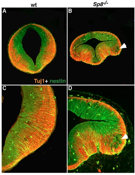
[image:4.612.138.563.60.288.2]To investigate whether the observed neural tube defects in the mid-and hindbrain could result in perturbations of neuronal differentiation, we examined the expression of the neuronal markers nestin and -III-tubulin (Tuj1). The results shown in Fig. 8 indicate that, in contrast to control embryos, in the midbrain of E11.5 Sp8 mutant embryos, nestin-positive cells, present as cell patches outside of the ventricular zone, still express nestin but are devoid of Tuj1 signal (Fig. 8D arrow). This indicates that these cells might Fig. 5. Otx2 and/or Gbx2 are not
required for the activation but for the restriction of Sp8 expression at the MHB. The expression of Sp8is shown, where possible, at different stages of development in wild-type (A,E,H), Gbx2–/– (B,F,I), Otx12/Otx12(C,G) and
Otx12/Otx12/Gbx2–/–(D) embryos. In situ hybridization was performed on sagittal sections and using 35S-labeled Sp8 riboprobe. Although the
Otx12/Otx12/Gbx2–/–double mutant is lethal between E9.2 and E.9.5 and therefore cannot be presented at E10.5 and E12.5, the in situ with Otx12/Otx12is shown at E10.5 as the E12.5 phenotype is essentially the same as at E10.5. As can be seen, Sp8is not downregulated in embryos lacking Gbx2 (B,F,I), Otx2 (C,G) or both proteins (D). Sp8is rather strongly expressed in Gbx2-deficient embryos in the rostral hindbrain committed to be transformed in an expanded posterior midbrain (Wassarman et al., 1997) (B,F,I). In Otx12/Otx12, the rostral
D
E
V
E
LO
P
M
E
N
T
correspond to neuronal progenitors that are not fully differentiated and could suggest that cell differentiation is at least delayed in the absence of Sp8.
[image:5.612.51.263.57.481.2]In order to obtain more insight into this process, we analyzed whether the differentiation of dopaminergic and serotonergic neurons was altered. The loss of Sp8 function provoked an alteration of the Otx2/Gbx2expression boundary and ectopic patches of cells expressing Fgf8, Otx2and/or Wnt1are found in the ventral part of the rostral hindbrain. This was further corroborated by the presence of transcripts of the homeobox gene Pitx3, a marker for midbrain dopaminergic neurons (Smidt et al., 2004), in this brain area of mutant embryos (Fig. 4J,K). Accordingly, the tyrosine hydroxylase Fig. 6. The loss of Sp8provoked overgrowth of the
[image:5.612.324.536.60.206.2]neuroepithelium.(A-M) Expression analysis of several markers of the ventral midbrain: Shh(A,B), Nkx6.1(C,D), Nkx2.2(E,F), Wnt5a(G,H), Wnt1(J,K) and Foxa2(L,M) on frontal vibratome sections (45 m) selected at the level of the posterior midbrain. The expression pattern of all these markers presented here reflects the expansion of the neuroepithelium and does not reveal a patterning defect at this stage of development. The overgrowth of the neuroepithelium results in reduced aqueduct space.
Fig. 7. Enhanced cell proliferation in the mid- and rostral hindbrain of Sp8-deficient embryos.(A-D) BrdU-labeled cells with a pulse of 30 minutes are shown in frontal sections, at the level of the posterior midbrain (A,B) and in sagittal hindbrain sections (C,D) of 10.5 wild-type and mutant embryos. In mutant embryos, a remarkable increase in the number of proliferating cells was evident, thus suggesting that Sp8negatively regulates cell proliferation. All images are at the same magnification. A and B are 5 m sections; C and D are 10 m sections.
[image:5.612.322.552.334.631.2]D
E
V
E
LO
P
M
E
N
T
protein (TH), detected by immunohistochemistry on sagittal sections at E11.5, E12.5 and E17.5, was also found at ectopic sites in the rostral hindbrain of Sp8–/–embryos (Fig. 9C, arrowheads; Fig. 9H, arrowhead; Fig. 9L, arrowhead; Fig. 9P, arrowhead). We then asked whether serotonergic neurons, normally positioned directly posterior to the MHB (Hynes and Rosenthal, 1999; Ye et al., 1998), were affected by the lack of Sp8. As shown in Fig. 9 on the same embryo sections together with dopaminergic neurons, serotonergic neurons were not detected at E11.5 (Fig. 9C,D). However, at E12.5 and E17.5, serotonergic neurons were found (Fig. 9G, see arrows in Fig. 9H,O,P) although the number of 5HT-positive cells is reduced in the area where some ectopic dopaminergic neurons emerge (Fig. 9H, arrowhead; see arrowheads in Fig. 9L,P). This suggests that the development of serotonergic neurons is at least delayed in Sp8 mutant embryos.
DISCUSSION
In the present study, we have analyzed the consequences of the loss of Sp8function on the development of the IsO and the midbrain. The lack of Sp8induced overgrowth of the mid- and rostral hindbrain.
Molecular marker analysis revealed that the positioning of the IsO is shifted posteriorly in Sp8mutant embryos. The observed defects were associated with ectopic Fgf8-, Otx2and/or Wnt1-expressing cells in the ventral part of the rostral hindbrain, and possibly affecting the differentiation of dopaminergic neurons in this area. Finally, we demonstrate that cell proliferation in the mid- and hindbrain is tightly controlled by Sp8activity.
[image:6.612.53.500.57.379.2]D
E
V
E
LO
P
M
E
N
T
Wurst and Bally-Cuif, 2001; Raible and Brand, 2004; Wassarman et al., 1997; Li and Joyner, 2001; Martinez-Barbera et al., 2001; Li et al., 2002; Li et al., 2005). In the absence of Gbx2or both Otx2and Gbx2, the expression domains of these secreted factors exhibit an overlapping disorganized pattern. Sp8loss of function provokes an altered positioning of the IsO, which can already be detected at E8.5. This malformation becomes less pronounced after E10.5. Thus, an Sp8-independent mechanism might be responsible for this process at later stages of gestation. Interestingly, the analysis of Gbx2 conditional knockout mutant in r1 uncovered a Gbx2-independent pathway to repress Otx2after E9 (Li et al., 2002).
Otx2 and Gbx2 were found to be responsible for the proper positioning of the IsO along the AP axis (Martinez-Barbera et al., 2001; Li and Joyner, 2001; Wurst and Bally-Cuif, 2001). Our findings clearly demonstrate that Sp8activation is independent of Otx2and/or Gbx2expression, although these genes are necessary for restricting the expression of Sp8at the MHB. Whether this effect is direct or indirect remains to be assessed.
The presence of ectopic patches of cells expressing Fgf8in the ventral and in the dorsal part, but Otx2, and/or Wnt1in the ventral part of the rostral hindbrain of Sp8mutant embryos indicates that its role may be restricted to the ventral neuroepithelium, where it might be required to refine cell identity by repressing the expression of Otx2, Wnt1and Fgf8. This suggests that Sp8may contribute to sharpen the molecular code of MHB region on the rostral side of the hindbrain.
By contrast, in Gbx2conditional mutant, where Gbx2function in r1 was abolished after E8.5, ectopic patches of cells expressing Fgf8, Otx2and/or Wnt1were described to reside in the dorsal part (alar plate) of r1 (Li et al., 2002). Altogether, this may indicate that there may be two separate pathways operating to maintain a normal expression of these genes in the dorsal and ventral part of the neural tube.
Sp8may restrict the Fgf8expression domain at
the MHB
The perturbed and overlapping expression domains of Fgf8and Wnt1in Sp8knockout embryos are consistent with a role for Sp8in mediating the segregation of these territories. The establishment of a juxtaposed Wnt1and Fgf8expression areas at the mes-met border is required for normal function of the IsO (Wassarman et al., 1997; Li and Joyner, 2001; Martinez-Barbera et al., 2001; Li et al., 2002). Otx2and Gbx2were shown to promote stable expression and to define the precise positioning of Fgf8at the MHB (Wassarman et al., 1997; Li and Joyner, 2001; Martinez-Barbera et al., 2001; Li et al., 2002; Li et al., 2005). In the absence of Otx2 and Gbx2, the expression of Sp8is similar to that reported for Fgf8and other genes transcribed at the MHB (Martinez-Barbera et al., 2001; Li and Joyner, 2001). By contrast, in Sp8-deficient embryos no downregulation of the expression of molecular determinant genes of the MHB was observed over time, as reported in embryos lacking one of these factors (Simeone et al., 2002; Wurst and Bally-Cuif, 2001; Raible and Brand, 2004; Chi et al., 2003). Sp8 (Sp8) and Sp9 were reported to positively regulate Fgf8 expression in the limb of mouse and chick as well as pectoral fin of zebrafish (Treichel et al., 2003; Bell et al., 2003; Kawakami et al., 2004). Given the highly GC-rich content of the putative promoter region of Fgf8, which contains Sp1-binding sites, Sp8 was suspected, together with Sp9, to directly interact with Fgf8(Kawakami et al., 2004). Therefore, one attractive interpretation of the disturbed Fgf8, Wnt1and Otx2 expression would be the requirement of Sp8to sustain a normal Fgf8 expression at the MHB. This is in agreement with the more
remarkable perturbation and spread of the Fgf8expression domain observed in the absence of Sp8, when compared to Wnt1and Otx2. Our findings therefore led us to propose that Sp8is necessary to restrict the expression of Fgf8 at the MHB. A disturbed Fgf8 expression was reported to be accompanied with Otx2and Wnt1 perturbed expression at the MHB (Wassarman et al., 1997; Li et al., 2002). In the limb of Sp8–/–embryos, Fgf8transcripts are properly activated but they gradually disappear during subsequent development, resulting in severe truncations of the appendages (Treichel et al., 2003; Bell et al., 2003; Kawakami et al., 2004). At the MHB, however, Fgf8expression is upregulated and expanded, suggesting a negative regulation of Fgf8 by Sp8in this region. Whether Sp8 is directly involved in this process remains to be assessed.
Sp8may control cell proliferation in the midbrain
and rostral hindbrain
D
E
V
E
LO
P
M
E
N
T
Finally, the zebrafish Sp/btd homolog Bts1 has been implicated in the regulation of Pax2 expression (Tallafuss et al., 2001). Whether Sp8 is involved in this process remains to be assessed. In summary, our study provides evidence that Sp8 is involved in positioning of the IsO. This factor is required to maintain a normal expression of Fgf8, Otx2 and Wnt1 at the MHB. Our findings are consistent with a role for Sp8 in sharpening the MHB expression code by restricting the expression of Fgf8, Otx2 and Wnt1 in this area. We further demonstrate that Sp8 controls cell proliferation in the midbrain and rostral hindbrain. Our findings sustain the notion that tissue patterning and cell proliferation and/or survival are tightly co-regulated.
We thank K. Schneider and I. Fischer for excellent technical assistance; Prof. D. Gallwitz for constant support; and C. Zeden, U. Teichmann and the animal house crew for help with the mice. We are indebted to A. Stoykova and H. Fukumitsu for fruitful discussions. We further thank Drs A. P. McMahon, A. L. Joyner, G. R. Martin, P. Burbach, M. Busslinger, J. F. Brunet, J. Nardelli, A. Kispert, F. Rijli, P. Charnay and R. Krumlauf for probes. This work was supported by the European community QLG3-CT-2000-01625 Brain Genes, Dance QLK3-CT-2001-02120, BMBF 01 GN 0102 and 01 GN 0510; by the DFG Center for Molecular Physiology of the Brain CMPB (Göttingen); by the Max-Planck Society, the Dr Helmut Storz- and Alte Leipziger-Stiftung (to A.M.); and by the Italian Association for Cancer Research (AIRC), the MRC (Grant number G0400410 ID71344) and the MIUR (ex DM 8/10/96 n. 623) (to A.S.).
Supplementary material
Supplementary material for this article is available at http://dev.biologists.org/cgi/content/full/133/9/1779/DC1
References
Acampora, D., Avantaggiato, V., Tuorto, F. and Simeone, A.(1997). Genetic control of brain morphogenesis through Otxgene dosage requirement.
Development124, 3639-3650.
Bell, S. M., Schreiner, C. M., Waclaw, R. R., Campbell, K., Potter, S. S. and Scott, W. J.(2003). Sp8 is crucial for limb outgrowth and neuropore closure.
Proc. Natl. Acad. Sci. USA100, 12195-12200.
Boulet, A. M., Moon, A. M., Arenkiel, B. R. and Capecchi, M. R.(2004). The roles of Fgf4 and Fgf8 in limb bud initiation and outgrowth. Dev. Biol.273, 361-372.
Broccoli, V., Boncinelli, E. and Wurst, W.(1999). The caudal limit of Otx2 expression positions the isthmic organizer. Nature401, 164-168. Chi, C. L., Martinez, S., Wurst, W. and Martin, G. R.(2003). The isthmic
organizer signal FGF8 is required for cell survival in the prospective midbrain and cerebellum. Development130, 2633-2644.
Crossley, P. H., Martinez, S. and Martin, G. R.(1996). Midbrain development induced by FGF8 in the chick embryo. Nature380, 66-68.
Donoghue, M. J., Lewis, R. M., Merlie, J. P. and Sanes, J. R.(1996). The Eph kinase ligand AL-1 is expressed by rostral muscles and inhibits outgrowth from caudal neurons. Mol. Cell Neurosci.8, 185-198.
Hirata, H., Tomita, K., Bessho, Y. and Kageyama, R.(2001). Hes1 and Hes3 regulate maintenance of the isthmic organizer and development of the mid/hindbrain. EMBO J.20, 4454-4466.
Hynes, M. and Rosenthal, A.(1999). Specification of dopaminergic and serotonergic neurons in the vertebrate CNS. Curr. Opin. Neurobiol.9, 26-36. Irving, C. and Mason, I.(2000). Signalling by FGF8 from the isthmus patterns anterior hindbrain and establishes the anterior limit of Hox gene expression.
Development127, 177-186.
Kawakami, Y., Esteban, C. R., Matsui, T., Rodriguez-Leon, J., Kato, S. and Belmonte, J. C.(2004). Sp8 and Sp9, two closely related buttonhead-like transcription factors, regulate Fgf8 expression and limb outgrowth in vertebrate embryos. Development131, 4763-4774.
Lee, S. M., Danielian, P. S., Fritzsch, B. and McMahon, A. P.(1997). Evidence that FGF8 signalling from the midbrain-hindbrain junction regulates growth and polarity in the developing midbrain. Development124, 959-969.
Li, J. Y. and Joyner, A. L.(2001). Otx2 and Gbx2 are required for refinement and not induction of mid-hindbrain gene expression. Development128, 4979-4991. Li, J. Y., Lao, Z. and Joyner, A. L.(2002). Changing requirements for Gbx2 in
development of the cerebellum and maintenance of the mid/hindbrain organizer. Neuron36, 31-43.
Li, J. Y., Lao, Z. and Joyner, A. L.(2005). New regulatory interactions and cellular responses in the isthmic organizer region revealed by altering Gbx2 expression.
Development132, 1971-1981.
Martinez, S., Crossley, P. H., Cobos, I., Rubenstein, J. L. and Martin, G. R. (1999). FGF8 induces formation of an ectopic isthmic organizer and isthmocerebellar development via a repressive effect on Otx2 expression.
Development126, 1189-1200.
Martinez-Barbera, J. P., Signore, M., Boyl, P. P., Puelles, E., Acampora, D., Gogoi, R., Schubert, F., Lumsden, A. and Simeone, A.(2001).
Regionalisation of anterior neuroectoderm and its competence in responding to forebrain and midbrain inducing activities depend on mutual antagonism between OTX2 and GBX2. Development128, 4789-4800.
McMahon, A. P. and Bradley, A.(1990). The Wnt-1 (int-1) proto-oncogene is required for development of a large region of the mouse brain. Cell62, 1073-1085.
Millet, S., Campbell, K., Epstein, D. J., Losos, K., Harris, E. and Joyner, A. L. (1999). A role for Gbx2 in repression of Otx2 and positioning the mid/hindbrain organizer. Nature401, 161-164.
Moorman, A. F., Houweling, A. C., de Boer, P. A. and Christoffels, V. M. (2001). Sensitive nonradioactive detection of mRNA in tissue sections: novel application of the whole-mount in situ hybridization protocol. J. Histochem. Cytochem.49, 1-8.
Panhuysen, M., Vogt Weisenhorn, D. M., Blanquet, V., Brodski, C., Heinzmann, U., Beisker, W. and Wurst, W. (2004). Effects of Wnt1 signaling on proliferation in the developing mid-/hindbrain region. Mol. Cell. Neurosci.26, 101-111.
Prakash, N., Brodski, C., Naserke, T., Puelles, E., Gogoi, R., Hall, A., Panhuysen, M., Echevarria, D., Sussel, L., Vogt Weisenhorn, D. M. et al. (2006). A Wnt1-regulated genetic network controls the identity and fate of midbrain-dopaminergic progenitors in vivo. Development133, 89-98. Puelles, E., Acampora, D., Lacroix, E., Signore, M., Annino, A., Tuorto, F.,
Filosa, S., Corte, G., Wurst, W., Ang, S. L. et al.(2003). Otx dose-dependent integrated control of antero-posterior and dorso-ventral patterning of midbrain.
Nat. Neurosci.6, 453-460.
Puelles, E., Annino, A., Tuorto, F., Usiello, A., Acampora, D., Czerny, T., Brodski, C., Ang, S. L., Wurst, W. and Simeone, A.(2004). Otx2 regulates the extent, identity and fate of neuronal progenitor domains in the ventral midbrain. Development131, 2037-2048.
Raible, F. and Brand, M.(2004). Divide et Impera – the midbrain-hindbrain boundary and its organizer. Trends Neurosci.27, 727-734.
Reifers, F., Bohli, H., Walsh, E. C., Crossley, P. H., Stainier, D. Y. and Brand, M. (1998). Fgf8 is mutated in zebrafish acerebellar (ace) mutants and is required for maintenance of midbrain-hindbrain boundary development and somitogenesis.
Development125, 2381-2395.
Rhinn, M., Dierich, A., Le Meur, M. and Ang, S.(1999). Cell autonomous and non-cell autonomous functions of Otx2 in patterning the rostral brain.
Development126, 4295-4304.
Schwarz, M., Alvarez-Bolado, G., Dressler, G., Urbanek, P., Busslinger, M. and Gruss, P.(1999). Pax2/5 and Pax6 subdivide the early neural tube into three domains. Mech. Dev.82, 29-39.
Simeone, A., Puelles, E. and Acampora, D.(2002). The Otx family. Curr. Opin. Genet. Dev. 12, 409-415.
Smidt, M. P., Smits, S. M., Bouwmeester, H., Hamers, F. P., van der Linden, A. J., Hellemons, A. J., Graw, J. and Burbach, J. P.(2004). Early developmental failure of substantia nigra dopamine neurons in mice lacking the homeodomain gene Pitx3. Development131, 1145-1155.
Sun, X., Mariani, F. V. and Martin, G. R.(2002). Functions of FGF signalling from the apical ectodermal ridge in limb development. Nature418, 501-508.
Tallafuss, A., Wilm, T. P., Crozatier, M., Pfeffer, P., Wassef, M. and Bally-Cuif, L.(2001). The zebrafish buttonhead-like factor Bts1 is an early regulator of pax2.1 expression during mid-hindbrain development. Development128, 4021-4034.
Thomas, K. R. and Capecchi, M. R.(1990). Targeted disruption of the murine int-1 proto-oncogene resulting in severe abnormalities in midbrain and cerebellar development. Nature346, 847-850.
Treichel, D., Schock, F., Jackle, H., Gruss, P. and Mansouri, A.(2003). mBtd is required to maintain signaling during murine limb development. Genes Dev.17, 2630-2635.
Trumpp, A., Depew, M. J., Rubenstein, J. L., Bishop, J. M. and Martin, G. R. (1999). Cre-mediated gene inactivation demonstrates that FGF8 is required for cell survival and patterning of the first branchial arch. Genes Dev.13, 3136-3148.
Urbanek, P., Fetka, I., Meisler, M. H. and Busslinger, M.(1997). Cooperation of Pax2 and Pax5 in midbrain and cerebellum development. Proc. Natl. Acad. Sci. USA94, 5703-5708.
Wassarman, K. M., Lewandoski, M., Campbell, K., Joyner, A. L., Rubenstein, J. L., Martinez, S. and Martin, G. R.(1997). Specification of the anterior hindbrain and establishment of a normal mid/hindbrain organizer is dependent on Gbx2 gene function. Development124, 2923-2934.
Wilkinson, D. G., Bhatt, S., Chavrier, P., Bravo, R. and Charnay, P.(1989a). Segment-specific expression of a zinc-finger gene in the developing nervous system of the mouse. Nature337, 461-464.
Wilkinson, D. G., Bhatt, S., Cook, M., Boncinelli, E. and Krumlauf, R.(1989b). Segmental expression of Hox-2 homoeobox-containing genes in the developing mouse hindbrain. Nature341, 405-409.
D
E
V
E
LO
P
M
E
N
T
homologue of human Sp1 is a head-specific segmentation gene. Nature366, 690-694.
Wurst, W. and Bally-Cuif, L.(2001). Neural plate patterning: upstream and downstream of the isthmic organizer. Nat. Rev. Neurosci.2, 99-108. Wurst, W., Auerbach, A. B. and Joyner, A. L.(1994). Multiple developmental
defects in Engrailed-1 mutant mice: an early mid-hindbrain deletion and patterning defects in forelimbs and sternum. Development120, 2065-2075.
Ye, W., Shimamura, K., Rubenstein, J. L., Hynes, M. A. and Rosenthal, A. (1998). FGF and Shh signals control dopaminergic and serotonergic cell fate in the anterior neural plate. Cell93, 755-766.