Evaluation of Serum Trace Elements and Vitamin Levels in
Hashimoto’s Thyroiditis: Single Centre Experience
from Turkey
Fatma Dilek Dellal1*, Mutlu Niyazoglu2,Esranur Ademoglu1, Suheyla Gorar1, Zehra Candan1, Handan Bekdemir1, Ziynet Alphan Uc1, Mehmet Senes3, Aysenur Ozderya4, Yalcin Aral1
1Endocrinology Department, Ankara Training and Research Hospital, Ankara, Turkey 2Endocrinology Department, Cerrahpasa Medical Faculty, Ankara, Turkey 3Biochemistry Department, Ankara Training and Research Hospital, Ankara, Turkey
4Endocrinology Department, Sirnak Government Hospital, Ankara, Turkey
Email: *[email protected]
Received April 14, 2013; revised May 14, 2013; accepted June 14,2013
Copyright © 2013 Fatma Dilek Dellal et al. This is an open access article distributed under the Creative Commons Attribution Li-cense, which permits unrestricted use, distribution, and reproduction in any medium, provided the original work is properly cited.
ABSTRACT
Aim: To determine levels of serum trace elements and vitamins, and to find out possible correlations between these elements and vitamins with thyroid function tests and thyroid autoantibody levels in patients having Hashimoto’s thyroiditis (HT). Methods: The study included 51 premenauposal women with untreated HT, aged 18 to 56 years without any known chronic diseases or chronic medicine usage, and 27 healthy premenauposal women aged 19 to 42 years old. Trace elements (selenium, zinc, copper, iron levels) and vitamins [A, E, B12, 25-OH-D, 1,25(OH)2D and folic acid levels] were evaluated in patient and control groups. Results: Consequently, serum trace elements and vitamin B12 levels did not significantly differ in patients with HT and control group. Thyroid functioning tests and autoantibody levels did not show any correlation with the levels of trace elements, vitamin A, vitamin E and 25-OH vitamin D. A correlation was detected between vitamin B12 and Anti thyroid peroxidase levels. Conclusion: The negative correlation between vitamin B12 and Anti thyroid peroxidase levels may demonstrate the necessity to screen the patients with HT for atrophic gastritis. We believe that more comprehensive studies with larger sample sizes are needed in which patients are randomized according to their nutritional status.
Keywords: Hashimoto’s Thyroiditis; Trace Element; Vitamin, Vitamin B12, Atrophic Gastritis; Anti Thyroid Peroxidase Antibody
1. Introduction
Hashimoto’s thyroiditis (HT) is an autoimmune disease caused by the destruction of thyroid gland in various de- grees via numerous immune mechanisms. HT is diag- nosed with the elevated thyroid antibodies in serum. Auto- antibodies, genetic tendency, intracellular oxidative me- chanisms and cytokines lead to cellular apoptosis and thus result in follicular destruction. Hypothyroidism may de- velop due to the destruction of thyroid gland in HT.
Trace elements are necessary for development, growth and physiology of the organism. They take part in various mechanisms in the body, but cannot be synthesized in the organism. These elements are iron, zinc, copper, selenium, chloride, florid, iodine, chrome, manganese, bore, cobalt,
molybdenum, vanadium, spelter and silisium. They take part in functions like immune regulation, nerve conduc- tion, regulation of membrane potential and maintenance of mithocondrial activity.
Zinc is an essential element for thyroid hormone func- tions [1].Copper is the vital component of numerous oxi- dative enzymes. [2]. Free copper takes role on cellular membranes as a pro-oxidant agent. Selenium is essential for deiodinase activities and thyroid hormone synthesis and metabolism. Selenium acts as a co-factor in the struc- ture of glutation peroxidase which has anti-oxidant fea- tures. Glutation peroxidase takes role in degradation of hydrogen peroxide to water. After interacting with vita- min E, selenium protects the cellular membrane against oxidative damages caused by lipid metabolism [3]. Iron takes place in structures of many enzymes in the body.
Vitamins are the essential elements which are necessary for occurrence of metabolic events and maintenance of health status, while they cannot be synthesized in the body or synthesized inadequately, and need to be intaken.
Non-enzymatic anti-oxidants, like vitamin E and A, contribute to decrease the oxidative damage caused by oxygen radicals by taking their high-energy electrons [4]. Another function of vitamin E is to increase the absorp- tion of vitamin A from the intestines and its level in the tissues. Concomitance of hypothyroidism and pernicious anemia is very frequent, and vitamin B12 deficiency is observed in pernicious anemia. Due to its antiinflam- matory and immunomodulatory features and potential effects on cytokine levels, decreased levels of vitamin D is associated with the increased risks of many disorders, particularly autoimmune diseases [5,6]. Folic acid, which is actually a pro-vitamin, is changed to dihydrofolat by dehydrofolat reductase enzyme after being absorbed, and then it is converted to tetrahydrofolate. Using single car- bon units, the nascent tetrahydrofolate transfers single carbon to some endogenous substances via various oxi- dating mechanisms.
In this study, we aimed to determine the levels of serum trace elements like selenium, zinc, copper and iron, and vitamins like A, E, B12, 25-OH-D, 1,25(OH)2D, folic acid in patients with HT and evaluate the association between thyroid antibodies and these elements and vitamins.
2. Material and Methods
This prospective study included 51 premenopausal wo- men aged between 18 to 56 years and 27 healthy pre- menopausal women aged between 19 to 42 years, who had applied to our clinic. Only female participants are involved in order to create a homogeneous group. Pa- tients were newly diagnosed and untreated with L-thy- roxine. Patients with any known diseases (diabetes mel- litus, hypertension, hyperlipidemia, coronary artery dis- ease, chronic liver or kidney diseases, gastrointestinal absorption problems, collagen tissue disease, bone me- tabolism disease, thyroid disease or malignancy) or chro- nic medicine users were excluded. Demographic charac- teristics, information on current smoking and alcohol consumption and personal and familial histories were re- corded, and detailed physical examinations were per- formed. Informed consent forms were obtained from all patients. Approval of Local Ethics Committee was taken. The study was performed in accordance with Helsinki Declaration and Good Clinical Practice.
HT was diagnosed by elevated anti thyroid peroxidase (anti-TPO), anti thyroglobulin (anti-TG) levels and thy- roid ultrasonography evaluation which revealed hetero- geneity and fibrotic bands in thyroid glands [7]. Subjects with normal thyroid antibodies were considered to be healthy.
Weight, waist circumference, hip circumference and height were measured in fasting status and with daily clothes by the same person. The waist circumference was accepted as the narrowest diameter between the arcus costarum and spina iliaca anterior superior, and the hip circumference was considered as the largest diameter over the gluteus maximus posteriorly and symphysis pubis anteriorly. Body mass index (BMI) was the ratio of the weight to the square of height (weight/heigh2- kg/m2).
Blood samples were collected following 12 hours of fasting. In order to study selenium levels (N: 46 - 143 µg/L), the collected blood samples were centrifuged at 5000 rpm/min after coagulation and stored at −80˚C until testing. The test was performed manually by AAS Hydro System Management. Blood samples were collected from antecubital veins to evaluate iron, copper, zinc, albumin, thyroid stimulating hormone (TSH), Anti-TG, Anti-TPO, TSH receptor antibody (TRAB), vitamin A, vitamin E, vitamin B12, 25-OH vitamin D, 1.25-(OH)2 vitamin D and folic acid. Iron (N: 70 - 180 µg/dL), copper (N: 12.6 - 24 µmol/L) and zinc (N: 10.4 - 22.9 µmol/L) were de-tected by Olympus AU 2700 equipment using the original kits. 25-OH vitamin D (N: 10 - 80 ng/mL), 1.25-(OH)2 vitamin D (N: 10 - 60 pg/mL) and TRAB (N: 0 - 14 U/L) levels were detected by radioimmunaassay (RIA) method using Dia Source kit. fT3 (N: 2.3 - 4.2 pg/mL), sT4 (N: 0.7 - 1.76 ng/dL), TSH (N: µIU/mL), anti-TPO (N: 0 - 60 U/mL), anti-TG (N: 0 - 60 U/mL), vitamin B12 (N: 190 - 911 pg/mL), folic acid (N: 5.38 - 20 ng/mL) levels were detected using Advia Centaur System (Siemens) and its original kits by chemiluscent method. Vitamin A (N: 1.05 - 2.8 µmol/L) and vitamin E (N: 11.6 - 46.4 µmol/L) were tested by HPLC method using Agilent 1200 equipment and Chromosystems kits.
Statistical analyzes were performed by SPSS 16 pro-gram. The values were presented as mean ± standard de- viation. Mann Whitney U test was used to compare the means between the two groups. P < 0.05 was accepted to be significant.
3. Results
Features of Hashimoto and control groups are presented on Table 1.
While there was a significant difference between Has- himoto and control groups regarding TSH, fT4, Anti- TPO, Anti-TG and TRAB, there was no significant dif- ference in terms of FT3 (Table 2).
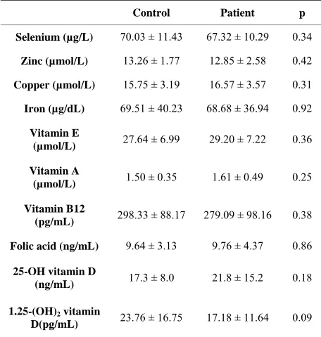
The levels of serum selenium, zinc, copper, iron, vita- min E, vitamin A, vitamin B12, folic acid, 25-OH vita- min D and 1.25-(OH)2 vitamin D did not differ between Hashimoto and the control groups (Table 3).
Table 1. Features of patient and control group.
Control Patient p
Age (year) 33.14 ± 6.87 35.37 ± 8.00 0.2
BMI (kg/m²) 26.76 ± 7.08 27.48 ± 5.17 0.64
Waist circumference
(cm) 87.08 ± 14.14 87.14 ± 11.68 0.98
Hip circumference
[image:3.595.58.287.248.389.2](cm) 105.57 ± 12.23 106.89 ± 9.49 0.63
Table 2. Thyroid function tests and thyroid autoantibody levels of patient and control group.
Control Patient p
TSH (µIU/mL) 1.99 ± 1.22 5.64 ± 6,32 0.0001
fT3 (pg/mL) 3.37 ± 0.33 3.26 ± 0.27 0.132
fT4 (ng/dL) 1.04 ± 0.15 0.91 ± 0.16 0.002
Anti-TPO
(U/mL) 41.56 ± 13.72 236.74 ± 266.95 0.0001
Anti-TG
(U/mL) 31.53 ± 12.48 145.03 ± 118.98 0.0001
TRAB (U/L) 7.4 ± 4.16 4.92 ± 3.32 0.019
Normal ranges: TSH: µIU/Ml, fT3: 2.3 - 4.2 pg/mL, fT4: 0.7 - 1.76 ng/dL, anti-TPO: 0 - 60 U/mL, anti-TG: 0 - 60 U/mL, TRAB: 0 - 14 U/L.
Table 3. Trace elements and vitamin levels of patient and control group.
Control Patient p
Selenium (µg/L) 70.03 ± 11.43 67.32 ± 10.29 0.34
Zinc (µmol/L) 13.26 ± 1.77 12.85 ± 2.58 0.42
Copper (µmol/L) 15.75 ± 3.19 16.57 ± 3.57 0.31
Iron (µg/dL) 69.51 ± 40.23 68.68 ± 36.94 0.92
Vitamin E
(µmol/L) 27.64 ± 6.99 29.20 ± 7.22 0.36
Vitamin A
(µmol/L) 1.50 ± 0.35 1.61 ± 0.49 0.25
Vitamin B12
(pg/mL) 298.33 ± 88.17 279.09 ± 98.16 0.38
Folic acid (ng/mL) 9.64 ± 3.13 9.76 ± 4.37 0.86
25-OH vitamin D
(ng/mL) 17.3 ± 8.0 21.8 ± 15.2 0.18
1.25-(OH)2 vitamin
D(pg/mL) 23.76 ± 16.75 17.18 ± 11.64 0.09
Normal ranges: selenium: 46 - 143 µg/L, zinc: 10.4 - 22.9 µmol/L, iron: 70 - 180 µg/dL, copper: 12.6 - 24 µmol/L, vitamin E: 11.6 - 46.4 µmol/L, vita- min A: 1.05 - 2.8 µmol/L, vitamin B12: 190 - 911 pg/mL, folic acid: 5.38 - 20 ng/mL, 25-OH vitamin D: 10 - 80 ng/mL, 1.25-(OH)2 vitamin D: 10 - 60 pg/mL.
4. Discussion
Levels of serum trace elements and vitamins did not dif- fer between HT and control groups in our study. We also did not determine a correlation between the levels of trace elements and thyroid antibodies. We determined a negative correlation only between vitamin B12 and Anti- TPO levels.
In the recent studies, oxidative mechanisms are con- sidered to play a role in thyroid autoimmunity. In this case, thyroid autoantibodies could be expected to be as- sociated with selenium, vitamin E and vitamin A, which are called antioxidants, as well as zinc and copper, which are called antioxidant enzyme co-factors. However, we could not detect such an association in our study. There was also no association between vitamin D, folic acid and iron with thyroid autoantibodies. We did not find any reports demonstrating correlations between thyroid auto- antibodies and vitamin E, vitamin A, vitamin, D folic acid, copper and zinc. While some reports showed an association between the levels of selenium [8] and auto- antibodies, some did not [9-11]. Different outcomes in these studies might be due to nutrition alterations among the individuals, genetic tendency, different stages of thy- roid diseases, differences in numbers of patients and their distributions and/or alterations in laboratory techniques.
There are a few reports regarding the association be- tween thyroid and vitamin B12. It is known that HT may accompany other autoimmune diseases. Pernicious ane- mia is one of them. Of the patients with hypothyroidism, 7% to 12% have evident pernicious anemia and 10% have latent pernicious anemia [12]. Vitamin B12 defi- ciency is observed in pernicious anemia [13]. Ness- Abramof et al. suggested that patients with autoimmune diseases should be screened for pernicious anemia by screening vitamin B12 levels in every 3 or 5 years [14]. We did not determine any vitamin B12 deficiency among patients in our study, but detected a correlation between vitamin B12 and Anti-TPO levels (r = −0.226). This rela- tionship might be indicative of an underlying similar autoimmune pathology. By these results, necessities ap- pear for the investigation of the level of gastric parietal cell antibody and for the determination of the co-mor- bidity of atrophic gastritis, which also an autoimmune disease, in patients with high levels of Anti-TPO.
Our study had some limitations. Selenoprotein levels for selenium status, seruloplasmin for copper status, and zinc levels in urine, erythrocyte for zinc status and parie- tal cell antibody could not be detected. Dietary habits of patients were not asked.
[image:3.595.57.285.449.689.2]Figure 1. Vitamin B12 and Anti-tpo levels in patient and control groups.
ments and vitamins. Thyroid autoantibody levels did not show any correlation between the levels of trace ele- ments and vitamin A, vitamin E and 25-OH vitamin D. A correlation was detected between vitamin B12 and Anti- TPO levels. This result may show that patients with HT should be screened for atrophic gastritis. We believe that more comprehensive studies with larger sample sizes are needed.
REFERENCES
[1] A. S. Prasad and A. Miale, “Zinc Metabolism in Patiends with the Sendrome of Iron Deficiency Anemia, Hypogo- nadism and Dwarfism,” Journal of Laboratory and Clinical Medicine, Vol. 61, 1963, pp. 537-549.
[2] M. Usdal, H. Pasaoglu and S. Muhtaroglu, “Biyokimya, su ve Elementler,” Erciyes Üniversitesi Yayınları, Kayseri, 1991.
[3] M. Gebre-Medhin, U. Ewald and L. Platin, “Elevated Serum Selenium in Diabetic Children,” Acta Paediatrica Scandinavica, Vol. 73, 1984, pp. 109-114.
doi:10.1111/j.1651-2227.1984.tb09907.x
[4] F. Karatas, U. Askin, I. Halifeoglu and E. Donder, “Gua- tr’lı Hastalarda Antioksidan Vitaminler (A, E ve C), Selenyum ve Glutatyon Peroksidaz(GSH-Px) Düzeyle- rinin Araştırılması,” Fırat Üniversitesi Sağlık Bilimleri Dergisi (Tıp), Vol. 20, No. 4, 2006, pp. 277-280.
[5] M. F. Holick, “Vitamin D: Importance in the Prevention of Cancers, Type 1 Diabetes, Heart Disease, and Osteo- porosis,” American Journal of Clinical Nutrition, Vol. 79,
2004, pp. 362-371.
[6] P. Bordelon, M. V. Ghetu and R. Langan, “Recognition and Management of Vitamin D Deficiency,” American Family Physician, Vol. 80, No. 8, 2009, pp. 841-846. [7] R. I. Henkin, “Trace Metals in Endocrinology,” TheMe-
dical Clinics of North America, W.B. Saunders Co, Phi- ladelphia, 1976.
[8] P. Zagrodzki and E. Przybylik-Mazurek, “Selenium and Hormone İnteractions in Female Patients with Hashimoto Disease and Healthy Subjects,” Endocrine Research, Vol. 35, No. 1, 2010, pp. 24-34.
doi:10.3109/07435800903551974
[9] P. Zagrodzki, F. Nicol, J. R. Arthur and M. Słowiaczek, “Selenoproteins in Human Thyroid Tissues,” Biofactors, Vol. 14, No. 1-4, 2001, pp. 223-227.
doi:10.1002/biof.5520140128
[10] P. Zagrodzki, F. Nicol, J. R. Arthur, M. Słowiaczek, S. Walas, H. Mrowiec and R. Wietecha-Posłuszny, “Seleno- enzymes, Laboratory Parameters, and Trace Elements in Different Types of Thyroid Tumour,” Biological Trace Element Research, Vol. 134, No. 1, 2010, pp. 25-40. doi:10.1007/s12011-009-8454-2
[11] P. Zagrodzki and R. Ratajczak, “Selenium Status, Sex Hormones, and Thyroid Function in Young Women,”
Journal of Trace Elemement and Medicine in Biology, Vol. 22, No. 4, 2008, pp. 296-304.
doi:10.1016/j.jtemb.2008.07.001
[13] S. H. Chen, C. S. Hung, C. P. Yang, F. S. Lo and H. H. Hsu, “Coexistence of Megaloblastic Anemia and Iron Deficiency Anemia in a Young Woman with Chronic Lymphocytic Thyroiditis,” International Journal of He- matology, Vol. 84, No. 3, 2006, pp. 238-241.
doi:10.1532/IJH97.A10518
[14] R. Ness-Abramof, D. A. Nabriski, L. E. Braverman, L.
Shilo, E. Weiss, T. Reshef, M. S. Shapiro and L. Shenk- man, “Prevalence and Evaluation of B12 Deficiency in Patients with Autoimmune Thyroid Disease,” The Ame- rican Journal of The Medical Sciences, Vol. 332, No. 3, 2006, pp. 119-122.
doi:10.1097/00000441-200609000-00004
Abbreviations
Anti-TG: Anti thyroglobulin; Anti-TPO: Anti thyroid peroxidase; BMI: Body mass index;
HT: Hashimoto’s thyroiditis; TSH: Thyroid stimulating hormone;