N A N O E X P R E S S
Open Access
Fe effect on the optical properties of TiO
2
:Fe
2
O
3
nanostructured composites supported on SiO
2
microsphere assemblies
Jesús I Peña-Flores
1, Abraham F Palomec-Garfias
1, César Márquez-Beltrán
1*, Enrique Sánchez-Mora
1,
Estela Gómez-Barojas
2and Felipe Pérez-Rodríguez
1Abstract
The effect of Fe ion concentration on the morphological, structural, and optical properties of TiO2films supported on silica (SiO2) opals has been studied. TiO2:Fe2O3films were prepared by the sol-gel method in combination with a vertical dip coating procedure; precursor solutions of Ti and Fe were deposited on a monolayer of SiO2opals previously deposited on a glass substrate by the same procedure. After the dip coating process has been carried out, the samples were thermally treated to obtain the TiO2:Fe2O3/SiO2composites at the Fe ion concentrations of 1, 3, and 5 wt%. Scanning electron microscopy (SEM) micrographs show the formation of colloidal silica microspheres of about 50 nm diameter autoensembled in a hexagonal close-packed fashion. Although the X-ray diffractograms show no significant effect of Fe ion concentration on the crystal structure of TiO2, theμ-Raman and reflectance
spectra do show that the intensity of a phonon vibration mode and the energy bandgap of TiO2decrease as the
Fe+3ion concentration increases.
Keywords:TiO2:Fe2O3films; Nanostructured composites; SiO2microsphere assemblies; Sol-gel; Dip coating
Background
SiO2microsphere assemblies coated with metal oxide films
have been considered as promising candidate materials for applications in photocatalysis due to their large surface area, excellent accessibility to the inner surface, and suit-able morphology in comparison with powder materials. For example, mesoporous TiO2films perform quite well
in the decomposing of pollutants and in the generation of hydrogen by water splitting [1]. Also, the TiO2is the most
promising catalyst due to its high efficiency, stability, and low cost. It has been used widely in the photocatalytic degradation of phenol. However, one disadvantage of TiO2 for industrial applications is the need of filtration
after the photodegradation process. Recently, the research has been focused on modifying the surface or the bulk of a semiconductor catalyst by adding transition metal impurities, which give rise to the mixed oxide semicon-ductor formation [2].
However, few research studies have been concerned to surface wetting application. One interest point in our current research is to observe the effect of light adsorption on surface materials with respect to their wetting pro-perties particularly of the Fe2O3:TiO2/SiO2composites;
this topic has attracted significant scientific attention particularly for biological systems. A method for the fabrication of thin films using sol-gel is the dip coating procedure, and it is an appropriate technique to obtain TiO2 [3], Fe2O3:TiO2 [4], and TiO2/SiO2 [5], among
others. This method has some advantages, for example, low investment cost for production facilities, large variety of coating materials, and high uniformity on large coating areas. Furthermore, an additional advantage of this tech-nique is the fact that both sides of the substrate can be coated simultaneously. The versatility of this technique allows us to control the vertical dipping velocity, which is very important in the wetness of the forming layer and in the gelation of the layer by the solvent evaporation in order to get uniformity in the film thickness. Theα-Fe2O3
has the important feature of absorbing a large part of the visible solar light due to its energy bandgap of 2.1 eV [6].
* Correspondence:[email protected]
1
Instituto de Física, Benemérita Universidad Autónoma de Puebla, Apdo. Post. J-48, Puebla Pue. 72570, México
Full list of author information is available at the end of the article
structural and optical properties of these composites.
Methods
Tetraethylorthosilicate (TEOS, 99%), ammonium hydroxide (28%), and titanium(IV) buthoxide (97%) were purchased from Sigma-Aldrich (St. Louis, MO, USA); ethyl alcohol (99.5%), iron nitrate nonahydrate (99.8%), hydrochloric acid (38%), and monoethanolamine (99.4%) were purchased from J.T.Baker (Center Valley, PA, USA); 2-methoxyethanol was supplied by Fluka (St. Louis, MO, USA). All chemicals were used without additional purification. In all experi-ments, we have used ultrapure water (Easy Pure II System, Thermo Fisher Scientific, Waltham, MA, USA).
The synthesis of SiO2microparticles was carried out
by the Stöber method. Following this method, it is possible to control the diameter of the spheres from the TEOS concentration. First, we made a solution by mixing ammonium hydroxide, ethanol, and water with volumes of 20, 38.4, and 41.6 ml, respectively. Then, we made a second solution with 6.6 ml of TEOS and 93.4 ml of ethanol, and later on, we mixed both solutions and stirred it for 1 h at room temperature. The TiO2
precursor solution was made by mixing and stirring the following chemical compounds for 2 h: 19.2 ml of ethanol, 3.8 ml of hydrochloric acid, 7.7 ml of water, and 19.2 ml of titanium(IV) buthoxide at room temperature. The Fe2O3 precursor solution was obtained as follows:
15.15 g of iron nitrate was dissolved in 60 ml of 2-methoxyethanol and 6.8 ml of monoethanolamine, and this solution was stirred for 2 h at room temperature. Finally, to have solutions to the 1, 3, and 5 wt% of Fe3+with
coating procedure at 1.5 mm/min rate. All immersion procedures were done at room temperature and normal pressure. Then, the sample containing the TiO2coating
was annealed at 500°C with air flux 1 ml/s for 6 h. The Fe2O3 coating of the SiO2opals was obtained by
following the same dip coating procedure with the Fe precursor solution under the same conditions. Finally, the TiO2:Fe2O3 (1, 3, 5 wt%) films supported on the
SiO2 opals were obtained by adding the appropriate
amounts of ferric nitrate nonahydrate into the TiO2
pre-cursor solution, and the same dipping procedure was carried out.
The samples were characterized by the technique: X-ray diffraction (XRD; D8 Discover CuKα(λ= 1.5406 Å), Bruker AXS, Inc., Madison, WI, USA),μ-Raman spectroscopy (RS; Horiba Jobin Yvon Lab Ram HR, HORIBA, Ltd., Kyoto, Japan), and diffuse reflectance spectroscopy (Varian Cary 100, Agilent Technologies, Inc., Santa Clara, CA, USA). The surface morphology of the samples was studied with a scanning electron microscope (JEOL JSM-6610LV, JEOL Ltd., Akishima-shi, Japan).
Results and discussion
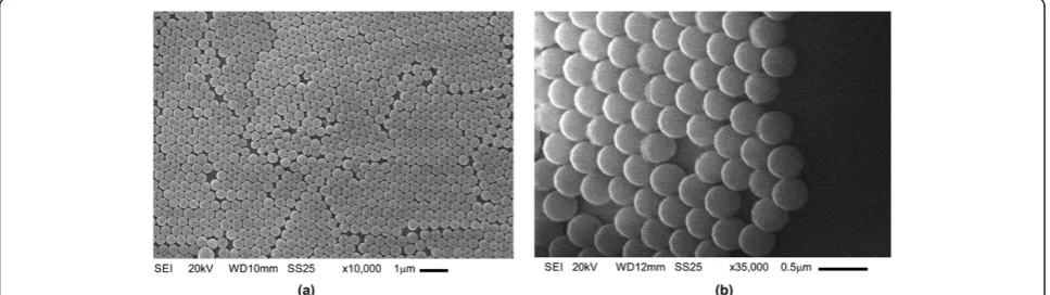
Figure 1a shows a scanning electron microscopy (SEM) micrograph at ×10,000 of the surface morphology of the SiO2 microspheres deposited on a glass substrate. It is
seen in this figure that in some regions, the SiO2
[image:2.595.57.541.567.703.2]micro-spheres form a close-packed arrangement, and in others, some fissures are present.
Figure 1b shows a lateral view at ×35,000 of the SiO2
microparticles. It is seen that the SiO2spheres are smooth
Figure 1SiO2microspheres of 365 nm average size deposited on glass substrate. (a)SEM micrograph at ×10,000 and(b)lateral view of
Figure 2A SiO2microsphere layer coated with TiO2:Fe2O3. (a)Formation of‘micro-shavings’(×2500) dispersed on the glass substrate,
(b)lateral view at ×25,000 of the SiO2spheres on glass substrate, and(c)lateral view at ×65,000 of the SiO2spheres on glass substrate.
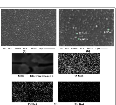
Figure 3SEM images of the surface morphology of the TiO2:Fe2O3composite supported on a SiO2microsphere layer.SEM images at
(a)×50,000,(b)×80,000, and(c)EDS mapping distribution of elements of the TiO2:Fe2O3/SiO2composite: The upper left image corresponds to
[image:3.595.59.541.244.678.2]homogeneous; this ruptures on the surface are probably produced by the deposition silica method, that is, the glass substrate is immersed into an aqueous solution where the silica microparticles are dispersed and thus the hexagonal closed packed is not very well controlled.
Guo et al. [7] described a controllable method to fabri-cate hexagonal close-packed Langmuir-Blodgett silica particulate monolayers modifying the silica surface by using a binary surfactant and solvent systems, reducing in this way fissures along the surface layer. The difficulty of this method to be applied in our work is that the TiO2 adsorption on the silica-modified surface is not
possible due to the hydrophilic character of TiO2 and
the hydrophobic character of the silica.
Figure 2 shows SEM images of the SiO2microspheres
coated with TiO2:Fe2O3. In Figure 2a at ×2,500, it is
observed the presence of ‘micro-shavings’dispersed on the glass substrate. This is due to the incorporation of TiO2:Fe2O3 coating, which destabilizes the SiO2
close-packed arrangement as shown in Figure 1. In Figure 2b, a lateral view at ×25,000 shows with more detail the curv-ing of the SiO2 single layer. In Figure 2c, a micrograph
the mapping distribution of elements of the composite shows the presence of oxygen, titanium, and iron ele-ments. The standardless EDS quantification is 56.78 wt% oxygen, 38.27 wt% silicon, 4.74 wt% titanium, and 0.20 wt% iron.
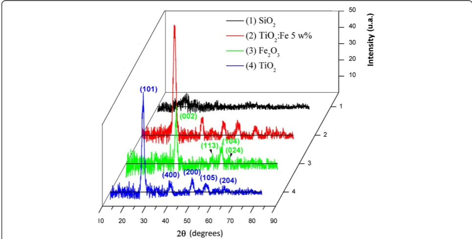
Figure 4 shows X-ray diffractograms of all prepared samples. The Fe2O3 X-ray diffractogram presents peaks
corresponding to the alpha phase of this compound according to the JCPDS data card. It is observed that the peak intensity is enhanced as the Fe concentration is increased. The most intense peak appears at 28° and corresponds to the (101) plane of the anatase TiO2
structure. It is also observed that there is no significant effect of Fe on the TiO2crystalline structure. However,
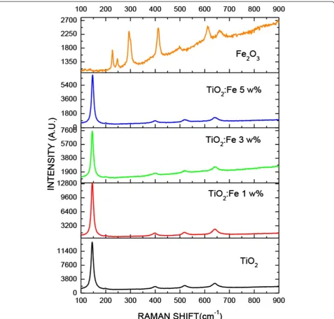
there is a considerable effect of iron concentration on the TiO2phonon modes of vibration as is shown in the
μ-Raman spectra (see Figure 5).
The Raman lines located at 397, 522, and 642 cm−1are, respectively, assigned to the B1, B1g, and A1g vibration
modes of TiO2. According to the analysis of group theory,
the TiO2anatase phase has six Raman active modes. The
[image:4.595.55.538.471.715.2]Raman spectrum of a TiO2anatase single crystal has been
investigated by Ohsaka et al. [8]; they have concluded that the six characteristic allowed modes appear at 146, 197, 400, 516, 520, and 641 cm−1.
The Raman lines appearing at 225, 245, 291, 410, 495, 611, and 1318 cm−1 are a characteristic of α-Fe2O3, i.e.,
the lines at 225 and 495 cm−1 are assigned to the A1g
vibration mode and the four peaks at about 245, 291, 410, and 611 cm−1 are attributed to the Eg vibration
mode [9]. It should be noted that there is a line at 663 cm−1 which is typical for Fe3O4[10]. In our study,
the Raman spectra of the TiO2 film supported on SiO2
opals are slightly shifted with respect to the TiO2anatase
phase obtained from TiO2bulk; this shift could be due
to the presence of Fe ions in the composite. In Figure 5, it is observed that the intensity of the phonon vibration mode at 149 cm−1is decreased as the Fe+3concentration is increased. These results suggest that when the Fe ions are incorporated into TiO2crystal structure, species
-Ti-O-Fe-O-Ti-O- type are formed. On the other side, in the Raman spectra of the TiO2:Fe2O3 composites, phonon
lines corresponding toα-Fe2O3are not observed.
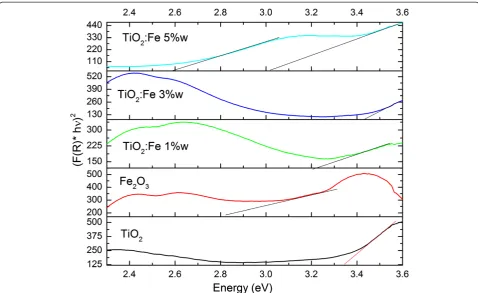
Reflectance spectra of the samples TiO2, Fe2O3, and
TiO2:Fe2O3 composites at 1, 3, and 5 wt%, respectively,
of Fe supported on the SiO2microspheres are shown in
[image:5.595.60.538.88.544.2]Figure 6. The diffuse reflectance spectra were fitted using the Kubelka-Munk theory where the intersection
of the fitted straight line and the photon energy axis gives the energy bandgap value [11,12]. Table 1 lists the energy bandgaps of TiO2, TiO2:Fe2O3(1, 3, and 5 wt%),
and Fe2O3 composites supported on SiO2 microsphere
assemblies. It is seen that the energy bandgap values of TiO2:Fe2O3composites lie between Eg = 3.5 eV of TiO2
bulk and Eg = 2.4 eV of Fe2O3. The energy bandgap
values of the whole set of samples are listed in Table 1. This effect has been observed in a previous work [12].
The decrement in energy bandgap of the TiO2-Fe2O3
composites with respect to the one of TiO2bulk is probably
due to the fact that Fe ions have been incorporated in the TiO2lattice as indicated in the Raman section.
In the case of the Fe2O3sample, we have observed at
2.44 eV a maximum which is considered to be the result of the pair excitation processes6A1+6A1→4T1(4G) +4T1
(4G) [13]; in the case of 2.62 eV, other maximum in the reflection spectrum is observed due to the6A1→4E,4A1
(4G) transition, in accordance to the Tanabe-Sugano dia-gram [13]. Here, we have also observed a reflection edge between 3.00 and 3.25 eV (413 and 381 nm), which could be due to the transition from the oxygen 2p orbital to the iron 3d orbital [14] or by the transition from the valence band to the conduction band of iron oxide. The bandgap value found for this oxide was 2.33 eV, which is a little greater than the one of reference value of Fe2O3in bulk
(2.22 eV). Additionally, there exists a maximum at 3.43 eV which is due to the contributions of the Fe3+ligand field transitions 6A1→4E(4D) and6A1→4T2(4D) [13].
Other-wise, when the TiO2:Fe concentration is 1 and 3 wt%, the
same maxima have been observed between 2.44 and 2.62 eV, which are related to the formation of Fe2O3
into the TiO2lattice supported by the SiO2microsphere
assembly. However, the maximum at 3.43 eV disappears because more Fe ions (5 wt%) are introduced into the TiO2 lattice diminishing the energy bandgap of TiO2
which induces optical transitions in the visible region.
Conclusions
An important effect of the Fe ion concentration on the morphological and optical properties of the TiO2 films
supported on the single layer of SiO2 microsphere
[image:6.595.58.537.88.381.2]assemblies has been found. We have prepared silica
Figure 6Kubelka-Munk transformed reflectance spectra of the samples TiO2, Fe2O3, and TiO2:Fe2O3at different Fe weight percentages.
Table 1 Energy bandgap values of TiO2, Fe2O3, and TiO2:
Fe2O3at 1, 3, and 5 wt% composites supported on SiO2
microsphere assemblies
Samples Eg (eV) λ(nm)
TiO2 3.25 381
TiO2:Fe2O31 wt% 2.79 444
TiO2:Fe2O33 wt% 2.66 466
TiO2:Fe2O35 wt% 2.53 489
[image:6.595.55.290.650.733.2]microparticles of about 364 nm diameter by the Stöber method. We have been able to fabricate a single layer of SiO2microspheres assembled in hexagonal close-packed
fashion on glass substrate with a controlled vertical dip coating procedure.
We have observed that the TiO2film formation affects
the stability of the SiO2 layer cracking the layer and
giving rise to the formation of‘micro-shavings’dispersed on the glass substrate, presenting a granular surface morphology with grain diameter of about 50 nm. The X-ray diffractograms do not show any significant effect of the Fe concentration into the TiO2 lattice structure.
However, a considerable effect of iron concentration on the intensity of the TiO2 phonon vibration modes as
shown in theμ-Raman spectra results has been observed. Furthermore, the diffuse reflectance spectra have shown that the energy bandgap of the TiO2:Fe2O3/SiO2
compos-ites are located in the range between the energy bandgap of TiO2and the one of Fe2O3bulk.
Competing interests
The authors declare that they have no competing interests.
Authors’contributions
AFPG and JIPF carried out the synthesis nanoparticle of SiO2. CMB carried
out the studies of SEM and coordinated and helped to draft the manuscript. ESM participated in the project development and the experimental results of optical properties using spectrophotometry. EGB helped us in the interpretation of Raman results. FPR helped us in the interpretation of reflectivity spectra and participated in the project development. All authors read and approved the final manuscript.
Authors’information
C. Marquez-Beltran, Ph.D. is a professor research fellow at the Puebla University, Puebla, Mexico. The goal of his research is to master the nucleation and aggregation of nanoparticles in order to get a full morphological control in the production of nanostructured materials. J. I. Peña-Flores and A. F. Palomec-Garfias are graduate students. Estela Gómez-Barojas, Ph.D. is a researcher at the Puebla University, Puebla, Mexico. Her research area is synthesis and study of morphological and optical properties of semiconductors. Enrique Sanchez-Mora, Ph. D. is a researcher at the Puebla University, Puebla, Mexico. His research area is synthesis of metal oxides and their study of photocatalytic, chemical, and optical properties. Felipe Pérez-Rodríguez, Ph. D., is a researcher at the Puebla University, Puebla, Mexico. His research areas are physical properties of advanced materials and development of theoretical models to describe the phononic properties of metamaterials.
Acknowledgements
This work was financially supported by VIEP-BUAP (VIEP/EXC-G/2014-225, VIEP/EXC-I/2014-133), PRODEP-BUAP 2014, and the Advanced Materials Research Group (BUAP-CA-250). The authors are thankful toDr. Efraín Rubio Rosasfor his help in taking SEM micrographs of the samples.
Author details
1Instituto de Física, Benemérita Universidad Autónoma de Puebla, Apdo. Post. J-48, Puebla Pue. 72570, México.2CIDS-IC, Benemérita Universidad Autónoma de Puebla, Apdo. Post. 196, Puebla Pue. 72000, México.
Received: 13 May 2014 Accepted: 6 September 2014 Published: 15 September 2014
References
1. Wang KX, Yao BD, Morris MA, Holmes JD:Supercritical fluid processing of thermally stable mesoporous titania thin films with enhanced photocatalytic activity.Chem Mater2005,17:4825.
2. Lu G, Yates JT Jr:Photocatalysis on TiO2Surfaces: principles, mechanisms,
and selected results.J Chem Rev1995,95:735.
3. Jung KY, Park SB:Enhanced photoactivity of silica-embedded titania particles prepared by sol–gel process for the decomposition of trichloroethylene.
J Appl Catal B: Environ2000,25:249.
4. Navio JA, Colon G, Macias M, Real C, Litter MI:Iron-doped titania semiconductor powders prepared by a sol–gel method. Part I: synthesis and characterization.Appl Catal A: Gen1999,177:111.
5. Lepore GP, Persaud L, Langford CH:Supporting titanium dioxide photocatalysts on silica gel and hydrophobically modified silica gel.
J Photochem Photobiol A Chem1996,98:103.
6. Cesar I, Kay A, Martinez JG, Gratzel M:Translucent thin film Fe2O3
photoanodes for efficient water splitting by sunlight: nanostructure-directing effect of Si-doping.J Am Chem Soc2006,128:4582.
7. Guo Y, Tang D, Du Y, Liu B:Controlled fabrication of hexagonally close-packed Langmuir–Blodgett silica particulate monolayers from binary surfactant and solvent systems.Langmuir2013,29(9):2849. 8. Oshaka T:Temperature dependence of the Raman spectrum in anatase
TiO2.J Phys Soc Jpn1980,48:1661.
9. de Faria DLA, Venanuncio Silva S, de Oliveira MT:Raman microspectroscopy of some iron oxides and oxyhydroxides.J Raman Spectrosc1997,28:873. 10. Bersani D, Lottici PP, Montenero A:Micro-Raman investigation of iron
oxide films and powders produced by sol–gel syntheses.J Raman
Spectrosc1999,30:355.
11. Escobedo Morales A, Sanchez Mora E, Pal U:Use of diffuse reflectance spectroscopy for optical characterization of un-supported nanostructures.
Rev Mex Fis, S2007,53(5):18.
12. López R, Gómez R:Band-gap energy estimation from diffuse reflectance measurements on sol–gel and commercial TiO2: a comparative study.
J Sol–gel, Sci Technol2012,61:1.
13. Sánchez E, Gómez E, Rojas E, Silva R:Morphological, optical and photocatalytic properties of TiO2–Fe2O3multilayers.Sol Energy Mater Sol Cells2007,91:1412.
14. Chernyshova I, Ponnurangam S, Somasundaran P:On the origin of an unusual dependence of (bio)chemical reactivity of ferric hydroxides on nanoparticle size.Phys Chem Chem Phys2010,12:14045.
doi:10.1186/1556-276X-9-499
Cite this article as:Peña-Floreset al.:Fe effect on the optical properties of
TiO2:Fe2O3nanostructured composites supported on SiO2microsphere assemblies.Nanoscale Research Letters20149:499.
Submit your manuscript to a
journal and benefi t from:
7 Convenient online submission
7 Rigorous peer review
7 Immediate publication on acceptance
7 Open access: articles freely available online
7 High visibility within the fi eld
7 Retaining the copyright to your article