R E S E A R C H A R T I C L E
Open Access
Standardized extract of
Ficus deltoidea
stimulates
insulin secretion and blocks hepatic glucose
production by regulating the expression of
glucose-metabolic genes in streptozitocin-induced
diabetic rats
Elham Farsi
1*, Mariam Ahmad
2, Sook Yee Hor
1, Mohamed B Khadeer Ahamed
1*, Mun Fei Yam
1and Mohd Zaini Asmawi
1Abstract
Background:Recently, there has been increasing interest inFicus deltoideaJack. (Moraceae) due to its chemical composition and the potential health benefits. The present study was undertaken to investigate the effect of extracts ofF. deltoidealeaves on diabetes.
Methods:The petroleum ether, chloroform and methanol extracts ofF. deltoideawere prepared and subjected to standardization using preliminary phytochemical and HPLC analysis. Dose selection was made on the basis of acute oral toxicity study (50–5000 mg/kg b. w.) as per OECD guidelines. Diabetes mellitus was induced with
streptozotocin and rats found diabetic were orally administered with the extract (250, 500 and 1000 mg/kg) for 14 days. Levels of blood glucose and insulin were measured in control as well as diabetic rats on 0, 7 and 14thday. In addition, glucose metabolism regulating gene expression was assessed using RT-PCR.
Results:HPLC analysis revealed that the methanol extract is enriched with C-glycosylflavones particularly, vitexin and isovitexin. In oral glucose tolerance test, oral administration of the methanol extract increased the glucose tolerance. The methanol extract showed significant (P < 0.01) antidiabetic activity. The extract treatment caused significant reduction (p < 0.01) in elevated fasting blood glucose level in streptozotocin-induced diabetic rats. The streptozotocin-related weight loss in rats was noticeably reversed by the extract treatment. Finally, RT-PCR analysis revealed a novel mechanisms for the anti-diabetic action of methanol extract ofF. deltoidea. The extract exerted its effect via an increase of insulin secretion which impeded the hepatic glucose production, via down-regulation of phosphoenolpyruvate carboxykinase and glucose-6-phosphatase genes expression on one hand, and up-regulation of hepatic GK and PPARγgenes expression on the other hand. The extract caused an increased expression of GLUT-4 gene expression in skeletal muscles which leads to normalize the hyperglycemia. The extract also nullified the toxic effects of streptozitocin by blocking its entry into the isletβ-cells through reducing the expression of GLUT-2 gene.
Conclusion:It can be concluded that,F. deltoideacould potentially inhibits the streptozitocin-induced
hyperglycemia in rats. Further the herb can be utilized as useful remedy for alleviation of diabetes complications.
Keywords:Ficus deltoidea, Streptozotocin, Diabetes, C-glycosylflavone, Glucose metabolism regulating genes, RT-PCR
* Correspondence:[email protected];[email protected]
1
Department of Pharmacology, School of Pharmaceutical Sciences, Universiti Sains Malaysia, 11800 Penang, Malaysia
Full list of author information is available at the end of the article
Background
Diabetes is a disease characterized by chronic hypergly-cemia resulting from impaired carbohydrate metabolism with an absolute or relative lack of insulin [1]. Insulin is the key regulator of glucose uptake. The rise in insulin level in the body leads to the suppression of hepatic gly-cogenolysis and stimulates insulin-dependent glucose uptake into muscle, liver and adipose tissue. The rate of glucose uptake into muscle cells is dependent on the action of insulin on the glucose transporter-4 (GLUT4) [2]. The GLUT4 receptor is localized in intracellular ves-icles, but when insulin binds to its own membrane re-ceptor, GLUT4 is transported to the plasma membrane to permit glucose uptake into muscle cells.
In particular, the liver plays a vital role in maintaining glucose homeostasis and glucose-transporter-2 (GLUT2) is the main facilitative glucose transporter in the liver [3]. GLUT2 carries out insulin-independent transport of glucose. The equilibrium between extracellular and in-tracellular glucose concentration is maintained due to the high capacity of GLUT2 for glucose. Glucose absorp-tion activates hepatic glucokinase (GK) which phosphor-ylates glucose, trapping it in liver cells for conversion to glycogen during the postprandial state. During fasting, liver produces glucose via glycogenolysis or by gluco-neogenesis from non-carbohydrate precursors, such as lactate, amino acids and glycerol [3]. The rate of glyco-genesis is due to the phosphorylase which is controlled by hormones whereas the rate of gluconeogenesis is con-trolled principally by the activities of unidirectional enzymes, such as phosphoenolpyruvate carboxykinase (PEPCK) and glucose-6-phosphatase (Glc-6-Pase). PEPCK catalyses the conversion of oxaloacetate to phosphoenol pyruvate (PEP), a rate limiting step of gluconeogenesis. Whereas, Glc-6-Pase catalyzes the final step of gluconeo-genesis, the production of free glucose from glucose-6-phosphate (G6P). In addition, the expression of PEPCK and GK the key enzymes of liver gluconeogenesis and gly-colysis, is under the control of insulin [4]. It is reported that insulin also regulates GLUT2 expression in the liver [5]. However, glucagon affects glucose metabolism main-ly by inducing gmain-lycogen breakdown and glucose-6-phosphatase on one hand whereas, on the other hand glucagon contributes the hyperglycemia by inhibiting the enzyme pyruvate kinase [6].
Diabetes mellitus is the chief contributor to global mor-bidity and mortality. It is a metabolic disorder that is char-acterized by hyperglycemia. Among the several factors, deficiency in insulin secretion is one of the principal fac-tors that leads to hyperglycemic condition. Emerging evi-dence indicates that a nuclear receptor, the peroxisome proliferator-activated receptorγ (PPARγ), enhances insu-lin signainsu-ling and glucose uptake in muscle upon binding PPARγ agonists [7]. Pharmacological evidence indicates
that PPARγis the major target for glitazones (TZDs). Se-veral hypoglycemic agents that increase the insulin sensi-tivity, such as metformin, gliclazide and glitazones are currently being used to normalize glucose metabolism in diabetes treatment. However, the limitations related with these drugs, including adverse effects and excessive cost, have spurred the investigations of new therapeutic agents derived from plants. Due to worldwide growth of herbal products as alternative medicine, their efficacy and safety has become great concern. In recent years much attention has been paid to phytochemicals, which are widely dis-tributed in medicinal plants and easily metabolized by humans, due to their negligible side effects and compara-tively lower cost than synthetic drugs [8,9]. However, the therapeutic mechanism of these phytochemicals is not yet well clarified; therefore more attention has been drawn to evaluate the preventive and therapeutic effects of herbal medicines in the suitable experimental models.
Ficus deltoidea Jack. (Moraceae) is a native wild ever-green shrub in Malaysia and widely distributed in Southeast Asian countries. It is locally known as Emas Cotek and Mas Cotek [10]. Decoction ofF. deltoidealeaves was exten-sively utilized in folk medicine to decrease symptom of dia-betes, hyperlipidemia and hypertension [11]. Herbal healers often recommend leaves of both male and female species as libido booster, aphrodisiac, also as postpartum treatment to strengthen the uterus [12]. The beneficial effects of F. deltoideaon hepatoprotection, hypertension, inflammation, ulcer, wound healing, inhibition of carbohydrate hydro-lyzing enzyme and antinociceptive activities were veri-fied [10,11,13].
Although the herb is widely used in traditional medi-cine to treat diabetes and recent studies revealed that the herb has potential antidiabetic property [13,14], yet the possible mechanism responsible for the antidiabetic property of F. deltoidea is not elucidated. However, the recent studies correlated the remarkable antioxidant and
α-glucosidases inhibitory activities of F. deltoidea with its antidiabetic property [10,15]. As circumstance this study aims to investigate the possible mechanism res-ponsible for glucose lowering effect of F. deltoidea by studying the expression of GK, Glc-6-Pase, PEPCK, GLUT2 and PPARγgenes in liver and GLUT4 in muscle of streptozitocin-induced diabetic rats. To establish relation between pharmacological properties and phyto-chemical content of plant extract, the screening and quan-titative measurement of two active principles, vitexin and isovitexin was performed.
Methods
Preparation ofF. deltoidealeaf extracts
The leaves were purchased from Herbagus Sdn. Bhd. Penang-Malaysia. The floral characteristics ofF. deltoidea
Shanmugan, School of Biological Sciences, USM. The specimens (Voucher No.: 11204) were then deposited at the herbarium of the School of Biology, Universiti Sains Malaysia. Oven dried leaves were pulverized to a fine powder and chemical components were extracted in pet-roleum ether, chloroform, methanol and water succes-sively at 37°C - 40°C. All the extracts were concentratedin vacuo using rotavapor (Buchi Rotavapor Model R-210, Flawil, Switzerland). The extraction yield calculated was 4.7% (petroleum ether extract), 3.6% (chloroform extract), 8.2% (methanol extract) and 5.5% (water extract). All the extracts were lyophilized and kept in tightly closed glass containers and stored at−80°C until the time of use.
Forin vivostudies, all the extracts were dissolved in 0.5% carboxymethyl cellulose (CMC) to obtain 1000 mg/mL stock solution and stored at 4°C. Further dilutions were prepared using distilled water to obtain the final concentra-tions of 500 and 250 mg/ml. The vehicle, 0.5% CMC is used as negative control.
Animals
For acute toxicity study, healthy adult female Sprague– Dawley rats (200–225 g) were used. Rats (200–230 g) of both sexes were used for pharmacological studies. All the animals were maintained in polypropylene cages at room temperature for adaptation. The rats were fed with standard rodent diet ad libitum and had free access to tap water. The experimental procedures of the animals were approved by the Animal Ethics Committee, Univer-siti Sains Malaysia (AECUSM) with reference number of PPSG/07 (a)/044/(2010).
Acute toxicity study
The study was conducted according to the OECD guide-line (425) by Up-and-Down Procedure as reported earl-ier [16]. Separate groups of rats were orally treated with five different single doses (55, 175, 550, 1750, 5000 mg/kg) of the extract. After the oral administration of the extract, signs of possible toxicity were observed every hour for the first six hours and every day for 14 days. The body weights of all animals were recorded daily. Treatment related mor-tality or signs of toxicity were observed for up to 14 days. At the end of the study, LD50value for the extract was
es-timated. For pharmacological studies three doses were se-lected. One was a safer dose (1/10thof LD50= 500 mg/kg),
second was higher than the safer dose (1000 mg/kg) and third was lower than the safer dose (250 mg/kg).
Phytochemical analysis ofF. deltoidealeaf extracts Phytochemical such as protein, polysaccharide, glyco-saponin, phenolics, flavonoids and tanins were analyzed in the F. deltoidea leaf extracts. The total contents of proteins, polysaccharides and glycosaponin in the ex-tracts were estimated calorimetrically, according to the
previous study [16]. Total phenolics were determined using the Folin-Ciocalteau reagent with gallic acid as a standard and the results were expressed as mg of gallic acid equivalents, whereas total flavonoids were deter-mined using the AlCl3 colorimetric method with
quer-cetin (QNT) as a standard and the results were expressed asμg of QNT equivalent. The amount of total condensed tannins was expressed as (+) catechin equivalents [CT, mg (+) catechin/g sample].
High performance liquid chromatography (HPLC) analysis of the extracts
A 100 mg portion of the extracts of F. deltoidea was dis-solved in 15 ml methanol and subjected to ultrasonica-tion for 10–15 min. Then the volume of the contents was made up to 25 ml using a volumetric flask. Finally, all the samples were filtered through a 0.45 mm Whatman filter paper. Similarly, the reference standards (5 mg) were dissolved in 5 ml methanol and then filtered. The stock solutions were used to prepare further dilutions.
Vitexin and isovitexin concentrations were determined by an Eclipse C18 reverse-phase (250-mm × 4.6 mm) column in an HPLC system equipped with a UV de-tector, quaternary pump, online degasser, auto sampler, automatic injector, column heater and photodiode array detector (Agilent Tech, Palo Alto, CA). The contents of vitexin and isovitexin were determined by the HPLC method described by Fu et al., [17]. The calibration curves were set up at 330 nm by subjecting the isovi-texin and viisovi-texin standard solutions to the isocratic mo-bile phase, which consisted of methanol/water/formic acid (33:66.37:0.67, v/v/v), at a flow rate of 1 ml/min at 30°C with an injection volume of 10 μl. Identification and calibration of the samples were performed by com-paring absorbance spectra and retention times with those of the standard reference. The concentrations of vitexin and isovitexin in the samples were estimated based on the regression lines of vitexin and isovitexin in the range of 5–200 μg/ml, which were Y = 28.305 X. 28.245 (R2 = 0.9982, n = 6) and Y = 23.90 X. 65.442 (R2 = 0.9992, n = 6), respectively, where Y is the peak area of the analyte and X is the concentration of the analyte (μg/ml).
Assessment of hypoglycemic activity ofF. deltoidealeaf extracts in rats
Advantage blood glucose meter. The blood samples were collected from the tail vein at 1, 2, 3, 5 and 7 h after ad-ministering the extracts to the overnight-fasted rats [18].
Assessment of antihyperglycemic activity ofF. deltoidea leaf extracts in rats
The antihyperglycemic activity was assessed using an oral glucose tolerance test that was performed in the overnight-fasted rats [19]. The rats were divided into six groups of six rats each. Group I served as the negative control and received 10 ml/kg of vehicle (0.5% of CMC) orally. Group II served as the positive control and was orally administered with 500 mg/kg metformin. The ani-mals of groups III, IV, V and VI received 1000 mg/kg of petroleum ether, chloroform, methanol and water extracts, respectively, via oral gavage daily. The Fasting blood glu-cose levels were checked initially using an ACCU-CHEKR Advantage blood glucose meter. The rats were then dosed intraperitoneally with 500 mg/kg of glucose one hour after administration of extracts and their blood glucose levels were examined again at 0, 30, 60, 90 and 120 min after glucose loading.
Assessment of blood glucose and plasma insulin levels of F. deltoidealeaves extracts in the streptozitocin-induced diabetic rats
Diabetes was induced in 50 overnight-fasted rats with free access to water by a single IP injection of freshly prepared streptozitocin in ice-cold normal saline at a dose of 55 mg/kg. After 72 h, the rats with a blood glu-cose level of 15 mM were considered diabetic and used in the experiment. Diabetic rats were divided into seven groups of six rats each (n = 6). Group I consisted of nor-mal rats that received 10 ml/kg of vehicle. Group II con-sisted of diabetic rats that received 10 ml/kg of vehicle. Group III of diabetic rats served as positive control and received 500 mg/kg metformin. Groups IV, V, VI and VII consisted of diabetic rats treated with 1000 mg/kg of petroleum ether, chloroform, methanol and water ex-tracts, respectively. The treatment was given twice a day for fourteen days. Separate groups of diabetic rats were also treated with 500 mg/kg or 250 mg/kg of the most active extract (methanol extract), administered as de-scribed above. Tail vein blood samples were collected on days 1, 3, 6, 9, 12 and 15 for blood glucose analysis, and the body weights were taken [20]. The blood samples collected were also used to analyze the fasting plasma insulin levels using the Mercodia Ultra-sensitive Rat In-sulin ELISA kit (Sweden). The animals from vehicle and.
Assessment of expression of diabetic-associated gene transcripts in the streptozitocin-induced diabetic rats To assess the effect of methanol extract of F. deltoi-dea leaves on expression of diabetic-associated gene
transcripts, diabetes was induced in 20 overnight-fasted rats by a single IP injection of freshly prepared streptozito-cin in ice-cold normal saline at a dose of 55 mg/kg. After 72 h, the rats with a blood glucose level of 15 mM were considered diabetic and used in the experiment. Diabetic rats were divided into 2 groups of six rats each (n = 6). Group I received 10 ml/kg of 0.5% carboxymethyl cellu-lose which considered as untreated group. Group II re-ceived 1000 mg/kg of methanol extract of F. deltoidea. The samples were orally administered twice daily for 2 weeks to diabetic rats. At the end of the experiment, all the rats were euthanized using carbon dioxide. Liver and skeletal muscle tissues were removed under aseptic condi-tions and immediately frozen in liquid nitrogen. One hun-dred milligrams of each tissue was homogenized in 1 ml TRI REAGENTR® and total RNA was extracted, follow-ing the manufacturer’s instructions (Molecular Research Center Inc, USA). Total RNA concentrations were de-termined by reading the absorbance at 260/280 nm using an Eppendorf spectrophotometer. The RNA in-tegrity was verified by electrophoresis on 1% denaturing agarose gels followed by gel green staining. Single-strand cDNA was generated from total RNA (1μg) with the RevertAid First Strand cDNA synthesis kit, follow-ing the manufacturer’s instructions (Fermentas, Inter-national Inc. Turkey). One μl of reverse transcription product was used to amplify the GK, PEPCK, Glc-6-Pase, GLUT2, GLUT4 and PPARγ genes in RT-PCR. The RT-PCR reactions were initiated at 94°C for 10 min followed by 40-cycles of denaturation at 94°C for 20 sec, primer annealing at 55°C for 20 sec, and extension at 72°C for 30 sec.β-actin was used as an internal con-trol for the PCR. The primer sequences of the PCR are given in Table 1.
Statistical analyses
The data are represented as the mean ± SEM. The differ-ences between treated and control groups were analyzed by one-way ANOVA followed by post hoc Dunnett’s test and accepted as significant atp< 0.05- 0 < 0.01.
Results
The total contents of proteins, polysaccharides, glycosaponins, phenolics, flavonoids and tannins in F. deltoidealeaf extracts
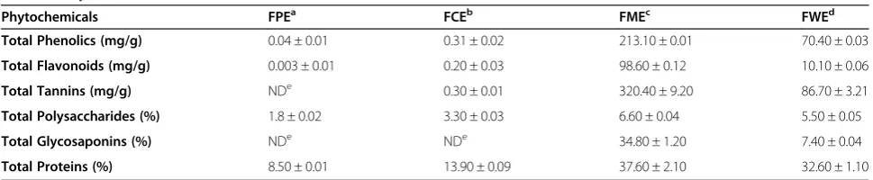
The results of quantitative analysis of total proteins, po-lysaccharides, glycosaponins, phenolics, flavonoids and tannins present in the extracts of F. deltoidea leaves is given in Table 2. The methanol extract of F. deltoidea
flavonoids, tannins, polysaccharides, glycosaponins and proteins.
HPLC analysis ofF. deltoidealeaf extracts
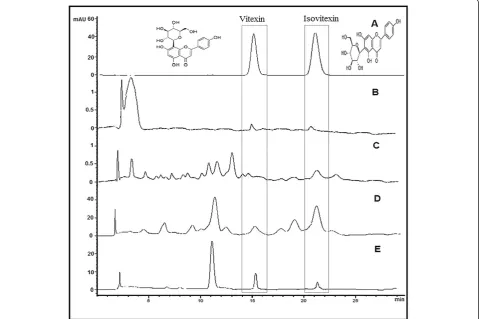
Vitexin and isovitexin in the F. deltoidealeaves extracts were detected and quantified by HPLC. The contents of vitexin and isovitexin varied between the different ex-tracts of the plant (Table 3). HPLC chromatograms of the extracts taken at 330 nm along with the mixed stan-dards are shown in Figure 1 the results indicated that the methoanolic extract showed higher contents of vitexin and isovitexin than compared to the other extracts.
Acute toxicity studies
Rats were orally treated with five different single doses of the extract and screened for signs of toxicity two weeks after administration. No treatment-related mor-tality was observed at all the tested doses. Throughout the 14 days observation period, no significant changes in behavior such as apathy, hyperactivity, morbidity, etc., were recorded in the treated animals. No abnormal
changes attributable to the treatment were noticed in body weights, respiration rate and heart rate. The present study demonstrated that the methanol extract ofF. deltoi-dea found to be safe at dose level of 5000 mg/kg and therefore, the LD50value for oral toxicity is considered to
be more than 5000 mg/kg.
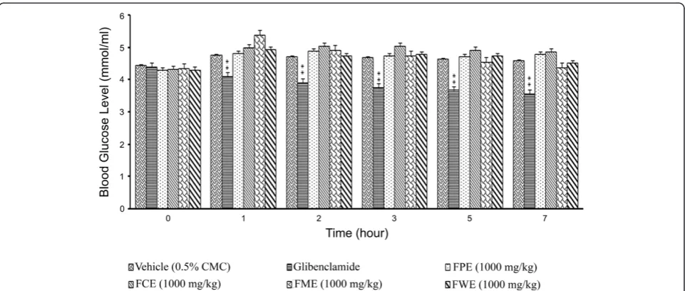
F. deltoidealeaf extracts failed to cause hypoglycemia in fasted rats but reduced hyperglycemia in glucose loaded rats
[image:5.595.62.538.100.309.2]Treatment of the animals with 1000 mg/kg of plant ex-tracts did not produce any significant hypoglycemic ef-fects (data not shown). Thus, the extracts will not cause blood glucose levels to fall below the standards required by the body. In oral glucose tolerance test, all tested plant extracts at a dose of 1000 mg/kg showed sig-nificant (p< 0.01) reduction in plasma glucose levels (Figure 2). Particularly, the methanol extract significantly (p< 0.01) reduced the plasma glucose level in a time-dependent manner. About 22.75% reduction of plasma glucose level was observed, as early as 15 min after Table 1 List of primers, the primer sequences and the primer melting temperature (Tm)
Gene Primer probe Sequence Annealing Tm°C
GK Forward 5′-CATATGTGCTCCGCAGGACTAG-3′ 61.7
Reverse 5′-CTTGTACACGGAGCCATCCA-3′
PEPCK Forward 5′-GCAACTTCTCTCGGCTCGTT-3′ 62.7
Reverse 5′-TGGCAGTTCTACTGGGCTACAC-3′
G6Pase Forward 5′-GGATCTACCTTGCGGCTCACT-3′ 62.7
Reverse 5′-TGTAGATGCCCCGGATGTG-3′
GLUT 2 Forward 5′-CATCAAAACGTAGAGCACGGTAA-3′ 63.4
Reverse 5′-TATGGGCATTTAGTCTGCACGTA-3′
GLUT4 Forward 5′-GCTTGGCTCCCTTCAGTTTG-3′ 63.4
Reverse 5′-CCTACCCAGCCAAGTTGCAT-3′
PPARγ Forward 5′-CATGAGTTCTTGCGCAGTATCC-3′ 63.4
Reverse 5′-AGAGCATTGAACTTGACAGCAAAC-3′
Β-actin Forward 5′-GCTCTGGCTCCTAGCACCAT-3′ 55
Reverse 5′-GCTGATCCACATCTGCTGGAA-3′
GK: Glucokinase, PEPCK: Phosphoenolpyruvate carboxykinase, G6Pase: Glucose-6-phosphatase, GLUT2: Glucose transporter type 2, GLUT4: Glucose transporter type 4, PPARγperoxisome proliferator-activated receptors-gamma.
Table 2 Phytochemical contents ofF. deltoidealeaf extracts
Phytochemicals FPEa FCEb FMEc FWEd
Total Phenolics (mg/g) 0.04 ± 0.01 0.31 ± 0.02 213.10 ± 0.01 70.40 ± 0.03
Total Flavonoids (mg/g) 0.003 ± 0.01 0.20 ± 0.03 98.60 ± 0.12 10.10 ± 0.06
Total Tannins (mg/g) NDe 0.30 ± 0.01 320.40 ± 9.20 86.70 ± 3.21
Total Polysaccharides (%) 1.8 ± 0.02 3.30 ± 0.03 6.60 ± 0.04 5.50 ± 0.05
Total Glycosaponins (%) NDe NDe 34.80 ± 1.20 7.40 ± 0.04
Total Proteins (%) 8.50 ± 0.01 13.90 ± 0.09 37.60 ± 2.10 32.60 ± 1.10 a
petroleum ether extract (FPE),b
chloroform extract (FCE),c
methanol extract (FME) andd
water extract (FWE).
e
[image:5.595.56.541.616.716.2]glucose administration, whereas the plasma glucose level was further reduced by up to 33.30% after 120 min of glucose administration.
Effect ofF. deltoidealeaf extracts on blood glucose in streptozitocin -induced diabetic rats
A preliminary screening test was carried out with the all the extracts at 1000 mg/kg. The result revealed that only methanol extract demonstrated significant (p < 0.01) anti-diabetic activity when compared to the negative control as well as the other test groups. The results showed that, the extract produced a marked reduction in fasting plas-ma glucose level in streptozitocin-induced diabetic rats. About 30.55% reduction was noticed after 3 days of oral administration of methanol extract (1000 mg/kg), whereas
it was further reduced to 60.19% by the end of the ex-periment (after 15 days treatment). The activity was sig-nificantly (p < 0.01) comparable to that of the standard reference, metformin 71.34%. Figure 3 shows the blood glucose levels in streptozitocin-induced diabetic rats over 15 days after the administration of different extracts ofF. deltoidealeaves.
Furthermore, treatment with methanol extract at 250 and 500 mg/kg resulted in a dose-dependent depletion in blood glucose level by 13.89 and 23.14%, respectively after 3 days of repeated administration of the extract in streptozitocin-induced diabetic rats (Figure 4). The re-duction in blood glucose levels was persisted throughout the period of treatment and after 15 days of treatment with methanol extract (250 and 500 mg/kg), the blood glucose level was further reduced by 41.38 and 62.63%, respectively.
Stimulatory effect of methanol extract ofF. deltoidea leaves on plasma insulin levels
[image:6.595.56.290.111.152.2]The initial and final levels of plasma circulating insulin are presented in Figure 5. Except the control, no effect was observed in the plasma insulin levels of the animals Table 3 Quantitative estimation of vitexin and isovitexin
(mg/g) in different extracts ofF. deltoidealeaf
Markers FPEa FCEb FMEc FWEd
Vitexin 0.69 ± 0.12 0.72 ± 0.08 6.29 ± 0.18 2.43 ± 0.02
Isovitexin 0.27 ± 0.09 0.41 ± 0.02 14.34 ± 0.26 0.98 ± 0.09 a
petroleum ether extract (FPE),b
chloroform extract (FCE),c
methanol extract (FME) andd
water extract (FWE).
[image:6.595.59.539.374.693.2]on the first day of treatment. The plasma insulin levels in streptozitocin-induced diabetic rats were significantly increased by 34.13, 86.01 and 115% in response to the treatment with methanol extract at concentrations of 250, 500 and 1000 mg/kg, respectively.
Effect of methanol extract ofF. deltoidealeaves on body weights
Figures 6 and 7 depicted the changes in the body weights of streptozitocin-induced diabetic rats administered with different doses of methanol extract (250, 500 and
1000 mg/kg). A significant effect of suppression in body weight gain was recorded after 9 days of treatment with 500 and 1000 mg/kg. However, the low dose (250 mg/kg) group of animals produced a significant weight loss only at the end of the experiment.
The effect of methanol extract on the expression of diabetic-associated gene transcripts in streptozitocin-induced diabetic rats
[image:7.595.58.539.88.292.2]The expression of diabetic-associated, glycolytic and glu-coneogenic genes, in the streptozitocin-induced diabetic
Figure 2The effect of oral administration ofF. deltoidealeaf extracts (1000 mg/kg) on the blood glucose levels of rats dosed with 1000 mg/kg of glucose.FPE = Petroleum ether extract; FCE = Chloroform extract; FME = Methanol extract and FEW = Water extract. The values are expressed as mean ± SEM (n = 6 in each group). **p< 0.01 compared to the control group (10 ml/kg vehicle).
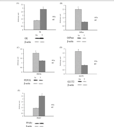
rats treated with methanol extract was determined using RT-PCR. As the loading control, the band density of each target gene was normalized to the band density of
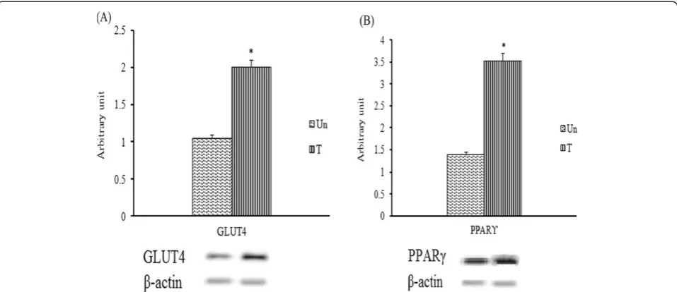
β-actin of the sample. Treatment with the methanol ex-tract significantly (p< 0.05) enhanced the expression of hepatic GK and PPARγtranscripts by 1.79 and 2.04-fold, respectively (Figure 8). Conversely, the treatment sig-nificantly (p< 0.05) reduced the expression of hepatic Glc-6-Pase, PEPCK and GLUT2 transcripts by 2.38-fold, 1.58-fold and 2.38-fold, respectively, compared to the untreated group. On the other hand, the expression of muscle GLUT4 and PPARγ showed altered expres-sion profiles in the streptozitocin-induced diabetic
rats following 2-weeks treatment of methanol extract. The expression of muscle GLUT4 transcript was sig-nificantly (p< 0.05) elevated by 1.9 fold compared to that of untreated group (Figure 9).
Discussions
[image:8.595.59.540.88.268.2]Regardless of the popularity of the traditional medicinal uses of F. deltoidea for diabetes related symptoms, very few studies have been reported to determine the molecu-lar mechanism and genes expression involve in the remed-ial effects of the herb. The reports on traditional uses of the herb to treat diabetes and the evidences that the plant is naturally gifted with abundance of antioxidant-rich
Figure 4The effect of daily oral administration ofF. deltoidealeaf methanol extract (FME) on the blood glucose levels of streptozitocin-induced diabetic rats.The methanol extract (FME) was administered to the animals at different doses (500 mg/kg or 250 mg/kg). The values are expressed as mean ± SEM (n = 6 in each group). *p< 0.05 and **p< 0.01 compared to the diabetic control group (10 ml/kg vehicle).
[image:8.595.58.540.499.686.2]bioactive constituents, have drawn attention toward hy-pothesizing thatF. deltoideacould be useful as a promis-ing remedy against diabetes if its efficacy is investigated and scientifically proven using the animal models with standard experimental protocols. Accordingly, the present study was designed to evaluate the acute and chronic anti-hyperglycemic effects ofF. deltoideaextracts in rats. The present study demonstrates that the methanol extract noticeably reduced the plasma glucose level in a time-dependent manner in oral glucose tolerance test conducted
in rats. Furthermore, the methanol extract significantly de-creased the fasting plasma glucose level and inde-creased the plasma insulin level in the streptozitocin-induced diabetic rats. In addition, the extract suppressed the body weight gain in the tested animals.
[image:9.595.58.540.89.280.2]Repeated (14 days) administration of the methanol ex-tract alleviated hyperglycemia and simultaneously in-creased plasma insulin levels in the streptozitocin-induced diabetic rats compared with those of the control diabetic rat. The antihyperglycemic activity of the methanol extract
Figure 6The effect of daily oral administration ofF. deltoidealeaf extracts (1000 mg/kg) on the body weight gain of streptozitocin-induced diabetic rats.FPE = Petroleum ether extract; FCE = Chloroform extract; FME = Methanol extract and FEW = Water extract. The values are expressed as mean ± SEM (n = 6 in each group). *p< 0.05 and **p< 0.01 compared to the diabetic control group (10 ml/kg vehicle).
[image:9.595.59.538.473.694.2]may partly due to its anti-oxidant and antagonistic effects against the toxic effects induced by streptozitocin [21,22]. The phytochemical analysis in the present study revealed that the methanol extract of F. deltoidea contains the highest content of flavonoids, phenolics, and tannins.
[image:10.595.59.536.87.617.2]HPLC analysis showed that the extract was particularly enriched with bioactive flavone C-glycosides, vitexin and isovitexin. The presences of flavonoids, which are efficient antioxidants, play a crucial role in cytoprotection and scavenging of free radicals, thereby protect the β-cells
from oxidative damage. Evidence of the C-glycosyl fla-vones in plants with antidiabetic properties and their effi-cacy in the treatment and prevention of diabetes has been extensively documented [21-24]. According to the Malaysian herbal monograph, vitexin and isovitexin are considered as the characteristic chemical markers of F. deltoidealeaves. In addition, it is reported that vitexin and isovitexin significantly reduced postprandial blood glucose level in mice and showedin vivoα-glucosidase inhibitory activity. Therefore, in the present study the activity of the extracts was correlated with quantitative estimation of the marker compounds. HPLC analysis revealed that quantity of both marker compounds was higher in methanol ex-tract than compared to the other exex-tracts ofF. deltoidea
leaves [25].
The antagonistic effects demonstrated by the methanol extract ofF. deltoideaagainst streptozitocin-induced hy-perglycemia were quite comparable with that of standard reference, metformin. Streptozitocin is a cytotoxic glu-cose analogue that is selectively absorbed by insulin pro-ducing β-cells through GLUT2 and it induces the death of β-cells by donating nitric oxide, generating reactive oxygen species and disrupting the actions of pancreatic antioxidant enzymes [26]. Pancreaticβ-cells, which store and release insulin, are the main regulator of glycogen-esis in muscle and liver. The absence of activeβ-cells is associated with a decrease in insulin level and an in-crease in peripheral insulin resistance, which results in excess glucose in the circulating blood. IP injection of streptozitocin (55 mg/kg) in this study effectively in-duced diabetes in normal rats. Streptozitocin injection produced a significant increase in the blood glucose level
and body weight and lowered the plasma insulin level in the rats after 72 hours. On the one hand, the methanol extract was observed to reduced hyperglycemia by sup-pressing hepatic gluconeogenesis through inducing the expression of PPARγ and GK, and on the other hand, the extract demonstrated remarkable decreased in ex-pression of the hepatic gluconeogenic genes PEPCK and Glc-6-Pase. These results have a similar effect as met-formin in the streptozitocin-induced diabetic rats [27]. Thus it is predicted that the blood-glucose lowering effect of the methanol extract of F. deltoidea could be attributed to the metformin-similar mechanism of sup-pression of hepatic glucose output [7,27,28]. Similar to metformin, the extract improved cellular sensitivity towards insulin by activating the insulin transduction pathway and consequently, the regulation of glucose absorption and metabolism in peripheral tissues [29,30]. Similarly, repeated treatment with the methanol extract of
[image:11.595.59.538.89.296.2]F. deltoidea in streptozitocinduced diabetic rats in-creased the mRNA transcript level of GLUT4 in muscles. Additionally, reduction of mRNA transcripts of hepatic Glc-6-Pase and PEPCK was also noticed in the same group of streptozitocin-induced diabetic rats. Interest-ingly, the expression of the glucose transporter Glut2 in liver which is insulin-independent was found to be re-duced in the extract treated streptozitocin-inre-duced dia-betic rats. The decrease in GLUT2 expression is related to a decrease in hepatic glucose output and plays a critical role in glucose homeostasis [31]. GLUT2 is the sole trans-porter that specifically transports streptozotocin in pan-crease [32]. Thus it could be postulated that methanol extract of F. deltoidea besides being strong antioxidant,
obstructs the cytotoxic effect of streptozitocin on islet
β-cells of pancreas by inhibiting GLUT2 gene expression. Although several preliminary studies on antidiabetic effects ofF. deltoieahave been carried out, the mechan-ism of action at genetic level have yet to be investigated. PPARγ has been reported to regulate the expression of genes involved in glucose metabolism [33]. In the pre-sent study, it was observed that the methanol extract en-hanced PPARγexpression in the liver of diabetic rats. In turn, PPARγcan directly activate hepatic GK expression. GK works as the glucose sensor in glucose homeostasis by phosphorylating glucose to glucose-6-phosphate, is the first step for both glycogen synthesis and glycolysis [34]. In the present study, administration of theF. deltoi-deamethanol extract increased the expression of hepatic GK and therefore, it can improve excess glucose toler-ance. There is a report that, over expression of hepatic GK reduces blood glucose in the body [35]. Similarly, a research study showed that the activity of Glc-6-Pase was increased in streptozitocin-induced diabetic rats [36]. In this study, Glc-6-Pase activity was markedly in-creased in the streptozitocin-induced diabetic rats when compared to that of normal control. However, the tran-script level of Glc-6-Pase was significantly decreased after administration of the methanol extract, which pro-bably due to the reduced extent of gluconeogenesis. This clearly indicates that methanol extract of F. deltoidea
produces anti-hyperglycemia in the animal model by pro-moting glucose utilization and suppressing the glucose production [37].
Conclusions
In conclusion, our results provide novel mechanisms for the plasma glucose-lowering action of methanol extract of
F. deltoidea. The extract produced its anti-hyperglycemic effect via increased secretion of insulin. Further it is con-firmed that the extract suppressed the transcription of genes involved in hepatic glucose production, such as phosphoenolpyruvate carboxykinase (PEPCK) and Glc-6-Pase. In addition, the extract stimulated the hepatic GK and PPARγgene expression, which consequently leads to an increased expression of GLUT-4 gene expression in skeletal muscles of streptozitocin-induced diabetic rats. Altogether, the extract potentially displayed antidiabetic activity by inhibiting hepatic glucose production and pro-moting glucose utilization. The extract also nullifies the hyperglycemic effects of streptozitocin which was ob-served through the reduced expression of GLUT-2 gene. Altogether, it can be concluded that the methanol extract ofF. deltoideahas the potential to be developed as an an-tidiabetic agent.
Competing interests
The authors declare no financial or commercial conflicts of interest.
Authors’contributions
MZA, MBKA, and EF designed the experiments. EF and SYH carried out the phytochemical analysis. EF and SYH performed thein vivostudies. EF, MBKA, MZA and MYF participated in gene analysis and drafted the manuscript. EF, BKA and MA analyzed all data and interpreted the results. All authors read and approved the final manuscript.
Acknowledgments
The authors would like to thank the Universiti Sains Malaysia for providing a grant to support this work (Grant No: 1001/PFARMASI/833016). The first author was supported by a USM Graduate Assistant fellowship from the Institute of Postgraduate Studies (IPS). This work was also partly supported by a Fundamental Research Grant Scheme (FRGS) Fasa 1/2008 (Grant No. 203/CIPPM/6711119) from the Ministry of Higher Education (MoHE).
Author details
1
Department of Pharmacology, School of Pharmaceutical Sciences, Universiti Sains Malaysia, 11800 Penang, Malaysia.2Department of Physiology, School of Pharmaceutical Sciences, Universiti Sains Malaysia, 11800 Penang, Malaysia.
Received: 11 September 2013 Accepted: 25 June 2014 Published: 4 July 2014
References
1. Eliza J, Daisy P, Ignacimuthu S, Duraipandiyan V:Antidiabetic and antilipidemic effect of eremanthin fromCostus speciosus(Koen.) Sm., in STZ-induced diabetic rats.Chem Biol Interact2009,182(1):67–72. 2. Rose AJ, Richter EA:Skeletal muscle glucose uptake during exercise: how
is it regulated?Physiology2005,20(4):260–270.
3. Postic C, Dentin R, Girard J:Role of the liver in the control of
carbohydrate and lipid homeostasis.Diabetes Metabol2004,30(5):398–408. 4. Engström L:The regulation of liver pyruvate kinase by
phosphorylation-dephosphorylation.Curr Top Cell Regul1978,13:28–51.
5. Granner D, Pilkis S:The genes of hepatic glucose metabolism.J Biol Chem
1990,265(18):10173–10176.
6. Postic C, Burcelin R, Rencurel F, Pegorier JP, Loizeau M, Girard J, Leturque A: Evidence for a transient inhibitory effect of insulin on GLUT2 expression in the liver: studiesin vivoandin vitro.Biochem J1993,293(Pt 1):119–124. 7. Desvergne B, Wahli W:Peroxisome proliferator-activated receptors:
nuclear control of metabolism.Endocr Rev1999,20:649–688. 8. Bhathena SJ, Velasquez MT:Beneficial role of dietary phytoestrogens in
obesity and diabetes.Am J Clin Nutr2002,76(6):1191–1201.
9. Cheng AY, Fantus IG:Oral antihyperglycemic therapy for type 2 diabetes mellitus.Can Med Assoc J2005,172(2):213–226.
10. Farsi E, Shafaei A, Hor SY, Ahamed MBK, Yam MF, Idress H:Correlation between enzymes inhibitory effects and antioxidant activities of standardized fractions of methanolic extract obtained fromFicus deltoidealeaves.Afr J Biotechnol2011,10(67):15184–15194. 11. Abdulla MA, Ahmed KA-A, Abu-Luhoom FM, Muhanid M:Role ofFicus
deltoideaextract in the enhancement of wound healing in experimental rats.Biomed Res2010,21(3):241–245.
12. Sulaiman M, Hussain M, Zakaria Z, Somchit M, Moin S, Mohamad A, Israf D: Evaluation of the antinociceptive activity ofFicus deltoideaaqueous extract.Fitoterapia2008,79(7):557–561.
13. Adam Z, Hamid M, Ismail A, Khamis S:Effect ofFicus deltoideaextracts on hepatic basal and insulin-stimulated glucose uptake.J Biol Sci2009, 9(8):796–803.
14. Aminudin N, Sin CY, Chee ES, Nee KI, Renxin L:Blood glucose lowering effect ofFicus deltoideaaqueous extract.Malays J Sci2007,26(1):73–78. 15. Misbah H, Aziz AA, Aminudin N:Antidiabetic and antioxidant properties
ofFicus deltoideafruit extracts and fractions.BMC Compl Alternative Med
2013,13(1):118.
16. Farsi E, Shafaei A, Hor SY, Ahamed MBK, Yam MF, Asmawi MZ, Ismail Z: Genotoxicity and acute and subchronic toxicity studies of a standardized methanolic extract ofFicus deltoidealeaves.Clinics2013,68(6):865–875. 17. Fu Y, Zu Y, Liu W, Hou C, Chen L, Li S, Shi X, Tong M:Preparative
separation of vitexin and isovitexin from pigeonpea extracts with macroporous resins.J Chrom2007,1139(2):206–213.
18. Jaiswal D, Kumar Rai P, Kumar A, Mehta S, Watal G:Effect ofMoringa oleiferaLam. leaves aqueous extract therapy on hyperglycemic rats.
19. Shirwaikar A, Rajendran K, Barik R:Effect of aqueous bark extract of
Garuga pinnataRoxb. in streptozotocin-nicotinamide induced type-II diabetes mellitus.J Ethnopharmacol2006,107(2):285–290.
20. Eidi A, Eidi M:Antidiabetic effects of sage (Salvia officinalisL.) leaves in normal and streptozotocin-induced diabetic rats.Diabetes Metab Syndr Clin Res Rev2009,3(1):40–44.
21. Jayaprakasam B, Vareed SK, Olson LK, Nair MG:Insulin secretion by bioactive anthocyanins and anthocyanidins present in fruits.J Agr Food Chem2005,53(1):28–31.
22. Pinent M, Blay M, Blade M, Salvado M, Arola L, Ardevol A:Grape seed-derived procyanidins have an antihyperglycemic effect in streptozotocin-induced diabetic rats and insulinomimetic activity in insulin-sensitive cell lines.Endocrinology2004,145(11):4985–4990. 23. Sezik E, Aslan M, Yesilada E, Ito S:Hypoglycaemic activity ofGentiana
olivieriand isolation of the active constituent through bioassay-directed fractionation techniques.Life Sci2005,76(11):1223–1238.
24. Dos Santos R, Dos Santos M, Schenkel E:Analysis of the plant drug Wilbrandia ebracteata (Cogn.) Cogn.Pharmaceut Biol1996,34(4):300–302. 25. Choo CY, Sulong NY, Man F, Wong TW:Vitexin and isovitexin from the
leaves ofFicus deltoideawithin vivoaglucosidase inhibition.
J Ethnopharmacol2012,142(3):776–781.
26. He Y, Martinez-Fleites C, Bubb A, Gloster TM, Davies GJ:Structural insight into the mechanism of streptozotocin inhibition ofO-GlcNAcase.
Carbohydr Res2009,344(5):627–631.
27. Meyer F, Ipaktchi M, Clauser H:Specific inhibition of gluconeogenesis by biguanides.Diabetes2008,57(2):306–314.
28. Radziuk J, Zhang Z, Wiernsperger N, Pye S:Effects of metformin on lactate uptake and gluconeogenesis in the perfused rat liver.Diabetes1997, 46(9):1406–1413.
29. Kahn S:The relative contributions of insulin resistance and beta-cell dysfunction to the pathophysiology of type 2 diabetes.Diabetologia
2003,46(1):3–19.
30. Stith B, Goalstone M, Espinoza R, Mossel C, Roberts D, Wiernsperger N:The antidiabetic drug metformin elevates receptor tyrosine kinase activity and inositol 1, 4, 5-trisphosphate mass in Xenopus oocytes.Endocrinology
1996,137(7):2990–2999.
31. Roden M, Bernroider E:Hepatic glucose metabolism in humans—its role in health and disease.Best Pract Res Clin Endocrinol Metabol2003, 17(3):365–383.
32. Schnedl WJ, Ferber S, Johnson JH, Newgard CB:STZ transport and cytotoxicity: specific enhancement in GLUT2-expressing cells.Diabetes
1994,43(11):1326–1333.
33. Kim J-S, Kwon C-S, Son KH:Inhibition of alpha-glucosidase and amylase by luteolin, a flavonoid.Biosci Biotechnol Biochem2000,64(11):2458–2461. 34. Brocklehurst K, Davies R, Agius L:Differences in regulatory properties
between human and rat glucokinase regulatory protein.Biochem J2004, 378:693–697.
35. Ferre T, Riu E, Bosch F, Valera A:Evidence from transgenic mice that glucokinase is rate limiting for glucose utilization in the liver.FASEB J
1996,10(10):1213–1218.
36. Reaven GM:Role of insulin resistance in human disease.Diabetes1988, 37(12):1595–1607.
37. Arion WJ, Wallin BK, Lange AJ, Ballas LM:On the involvement of a glucose 6-phosphate transport system in the function of microsomal glucose 6-phosphatase.Mol Cell Biochem1975,6(2):75–83.
doi:10.1186/1472-6882-14-220
Cite this article as:Farsiet al.:Standardized extract ofFicus deltoidea
stimulates insulin secretion and blocks hepatic glucose production by regulating the expression of glucose-metabolic genes in streptozitocin-induced diabetic rats.BMC Complementary and Alternative Medicine
201414:220.
Submit your next manuscript to BioMed Central and take full advantage of:
• Convenient online submission
• Thorough peer review
• No space constraints or color figure charges
• Immediate publication on acceptance
• Inclusion in PubMed, CAS, Scopus and Google Scholar
• Research which is freely available for redistribution