Investigating Calcium Channel Blockers as
Antimalarials
May Rajab
School of Environment and Life Sciences
University of Salford
Salford, UK
ii
Declaration
I certify that this thesis, submitted to the University of Salford in partial fulfilment of the
requirements for a Degree of Doctor of Philosophy, is a presentation of my own research work
and has been funded by the University of Salford’s Pathway to Excellence studentship. The
content of this thesis has not been submitted for any degree or other purpose at this or any
other university.
Although the author has carried out most experiments and analysis of this research, some
parts of this thesis were carried out in collaboration with other colleagues. Wherever
contributions of others are involved, every effort is made to indicate this clearly with due
reference to the literature and acknowledgment of collaborative research and discussions. Dr
Steve Rossington carried out the synthesis of some of the fendiline analogues which have
iii
Table of contents
Declaration ... ii
Table of contents ... iii
List of figures ... viii
List of tables ... x
Acknowledgments ... xi
Abbreviations ... xiii
Abstract ... xvii
Chapter 1 ... 1
Introduction ... 1
1.1 Global impact of malaria ... 1
1.2 Biology of the parasite ... 2
1.3 Clinical symptoms of malaria ... 4
1.4 Malaria control and prevention ... 5
1.4.1 Vector control ... 6
1.4.2 Vaccines ... 7
1.4.3 Prophylaxis ... 8
1.5 Malaria chemotherapy ... 9
1.5.1 Quinoline derivatives ... 9
1.5.2 Antifolates ... 12
DHFR Inhibitors ... 13
DHPS Inhibitors ... 15
1.5.3 Artemisinins ... 16
1.5.4 Antibiotics and antiparasitic drugs ... 18
1.6 Antimalarial drug discovery ... 20
1.6.1 Traditional drug development ... 20
1.6.2 Drug repositioning ... 22
1.7 Calcium and calmodulin ... 23
1.7.1 Importance of calcium ... 23
1.7.2 Importance of calmodulin ... 26
iv
1.8.1 Voltage-gated calcium channels ... 28
1.8.2 Calcium channel blockers ... 30
1.8.3 Fendiline ... 32
1.9 Aims and Objectives ... 34
Chapter 2 ... 36
Materials and methods ... 36
2.1 In vitro cultivation of P. falciparum ... 36
2.1.1 Parasite strains: ... 36
2.1.2 Complete media: ... 36
2.1.3 Wash media: ... 36
2.1.4 Processing human RBCs for parasite culture ... 37
2.1.5 Maintaining parasite culture ... 37
2.1.6 Estimating parasitaemia... 37
2.1.7 Preservation of parasites in liquid nitrogen ... 38
2.1.8 Thawing of parasites from liquid nitrogen ... 38
2.1.9 Culture synchronisation ... 38
2.1.10 Percoll purification ... 39
2.2 Drug susceptibility assays ... 39
2.2.1 Optimising the haematocrit levels of SG plate reader assay ... 39
2.2.2 Optimisation of media levels for SG plate reader assay ... 40
2.2.3 Dose-response assay ... 41
2.2.4 Adopted SG plate reader method ... 41
2.2.5 SG flow cytometer method ... 42
2.2.6 Validating the SG plate reader method ... 42
2.3 Plate reader optimisation results ... 43
2.3.1 Optimising the haematocrit levels of SG plate reader assay ... 43
2.3.2 Optimisation of media levels for SG plate reader assay ... 44
2.3.3 Validating the SG plate reader method ... 44
2.4 Cytotoxicity test on mammalian cells ... 46
2.4.1 Cell culture media ... 46
2.4.2 Cultivation of HepG2 cells ... 46
2.4.3 MTT (Methylthiazol tetrazolium) assay ... 46
2.4 Drug Stocks ... 47
v
Chapter 3 ... 49
Investigating the antimalarial efficacy of fendiline and combinatorial regimes with commercially available drugs ... 49
3.1 Introduction ... 49
3.1.1 Combination therapy ... 49
3.1.2 Chloroquine reversal drugs ... 53
3.1.3 Aims... 55
3.2 Methods ... 55
3.2.1 Dose-response assay for IC50 determination ... 55
3.2.2 CalcuSyn combination assay for malaria ... 55
3.2.3 Chloroquine potentiation assay ... 56
3.2.4 Cytotoxicity assay ... 56
3.3 Results ... 57
3.3.1 Dose-response assay ... 57
3.3.2 CalcuSyn combination assay for malaria ... 58
Validating the combination assay using an ATQ-PG control ... 58
Combination of fendiline with existing antimalarial drugs ... 60
Combination of fendiline with existing calcium channel blockers ... 67
3.3.3 Chloroquine potentiation assay ... 71
3.3.4 Cytotoxicity assay ... 72
3.4 Discussion ... 73
3.4.1 CalcuSyn combination assay ... 73
3.4.2 Chloroquine potentiation assay ... 75
3.4.3 Conclusion ... 78
Chapter 4 ... 79
Synthesis and evaluation of fendiline analogues ... 79
4.1 Introduction ... 79
4.1.1 Historical perspective ... 79
4.1.2 Synthesis of fendiline analogues ... 83
Organolithium chemistry and ozonolysis... 85
Current procedure: Palladium coupling ... 86
Reductive amination ... 87
4.1.3. Aims... 88
vi
4.2.1 General Experimental ... 88
4.2.2 Synthesis of the aldehyde via Palladium coupling ... 89
4.2.3 Reductive amination ... 91
4.2.4 Antimalarial activity ... 97
4.2.5 Cellular toxicity... 97
4.2.6 hERG channel inhibition assay ... 97
4.2.7 Time-course analysis ... 98
4.2.8 Stage-specificity assay ... 98
4.3 Results ... 99
4.3.1 Screening ... 99
4.3.2 hERG channel inhibition assay ... 102
4.3.3 Time-course analysis ... 102
4.3.4 Stage-specificity assay ... 103
4.4 Discussion ... 104
4.4.1 Synthesis of fendiline analogues ... 104
4.4.2 Screening ... 105
4.4.3 hERG channel inhibition assay ... 107
4.4.4 Time course analysis and stage-specificity assay ... 108
4.4.5 Conclusion and future work ... 110
Chapter 5 ... 112
Optimising a flow cytometry-based calcium fluctuation assay ... 112
5.1 Introduction ... 112
5.1.1 Calcium regulation in mammalian cells vs malaria parasites ... 112
5.1.3 Methods to detect intracellular calcium ... 115
5.1.3 Aims... 117
5.2 Experimental ... 118
5.2.1 Optimisation of a Percoll separation method ... 118
5.2.2 Fluo-8 staining of RBCs ... 120
5.2.3 Optimisation using long-wavelength DNA fluorescent dyes ... 121
5.2.4 Comparison between Fluo-8, TOTO-3 and SG via Percoll purification ... 123
5.2.5 Comparison between Fluo-8, TOTO-3 and SG in a serially diluted culture ... 125
5.2.6 DRAQ5 staining of RBCs ... 127
5.2.7 Dual staining with Fluo-8 and DRAQ5 ... 128
vii
5.2.9 Measuring variances in calcium concentrations ... 133
5.2.10 Measuring calcium fluctuations using flow cytometry ... 134
5.2.11 Measuring calcium fluctuations in non-infected blood ... 139
5.2.12 Measuring calcium fluctuations in saponin treated cultures ... 140
5.3 Discussion ... 141
5.3.1 Percoll separation gradient ... 141
5.3.2 Fluorescent dyes ... 142
Fluo-8 calcium dye ... 142
Long-wavelength DNA dye ... 143
5.3.3 Detecting calcium fluctuations ... 143
5.3.5 Conclusion and future work ... 151
Chapter 6 ... 152
General Discussion ... 152
6.1 General discussion ... 152
Appendix I ... 156
Appendix II ... 174
Appendix III ... 176
viii
List of figures
FIGURE 1.1- ESTIMATED NUMBER OF MALARIA CASES (A) AND DEATHS (B) BETWEEN 2000 AND 2015………....2
FIGURE 1.2- A SCHEMATIC SHOWING THE LIFE CYCLE OF THE PLASMODIUM PARASITE...4
FIGURE 1.3- AN OVERVIEW OF THE MAIN AREAS OF RESEARCH THAT ARE ONGOING TO CONTROL THE SPREAD OF MALARIA...6
FIGURE 1.4- CHEMICAL STRUCTURES OF QUININE AND SOME OF ITS ANALOGUES...12
FIGURE 1.5- A BRIEF SCHEMATIC OF THE FOLATE PATHWAY, HIGHLIGHTING THE SITES OF ACTION OF THE ANTI-FOLATE DRUGS...13
FIGURE 1.6- CHEMICAL STRUCTURES OF PYRIMETHAMINE AND THE TWO PRODRUGS PROGUANIL AND CHLORPROGUANIL ALONG WITH THEIR ACTIVE FORMS………...………...14
FIGURE 1.7– CHEMICAL STRUCTURE OF ARTEMISININ AND SOME OF ITS SYNTHETIC ANALOGUES………...16
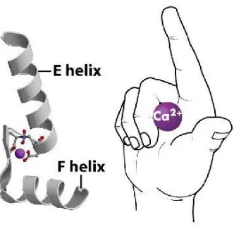
FIGURE 1.8- A SCHEMATIC SHOWING THE EF HAND MOTIF OF CALMODULIN……….27
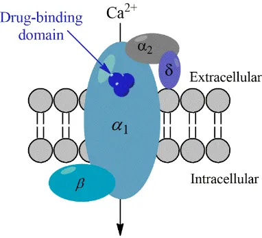
FIGURE 1.9- SCHEMATIC OF A VOLTAGE GATED CALCIUM CHANNEL AND ITS SUBUNITS WITHIN A CELL MEMBRANE...29
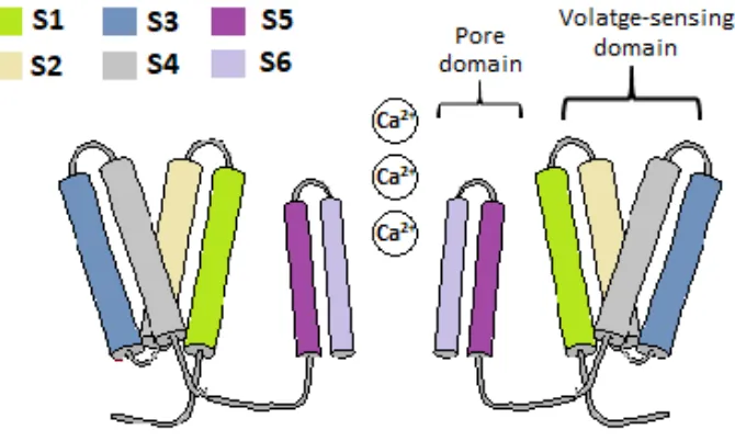
FIGURE 1.10- SCHEMATIC SHOWING THE DIFFERENT SEGMENTS WITHIN THE α1 SUBUNIT OF VOLTAGE GATED ION CHANNELS...30
FIGURE 1.11- MOLECULAR STRUCTURE OF FENDILINE...32
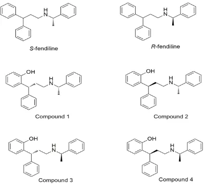
FIGURE 1.12- MOLECULAR STRUCTURES OF S- AND R-FENDILINE ALONG WITH FOUR HYDROXY ANALOGUES...33
FIGURE 2.1- INITIAL OPTIMISATION RESULTS OF INFECTED RBCS DILUTED IN DIFFERENT MEDIUMS WITH VARYING HAEMATOCRIT LEVELS...43
FIGURE 2.2- COMPARISON BETWEEN THE PLATE READER RESULTS AND THE FLOW CYTOMETER DATA...44
FIGURE 2.3 - COMPARISON OF RESULTS OBTAINED USING FLOW CYTOMETRY, PLATE READER AND GIEMSA-STAINED MICROSCOPE SLIDES...45
FIGURE 3.1- EXAMPLE OF AN ISOBOLOGRAM...52
FIGURE 3.2 - RESULTS OF THE POSITIVE CONTROL COMBINATION ASSAY BETWEEN ATOVAQUONE (ATQ) AND PROGUANIL (PG), INCUBATED AT A CONSTANT RATIO (1:7500)…...59
FIGURE 3.3- THE CALCUSYN-BASED COMBINATION ASSAY BETWEEN FENDILINE (FHCL) AND ARTEMETHER (ART)...61
FIGURE 3.4- THE CALCUSYN-BASED COMBINATION ASSAY BETWEEN FENDILINE AND ATOVAQUONE (ATQ)...62
FIGURE 3.5- THE CALCUSYN-BASED COMBINATION ASSAY BETWEEN FENDILINE AND CHLOROQUINE (CQ)...63
FIGURE 3.6- THE CALCUSYN-BASED COMBINATION ASSAY BETWEEN FENDILINE AND DOXYCYCLINE (DOX)...64
FIGURE 3.7- THE CALCUSYN-BASED COMBINATION ASSAY BETWEEN FENDILINE AND MEFLOQUINE (MEF)...65
FIGURE 3.8- THE CALCUSYN-BASED COMBINATION ASSAY BETWEEN FENDILINE AND PROGUANIL (PG)...66
FIGURE 3.9- THE CALCUSYN-BASED COMBINATION ASSAY BETWEEN FENDILINE AND DILTIAZEM (DIL)...68
FIGURE 3.10- THE CALCUSYN-BASED COMBINATION ASSAY BETWEEN FENDILINE AND NICARDIPINE (NIC)...69
FIGURE 3.11- THE CALCUSYN-BASED COMBINATION ASSAY BETWEEN FENDILINE AND VERAPAMIL (VP)...70
FIGURE 3.12- RESULTS OF THE CHLOROQUINE POTENTIATION ASSAY OF VERAPAMIL AND FENDILINE...72
ix
FIGURE 4.1- EXAMPLES OF SEMI-SYNTHETIC ARTEMISININ DERIVATIVES...80
FIGURE 4.2- AN EXAMPLE OF ONE OF THE ARTEMISININ ANALOGUES SYNTHESISED BY MODIFYING THE PEROXY GROUP...81
FIGURE 4.3- TWO ARTEMISININ DERIVED COMPOUNDS TESTED AGAINST CANCER CELLS...82
FIGURE 4.4-THE TWO SYNTHETIC ROUTES UTILISED TO PRODUCE THE FENDILINE ANALOGUES...84
FIGURE 4.5- SCHEMATIC OF THE SYNTHETIC ROUTE FOLLOWED IN THE SYNTHESIS OF THE INTERMEDIATE ALDEHYDE...86
FIGURE 4.6- THE PROPOSED MECHANISM OF THE PALLADIUM COUPLING REACTION...87
FIGURE 4.7- THE REDUCTIVE AMINATION REACTION FOR THE SYNTHESIS OF FENDILINE...88
FIGURE 4.8- PRODUCTS OBTAINED FROM THE PALLADIUM COUPLING REACTIONS...90
FIGURE 4.9- THE FENDILINE ANALOGUE (4C) TAKEN FORWARD FOR FURTHER INVESTIGATION...102
FIGURE 4.10- GRAPH SHOWING THE RESULTS OF THE HERG INHIBITION ASSAY...102
FIGURE 4.11- DOSE-RESPONSE OF COMPOUND 4C...103
FIGURE 4.12- RESULTS OF THE STAGE-SPECIFICITY ASSAY...104
FIGURE 5.1- SCHEMATIC OF THE DIFFERENT CALCIUM CHANNELS THAT CAN BE FOUND WITHIN A MAMMALIAN CELL...113
FIGURE 5.2- GIEMSA-STAINED MICROSCOPE SLIDE IMAGES OF THE CULTURES PRIOR TO (A) AND AFTER (B) PERCOLL GRADIENT SEPARATION...119
FIGURE 5.3- GIEMSA-STAINED SLIDES OF THE CULTURES BEFORE (A) AND AFTER (B) PERCOLL PURIFICATION...125
FIGURE 5.4- RESULTS OF THE SERIAL DILUTION CARRIED OUT USING THE THREE FLUORESCENT DYES (SG,TOTO-3 AND FLUO-8)....126
FIGURE 5.5- DENSITY GRAPHS VISUALISED ON THE APC-CY7-A CHANNEL DISPLAYING INFECTED AND NON-INFECTED RBCS STAINED WITH DRAQ5...127
FIGURE 5.6- COMPARISON OF PARASITAEMIA ESTIMATION BETWEEN TWO DNA BINDING FLUORESCENT DYES, SG AND DRAQ5, VIA SERIAL DILUTION...128
FIGURE 5.7- GATING STRATEGY USED TO VISUALISE CALCIUM CHANGES WITHIN AN INFECTED CULTURE...131
FIGURE 5.8- ANALYSIS OF VARYING CALCIUM CONCENTRATIONS ON INFECTED RBCS...133
FIGURE 5.9- CALCIUM FLUX EXPERIMENTS IN RESPONSE TO THE CALCIUM IONOPHORE A23187 AND EDTA ON INFECTED RBCS...135
FIGURE 5.10- CALCIUM FLUCTUATION EXPERIMENTS CARRIED OUT ON P. FALCIPARUM INFECTED RBCS AT BOTH RING (CIRCLES) AND LATE TROPHOZOITE/SCHIZONT (SQUARES) STAGES...137
FIGURE 5.11- GRAPHICAL ANALYSIS OF CALCIUM FLUX IN RESPONSE TO BOTH A) FPL 64176(FPL) AND B) BAY K8644(BAY)…...138
FIGURE 5.12- RESULTS OF THE SOLVENT CONTROL EXPERIMENTS...139
FIGURE 5.13- CALCIUM FLUX EXPERIMENTS IN RESPONSE TO THE CALCIUM IONOPHORE A23187 ON BLOOD ONLY SAMPLES...140
x
List of tables
TABLE 2.1- COMPARISON OF THREE IC50 VALUES OBTAINED USING DIFFERENT METHODS...45
TABLE 3.1- INTERPRETATION OF THE CI VALUES THAT ARE PRODUCED BY THE CALCUSYN SOFTWARE...56
TABLE 3.2- IC50 VALUES OF CURRENT ANTIMALARIAL DRUGS AND CCBS AGAINST P. FALCIPARUM K1 STRAIN PARASITES ALONG WITH PUBLISHED VALUES...57
TABLE 3.3- THE COMBINATION INDEX (CI) VALUES FOR THE CONTROL EXPERIMENT BETWEEN ATOVAQUONE AND PROGUANIL...59
TABLE 3.4- THE CI VALUES DETERMINED BY THE CALCUSYN SOFTWARE FOR THE COMBINATION OF FENDILINE AND ARTEMETHER...61
TABLE 3.5- THE CI VALUES DETERMINED BY THE CALCUSYN SOFTWARE FOR THE COMBINATION OF FENDILINE AND ATOVAQUONE...62
TABLE 3.6- THE CI VALUES DETERMINED BY THE CALCUSYN SOFTWARE FOR THE COMBINATION OF FENDILINE AND CHLOROQUINE...63
TABLE 3.7- THE CI VALUES DETERMINED BY THE CALCUSYN SOFTWARE FOR THE COMBINATION OF FENDILINE AND DOXYCYCLINE...64
TABLE 3.8- THE CI VALUES DETERMINED BY THE CALCUSYN SOFTWARE FOR THE COMBINATION OF FENDILINE AND MEFLOQUINE...65
TABLE 3.9- THE CI VALUES DETERMINED BY THE CALCUSYN SOFTWARE FOR THE COMBINATION OF FENDILINE AND PROGUANIL...66
TABLE 3.10- THE CI VALUES DETERMINED BY THE CALCUSYN SOFTWARE FOR THE COMBINATION OF FENDILINE AND DILTIAZEM...68
TABLE 3.11- THE CI VALUES DETERMINED BY THE CALCUSYN SOFTWARE FOR THE COMBINATION OF FENDILINE AND NICARDIPINE...69
TABLE 3.12- THE CI VALUES DETERMINED BY THE CALCUSYN SOFTWARE FOR THE COMBINATION OF FENDILINE AND VERAPAMIL...70
TABLE 3.13 - SUMMARY OF THE CI VALUES OF ALL COMBINATION ASSAYS CARRIED OUT BETWEEN FENDILINE AND EXISTING ANTIMALARIALS AND CCBS...71
TABLE 3.14- IC50 VALUES OF CHLOROQUINE ALONE AND IN COMBINATION WITH VERAPAMIL OR FENDILINE...72
TABLE 4.1-PD(OAC)2 CATALYSED CONJUGATE ADDITIONS OF ARYLBORONIC ACIDS TO TRANS-CINAMMALDEHYDE...90
TABLE 4.2- ANTI-PLASMODIAL ACTIVITY (P. FALCIPAUM K1 STRAIN) AND HEPG2 CYTOTOXICITY OF THE FIRST SERIES OF FENDILINE ANALOGUES...100
TABLE 4.3- ANTIMALARIAL ACTIVITY AND HEPG2 CYTOTOXICITY OF THE SECOND SERIES OF FENDILINE ANALOGUES...101
TABLE 4.4- IC50 VALUESOF COMPOUND 4C OBTAINED AT THREE DIFFERENT TIME POINTS...103
TABLE 5.1- RESULTS OF THE PERCOLL GRADIENT SEPARATION...119
TABLE 5.2- COMPARISON OF UNSTAINED AND FLUO-8 STAINED RBCS (INFECTED AND NON-INFECTED)...120
TABLE 5.3- COMPARISON BETWEEN SG AND A RANGE OF LONG-WAVELENGTH DNA BINDING FLUORESCENT DYES...122
TABLE 5.4 - DENSITY GRAPHS SHOWING SG, TOTO-3 AND FLUO-8 STAINED INFECTED RBCS BEFORE AND AFTER PERCOLL PURIFICATION...124
TABLE 5.5- COMPARISON BETWEEN DIFFERENT DYES OF THE FOLD-INCREASE DETECTED IN PARASITE POPULATION BEFORE AND AFTER PERCOLL GRADIENT SEPARATION...125
TABLE 5.6- IMAGE SHOWING THE GATED POPULATION OF INFECTED CELLS (SG AND TOTO-3) AND THE PRESUMED TO BE INFECTED POPULATION STAINED WITH FLUO-8 LABELLED (P2)...126
TABLE 5.7 - FLOW CYTOMETRY PRODUCED DENSITY GRAPHS DISPLAYING THE APC-CY7-A (DNA)/FITC-A(CALCIUM) FILTERS OF AN ONGOING CULTURE BEFORE AND AFTER PERCOLL TREATMENT...129
TABLE 5.8- FLOW CYTOMETRY PRODUCED IMAGES AND GIEMSA-STAINED SLIDES OF AN ONGOING CULTURE AT DIFFERENT TIME POINTS...132
xi
Acknowledgments
Many people have made this research possible and I would like to take the time to thank
everyone. I would like to acknowledge the University of Salford’s Pathway to Excellence
studentship that funded this research and the PGR Fund to attend conferences. Initial thanks
go to my supervisor Niroshini. Without you this research simply would not have been possible.
Thank you for choosing me to do this project and for your guidance along the way.
Jim, where do I begin. I could dedicate the entire acknowledgements section to you for all
your support, help, encouragement and, most importantly, your belief in me. The past nine
years have been a rollercoaster ride; from telling you I was leaving to do a degree in Physics
in 2009 to standing in your office complaining about how much I hated the forever-breaking
flow cytometer a few months ago. Your patience and diplomacy are astounding. You never
doubted me, and for that I can’t thank you enough. I’ve said this before and I truly mean it, I
will forever be in your debt.
Steve, without you this research surely would not have happened. I owe a lot of my lab skills
to you. You’re a generous man with a good heart, even though most people think you are a
phantom!
Gemma Lace, you’re an absolute star! You have done right by me many times over the years.
I won’t forget your words of advice or your encouraging comments, nor will I forget the little
diagram you drew showing me how far I had come in my academic life. That really did put it
all into perspective.
A big thank you to Basmah Allarakia, Maria Morlan-Mairal and Matthew Jones for the advice
and suggestions with the calcium experiments.
The technicians; Mark, Sally, Tony, Cath, Manisha, Marian, Andrew, Nicki, Andy and Abby, if
it weren’t for you I’d still be wondering around Cockcroft looking for random bits of glassware,
machinery, reagents and my sanity! You’re an amazing team. Your hard work, compassion
and endless supply of biscuits never went unnoticed. Thank you for always telling me I could
xii
Manisha you will always be a dear friend. You once said that you would carry me through to
the end of my PhD if that’s what it took. Little did you know it was that comment that got me
through a very dark time.
To my lab colleagues, Basmah, Muna, Priyanka and Matthew, it’s been a journey. We’ve had
our ups and downs. At times it felt like more downs than ups, but we got through it and that’s
the main thing. I wish you every success in your future.
To my fellow postgraduate friends (Jess Hall, Dr Laura Brettell, Dr Maria Morlan-Mairal, Dr
Paz-Aranega Bou, Dr Kamila Schmidt, Dr Ana Cubas Atienzar and Natalie Barnes) thanks for
listening to my never-ending rants, for your advice (in more areas than just science!) and for
all the laughter. Most importantly though thank you for the lovely adventures up Ben Nevis,
Scafell, Snowdon, Mam Tor, our walk along the Tolkein trail and the amazing holidays in
Barcelona and Benicassim. It was these times that made the PhD journey worthwhile.
To my Uncle Peter, thank you for being my professional proof-reader. My placement in
Uganda would not have been possible if it weren’t for you and Aunty Marti. An adventure
that has been a turning point in my life.
Hazel and Graham, thank you for giving me a home, for providing me with an endless supply
of teacakes and for putting up with a very moody me while writing my thesis. My gratitude,
respect and love for you is endless.
Dave, my partner in crime and my confidant. I admire the dedication and perfection you give
to any project you have, even though that comes with a high level of stubbornness. I should
really learn some lessons from you. You have stood by me day in and day out, through thick
and thin and never once gave up faith or belief in me. I love you dearly and totally understand
if you never want to hear the words fendiline, calcium and flow cytometry again!
Last but not least, my family. I am who I am today because of you. Grandad, we love you very
much even though we are constantly nagging you. Hanni, thanks for being the best brother I
could have wished for, in spite of all the tormenting. Dad, my inquisitiveness and love for
science no doubt came from you. For that and for so many other things I will always be
grateful. Mum, if I become half the woman you are in strength and selflessness then I have
xiii
Abbreviations
2-APB 2-aminoethyl diphenylborinate
ACT Artemisinin combination therapy
AM Acetoxymethyl
ART Artemether
ATQ Atovaquone
BAPTA 1,2-Bis(2-aminophenoxy)ethane-tetraacetic acid
BIPY 2,2’-bipyridyl
CaMK Calmodulin kinase
CCB Calcium channel blocker
CDPK Calcium-dependant protein kinase
CI Combination index
CM Complete media
CQ Chloroquine
DDE Delayed-death effect
DDT Dichlorodiphenyltrichloroethane
DHFR Dihydrofolate reductase
DHPS Dihydropteroate synthase
DIL Diltiazem
DMSO Dimethyl sulfoxide
dTMP Deoxythymidine monophosphate
DV Digestive vacuole
DOX Doxycycline
xiv
ED Effective dose
EDTA Ethylenediamine tetraacetic acid
EGTA Ethylene glycol tetraacetic acid
ELISA Enzyme-linked immunosorbent assay
ER Endoplasmic reticulum
FDA Food and drug administration (USA)
FHCl Fendiline hydrochloride
FIC Fractional inhibitory concentration
FSC Forward scatter
Fu Fraction unaffected
G6PD Glucose-6-phosphate dehydrogenase
GSK GlaxoSmithKline
hERG human ether-à-gogo-related gene
IC50 Half maximal inhibitory concentration
ICAM-1 Intercellular adhesion molecule-1
IP3 Inositol 1,4,5-trisphosphate
IP3R Inositol-1,4,5-trisphosphate receptor
IPT Intermittent preventative treatment
IRS Indoor residual spraying
LLINs Long-lasting insecticidal nets
LTCCs L-type calcium channels
MDA Mass drug administration
mdr1 Mammalian multi-drug resistant gene
MEF Mefloquine
MTT Methylthiazol tetrazolium assay
xv
NMDA N-methyl-D-aspartate
NMDAR N-methyl-D-aspartate receptor pABA para-aminobenzoic acid
PBS Phosphate-buffered saline
PCT Parasite clearance time
PfCHA P. falciparum Ca2+/H+ antiporter
PfCRT P. falciparum chloroquine resistance transporter
PfEMP1 P. falciparum erythrocyte membrane protein 1
PfHRP P. falciparum histidine-rich protein
PfMDR1 P. falciparum multi-drug resistant protein-1
PfMRP P. falciparum multidrug resistance-associated protein
PG Proguanil
PMCA Plasma membrane Ca2+ ATPase
PRR Parasite reduction ratio
PV Parasitophorous vacuole
PVM Parasitophorous vacuole membrane
RBCs Red blood cells
RFU Relative fluorescence units
rt Room temperature
RTCCs R-type calcium channels
RYR Ryanodine receptor
SEC Single exposure chemoprotection
SERCA Sarco/endoplasmic reticulum Ca2+ ATPase pumps
SG SYBR Green I
SOCE Store-operated calcium entry
xvi
SR Sarcoplasmic reticulum
SSC Side scatter
TPC Two-pore channels
TRP Transient receptor potential channels
VP Verapamil
WHO World Health Organisation
xvii
Abstract
The rise in resistance to current antimalarial drugs has led researchers to consider drug
repositioning as a quicker alternative for drug development and discovery. Preliminary drug
repositioning screens carried out at the University of Salford identified calcium channel
blockers (CCBs) as potential antimalarial agents. A growing body of evidence has
demonstrated the importance of calcium within the Plasmodium life cycle. Studies have
shown CCBs and calmodulin inhibitors to exhibit antimalarial activity. The research carried
out in this project aims to evaluate the antimalarial efficacy, safety profile, mode of action
and drug interactivity of the commercially available CCB and calmodulin inhibitor fendiline,
and a range of its synthetic analogues.
Initial screening of fendiline alone and in combination with commercially available drugs was
carried out using a SYBR Green (SG) plate reader assay. Both CalcuSyn-based combination
studies and a chloroquine potentiation assay were carried out. This was succeeded by the
synthesis of fendiline analogues, which were carried out via a two-step synthetic route
starting with a palladium catalysed coupling reaction followed by a reductive amination. Both
the antimalarial activity and the cytotoxicity of the synthesised compounds were evaluated
which led to a lead candidate to be selected (the hydroxy fendiline analogue, 4c). Further
investigations into the activity, stage specificity and the effect compound 4c has on the hERG
channel was carried out to develop a preliminary understanding of the mode of action of the
compound. Finally, optimisation experiments to develop a flow cytometry-based assay that
would detect fluctuations in calcium levels within infected red blood cells (RBCs) were
performed.
The conducted research showed the commercially available fendiline to have activity towards
the multi-drug resistant Plasmodium falciparum K1 strain within the micromolar range (IC50 =
3.74 ± 0.64 µM). CalcuSyn-based combinations studies showed fendiline to have either an
antagonistic or additive effect with currently available drugs. Interestingly, fendiline was
xviii
concentration required for verapamil. Furthermore, the range of synthesised fendiline
analogues identified several compounds that exhibited more activity towards the P.
falciparum infected RBCs. The 2’ hydroxyl fendiline analogue (4c) was 5.6-fold more potent
than fendiline itself (IC50 = 0.67 ± 0.21 µM) on the P. falciparum K1 strain, with an almost
one-hundred-fold difference between antimalarial activity and cytotoxicity. The compound was
found to be slow acting that targets the schizont stages of the parasite blood stages. The hERG
channel inhibition assay gave an IC50 of 4.03 ± 0.52 µM, which is within the range that most
small compounds fall within (1-10 µM). Finally, the optimisation experiments showed the
developed method was only sensitive to dramatic calcium changes within RBCs and not within
the parasites themselves. Further work is required to improve the sensitivity of the assay.
In conclusion, the hydroxy fendiline compound provides an interesting candidate to
investigate further as a combinatory partner with other antimalarials, and as a scaffold to
Chapter 1
Introduction
1.1 Global impact of malaria
Malaria is a major infectious disease that has affected the human population for thousands
of years (Hong, 2013). Despite efforts to treat and eradicate the disease it remains a major
public health concern. According to the World Health Organisation (WHO), there was an
average of 216 million malaria cases with an estimated 445,000 deaths in 2016, the majority
of which were in sub-Saharan Africa. Approximately 70% of the deaths in that year occurred
in children under the age of five (WHO, 2017c).
Despite ongoing efforts to treat the disease and control its spread, the situation remains
problematic. According to Walker, Nadjm and Whitty (2018), in parts of Africa an average
person can become infected with malaria four times a year, if not more. The WHO states that
since 2010 there has been progress in reducing incidences of malaria worldwide (WHO,
2017c). Cases of malaria fell from 243 million in 2010 to 210 million in 2014. However, since
then progress has stalled and, in fact, reversed somewhat in 2016, when the number of cases
increased compared with the previous year (WHO, 2015, 2017c). The graphs in Figure 1.1 (A
and B) show the number of malaria cases and deaths over a range of years between 2000 and
2015.
The effect of malaria is not solely limited to human health, mortality and morbidity, but also
affects the economics, productivity, national growth and development of the areas worst hit
by the disease, and frequently also the social aspects of life - an effect often overlooked (Jones
and Williams, 2004; Ovadje and Nriagu, 2011; Hong, 2013).
The wide and unregulated use of effective drugs to treat infected people, as well as extensive
use of insecticides to kill the mosquito vector, has led to one of the biggest problems faced
2
first is the parasite’s resistance to chemotherapy and the second is resistance of the
mosquitoes to insecticide (Cobo, 2014).
E s t im a t e d N u m b e r o f M a la r i a C a s e s
Y e a r
2 0 0 0 2 0 0 5 2 0 1 0 2 0 1 5 0
5 0 1 0 0 1 5 0 2 0 0 2 5 0 3 0 0
2 6 2 2 6 4
2 4 3
2 1 4
M a la r ia I n c id e n c e (M il li o n )
E s t im a t e d N u m b e r o f M a la r ia D e a t h s
M a la r ia D e a th s (T h o u s a n d )
2 0 0 0 2 0 0 5 2 0 1 0 2 0 1 5 0
2 0 0 4 0 0 6 0 0 8 0 0 1 0 0 0
8 3 9
7 3 8
5 5 4
4 3 8
Y e a r
Figure 1.1 – Estimated number of malaria cases (A) and deaths (B) between 2000 and 2015. Source (WHO, 2015)
1.2 Biology of the parasite
Despite malaria being in existence for thousands of years, the causative Plasmodia protozoan
was not identified until the 1880s (Aditya et al., 2013). Amongst the 120 known species, P.
malariae, P. falciparum, P. ovale, P. vivax and P. knowlesi are linked to human malaria. P. malariae and P. knowlesi are known to infect other animals as well as humans, whereas the
other malaria causing parasites are believed to only infect humans (Aditya et al., 2013; Singh
and Daneshvar, 2013; Sutherland, 2016).
The parasite is transferred from one host to another via the Anopheles mosquito, which was
found to be the vector of malaria in the 1890s (Urscher, Alisch and Deponte, 2011). Malaria
occurs when an infected female mosquito injects the parasites into a human during a blood
meal. The parasites at this stage are in the form of “sporozoites” and are found in the
mosquito’s salivary glands. Once inside the human the parasites travel to the liver and infect
the hepatocytes, which develop into exoerythrocytic schizonts after an incubation period
which can last anything from a few days, a few months or even years (Perlmann and
3
It is important to note that P. vivax and P. ovale are two strains that have the ability to
hibernate in the liver for long periods of time in the form of hypnozoites before being
reactivated and released into the bloodstream. It is this dormant stage in these species that
cause relapses of the infection even after the initial blood stage infection is cleared (Perlmann
and Troye-Blomberg, 2002; Sutherland, 2016).
Exoerythrocytic schizonts contain up to 30,000 merozoites which are released into the blood
stream and invade red blood cells (RBCs) starting the erythrocytic cycle (Cobo, 2014; Leroy et
al., 2014). This is the asexual stage in the parasite’s life cycle and the phase that results in the
pathology associated with malaria (Leroy et al., 2014). The parasites develop in the RBC
through three main stages; ring, trophozoite and schizont. Initially the parasite digests
haemoglobin present in the RBC during the ring stage; it cannot, however, digest the toxic
heme part of haemoglobin and thus converts it to insoluble crystalline packages known as
hemozoin. The build-up of hemozoin signifies the trophozoite stage (Grimberg, 2011). Finally,
during the schizont stage the parasite begins DNA synthesis: it assembles and packages a copy
of the genome into each merozoite and eventually the infected RBC ruptures and the cycle is
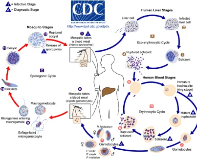
repeated (Grimberg, 2011). A schematic of the life cycle is shown in Figure 1.2.
Within the RBCs the parasites can sometimes differentiate into sexually reproducing cells
(gametocytes). If an Anopheles mosquito takes a blood meal when these cells are present the
sexual stage of the parasite’s life cycle begins (Marcus, 2009). Once inside the mosquito, the
parasites are released from the RBC and travel to the midgut where fertilization occurs to
form diploid zygotes. These then migrate to the intestine to undergo meisosis and form
haploid cells (oocytes) (Wahlgren and Perlmann, 2003; Marcus, 2009). Up to ten oocytes can
be carried by an infected mosquito that continue dividing and eventually travel to the
mosquito’s salivary glands in the form of sporozoites, which is where the cycle of human
infection begins. It is important to note that thousands of sporozoites are released by each
4
Figure 1.2 – A schematic showing the life cycle of the Plasmodium parasite.
Source: https://www.cdc.gov/malaria/about/biology/index.html
1.3 Clinical symptoms of malaria
Out of the malaria causing Plasmodium species, P. falciparum and P. vivax cause the most
infections worldwide, with P. falciparum being the most deadly (Ovadje and Nriagu, 2011;
Autino et al., 2012). Clinical symptoms occur as a result of the asexual erythrocytic cycle of
the parasite. Symptoms of uncomplicated malaria include general flu-like symptoms,
encompassing headache, fever, vomiting, fatigue and dizziness. Whereas symptoms of severe
malaria include high fever, respiratory failure, lactic acidosis, progressing anaemia, cerebral
malaria and multiorgan dysfunction (Chen, Schlichtherle and Wahlgren, 2000; Trampuz et al.,
2003; Bartoloni and Zammarchi, 2012; Alister, Mustaffa and Pradeep, 2012; Eugene-Ezebilo
and Ezebilo, 2014). Fatality often occurs due to severe anaemia or cerebral malaria (Autino et
5
Severe anaemia due to malaria is multifactorial and complex. Infected RBCs rupture for the
release of schizonts therefore naturally reducing RBCs in circulation. Reduction of infected
and non-infected RBCs by splenic removal or immune mediated lysis also contributes to
anaemia. Other causes include reduced erythropoiesis, reduced erythropoietin, bone marrow
suppression and inadequate reticulocyte production (Phillips and Pasvol, 1992; Chen,
Schlichtherle and Wahlgren, 2000; Kai and Roberts, 2008; Haldar and Mohandas, 2009; Autino
et al., 2012; Alister, Mustaffa and Pradeep, 2012)
Mature parasitized RBCs can be removed from circulation and adhere to endothelial cells
(cytoadhesion) within the microvasculature of organs, a process known as sequestration.
They can adhere to the brain, lung, heart, kidney, liver and placenta (Autino et al., 2012). It is
thought sequestration helps infected RBCs avoid splenic removal. The P. falciparum
erythrocyte membrane protein 1 (PfEMP1) and the P. falciparum histidine-rich protein
(PfHRP) have long been attributed to the adhesion of infected RBCs to the host’s blood vessels
and tissues. Reports have suggested PfEMP1 interacts with human endothelial proteins CD36
and intercellular adhesion molecule-1 (ICAM-1) (Chen, Schlichtherle and Wahlgren, 2000;
Autino et al., 2012; Tuikue et al., 2017).
Cytoadhesion along with rosetting, which is the adhesion of infected RBCs to non-infected
RBCs, lead to vascular obstruction. These processes can result in local hypoxia, in addition to
the localised release of parasite toxins to the area causing a focused inflammatory response
which can lead to tissue damage (Chen, Schlichtherle and Wahlgren, 2000; Autino et al., 2012;
Alister, Mustaffa and Pradeep, 2012). Initially these effects were thought to be unique to P.
falciparum infections but have since been extended to P. vivax and P. knowlesi infections
(Carvalho et al., 2010; Fatih et al., 2012).
1.4 Malaria control and prevention
Many factors influence the development and spread of malaria, including the three main
agents required for the disease to occur (humans, mosquitos and the parasites). Examples of
other contributing factors include: climate, vector and human habitation, and most
6
An overview of the three main strategies to control the spread of malaria is shown in Figure
1.3.
Figure 1.3 - An overview of the main areas of research that are ongoing to control the spread of malaria.
1.4.1 Vector control
The ability of malaria to spread is directly linked to the survival of the vector. Climate plays a
major role in mosquito survival and although that cannot be influenced - environmental
control measures, personal protective measures and use of effective insecticides are all
methods that have been applied in order to constrain the mosquito and thus the spread of
malaria (Ovadje and Nriagu, 2011).
A breakthrough in mosquito control occurred when the insecticidal properties of
dichlorodiphenyltrichloroethane (DDT) were discovered, and the effects were large scale
after WWII when the use of DDT was widely adopted (Oliva et al., 2014). Despite the positive
effect, DDT was banned in the 1970s in some countries due to its toxicity to the environment
and was replaced by pyretheroids, but the emergence of mosquitos resistant to pyretheroids
and the urgent need to reduce malaria incidences led to DDT being reintroduced specifically
for Indoor Residual Spraying (IRS) (Van Dyk et al., 2010; van den Berg et al., 2012).
The use of IRS and long-lasting insecticidal nets (LLINs) (commonly known as bed nets) has
helped greatly in reducing the incidences of malaria, particularly in some regions of Africa
Malaria Control and
Treatment
Vector Control
Insecticides Bed nets
Vaccines
Pre-erythrocytic
stage vaccines
Liver stage vaccines
Transmission blocking vaccines
Chemotherapy
7
(Pinder et al., 2015). The spread of resistance to commonly used insecticides however has led
researchers to explore alternative and novel vector control tools. Research is ongoing into
developing novel insecticides and genetic modification of mosquitoes in addition to a variety
of mosquito odour baits and traps (Takken and Knols, 2009; Barreaux et al., 2017; Benelli and
Beier, 2017). In the meantime, the WHO’s core vector control recommendations are the use
of insecticide-treated nets (namely LLINs) and IRS (WHO, 2017a).
1.4.2 Vaccines
The complexity of the disease and the complexity of immunity towards it creates a major
hurdle in developing a vaccine for malaria (Drew and Beeson, 2015; Dunachie et al., 2015).
The WHO, along with Malaria Vaccines Funders Group, revised a strategic plan in 2013 under
the title “The Malaria Vaccine Roadmap” with the aim of developing vaccine by 2015 that
“has a protective efficacy of more than 50% against severe disease and death and lasts longer
than a year” (WHO, 2014).
There are three broad types of vaccines in development for malaria; pre-erythrocytic vaccines
to target the liver-stage sporozoites which would decrease, if not prevent, the amount of
blood stage infection, blood stage vaccines which target infected blood cells by recognising
antigens present on their surface, and transmission-blocking vaccines which would produce
antibodies that target the sexual or mosquito stages of the parasite life cycle (Moorthy et al.,
2007; Ballou and Vekemans, 2017). The latter type of vaccine would reduce the spread of
malaria, but would not prevent malaria developing in the vaccinated person (Ballou and
Vekemans, 2017).
Despite the ongoing research, there is currently no registered malaria vaccine (Ballou and
Vekemans, 2017). The only leading vaccine candidate, RTS,S/AS01 (MosquirixTM, GSK), has
completed phase III clinical trials and will enter a Malaria Vaccine Implementation Program in
three African countries (Malawi, Ghana and Kenya) this year (2018). This is a
WHO-coordinated pilot program that will closely monitor and evaluate the safety and efficacy of
the vaccine (WHO, 2017b). RTS,S/AS01 is based on a fragment of the P. falciparum
circumsporozoite protein, which is coexpressed and fused with Hepatitis B virus surface
8
circumsporozoite protein is transmitted from the mosquito to the host during infection
(Ballou and Vekemans, 2017).
Reports have stated that RTS,S/AS01 is a promising start for a malaria vaccine, although an
ideal candidate would be a multi-antigen vaccine that would both protect individuals from
infection and block transmission of multiple species and strains (Karunamoorthi, 2014; Drew
and Beeson, 2015; Hemingway et al., 2016; Ballou and Vekemans, 2017).
1.4.3 Prophylaxis
Malaria chemoprophylaxis is most commonly used for travellers to malaria endemic areas
(Castelli et al., 2010; Walker, Nadjm and Whitty, 2018). Ideally there would be a prophylaxis
programme for people living in malaria endemic areas, but the reality is not so simple. The
rise of resistant strains to cheap and readily available drugs (such as chloroquine) and the
existence of parasite strains resistant to different drugs depending on the geographical
location, are among the reasons why a single prophylactic drug is not available for malaria
endemic countries (Fernando, Rodrigo and Rajapakse, 2011a).
The WHO does however recommend intermittent preventative treatment (IPT) for pregnant
women and seasonal malaria chemoprevention (SMC) for children aged 3-59 months in
malaria endemic countries. Sulfadoxine-pyrimethamine is the recommended drug
combination for IPT, and sulfadoxine-pyrimethamine in addition to amodiaquine is
recommended for SMC (WHO, 2017c). According to the WHO (2017c), as of 2015 no country
had reported implementing an IPT strategy for pregnant women, whereas 15 million children
in 12 countries were included in SMC programmes.
There have been previous occasions where antimalarial drugs have been given to whole
populations (Mass Drug Administration, MDA) in attempts to eradicate malaria. For example,
the Italian government in 1900 administered free quinine to be taken by the population as a
prophylaxis and treatment. This did result in reduced malaria numbers and mortality, but
transmission was not stopped and elimination of malaria did not occur until after WWII when
strict DDT spraying, treatment and prophylaxis using quinine along with organised malaria
9
Another example, albeit in a slightly different manner, was seen in the form of medicated salt.
The method included the addition of antimalarial drugs, chloroquine or pyrimethamine, to
salt used for cooking (Wernsdorfer, 1994; Greenwood, 2008; Alonso et al., 2011). The method
was carried out as part of a malaria eradication programme by the WHO. It was tested in
Brazil between 1959 and 1961, and parts of Africa in the mid-1960s (da Silva and Hochman,
2011; Talisuna et al., 2015). Although some reports stated reduction in the prevalence of
malaria (Alonso et al., 2011), the method was withdrawn and the overall results were
inconclusive, if not somewhat negative (Greenwood, 2008; Talisuna et al., 2015). The inability
to control administration due to drug leakage and variation in dosage, as well as the rise of
resistance, are among the reasons suggested for this method’s failure (Alonso et al., 2011;
Talisuna et al., 2015). In fact, an article published in 1988 suggested the rise of chloroquine
resistant P. falciparum may largely be due to the medicated salt programme (Payne, 1988).
1.5 Malaria chemotherapy
Treating malaria by chemotherapy is one of the most important methods relied on to control
the disease. For the purpose of this review, drugs used to treat malaria have been broadly
grouped into four sections; quinoline derivatives, antifolates, artemisinins, and antibiotics
and antiparasitic agents. A few examples of some of the major drugs used from each group
are discussed in this section.
1.5.1 Quinoline derivatives
The use of natural products to treat malaria has been around for thousands of years. One of
the oldest antimalarial drugs that has been used in treatment since the 17th century is quinine,
a natural product derived from the bark of the Cinchona tree. Initially infusions of the bark of
the tree were used in treatment, however in 1820 the active ingredient quinine was extracted
from the Cinchona bark and soon after became the standardised malaria treatment (Saxena
et al., 2003; Achan et al., 2011; Adebayo and Krettli, 2011).
According to a published review by Achan et al. (2011), alkaloids from the Cinchona tree that
included quinine amongst others, were tested in one of the earliest recorded clinical trials
10
quinine in the bark were higher than the other extracts, and thus it remained the main source
of malaria treatment until the 1920s (Achan et al., 2011).
It is thought the quinoline component of quinine is important for its antimalarial activity, thus
several synthetic analogues of quinine have been made preserving this chemical moiety
(Jones, Panda and Hall, 2015). Of the synthetic compounds, the most important was
chloroquine which was synthesised in 1934 by Hans Andersag and his group at Bayer AG,
though not used for malaria until 1947 (Thomé et al., 2013; Mushtaque and Shahjahan, 2015).
From then onwards chloroquine became the treatment of choice, in fact it was the only drug
used for long periods of time in some areas in Africa due to it being cheap and effective with
acceptable side effects (Saxena et al., 2003; Thomé et al., 2013; Mushtaque and Shahjahan,
2015).
Due to the great success of chloroquine at the time, along with the successful use of vector
control using DDT, the eradication of malaria was highly anticipated (Mojab, 2012).
Regrettably the expectation was diminished when resistance to both chloroquine and DDT
developed in the 1960s, and by the 1980s strains of P. falciparum that were multi-drug
resistant had appeared in several countries (Mojab, 2012).
Several theories have been proposed for the mechanism of action of quinoline-derived drugs.
The most accepted of these is that it interferes with the digestion of haemoglobin in the
digestive vacuole (DV). As previously described (section 1.2), the parasite cannot digest heme
in haemoglobin and thus converts it to hemozoin crystals, which are toxic to the human host.
It is thought that the basic drug diffuses into the acidic environment of the DV where it
becomes protonated and is unable to diffuse out again. The drug prevents the conversion of
heme to hemozoin by binding to heme resulting in a build-up of toxic heme which kills the
parasite (Thomé et al., 2013; Jones, Panda and Hall, 2015; Mushtaque and Shahjahan, 2015).
The accumulation of chloroquine in the DV is greatly reduced in chloroquine resistant strains;
this is particularly seen in P. falciparum (Thomé et al., 2013; Jones, Panda and Hall, 2015). The
inability of the drug to be transported into the DV is believed to be the reason behind the
resistance. There have been several theories offered to explain the reduced chloroquine
levels, such as reduced influx, increased efflux and modified chloroquine binding site
11
transporter (PfCRT) gene is believed to be responsible for the resistance (Thomé et al., 2013;
Jones, Panda and Hall, 2015; Antony and Parija, 2016). A study carried out by Lakshmanan et
al. (2005) showed that replacing the mutated PfCRT gene with the wild type gene resulted in
the strains becoming chloroquine sensitive, thus supporting the theory. An important area of
research involves synthesizing compounds that would reverse chloroquine resistance, with
PfCRT protein being a potential target (Jones, Panda and Hall, 2015; Mishra et al., 2017).
Reports of mutations found in the P. falciparum multidrug resistance protein 1 (Pfmdr1) gene
and the P. falciparum multidrug resistance-associated protein (Pfmrp) gene have also been
linked with resistance to chloroquine. However studies have suggested that mutations in
these genes alone are not enough to cause the resistance (Djimde et al., 2001; Mu et al., 2003;
Duraisingh and Cowman, 2005; Raj et al., 2009; Antony and Parija, 2016).
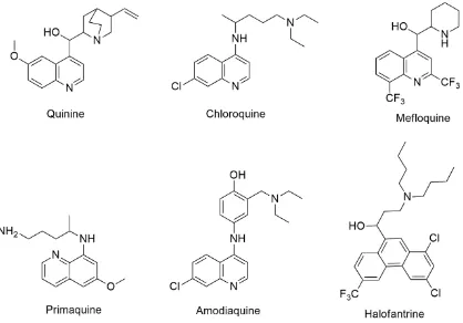
A number of modifications have been made to the quinine skeleton which includes
substituting the hydroxyl group, vinyl group, the quinoline and quinuclidine rings (Jones,
Panda and Hall, 2015). Some of the modified compounds either replaced chloroquine or are
given as second-line drugs, examples of which are amodiaquine, mefloquine, primaquine,
ferroquine and halofantrine (Figure 1.4 shows the chemical structures of some of the
analogues) (Duraisingh and Cowman, 2005; van Schalkwyk and Egan, 2006), but cross
resistance to some of these drugs has developed (Duraisingh and Cowman, 2005).
The 8-aminoquinoline, primaquine (Figure 1.4), should be mentioned due to it being the only
drug capable of clearing hypnozoites in early P. vivax and P. ovale. The drug however does
have the adverse side effect of haemolytic anaemia in patients with glucose-6-phosphate
dehydrogenase deficiency (G6PD). Screening patients before using the drug is therefore
necessary and its use is prohibited for pregnant women and children (Ramos Junior et al.,
2010; Fernando, Rodrigo and Rajapakse, 2011b; Myint et al., 2011; Andrews, Fisher and
Skinner-Adams, 2014; Ashley, Recht and White, 2014). Research into developing primaquine
analogues with reduced toxicity is ongoing, with the only prominent candidate to date being
tafenoquine, which is cited to be less toxic than primaquine but should also not be
administered to pregnant women (Zhu et al., 2007; Mishra et al., 2017). Another analogue,
known as elubaquine or CDRI 80/53, has completed Phase II/III clinical trials, but has only
12
Figure 1.4 - Chemical structures of quinine and some of its analogues.
1.5.2 Antifolates
For the last 70 years, antifolates have been used to prevent cell proliferation in cancer,
bacterial and parasitic infections such as malaria (Said, Jeffes and Weinstein, 1997;
Salcedo-Sora and Ward, 2013; Kuhlmann and Fleckenstein, 2017). The term folate encompasses
several folate derivatives which include, but are not limited to, folic acid, dihydrofolate and
tetrahydrofolate (Nzila et al., 2005).
Folic acid (vitamin B9) is essential in the folate pathway. In humans folic acid is obtained
through diet, but organisms such as bacteria and parasites have the ability to synthesise it
themselves (Litwack, 2008). Furthermore, folate derivatives are involved in providing one
carbon units needed for the synthesis of purines, thymidylate and some amino acids (Nelson
and Cox, 2008).
Both the conversion of the vitamins and the inhibition of synthesis are an attractive pathway
for drug development (Nzila, 2006a). The two folate enzymes targeted by antimalarial drugs
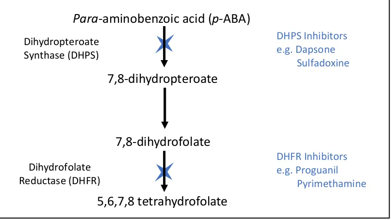
13
DHPS inhibitors and DHFR inhibitors are often given together in combination. Figure 1.5
shows a simple schematic of the folate pathways, highlighting both enzymes.
In brief, DHPS is an enzyme involved in synthesising dihydropteroate - a precursor to
dihydrofolate. Inhibiting DHPS activity ultimately results in reduced amounts of dihydrofolate,
the substrate of DHFR. The decrease in substrate allows the activity of the DHFR inhibitors to
increase, therefore blocking the pathway at two points results in a synergistic combination
(Yuthavong, 2002; Gregson and Plowe, 2005; Nzila, 2006a).
[image:31.595.97.500.273.500.2]
Figure 1.5 - A brief schematic of the folate pathway, highlighting the sites of action of the anti-folate drugs.
DHFR Inhibitors
Interest in agents that targeted this pathway occurred during WWII when the supply of
quinine was reduced and research into synthetic antimalarials was becoming necessary
(Gregson and Plowe, 2005). Around the same time cancer research showed that folic acid
played a major role in the proliferation of leukaemia, and agents that block the folate pathway
could reduce the spread of cancer (Nzila, 2006b; Müller and Hyde, 2013). The success of
antifolates in cancer treatment led to the development of the prodrug proguanil in 1945,
which was the first reported antifolate (DHFR Inhibitor) used for malaria (Nzila, 2006b).
Para-aminobenzoic acid (p-ABA)
7,8-dihydropteroate
7,8-dihydrofolate
5,6,7,8 tetrahydrofolate
Dihydropteroate Synthase (DHPS)
DHPS Inhibitors e.g. Dapsone Sulfadoxine
DHFR Inhibitors e.g. Proguanil Pyrimethamine
14
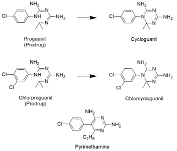
The effectiveness of proguanil led to the development of analogues such as chlorproguanil. It
also led to interest being taken in the 2,4-diaminopyrimidine family that identified
pyrimethamine – which was according to (Nzila, 2006b) “the most widely used antifolate
antimalarial agent”. A diagram showing the three mentioned drugs can be seen in Figure 1.6.
It is believed that the potency of these agents is in the chlorine(s) on the phenyl ring along
with the additional bond between the phenyl and the diaminopyrimidine ring – which can be
[image:32.595.123.477.233.536.2]seen in the active form of proguanil and chlorproguanil (Nzila, 2006b).
Figure 1.6 - Chemical structures of pyrimethamine and the two prodrugs proguanil and chlorproguanil along with their active forms.
As with other antimalarial agents, resistance also occurred with these DHFR inhibitors also.
Resistance to proguanil was reported rapidly. This may have been due to its heavy use as a
prophylactic drug in Southeast Asia in the 1940s and 1950s (Gregson and Plowe, 2005).
Reports stated that a 100 mg dose in 1947 would have a 100% cure rate, whereas in 1949
failure rose to 25% after a 300 mg dose (Davey and Robertson, 1957). According to Gregson
and Plowe (2005) both proguanil and pyrimethamine resistance were also reported during
15
Despite the parasites’ resistance to proguanil and pyrimethamine they are still used in
combination with other drugs, although their use is strategically planned (WHO, 2017c;
Walker, Nadjm and Whitty, 2018). For example, combination therapy that includes
pyrimethamine along with sulfadoxine (DHPS inhibitor) and artesunate (discussed in section
1.5.3) is given as first line treatment in India (WHO, 2017c). Pyrimethamine combined with
sulfadoxine is given as IPT for pregnant women in parts of Africa (WHO, 2017c). The
combination of proguanil and atovaquone (Malarone®) is recommended as a prophylaxis
drug for travellers to malaria endemic countries where resistance to chloroquine is high
(Waller and Sampson, 2018).
DHPS Inhibitors
A major class of DHPS inhibitors are the sulphonamides. The basic structure of sulphonamides
is similar to para-aminobenzoic acid (pABA), a DHPS substrate (Katzung, Masters and Trevor,
2012). Although they are important drugs in the treatment of malaria, the parasite has the
ability to salvage exogenous folate and pABA thus reducing the efficacy of these drugs (Delfino
et al., 2002; Yuthavong, 2002; Gregson and Plowe, 2005; Salcedo-Sora and Ward, 2013;
Talawanich et al., 2015). As previously discussed, using them in combination with DHFR
inhibitors results in a synergistic effect (Yuthavong, 2002; Nzila, 2006a).
Of the several DHPS inhibitors used dapsone is one of the more important agents. Although
synthesised in 1908, its antimicrobial activity was not identified until the 1930s (Nzila, 2006b).
It was administered with pyrimethamine in a drug known as Maloprim™(Müller and Hyde,
2013)and with chlorproguanil known as Lapdap (Lang and Greenwood, 2003; Alker et al.,
2005).
Another DHPS inhibitor, sulfadoxine, is also used in combination with pyrimethamine
(Fansidar™). Although this combination was used as a first line treatment in Africa for many
years, the development of resistance to it and to many of the above mentioned antifolates
led to them being replaced by other antimalarials. Resistance is identified by mutations in
both dhfr and dhps genes (Kublin et al., 2002; Alker et al., 2005; Plowe, 2009; Deloron et al.,
16
1.5.3 Artemisinins
Artemisinin, a natural product extracted from the Chinese herb qinghaor (Atermisia annua L),
is thought to have been used as a therapeutic to treat high fevers as far back as 2000 years
ago (Ploypradith, 2004; Krungkrai et al., 2010). The compound was isolated and its chemical
structure determined in 1972 by Chinese scientists who gave it the name Qinghaosu (now
known as artemisinin) (Engel and Straus, 2002; Ploypradith, 2004). The clinical importance of
artemisinin to treat malaria was reported in 1979 by the Qinghaosu Antimalarial Coordinating
Research Group. The compound was subsequently distributed to the rest of the world (Engel
and Straus, 2002; Krungkrai et al., 2010).
Since the discovery of its antimalarial activity, several synthetic derivatives of artemisinin
have been produced which are more soluble than artemisinin, examples of which are
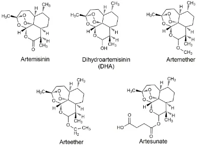
dihydroartemisinin, artemether, arteether and artesunate (Engel and Straus, 2002) (chemical
structures can be seen in Figure 1.7). All artemsinin-based drugs are converted in vivo to
[image:34.595.105.494.426.717.2]dihydroartemisinin, which is the metabolically active form (Eastman and Fidock, 2009).
17
The artemisinin-based compounds are fast-acting drugs with a very short half-life of one to
three hours (Liu, 2017). Due to their rapid elimination, the compounds cannot kill all parasites
during a short three-day treatment resulting in recrudescence (Giao et al., 2001; Liu, 2017;
Wang et al., 2017). The short half-life also eliminates their use for prophylaxis (Liu, 2017). In
order to reduce the rate of recrudescence, and in a bid to slow down the rate of parasite
resistance to artemisinin-based drugs, the WHO recommends the drug is not given as a
monotherapy but as a combinatory therapy (artemisinin-based combination therapy, ACT),
typically with a slower acting drug with a longer half-life (Giao et al., 2001; Eastman and
Fidock, 2009; Krungkrai et al., 2010; Cheng, Kyle and Gatton, 2012; Tripathy and Roy, 2014;
Severini and Menegon, 2015). According to Krungkrai et al. (2010) the WHO recommended
ACT as the first line treatment for uncomplicated P. falciparum malaria in Africa in 2001. This
remains the case to this day (WHO, 2018).
The mechanism of action of artemisinin and its derivatives is not fully understood, however
compounds lacking the endoperoxide bridge lose their antimalarial activity thus highlighting
its importance (Krungkrai et al., 2010; Mojab, 2012). A chemically engineered artemisinin that
allowed the fluorescent labelling of proteins covalently bound to the compound following
incubation with the parasites was synthesised (Wang et al., 2015). The proteins were then
identified by mass spectrometry. The chemically engineered analogue was as effective as the
unmodified artemisinin when tested on P. falciparum parasites. The results identified 124
protein targets, suggesting artemisinin has a diverse mechanism of action targeting several
cellular activities. Furthermore the study also suggested that artemisinin is activated by heme
within infected RBCs, which further supports previous suggestions that artemisinin’s
selectivity to infected RBCs is due to heme activation within the parasite (Klonis et al., 2011;
Klonis, Creek and Tilley, 2013). Other groups have since supported these findings (Ismail et
al., 2016; Zhang et al., 2016; Zhou, Li and Xiao, 2016). Although studies have shown
artemisinin to target hundreds of proteins, the contribution of each target to parasite death
is unknown (Wang et al., 2017).
It is also thought that the activation of artemisinin by heme within the parasitophorous
vacuole (PV) triggers a series of reactions that result in the build-up of oxygen centred and/or
18
leading to parasite death (Olliaro et al., 2001; Krishna, Uhlemann and Haynes, 2004; Saifi et
al., 2013; Severini and Menegon, 2015; Tilley et al., 2016).
Efforts to slow the rate of resistance to artemisinin-based drugs were made by providing
training for health care professionals in certain countries, both in diagnosis and in
distinguishing between severe and uncomplicated malaria. The WHO also provided treatment
guidelines and recommendations to protect their efficacy (Ndong et al., 2015). Nevertheless,
reports of delayed parasite clearance after treatment with artemisinin-based drugs has
emerged. Initially this was reported in Cambodia but has since become widespread across
Southeast Asia (Krungkrai et al., 2010; Cheng, Kyle and Gatton, 2012; Tun et al., 2015).
Initial studies by Ariey et al. (2014) showed mutations in the kelch gene (K13) to be associated
with the delayed clearance. A study carried out by Nyunt et al. (2015) in Myanmar found K13
mutated parasites in the eastern side of the country. Myanmar provides the only route for
the resistance to travel from Southeast Asia through to India and on to Africa, which is
believed to be the trajectory of previous antimalarial drug resistance (Tun et al., 2015).
Resistance to ACT threatens the world’s effort to control and eradicate malaria (Ariey et al.,
2014; Paloque et al., 2016), particularly as reports of artemisinin resistance spreading is
outpacing the implementation of new control measures and the development of new drugs
(Tun et al., 2015).
1.5.4 Antibiotics and antiparasitic drugs
The spread of parasites resistant to chloroquine in the 1960s prompted investigations into
alternative treatments, such as antibiotics (Tan et al., 2011). One of the most important class
of antibiotics used for malaria treatment are the tetracyclines, mainly doxycycline.
Doxycycline is a broad-spectrum antibiotic synthetically derived from tetracycline which is
produced by Streptomyces sp. As antimalarials, these drugs are slow acting. Infections take a
week to clear following administration; this is known as delayed-death effect (DDE). Due to
the delayed effect, the recommendation for both doxycycline and tetracycline is that they be
given in combination with quinine as a second line treatment for uncomplicated malaria
(Noedl, 2009; Tan et al., 2011; WHO, 2017c; Walker, Nadjm and Whitty, 2018). Doxycycline
19
pyrimethamine-sulfadoxine resistant strains (Tan et al., 2011; Walker, Nadjm and Whitty,
2018).
The malaria parasite has an apicoplast organelle which is a non-photosynthetic plastid that is
essential for the parasite’s survival and unique to apicomplexan parasites (Nzila, Ma and
Chibale, 2011; Chakraborty, 2016). This organelle carries out metabolic pathways in a manner
similar to that of prokaryotes. It has a circular genome, replicates DNA and carries out
transcription and translation in a prokaryote-like manner (Chakraborty, 2016). It is believed
that doxycycline targets the translation proteins of the apicoplast in the parasite, producing
a progeny with dysfunctional apicoplasts. The dysfunctional apicoplasts eventually result in
parasite death – hence the DDE (Nzila, Ma and Chibale, 2011; Chakraborty, 2016).
Clindamycin is another antibiotic used in malaria treatment. This drug is used as an antibiotic
to treat anaerobic and gram positive bacterial infections, as an antiparasitic to treat
toxoplasmosis and an anti-fungal to treat Pneumocystis carinii pneumonia (Obonyo and Juma,
2012). Clindamycin is another slow acting antimalarial believed to have a similar mechanism
of action to doxycycline (Chakraborty, 2016) and is recommended to be given in combination
with quinine to treat pregnant women with uncomplicated malaria (D ’alessandro et al.,
2018).
Another important class of drugs for malaria treatment are the naphthoquinones. These are
a group of naturally occurring compounds with reported antibacterial, antiparasitic and
antifungal activity (Riffel et al., 2002). The most important compound from this group is
atovaquone. Atovaquone is an analogue of ubiquinone, which is a lipophilic molecule found
in the mitochondria of cells and is involved in the transfer of electrons in the mitochondrial
respiratory chain (Riffel et al., 2002; El Hage et al., 2009; Müller and Hyde, 2010; Quinzii and
Hirano, 2010). Atovaquone is reported to inhibit the cytochrome bc1 complex in the parasite’s
mitochondria (Sutherland et al., 2008; Siregar et al., 2015). The effect of the drug is believed
to result in the collapsing of the mitochondrial membrane potential and the inhibition of the
parasite specific enzyme dihydroorotate dehydrogenase, which is involved in the synthesis of
pyrimidine. The parasite is unable to salvage pyrimidine from the host, the drug therefore
depletes the parasite from its pyrimidine pool preventing replication and eventually leads to