Effect of Light-tip Distance on the Shear Bond Strengths of
Composite Resin
Vittorio Cacciafestaa; Maria Francesca Sfondrinib; Andrea Scribantec;
Andreas Boehmed; Paul-Georg Jost-Brinkmanne
Abstract: The purpose of this study was to assess the effect of light-tip distance on the shear bond
strength and failure site of brackets cured with three different light curing units: a high-intensity halogen (Astralis 10, 10-second curing), a light-emitting diode (LED, e-Light, six-second curing), and a plasma arc (PAC System, four-second curing). One hundred and thirty-five bovine permanent mandibular incisors were randomly allocated to nine groups of 15 specimens each. Stainless steel brackets were bonded with a composite resin to the teeth, and each curing light was tested at zero, three, and six mm from the bracket. After bonding, all samples were stored in distilled water at room temperature for 24 hours and subsequently tested for shear bond strength. When the three light curing units were compared at a light-tip distance of zero mm, the three lights showed no significantly different shear bond strengths. At light-tip distances of three and six mm, no significant differences were found between the halogen and plasma arc lights, but both lights showed significantly higher shear bond strengths than the LED light. When evaluating the effect of the light-tip distance on each light curing unit, the halogen light showed no significant differences between the three distances. However, the LED light produced significantly lower shear bond strengths at a greater light-tip distance, and the plasma arc lamp showed significantly higher shear bond strengths at a greater light-tip distance. In hard-to-reach areas, the use of PAC system is suggested, whereas the LED evaluated in this study is not recommended. (Angle Orthod 2005;75:386–391.)
Key Words: Bond strength; Light-tip distance; Light curing units; Composite resins; High-intensity
light curing
INTRODUCTION
Light-cured composite resins provide a reduced risk of contamination, consistent handling characteristics, permit immediate archwire insertion, and give virtually unlimited working time.1 However, according to the manufacturers’
guidelines, visible light curing units require 20 seconds to cure orthodontic composite resins and 40 seconds to light cure resin-modified glass ionomers per bracket.2This
pro-aAssistant Clinical Professor, Department of Orthodontics,
Uni-versity of Insubria, Varese, Italy.
bAssistant Clinical Professor, Department of Orthodontics,
Uni-versity of Pavia, Pavia, Italy.
cPostgraduate Resident, Department of Orthodontics, University of
Pavia, Pavia, Italy.
dPostgraduate Resident, Department of Orthodontics and
Dento-facial Orthopedics, Humboldt University, Berlin, Germany.
eAssociate Professor, Department of Orthodontics and Dentofacial
Orthopedics, Humboldt University, Berlin, Germany.
Corresponding author: Vittorio Cacciafesta, DDS, MSc, PhD, c/o Studio Prof. Giuseppe Sfondrini, Via Liberta` 17, 27100 Pavia, Italy (e-mail: vcacciafesta@hotmail.com)
Accepted: June 2004. Submitted: May 2004.
q2005 by The EH Angle Education and Research Foundation, Inc.
longed curing time is uncomfortable for the patient, im-practical with children, and inconvenient for the clinician.3,4
Various attempts have been made to accelerate the speed of the light curing process by using a larger light guide or laser devices.5–9
In recent years, xenon plasma lights were introduced for high-intensity curing of orthodontic bonding materials.2,10–13
The advantage of this high-intensity light is that the same amount of total light energy can be delivered to the com-posite in a much shorter time period.12Claims of exposure
times of three to five seconds have been made for bonding brackets with the plasma arc light curing system, with shorter times for ceramic brackets.14 Previous investigations
gener-ally have reported no significant differences in bond strengths and failure sites of brackets cured with the plasma arc light compared with brackets cured with conventional halogen lights, both in vitro and in vivo.2,11–13,15,16
The innovative light-emitting diode (LED) technology, based on semiconductors, has opened new and interesting views in the field of photopolymerization. LEDs add the advantages of a soft-start polymerization, safety, efficiency, economy, and the longer lifetime of LED light.17 Despite
poly-TABLE 1. Light Tip Size and Intensity of the Three Light Curing Units Tested at Different Distances From the Bracket Base
Lighta
Tip Size, mm
Distance,
mm Group
Light Intensity, mW/cm2
HL
LED
7.5
7.5
0 3 6 0 3 6
1 2 3 4 5 6
1100 715 350 700 300 175
PAC 8 0
3 6
7 8 9
1200 1075 950
aHL indicates halogen light; LED, light-emitting diode; and PAC,
plasma arc.
merization qualitatively comparable with other light sourc-es18or slightly lower.19In addition, the temperature increase
is significantly lower and does not pose a threat to the pulp-al tissue.20,21 Previous investigations comparing the shear
bond strengths of brackets cured with LED with those of brackets cured with conventional halogen lights showed no significant differences.22,23 Other studies reported
signifi-cantly lower bond strengths with the LED when used for 10 seconds.24,25 However, in both studies all the bond
strengths were above eight MPa and therefore clinically ac-ceptable, even with a 10-second cure.
The degree to which these bonding materials cure de-pends on the intensity and quality of light to which they are exposed and the curing time. Once the light has left the curing unit, factors such as composite type, composite shade, thickness of resin increment or overlying tooth struc-ture, the distance and orientation of the light tip, and the diameter of the light tip may reduce intensity and provide a lower degree of polymerization.26 The inability to place
a light tip in close approximation to the composite resin may affect the resultant polymerization and clinical dura-bility.27
To date, there are no studies that have evaluated the ef-fect of light-tip distance on the shear bond strength of or-thodontic brackets cured with halogen, LED, and plasma arc lights. Accordingly, the purpose of this study was to evaluate the effects of different light-tip distances on the shear bond strength and site of bond failure of brackets bonded with a composite resin and cured with halogen, LED, and plasma arc lights. The null hypothesis of the study was that there is no significant difference in bond strength and debond site location among brackets bonded with the halogen light used for 10 seconds, the LED light used for six seconds, and the plasma arc light used for four seconds at different light-tip distances.
MATERIALS AND METHODS
Teeth
A total of 135 freshly extracted bovine permanent man-dibular incisors were collected from a local slaughterhouse and stored in a solution of 0.1% thymol for one month at 48C. The inclusion criteria included intact buccal enamel with no cracks from extraction and no caries. The teeth were randomly divided into nine different groups of 15 each as defined in Figure 1. The teeth were cleansed of soft tissue and embedded in cold-curing, fast-setting acrylic (SG 130, Ebalta, Rothenburg/Tauber, Germany) thus allowing the buccal surface of enamel to be exposed. Each tooth was oriented so that its labial surface was parallel to the shear-ing force.
Brackets
All 135 teeth were bonded with 0.018-inch stainless steel maxillary central incisor brackets (Victory Series,
3M/Un-itek, Monrovia, Calif) by one operator. The average bracket base surface area was reported by the manufacturer to be 11.7 mm2. This was verified by measuring it with a digital
caliper (Mitutoyo, Miyazaki, Japan). The area of 15 brack-ets was recorded, and the mean value was calculated for each group.
Bonding procedure
Before bonding, the facial surface of each incisor was cleaned for 10 seconds with a mixture of water and fluo-ride-free pumice in a rubber polishing cup, using a low-speed handpiece. The enamel surface was water-rinsed to remove pumice or debris and dried with an oil-free air stream.
Teeth were etched with 37% phosphoric acid gel (3M/ Unitek) for 30 seconds, followed by thorough washing and drying. A thin layer of XT primer (Transbond XT, 3M/ Unitek) was applied on the etched enamel, and the brackets were bonded with the composite resin (Transbond XT, 3M/ Unitek) near the center of the facial surface of the tooth. Sufficient pressure was used to express excess adhesive, which was removed from the margins of the bracket base with a scaler before polymerization.
Light curing units
[image:2.612.312.552.93.211.2]rep-FIGURE 1. Diagrammatic representation of study specimen
[image:3.612.61.300.67.137.2]group-ing.
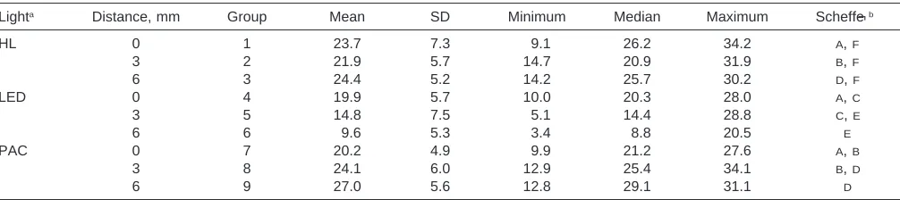
TABLE 2. Descriptive Statistics (in MPa) of Shear Bond Strengths of the Nine Groups Tested (Each Group Consisted of 15 Specimens)
Lighta Distance, mm Group Mean SD Minimum Median Maximum Scheffe´b
HL
LED
0 3 6 0 3 6
1 2 3 4 5 6
23.7 21.9 24.4 19.9 14.8 9.6
7.3 5.7 5.2 5.7 7.5 5.3
9.1 14.7 14.2 10.0 5.1 3.4
26.2 20.9 25.7 20.3 14.4 8.8
34.2 31.9 30.2 28.0 28.8 20.5
A,F B,F D,F A,C C,E E
PAC 0
3 6
7 8 9
20.2 24.1 27.0
4.9 6.0 5.6
9.9 12.9 12.8
21.2 25.4 29.1
27.6 34.1 31.1
A,B B,D D
aHL indicates halogen light; LED, light-emitting diode; and PAC, plasma arc. bScheffe´ grouping. Means with the same letter are not significantly different. resentation of the study design is illustrated in Figure 1.
The light-tip size (mm) and intensity (mW/cm2) of each
light curing unit at the three different distances are pre-sented in Table 1. Light intensity was measured with a ra-diometer (Demetron, SDS Kerr, Danbury, Conn).
Following the manufacturers’ instructions, the brackets were cured with the halogen light for five seconds on the mesial and five seconds on the distal side (total cure time: 10 seconds in high-power mode). The brackets were cured with the LED for three seconds on the mesial and three seconds on the distal side (total cure time: six seconds in fast-cure mode). The brackets were cured with the plasma arc light for two seconds on the mesial and two seconds on the distal side (total cure time: four seconds).
Debonding
After bonding, all samples were stored in distilled water at room temperature for 24 hours and subsequently tested in a shear mode on a universal testing machine (Erichsen 469 LE4, 500 N, Wuppertal, Germany), according to the draft of ISO specification TC 106/SC 2/WG 16. Specimens were secured in the lower jaw of the machine so that the bonded bracket base was parallel to the shear force direc-tion. Specimens were stressed in a gingivoocclusal direction at a crosshead speed of 1 mm/min, as in previous stud-ies.2,28,29The maximum load necessary to debond or initiate
bracket fracture was recorded in newtons and converted to megapascals as a ratio of newtons to surface area of the bracket.
Residual adhesive
After bond failure, the bracket bases and the enamel sur-faces were examined by the same operator. The Adhesive Remnant Index (ARI) was used to assess the amount of adhesive left on the enamel surface.30The ARI scores range
from 0 to 3: ‘‘0’’ indicates no adhesive remained on the tooth in the bonding area; ‘‘1,’’ less than half the adhesive remained on the tooth; ‘‘2,’’ more than half the adhesive remained on the tooth; and ‘‘3,’’ all adhesive remained on the tooth, with a distinct impression of the bracket mesh.
Statistical analysis
Descriptive statistics were calculated for each of the nine groups. A two-way analysis of variance (ANOVA) was ap-plied to determine whether significant differences in debond values existed among the various groups. For post hoc test, a Scheffe´’s test was used.
The chi-square (x2) test was used to determine significant
differences in the ARI scores among the different groups. The level of significance for all statistical tests was set at
P,.05. All statistical analyses were performed with Stata seven Program (Stata Corp, College Station, Tex).
RESULTS
Descriptive statistics for shear bond strengths are pre-sented in Table 2. The results of the ANOVA indicated the presence of significant differences among the various groups (P5.000). When comparing the three light curing units at a light-tip distance of zero mm (groups 1, 4, and 7), no significant differences were reported in terms of shear bond strengths (P5.17). On the other hand, at dis-tances of three mm (groups 2, 5 and 8) and six mm (groups 3, 6 and 9), no significant differences were found between the halogen and plasma arc lights (P . .44), but both showed significantly higher shear bond strengths than the LED light (P, .16).
[image:3.612.61.564.595.707.2]FIGURE 2. Mean shear bond strengths (MPa) of the three curing
[image:4.612.48.557.606.717.2]units under the three different light-tip distances (zero, three, and six mm). HL5halogen light; LED5light-emitting diode; PAC5plasma arc light.
TABLE 3. Frequency of Distribution of ARI Scores (%)a
Group ARI50 ARI51 ARI52 ARI53
Group 1—HL—0 mm Group 2—HL—3 mm Group 3—HL—6 mm Group 4—LED—0 mm Group 5—LED—3 mm
1 (6.7) 3 (20.0) 5 (33.3) 1 (6.7) 1 (6.7)
2 (13.3) 3 (20.0) 1 (6.7) 0 (0.0) 1 (6.7)
1 (6.7) 0 (0.0) 0 (0.0) 0 (0.0) 0 (0.0)
11 (73.3) 9 (60.0) 9 (60.0) 14 (93.3) 13 (86.7) Group 6—LED—6 mm
Group 7—PAC—0 mm Group 8—PAC—3 mm Group 9—PAC—6 mm
3 (20.0) 2 (13.3) 0 (0.0) 1 (6.7)
1 (6.7) 2 (13.3) 2 (13.3) 0 (0.0)
1 (6.7) 0 (0.0) 1 (6.7) 2 (13.3)
10 (27.0) 11 (73.3) 12 (80.0) 12 (80.0)
aARI indicates adhesive remnant score; HL, halogen light; LED, light-emitting diode; and PAC, plasma arc. distances (groups 1, 2, and 3). Using the LED light, no
significant differences (P . .087) were reported between groups 4 (zero mm) and 5 (three mm) and between groups 5 (three mm) and 6 (six mm). On the other hand, a signif-icant reduction (P 5 .000) in bond strength value was found when comparing group 4 (zero mm) with group 6 (six mm). Using the PAC system, no significant difference (P . .16) was found between groups 7 (zero mm) and 8 (three mm) and between groups 8 (three mm) and 9 (six mm), whereas a significant increase (P 5 .007) in bond strength was reported when comparing group 7 (zero mm) with group 9 (six mm) (Figure 2).
The ARI scores for the nine groups tested are listed in Table 3. When comparing the three lights for each light-tip distance, the x2 test results indicated no significant
differ-ences (P . .3) in ARI scores for the three lights at each tested distance (zero, three, and six mm). When evaluating the effect of the light-tip distance on each light curing unit, no significant differences (P . .41) in ARI scores were found among the three distances for all the light curing units (halogen, LED, and plasma arc).
DISCUSSION
The null hypothesis of the study was rejected. The pre-sent investigation demonstrated that the three light curing
units showed no statistically different shear bond strengths at a light-tip distance of zero mm. At greater distances of three and six mm, no significant differences were found between the halogen and plasma arc lights, but both re-vealed significantly higher shear bond strengths than the LED light.
There are no studies in the literature that have compared the shear bond strengths of halogen, LED, and plasma arc lights used at different light-tip distances from the brackets. Previous investigations that compared the shear bond strengths of brackets cured with LED with those of brackets cured with conventional halogen lights showed no signifi-cant differences between these two lights.22,23Other authors
compared the bond strength of halogen and plasma arc lights and reported no significant differences between these two lights.2,11–13 This is in agreement with the present
in-vestigation, where no significant differences between the three curing units were found at a light-tip distance of zero mm from the bracket.
Moreover, in the present study the effect of light-tip dis-tance on the shear bond strength of each light curing unit was investigated. When using the halogen light, the light-tip distance did not significantly affect the bond strength values. However, when using the LED light, a greater light-tip distance produced lower shear bond strengths, with a significant reduction at a light-tip distance of six mm from the bracket base. On the other hand, using the plasma arc light, a greater light-tip distance produced higher shear bond strengths, with a significant increase at six mm from the bracket base. As illustrated in Table 1, the light intensity of the LED light had a 58% decrease at three mm and a 75% decrease at six mm, compared with the intensity mea-sured at zero mm. On the other hand, the plasma arc light showed a 11% and 21% decrease in light intensity at three and six mm, respectively. Thus, it could be hypothesized that because the intensity of the plasma arc light is very high, increasing the distance of the light tip from the brack-et base could allow for curing a wider area of the enamel surface, without a significant decrease in power output.
Caldas et al26 evaluated the effect of light-tip distance on
the hardness of composite resins and reported that the hal-ogen, LED, and plasma arc units showed a decrease in the resin composite hardness as the light-tip distance increased. Another investigation evaluated the decrease in power out-put of LED curing devices with increasing distance to the filling surface.31The authors found that although blue LED
curing devices might have the same curing potential as a halogen device when placed in direct contact with a resin composite, blue LED curing devices may not provide a suf-ficient cure when placed at a distance of 10 mm to the resin composite surface. This decrease in power output can ex-plain the lower shear bond strength values achieved in our investigation when the LED curing light was placed at a light-tip distance of six mm.
Reynolds32suggested that minimum bond strength of six
to eight MPa was adequate for most clinical orthodontic needs. In the present work, the bond strengths of the three light curing units used at the three different light-tip dis-tances from the bracket base exceeded these limits.
No significant differences in ARI scores were reported among the three curing lights at the three distances tested, and no significant differences were found among the three light-tip distances for each light curing unit. A higher fre-quency of ARI scores of 3 was reported for all the groups tested, indicating that the entire adhesive remained on the tooth with a distinct impression of the bracket mesh. Pre-vious studies that compared the ARI scores of halogen and LED curing lamps showed no significant differences be-tween the two lights, in agreement with our findings.22,24,25
Other reports evaluated the ARI scores of halogen and plas-ma arc lamps and showed a significantly higher frequency of bond failure at the bracket-adhesive interface (ARI score of 3) when the composite resin was cured with a conven-tional halogen light.2,12The composite resin cured with the
xenon arc light showed a significantly higher frequency of bond failure at the enamel-adhesive interface (ARI score of 0), in contrast with the present findings. The variability of the results may be attributed to the differences in the me-chanical and physical properties of the materials tested in each study.
CONCLUSIONS
With the limitations of this in vitro study, the following conclusions may be drawn:
• When used at a distance of zero mm from the bracket, the three light curing units showed no statistically differ-ent shear bond strengths.
• At distances of three and six mm, no significant differ-ences were found between the halogen and plasma arc lights, but both had significantly higher shear bond strengths than the LED light.
• The halogen light showed that no significant differences in bond strength were found among the three distances.
Using the LED light, a greater light-tip distance produced significantly lower shear bond strengths, whereas using the plasma arc lamp, a greater light-tip distance caused significantly higher shear bond strengths. Therefore, in hard-to-reach areas, the use of PAC system is suggested, whereas the e-Light is not recommended.
• All the combinations tested showed bond strengths that could be clinically acceptable, although big differences existed among them.
• No significant differences in ARI scores were found among the various groups.
ACKNOWLEDGMENTS
3M/Unitek, GC Europe, Ivoclar-Vivadent, and American Dental Technologies are acknowledged for providing the materials tested in this study.
REFERENCES
1. Read MJ. The bonding of orthodontic attachments using a visible light cured adhesive. Br J Orthod. 1984;11:16–20.
2. Sfondrini MF, Cacciafesta V, Pistorio A, Sfondrini G. Effects of conventional and high-intensity light-curing on enamel shear bond strength of composite resin and resin-modified glass-ionom-er. Am J Orthod Dentofacial Orthop. 2001;119:30–35.
3. Pearson AI. Optimal light curing of adhesive precoated brackets.
J Clin Orthod. 1995;29:583–585.
4. Sunna S, Rock WP. Clinical performance of orthodontic brackets and adhesive systems: a randomized clinical trial. Br J Orthod. 1998;25:283–287.
5. Frost T, Norevall LI, Persson M. Bond strength and clinical ef-ficiency for two light guide sizes in orthodontic bracket bonding.
Br J Orthod. 1997;24:35–40.
6. Blankenau RJ, Kelsey WP, Powell GL, Shearer GO, Barkmeier WW, Cavel WT. Degree of composite resin polymerization with visible light and argon laser. Am J Dent. 1991;4:40–42. 7. Lalani N, Foley TF, Voth R, Banting D, Mamandras A.
Poly-merization with the argon laser: curing time and shear bond strength. Angle Orthod. 2000;70:28–33.
8. Talbot TQ, Blankenau RJ, Zobitz ME, Weaver AL, Lohse CM, Rebellato J. Effect of argon laser irradiation on shear bond strength of orthodontic brackets: an in vitro study. Am J Orthod
Dentofacial Orthop. 2000;118:274–279.
9. James JW, Miller BH, English JD, Tadlock LP, Buschang PH. Effects of high-speed curing devices on shear bond strength and microleakage of orthodontic brackets. Am J Orthod Dentofacial
Orthop. 2003;123:555–561.
10. Cacciafesta V, Sfondrini MF, Sfondrini G. A xenon arc light-cur-ing unit for bondlight-cur-ing and bleachlight-cur-ing. J Clin Orthod. 2000;34:94– 96.
11. Oesterle LJ, Newman SM, Shellhart WC. Rapid curing of bond-ing composite with a xenon plasma arc light. Am J Orthod
Den-tofacial Orthop. 2001;119:610–616.
12. Pettemerides AP, Ireland AJ, Sherriff M. An ex vivo investigation into the use of a plasma arc lamp when using a visible light-cured composite and a resin-modified glass poly (alkenoate) cement in orthodontic bonding. J Orthod. 2001;28:237–244.
13. Ishikawa H, Komori A, Kojima I, Ando F. Orthodontic bracket bonding with a plasma-arc light and resin-reinforced glass ionom-er cement. Am J Orthod Dentofacial Orthop. 2001;120:58–63. 14. Mayes JH. Curing lights: an overview. Clin Impressions. 2000;9:
15–17.
arc and conventional halogen curing lights for orthodontic bond-ing. Am J Orthod Dentofacial Orthop. 2004;125:30–35. 16. Sfondrini MF, Cacciafesta V, Scribante A, Klersy C. Plasma arc
versus halogen light curing of orthodontic brackets: a 12-month clinical study of bond failures. Am J Orthod Dentofacial Orthop. 2004;125:342–347.
17. La Torre G, Marigo L, Pascarella GA, Rumi G. Light-emitting diodes (LED) technology applied to the photopolymerization of resin composites. Minerva Stomatol. 2003;52:193–200.
18. Mills RW, Uhl A, Jandt KD. Optical power outputs, spectra and dental composite depths of cure, obtained with blue light emitting diode (LED) and halogen light curing units (LCUs). Br Dent J. 2002;193:459–463.
19. Dunn WJ, Bush AC. A comparison of polymerization by light-emitting diode and halogen-based light-curing units. J Am Dent
Assoc. 2002;133:335–341.
20. Hofmann N, Hugo B, Klaiber B. Effect of irradiation type (LED or QTH) on photo-activated composite shrinkage, strain kinetics, temperature rise, and hardness. Eur J Oral Sci. 2002;110:471– 479.
21. Yap AU, Soh MS. Thermal emission by different light-curing units. Oper Dent. 2003;28:260–266.
22. Dunn WJ, Taloumis LJ. Polymerization of orthodontic resin ce-ment with light-emitting diode curing units. Am J Orthod
Den-tofacial Orthop. 2002;122:236–241.
23. Bishara SE, Ajlouni R, Oonsombat C. Evaluation of a new curing light on the shear bond strength of orthodontic brackets. Angle
Orthod. 2003;73:431–435.
24. Swanson T, Dunn WJ, Childers DE, Taloumis LJ. Shear bond strength of orthodontic brackets bonded with light-emitting diode curing units at various polymerization times. Am J Orthod
Den-tofacial Orthop. 2004;125:337–341.
25. Usumez S, Buyukyilmaz T, Karaman AI. Effect of light-emitting diode on bond strength of orthodontic brackets. Angle Orthod. 2004;74:259–263.
26. Caldas DB, de Almeida JB, Correr-Sobrinho L, Sinhoreti MA, Consani S. Influence of curing tip distance on resin composite Knoop hardness number, using three different light curing units.
Oper Dent. 2003;28:315–320.
27. Rueggeberg FA, Jordan DM. Effect of light-tip distance on po-lymerization of resin composite. Int J Prosthodont. 1993;6:364– 370.
28. Jobalia SB, Valente RM, de Rijk WG, BeGole EA, Evans CA. Bond strength of visible light-cured glass ionomer orthodontic cement. Am J Orthod Dentofacial Orthop. 1997;112:205–208. 29. Millett DT, Cattanach D, McFadzean R, Pattison J, McColl J.
Laboratory evaluation of a compomer and a resin-modified glass ionomer cement for orthodontic bonding. Angle Orthod. 1999;69: 58–63.
30. A˚ rtun J, Bergland S. Clinical trials with crystal growth condi-tioning as an alternative to acid-etch enamel pretreatment. Am J
Orthod. 1984;85:333–340.
31. Meyer GR, Ernst CP, Willershausen B. Decrease in power output of new light-emitting diode (LED) curing devices with increasing distance to filling surface. J Adhes Dent. 2002;4:197–204. 32. Reynolds IR. A review of direct orthodontic bonding. Br J