0095-1137/96/$04.0010
Copyrightq1996, American Society for Microbiology
Detection by PCR of the nim Genes Encoding 5-Nitroimidazole
Resistance in Bacteroides spp.
STE´ PHANIE TRINHANDGILLES REYSSET* Unite´ des Anae´robies, Institut Pasteur, 75724 Paris ce´dex 15, France
Received 16 January 1996/Returned for modification 25 March 1996/Accepted 11 June 1996
A PCR method was developed for detection of the nim genes encoding 5-nitroimidazole resistance in
Bacteroides spp. Two PCR primers specific for nim genes were designed. They allowed amplification of a 458-bp
fragment from all characterized plasmid- and chromosome-borne metronidazole resistance genes. The spec-ificity of the method was tested with DNA from metronidazole-sensitive Bacteroides spp. strains and from other strains of unrelated species. Each DNA preparation was analyzed with and without an internal positive control to verify that the absence of PCR amplification product was not due to inhibition of the Taq polymerase inhibitors. By this technique, two newly discovered metronidazole-resistant clinical strains of Bacteroides
fragilis were shown to harbor resistance genes undetectable by Southern blotting. In spite of the sequence
divergence of the nim genes, the PCR method is thus suitable for epidemiological investigations. The ampli-fication method also revealed that nim-related resistance genes were not present in either Streptomyces strain S6670, a natural producer of 2-nitroimidazole, or in Enterococcus faecalis strains, which have been suggested to possess metronidazole-inactivating enzyme.
Bacteroides species are major members of the normal co-lonic human microflora. Species most frequently isolated from the flora are Bacteroides vulgatus, Bacteroides thetaiotaomicron, Bacteroides distasonis, and, less frequently, Bacteroides eggerthii and Bacteroides fragilis (10). These gram-negative anaerobic bacteria are often involved in various infectious diseases (6), and the choice of suitable antibiotics for therapy is limited because Bacteroides spp. are frequently resistant to clinically relevant antimicrobial agents, including tetracycline, clindamy-cin, andb-lactam antibiotics (7). In contrast, the 5-nitroimid-azole (5-NI) molecules are very potent anaerobicidal agents commonly used to treat or prevent Bacteroides infections. Met-ronidazole (MTR), ornidazole, and tinidazole are the com-pounds most often prescribed.
In spite of the wide use of these drugs during the last 30 years, few 5-NI-resistant strains have been isolated. A B. fragilis strain (NCTC11295) with a high level of resistance to MTR (MIC, 64mg ml21) was isolated in 1978 from a patient with
Crohn’s disease following a long-term MTR regimen (11). Since then, other resistant strains have been found in clinical specimens (21). They are very rare, and the resistance is non-transferable, such that the potential for epidemiological dis-semination is low. On the other hand, clinical Bacteroides spp. strains with reduced sensitivity to MTR (MICs of 5-NI lower than or near the standard breakpoint for MTR, i.e., 16 mg ml21) have also been isolated in France since 1986 (4). These Bacteroides strains present a new 5-Nirresistance phenotype
characterized by higher levels of resistance to tinidazole or ornidazole than to MTR (4, 19), contrasting with the situation for strain NCTC11295. Two epidemiological studies conducted in French hospitals by Breuil et al. (1) and Patey et al. (17) show that the incidence of such resistant strains among Bacte-roides isolates is increasing and is currently 3 to 5%. A subse-quent molecular study (20) demonstrated that the moderate
resistance phenotype of these clinical strains was in all cases associated with the presence of the 5-NI resistance gene (nim). Four related nim genes have now been identified in Bacte-roides species (8, 26). Three of them are located on low-copy-number mobilizable plasmids: pIP417 (7.7 kb, nimA) from B. vulgatus BV-17, pIP419 (10 kb, nimC) from B. thetaiotaomicron BT-13, and pIP421 (7.3 kb, nimD) from B. fragilis BF-F239. The fourth gene (nimB) was mapped to the chromosome of B. fragilis BF-8. In most other isolates (73%), the nim gene was also located on the chromosome and present in one or two copies (20). Most of both the plasmid-borne and chromosoma-lly determined resistance markers are transferable by a conju-gative process (2, 24). For example, the plasmid pIP417 has been transferred in vitro by conjugation into various sensitive strains and species of both Bacteroides and Prevotella (25). The transfer is presumed to have also occurred in vivo, as plasmids similar or identical to pIP417, first found in a B. vulgatus strain, have been detected in different clinical B. fragilis isolates (20). Thus, the mobilization of the nim genes could account for the spread of the 5-NI resistance among at least some gram-neg-ative anaerobic bacteria. Monitoring for nim genes is therefore recommended for clinically important anaerobic pathogens.
Detection of 5-NI-resistant strains by conventional suscep-tibility testing methods requires conditions of strict anaerobi-osis because of the usual inactivity of the drug in the presence of even low levels of oxygen (5, 13). This is particularly impor-tant for the moderately resisimpor-tant strains, because of their low MICs. Even if the nim genes protect the bacteria more effi-ciently against tinidazole or ornidazole than against MTR (4, 24), MTR is still the most common 5-NI compound used in susceptibility testing and MIC determinations in clinical labo-ratories. The presence of the nim genes can be also detected by Southern blotting of DNA with specific [32P]dATP-labeled
probe, although the method presents some limitations. One is the necessity of using several probes as some nim genes are different enough not to cross-hybridize in high-stringency con-ditions (20). Southern blotting is also time-consuming, requires purified DNA and handling of radioactive products, and is thus difficult for nonspecialized laboratories. One consequence of these technical considerations is that the prevalence of the * Corresponding author. Mailing address: Unite´ des Anae´robies,
Institut Pasteur, 75724 Paris ce´dex 15, France. Phone: 33 (1) 40 61 33 70. Fax: 33 (1) 40 61 31 23. Electronic mail address: greysset@pasteur .fr.
2078
on May 15, 2020 by guest
http://jcm.asm.org/
resistance nim genes among clinical isolates may be underes-timated. We describe here an alternative technique, based on PCR (22), for screening anaerobic bacteria for nim genes.
MATERIALS AND METHODS
Bacterial strains.The Bacteroides spp. strains resistant to 5-NI used in this study are listed in Table 1. The other strains used to test the specificity of the PCR method are listed in Table 2.
Media and culture conditions.Anaerobic bacterial strains were cultured in an anaerobic chamber (Celster, Velizy, France) under 5% H2–5% CO2–90% N2.
Both Trypticase glucose yeast extract (TGY) medium (18) and Wilkins-Chalgren (WC) medium (Oxoid, Basingstoke, Hampshire, United Kingdom) were used for bacterial growth. The MIC of MTR was determined on solid WC agar medium, by the E-test method according to the supplier’s instructions (AB BIODISK, Dalva¨gen, Sweden). Aerobic bacterial strains were cultured at 378C in Luria broth (23) or brain heart infusion (Difco Laboratories, Detroit, Mich.).
Strepto-myces strains were cultured in aerobic conditions at 378C in Pridham’s starch medium as described by P. Jeannin: 10 g of starch liter21; 1 g of K
2HPO4liter21;
1 g of MgSO4liter21; 1 g of NaCl liter21; 1 g of SO4(NH4)2liter21; 2 g of CaCO3
liter21.
DNA extraction from bacterial strains.Bacteria were harvested either by centrifugation of 2 ml of an overnight culture or by picking colonies from agar with a 1-ml inoculation loop. The target DNA was extracted as described by K. Wilson (27). The cells were resuspended in 600ml of TE buffer (13) (10 mM Tris hydrochloride [pH 7.4], 1 mM EDTA) containing proteinase K (to a final con-centration of 100mg ml21). The samples were incubated at 378C for 1 h. To
remove cell wall debris, polysaccharides, and other contaminating macromole-cules, the NaCl concentration of the DNA solution was adjusted to 0.7 M and hexadecyltrimethylammonium bromide (CTAB), in 0.7 M NaCl, was added to a final concentration of 1%. The reaction mixture was incubated for 10 min at 658C and then centrifuged at 13,000 rpm for 5 min. For DNA extraction, the super-natant was treated with 1 volume of chloroform-isoamyl alcohol (24:1) and then with 1 volume of phenol-chloroform-isoamyl alcohol (25:24:1). DNA was pre-cipitated from the aqueous phase with 2 volumes of 100% ethanol for 20 min at 2808C. DNA was then pelleted by centrifugation (13,000 rpm for 15 min), washed with 70% ethanol, air dried, and resuspended in 100ml of distilled water. A rapid DNA extraction procedure was also used. After centrifugation, the cells were resuspended in 100ml of TE buffer (13), pH 7.5. The bacterial suspension was incubated for 10 min at 958C and then centrifuged at 13,000 rpm for 5 min. The supernatant containing the DNA was collected.
Oligonucleotide primers for PCR.Multiple alignment of the nucleotide se-quences of the four nim genes (Fig. 1) was generated to select the following pair of primers: NIM-3 (59-ATG TTC AGA GAA ATG CGG CGT AAG CG-39) and NIM-5 (59-GCT TCC TTG CCT GTC ATG TGC TC-39). The corresponding sequences are at positions 1 to 26 and 436 to 458 for NIM-3 and NIM-5, respectively. Both were synthesized by Genset SA, Paris, France. The nucleotide sequences of the nim genes are available in the EMBL, GenBank, and DDBJ nucleotide sequence databases, under accession numbers X71444, X71443, X76948, and X76949 for nimA, nimB, nimC, and nimD genes, respectively. The pair of primers allows a 458-bp fragment amplification.
DNA amplification.Each PCR assay was performed in 100ml of reaction mixture containing 10ml of DNA sample, 25mM tetramethylammonium chlo-ride (Aldrich, Milwaukee, Wis.), 200mM each deoxynucleoside triphosphate, 1 mM each primer, 10ml of Taq polymerase buffer (10 mM Tris-HCl, 50 mM KCl, 1.5 mM MgCl2, 0.1% Triton X-100, 0.2 mg of bovine serum albumin or gelatin
ml21, pH 9), 1 U of Taq polymerase (Appligene Oncor), and distilled water.
Target DNAs were amplified in a thermal cycler (model Crocodile III; Appligene Oncor). After an initial denaturation step at 948C for 10 min, the reaction mixtures were subjected to 32 cycles of amplification consisting of denaturation at 948C for 30 s, annealing at 628C for 1 min, extension at 728C for 1 min, and a final extension step at 728C for 10 min.
To prevent cross-contamination with amplified DNA sequences, all reagents
were aliquoted, autoclaved, and irradiated with a UV box (model UV cross-linker RPN 2500; Amersham Life Science). Microcentrifuge tubes, racks, gloves, and micropipettes were also irradiated. Sterile tips with aerosol barriers (ATGC, Inc.) were used, and all reagents were premixed. One set of micropipettes was reserved for handling amplified DNA. A negative control containing no template DNA was added to each set of amplification reaction mixtures.
Gel electrophoresis and Southern blot hybridization.After amplification, 20 ml of each PCR product was analyzed on a 1.3% (wt/vol) agarose gel by hori-zontal electrophoresis in TBE buffer (89 mM Tris base, 89 mM boric acid, 2 mM Na2EDTA, pH 8.25). After electrophoresis, the gel was stained with a solution
containing 0.5mg of ethidium bromide ml21
, and DNA fragments were visual-ized under a UV light transilluminator (302 nm; UVP, Inc.). A 100-bp ladder (Gibco BRL) was used as a molecular weight marker: it consisted of 15 blunt-ended fragments between 100 and 1,500 bp in multiples of 100 bp and an additional fragment of 2,072 bp. Southern blot analysis was performed according to the method of Bryant and Tandeau de Marsac (3). The gel was denatured in 1.5 M NaCl–0.5 N NaOH and neutralized in 0.5 M Tris-HCl (pH 7.4)–1.5 M NaCl. The DNA fragments were transferred to a nylon membrane (Hybond-N1; Amersham International) and covalently linked by heating. The nylon mem-branes were prehybridized at 658C for 4 h in 10 ml of a solution containing 243 NET (0.36 M Tris-HCl [pH 7.5], 3.6 M NaCl, 24 mM EDTA) and 1003 Den-hardt solution (0.02% Ficoll, 0.02% polyvinylpyrrolidone, 0.02% bovine serum albumin, 10% sodium dodecyl sulfate). Hybridization was performed at 60 or 508C for 18 h in the prehybridization buffer containing the probe pFK726 radio-labeled with [32
[image:2.612.57.300.81.174.2]P]dATP with the Boehringer Mannheim nick-translation kit (Boehringer-Mannheim France SA, Meylan, France). The Southern blots were
TABLE 1. Bacteroides spp. strains resistant to 5-NIs
Sp. Strain MIC of
MTRa
5-Nir
determinant Reference
B. fragilis BF-8 34 Chromosome 20
B. vulgatus BV-17 4 pIP417 20
B. thetaiotaomicron BT-13 4 pIP419 20
B. fragilis BF-F239 8 pIP421 20
B. fragilis BF-152/94 32 Chromosome This study
B. fragilis BF-377/95 12 pIP417-like This study
B. fragilis BF-376/95 32 Unknown This study
aMICs of MTR were determined on solid agar WC medium (Oxoid) by the
E-test method and are expressed in micrograms per milliliter.
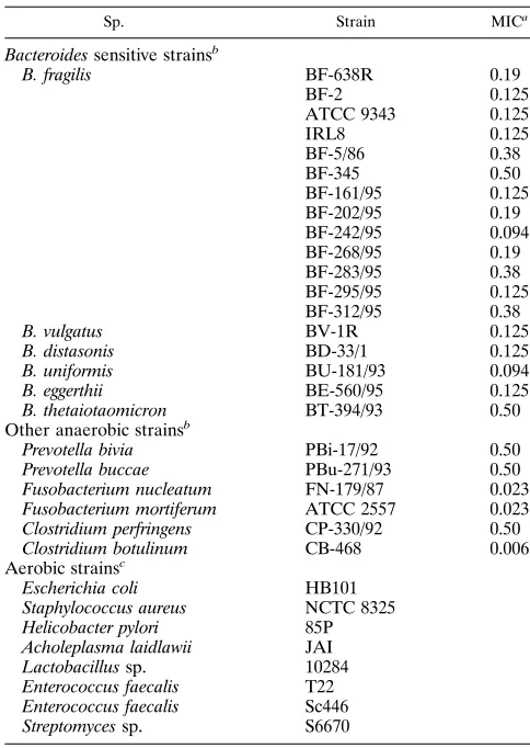
TABLE 2. Bacteroides spp. strains sensitive to 5-NIs and other strains used
Sp. Strain MICa
Bacteroides sensitive strainsb
B. fragilis BF-638R 0.19
BF-2 0.125
ATCC 9343 0.125
IRL8 0.125 BF-5/86 0.38 BF-345 0.50 BF-161/95 0.125 BF-202/95 0.19 BF-242/95 0.094 BF-268/95 0.19 BF-283/95 0.38 BF-295/95 0.125 BF-312/95 0.38
B. vulgatus BV-1R 0.125
B. distasonis BD-33/1 0.125
B. uniformis BU-181/93 0.094
B. eggerthii BE-560/95 0.125
B. thetaiotaomicron BT-394/93 0.50
Other anaerobic strainsb
Prevotella bivia PBi-17/92 0.50
Prevotella buccae PBu-271/93 0.50
Fusobacterium nucleatum FN-179/87 0.023
Fusobacterium mortiferum ATCC 2557 0.023
Clostridium perfringens CP-330/92 0.50
Clostridium botulinum CB-468 0.006
Aerobic strainsc
Escherichia coli HB101
Staphylococcus aureus NCTC 8325
Helicobacter pylori 85P
Acholeplasma laidlawii JAI
Lactobacillus sp. 10284
Enterococcus faecalis T22
Enterococcus faecalis Sc446
Streptomyces sp. S6670
aMICs of MTR were determined on WC medium (Oxoid) by the E-test
method and are expressed in micrograms per milliliter.
bThese strains were from the Anaerobes Reference Center, Pasteur Institute,
Paris, France.
cThe first four strains were obtained from various laboratories at the Pasteur
Institute, the Lactobacillus and Enterococcus strains were from E. Nagy, and the
Streptomyces sp. strain was from P. Jeannin, Rhoˆne-Poulenc-Rorer. All aerobic strains are resistant to up to 32mg of MTR ml21, the E-test limit value.
on May 15, 2020 by guest
http://jcm.asm.org/
[image:2.612.314.556.322.663.2]then washed three times for 15 min in 63NET and subjected to autoradiography for 1 or 2 h at2808C with Hyperfilm-MP (Amersham International).
RESULTS
Evaluation of the PCR method for detection of nim genes.In
preliminary studies, PCRs were performed with Bacteroides strains that carry the 5-Nir determinant either on a plasmid
(BV-17, BT-13, BF-F239) or on the chromosome (BF-8). The sensitivity of the PCR method for detection of nim genes was evaluated by examining serial dilutions of total DNA extracted by the procedure described by K. Wilson (27). The PCR prod-ucts generated with the primer pair NIM-3 and NIM-5 were analyzed on an ethidium bromide-stained electrophoresis gel (Fig. 2A). A distinct band with the expected size of 458 bp was detected in all strains carrying a nim gene, but with varying intensity. For BV-17 and BT-13, harboring the nimA and nimC genes, respectively, the highest template DNA dilution giving a visible fragment was 1025(lanes 13 and 20), whereas the
de-tection limit was a dilution of 1024 for BF-8 (nimB) (lane 6)
and only 1023for BF-F239 (nimD) (lane 24).
To confirm that the product amplified by the PCR method was indeed a nim gene fragment, the amplified target se-quences were transferred to a membrane by standard blotting and probed with [32P]dATP-labeled plasmid pFK726. The
re-combinant plasmid pFK726 contains the internal EcoRV-PvuII restriction fragment of the nimA gene in pUC19 (28). The restriction sites EcoRV and PvuII are 106 and 468 nucle-otides downstream from the ATG codon initiation of the nimA
gene (Fig. 1). Hybridization was performed at 608C, and mem-branes were exposed for 2 h (Fig. 2B). PCR products obtained from the nimA and nimC genes gave strong hybridization sig-nals (lanes 9 to 13 and 16 to 20, respectively), and those of the nimD gene gave a weaker signal (lanes 22 to 24). In contrast, the probe hybridized very poorly with the PCR products from the nimB gene: no band was visible after 2 h of exposure (lanes 3 to 7), and only a faint band was visible after 16 h of exposure (data not shown). Similar results were obtained with hybrid-ization assays performed at 508C (data not shown).
Specificity of the PCR method.The specificity of the PCR
[image:3.612.64.544.67.406.2]method was first tested with MTR-sensitive strains of the B. fragilis group: 13 of B. fragilis and 1 each of B. vulgatus, B. dis-tasonis, B. uniformis, B. eggerthii, and B. thetaiotaomicron. MICs of MTR were determined by the E-test method (Table 2). Each DNA sample was tested for amplification by the NIM-3–NIM-5 primer pair. The 458-bp fragment was not de-tected in any of the 18 strains tested by ethidium bromide-stained agarose gel electrophoresis (Fig. 3A). DNA samples from bacteria, phylogenetically closely or distantly related to the B. fragilis group, were also investigated with identical con-ditions. The strains included the anaerobes Prevotella bivia, Prevotella buccae, Fusobacterium nucleatum, Fusobacterium mortiferum, Clostridium perfringens, and Clostridium botulinum and the aerobes Escherichia coli, Helicobacter pylori, Staphylo-coccus aureus, Acholeplasma laidlawii, and Lactobacillus sp. None of these DNAs yielded an amplified fragment (Fig. 4A),
FIG. 1. Alignment of the nucleotide sequences of the four nim genes. The common nucleotides are indicated in boldface. The EcoRV and PvuII restriction sites
are underlined in the nimA sequence. Positions of the oligonucleotide primers NIM-3 and NIM-5 are indicated by arrows below the sequences.
on May 15, 2020 by guest
http://jcm.asm.org/
whereas DNA from B. vulgatus BV-17 used as a positive con-trol did (lane 2).
To eliminate false-negative results due to Taq polymerase inhibitors, PCR experiments were conducted with test DNA alone (diluted 101-fold) and test DNA plus positive control
DNA (diluted 102-fold) from BV-17 (Fig. 3B for sensitive Bacteroides spp. strains and Fig. 4B for the other strains). The 458-bp fragment was amplified from the added control DNA, but the intensity of the signal varied from strain to strain. This may be due to the presence of subinhibitory concentrations of Taq polymerase inhibitors or to nonspecific annealing of the primers to the DNA of the sensitive strain, thereby reducing PCR product yield. This was observed for strains B. fragilis BF-202/95 (Fig. 3, lane 12) and Lactobacillus sp. strain 10284 (Fig. 4, lane 14), the amplification product of which gave a very weak signal in standard loading conditions. When the PCR products were concentrated before loading, the 458-bp frag-ment was detected (data not shown).
Detection of nim genes in microorganisms carrying an
un-defined 5-NI resistance determinant.To validate the nim gene
PCR method, DNAs from three recent 5-Nirclinical isolates of B. fragilis (BF-376/95, BF-377/95, and BF-152/94) were exam-ined (Fig. 5A, lanes 11 to 13). The PCR test yielded the 458-bp amplified fragment for each of the three strains. In Southern blot analysis (Fig. 5B), the PCR product of strain BF-377/95 (lane 12) hybridized with the nimA gene probe, whereas no hybridization was observed for the two other strains (lanes 11 and 13). Subsequent genetic analysis of B. fragilis BF-377/95 has revealed that the amplified nim gene was located on a 7.7-kb plasmid, with the same restriction pattern as plasmid pIP417. The nim gene in BF-152/94 was located on the chro-mosome. The location of the nim gene from BF-376/95 has not yet been investigated.
[image:4.612.124.485.71.294.2]PCR amplifications were also performed with DNAs ex-tracted from two strains of Enterococcus faecalis which protect B. fragilis against the killing effect of MTR (15) and from
FIG. 2. Gel electrophoresis of PCR products from the four nim genes. (A) Ethidium bromide-stained gel of products amplified by the nim gene primers; (B) Southern blot hybridization with [32
P]dATP-labeled probe pFK726. Lanes 1 and 14, negative control (sterile distilled water); lanes 2, 8, 15, and 21, molecular size markers (fragments of 100 to 1,500 bp in multiples of 100 bp and an additional fragment of 2,072 bp); lanes 3 to 7, DNA from B. fragilis BF-8 serially diluted from 1021
to 1025
; lanes 9 to 13, DNA from B. vulgatus BV-17 serially diluted from 1021
to 1025
; lanes 16 to 20, DNA from B. thetaiotaomicron BT-13 serially diluted from 1021
to 1025
; lanes 22 to 26, DNA from B. fragilis BF-F239 serially diluted from 1021
to 1025
.
FIG. 3. Specificity of the PCR detection assay assessed with 5-NI-sensitive
Bacteroides spp. strains. Shown is gel electrophoresis of PCR products from a
1021dilution of DNA samples, without (A) and with (B) addition of a 1022
di-lution of positive control DNA (B. vulgatus BV-17). Lane 1, negative control (sterile distilled water); lane 2, positive control; lanes 3, 10, and 18, molecu-lar size markers (as in Fig. 2); lane 4, B. fragilis BF-638R; lane 5, B. fragilis BF-2; lane 6, B. fragilis ATCC 9343; lane 7, B. fragilis IRL8; lane 8, B. fragilis BF-5/86; lane 9, B. fragilis BF-345; lane 11, B. fragilis BF-161/95; lane 12, B. fragilis BF-202/95; lane 13, B. fragilis BF-242/95; lane 14, B. fragilis BF-268/95; lane 15,
B. fragilis BF-283/95; lane 16, B. fragilis BF-295/95; lane 17, B. fragilis BF-312/95;
lane 19, B. vulgatus BV-1R; lane 20, B. distasonis BD-33/1; lane 21, B. uniformis BU-181/93; lane 22, B. eggerthii BE-560/95; lane 23, B. thetaiotaomicron BT-394/ 93.
on May 15, 2020 by guest
http://jcm.asm.org/
Streptomyces strain S6670, a 2-nitroimidazole producer (12), which may carry a nitroimidazole resistance gene (Fig. 5A, lanes 4 to 6). None of these DNA samples gave PCR products, except when DNA of B. fragilis BV-17 was added to the reac-tion mixture as a positive control (lanes 7 to 9). The same results were obtained by hybridization assays performed at 608C with the nimA gene probe (Fig. 5B, lanes 4 to 9).
Validation of a routine nim gene amplification assay.The
PCR-based method for nim gene detection was improved by using an easier and more rapid DNA extraction procedure, involving heating a bacterial suspension and recovering the DNA in the supernatant (see Materials and Methods). The PCR method was tested with DNAs extracted from the 5-NI-resistant strain BV-17 and from the 5-NI-sensitive strain BF-638R. The strains were cultivated in both liquid and solid WC medium, and in TGY, a medium more routinely used for isolation and growth of anaerobic bacteria. Amplification re-actions were performed as described above, with the same set of primers (Fig. 6). In all growth media, the intensity of the signal was greater with DNAs extracted from 2 ml of an over-night culture (lanes 3, 4, 6, and 7) than with those from colo-nies scraped from plates (lanes 10, 11, 13, and 14). Moreover, better results were obtained when strains were grown in WC medium (lanes 3, 4, 10, and 11) than when they were grown in TGY medium (lanes 6, 7, 13, and 14). DNA extraction from bacteria cultivated in the rich TGY medium was less efficient, probably because of higher production of polysaccharides, which may also interfere with amplification reactions. Never-theless, the PCR-based method was specific: no band was ob-served for the negative control strain BF-638R (lanes 5, 8, 12,
and 15), whatever the medium or the DNA extraction proce-dure used.
DISCUSSION
A PCR-based method was developed for detection and screening of 5-NI resistance genes (nim) from Bacteroides spp. Moderate resistance to MTR, tinidazole, and ornidazole is conferred on clinical isolates by specific genes found either on the chromosome or on small mobilizable plasmids (19). A pair of primers (NIM-3 and NIM-5) were designed from the nucle-otide sequences of the four identified nim genes for in vitro amplification of a 458-bp fragment. The fragment was obtained with all DNA samples tested from 5-Nir Bacteroides strains.
The sensitivity of the method depended on the copy number of the nim genes. Plasmid-borne genes have higher copy numbers than chromosomal genes, and in view of the replication prop-erties of the 5-Nirplasmids (9), we may assume that there are
[image:5.612.79.271.72.315.2]about 10 to 20 copies per cell. In contrast, there are never more than two nim gene copies on the chromosome (19). This could explain why the sensitivity of the PCR method is 10-fold higher for both BV-17 and BT-13 strains (plasmid nim genes) than for strain BF-8 (chromosomal nim gene). The specificity of the primers also affects sensitivity. The sequences of the target DNA have to be complementary to the primers used for PCR amplification. In this case, the choice of primer sequences was limited by the high degree of variability (55%) between the nucleotide sequences of the resistance genes (Fig. 1). The primers are less similar to the nimD gene sequence than to the nimA gene sequence, and this may be why the sensitivity is FIG. 4. Specificity of the PCR detection assay assessed with 5-NI-sensitive
aerobic and anaerobic bacteria. Shown is gel electrophoresis of PCR products from a 1021
dilution of DNA samples, without (A) and with (B) addition of a 1022
dilution of positive control DNA (B. vulgatus BV-17). Lane 1, negative control (sterile distilled water); lane 2, positive control; lanes 3 and 10, molecular size markers (as in Fig. 2); lane 4, P. bivia PBi-17/92; lane 5, P. buccae PBu-271/ 93; lane 6, F. mortiferum ATCC 2557; lane 7, F. nucleatum FN-179/87; lane 8, C.
perfringens CP-408/92; lane 9, C. botulinum CB-468; lane 11, E. coli HB101; lane
12, S. aureus NCTC8325; lane 13, H. pylori 85P; lane 14, Lactobacillus sp. strain 10284; lane 15, A. laidlawii JAI.
FIG. 5. Agarose gel electrophoresis (A) and Southern blot hybridization (B) of amplification products from microorganisms carrying an uncharacterized 5-NI resistance determinant. Lane 1, negative control (sterile distilled water); lane 2, positive control (B. vulgatus BV-17); lanes 3 and 10, molecular size markers (as in Fig. 2); lane 4, E. faecalis T22; lane 5, E. faecalis SC446; lane 6, Streptomyces sp. strain S6670; lanes 7 to 9, as in lanes 4 to 6, with addition of a 1022dilution
of positive control DNA; lane 11, B. fragilis 152/94, lane 12, B. fragilis BF-377/95; lane 13, B. fragilis BF-376/95.
on May 15, 2020 by guest
http://jcm.asm.org/
[image:5.612.335.528.390.655.2]lower for BF-F239 than for BV-17 DNAs, although both genes are on a plasmid.
The technique is also very specific for the nim genes as no cross-reactivity was observed either with 5-NI-sensitive Bacte-roides spp. or with the other microorganisms tested. For the 5-NirBacteroides spp. DNAs, the PCR products were detected
by agarose gel electrophoresis and ethidium bromide staining, and the specificity was verified by hybridization with a nimA gene probe. The blotting detection was less sensitive than the gel-based technique: for example, amplified DNA from strain BF-8 was visible on the ethidium bromide-stained gel but not on the autoradiogram (Fig. 2B).
The presence of nim genes in recent 5-Nir isolates of B. fragilis, BF-377/95, BF-376/95, and BF-152/94, was detected by the PCR method. The resistance gene of strain BF-377/95 was shown to be similar to the nimA gene of B. fragilis BV-17 and located on a pIP417-like plasmid. The genes encoding 5-Nirin
strains BF-376/95 and BF-152/94 gave the expected 458-bp fragment by PCR, but which in both cases did not hybridize with the nimA probe even under low-stringency conditions. Thus, the PCR method permits detection of novel 5-NI-encod-ing resistance genes that confirmed the great divergence at the nucleotide level of the nim genes. Sequencing of the PCR products of the newly detected genes will indicate the true diversity of the resistance genes among the Bacteroides species. E. faecalis was first reported by Narikawa to inactivate in vitro MTR molecules (16). More recent studies have shown that E. faecalis strains protect B. fragilis group strains against the killing effect of MTR at concentrations up to four- to eightfold greater than the normal MIC (14). The protection is a characteristic specific to E. faecalis as strains of Streptococcus spp., Staphylococcus aureus, and S. epidermidis do not have the same effect (15). Sonicated cell extracts of E. faecalis inactivate MTR to the same extent as do living cells, suggesting enzy-matic modification of MTR to an inactive product (14). The nim genes are of unknown ancestry; therefore, we looked for the presence of related genes in the E. faecalis genome. No PCR amplification product was obtained from DNAs extracted from two strains of E. faecalis, suggesting that the nim genes of Bacteroides spp. were of different origin (Fig. 5A). The nim
genes might have been derived from an ancestral resistance gene in the genome of a natural producer organism. In fact, 5-NI molecules have never been found in living microorgan-isms, but a 2-nitroimidazole (azomycin) was identified in the Streptomyces sp. strain S6670 in Rhoˆne-Poulenc’s laboratories (12). The DNA of this strain was submitted to the nim gene PCR detection method, but no amplified product was obtained (Fig. 5A). The origin of the nim genes thus remains an open question.
The dissemination of 5-NI resistance is one of the most important antibiotic resistance issues for Bacteroides spp. It is important to monitor carefully the spread of the nim genes. The presence of moderate MTR resistance is sometimes dif-ficult to detect by conventional susceptibility testing methods. Consequently, we developed an easier and more rapid PCR detection method useful for large epidemiological studies. The technique does not involve extensive purification of DNA and may be performed with bacterial cells cultivated in classical anaerobic growth media.
In conclusion, the PCR technique described in this study is sensitive and specific for nim genes, in spite of their nucleotide sequence diversity. It could be used to identify 5-NI-resistant Bacteroides strains both for research and for epidemiological investigations and avoids the ambiguity associated with classi-cal MIC evaluations and the complexity of Southern blotting methods.
ACKNOWLEDGMENTS
This work was supported by the Professor Van Straelen legacy. We wish to thank M. Sebald for critical reading of the manuscript, D. Roulland-Dussoix for helpful advice throughout the work, the members of the “Groupe d’E´ tudes E´pide´miologiques et Prophylac-tiques” for providing the 5-NI-resistant Bacteroides strains used in this study, A. Andremont for the gift of B. fragilis BF-376/95 and BF-377/ 95, E. Nagy for providing the Enterococcus and Lactobacillus strains, and P. Jeannin for providing the Streptomyces sp. strain S6670. We also thank V. Lorin and V. Briolat for excellent technical assistance.
REFERENCES
1. Breuil, J., C. Burnat, O. Patey, and A. Dublanchet. 1989. Survey of
Bacte-roides fragilis susceptibility patterns in France. J. Antimicrob. Chemother.
24:69–75.
2. Breuil, J., A. Dublanchet, N. Truffaut, and M. Sebald. 1989. Transferable resistance in the Bacteroides fragilis group. Plasmid 21:151–154.
3. Bryant, D. A., and N. Tandeau de Marsac. 1988. Isolation of gene encoding components of photosynthetic apparatus. Methods Enzymol. 167:755–765. 4. Dublanchet, A., J. Caillon, J. P. Emond, N. H. Chardon, and H. B. Drugeon.
1986. Isolation of Bacteroides strains with reduced sensitivity to 5-nitroimid-azoles. Eur. J. Clin. Microbiol. 5:346–347.
5. Edwards, D. I. 1993. Nitroimidazole drugs—action and resistance mecha-nisms. I. Mechanisms of action. J. Antimicrob. Chemother. 31:9–20. 6. Finegold, S. M. 1977. Anaerobic bacteria in human disease. Academic Press,
New York.
7. Garcı´a-Rodrı´guez, J. A., J. E. Garcı´a-Sa´nchez, and J. L. Mun˜oz-Bellido. 1995. Antimicrobial resistance in anaerobic bacteria: current situation. Anaerobe 1:69–80.
8. Haggoud, A., G. Reysset, H. Azzedoug, and M. Sebald. 1994. Nucleotide sequence analysis of two 5-nitroimidazole resistance determinants from
Bac-teroides strains and of a new insertion sequence upstream of the two genes.
Antimicrob. Agents Chemother. 38:1047–1051.
9. Haggoud, A., S. Trinh, M. Moumni, and G. Reysset. 1995. Genetic analysis of the minimal replicon of plasmid pIP417 and comparison with the other encoding 5-nitroimidazole resistance plasmids from Bacteroides spp. Plasmid 34:132–143.
10. Holdeman, L. V., I. J. Good, and W. E. C. Moore. 1976. Human fecal flora: variation in bacterial composition within individuals and a possible effect of emotional stress. Appl. Environ. Microbiol. 31:359–375.
11. Ingham, H. R., S. Eaton, C. W. Venables, and P. C. Adams. 1978. Bacteroides
fragilis resistant to metronidazole after long-term therapy. Lancet i:214.
12. Jolles, G. E. 1977. Origin and anti-infective activities of metronidazole, p. 3–11. In S. M. Finegold (ed.), Metronidazole. Experta Medica, Amsterdam. 13. Mu¨ller, M.1986. Reductive activation of nitroimidazoles in anaerobic
mi-croorganisms. Biochem. Pharmacol. 35:37–41. FIG. 6. Gel electrophoresis of PCR products amplified from DNA prepared
by the rapid extraction procedure from the 5-Nirstrain BV-17 and the 5-Nis
strain BF-638R. Both strains were grown in WC (lanes 3 to 5) and TGY (lanes 6 to 8) liquid media and on solid agarose plates of WC (lanes 10 to 12) and TGY (lanes 13 to 15). Lane 1, negative control (sterile distilled water); lanes 2 and 9, molecular size markers (as in Fig. 2); lanes 3, 6, 10, and 13, a 1021dilution of
DNA from BV-17; lanes 4, 7, 11, and 14, a 1023dilution of DNA from BV-17;
lanes 5, 8, 12, and 15, a 1021dilution of DNA from BF-638R.
on May 15, 2020 by guest
http://jcm.asm.org/
14. Nagy, E., and J. Fo¨ldes.1991. Inactivation of metronidazole by Enterococcus
faecalis. J. Antimicrob. Chemother. 27:63–70.
15. Nagy, E., H. Werner, and W. Heizmann. 1990. In vitro activity of daptomycin-metronidazole combinations against mixed bacterial cultures: reduced activ-ity of metronidazole against Bacteroides species in the presence of
Entero-coccus faecalis. Eur. J. Clin. Microbiol. Infect. Dis. 9:287–291.
16. Narikawa, S. 1986. Distribution of metronidazole susceptibility factors in obligate anaerobes. J. Antimicrob. Chemother. 18:565–574.
17. Patey, O., E. Varon, T. Prazuck, I. Podglajen, A. Dublanchet, L. Dubreuil, and J. Breuil.1994. Multicentre survey in France of the antimicrobial sus-ceptibilities of 416 blood culture isolates of the Bacteroides fragilis group. J. Antimicrob. Chemother. 33:1029–1034.
18. Privitera, G., A. Dublanchet, and M. Sebald. 1979. Transfer of multiple antibiotic resistance between subspecies of Bacteroides fragilis. J. Infect. Dis. 139:97–101.
19. Reysset, G. 1996. Genetics of 5-nitroimidazole resistance in Bacteroides spe-cies. Anaerobe 2:59–69.
20. Reysset, G., A. Haggoud, and M. Sebald. 1993. Genetics of resistance of
Bacteroides species to 5-nitroimidazole. Clin. Infect. Dis. 16(Suppl.):S401–
S403.
21. Reysset, G., W. J. Su, and M. Sebald. 1992. Genetics of 5-nitroimidazole resistance in Bacteroides, p. 494–504. In M. Sebald (ed.), Genetics and molecular biology of anaerobic bacteria. Springer-Verlag, New York. 22. Saiki, R. K., D. H. Gelfand, S. Stoffel, S. J. Scharf, R. Higuchi, G. T. Horn,
K. B. Mullis, and H. A. Erlich.1988. Primer-directed enzymatic amplifica-tion of DNA with a thermostable DNA polymerase. Science 239:487–491. 23. Sambrook, J., E. F. Fritsch, and T. Maniatis. 1989. Molecular cloning: a
laboratory manual, 2nd ed. Cold Spring Harbor Laboratory, Cold Spring Harbor, N.Y.
24. Sebald, M., G. Reysset, and J. Breuil. 1990. What’s new in 5-nitro-imidazole resistance in the Bacteroides fragilis group? p. 217–225. In S. P. Borriello (ed.), Clinical and molecular aspects of anaerobes. Wrightson Biomedical Publishing Ltd., Petersfield, U.K.
25. Trinh, S., A. Haggoud, and G. Reysset. Conjugal transfer of the 5-nitroim-idazole resistance plasmid pIP417 from Bacteroides vulgatus BV-17: charac-terization and nucleotide sequence analysis of the mobilization region. Sub-mitted for publication.
26. Trinh, S., A. Haggoud, G. Reysset, and M. Sebald. 1995. Plasmids pIP419 and pIP421 from Bacteroides: 5-nitroimidazole resistance genes and their upstream insertion sequence elements. Microbiology 141:927–935. 27. Wilson, K. 1994. Miniprep of bacterial genomic DNA, p. 2.4.1–2.4.2. In F. M.
Ausubel, R. Brent, R. E. Kingston, D. D. Moore, J. G. Seidman, J. A. Smith, and K. Struhl (ed.), Current protocols in molecular biology. John Wiley and Sons, Inc., New York.
28. Yanisch-Perron, C., J. Vieira, and J. Messing. 1985. Improved M13 phage cloning vectors and host strains: nucleotide sequences of the M13mp18 and pUC19 vectors. Gene 33:103–119.