Copyrightq1997, American Society for Microbiology
Differentiation of
Mycobacterium tuberculosis
Isolates by Spoligotyping
and IS
6110
Restriction Fragment Length Polymorphism
M. GOYAL,1* N. A. SAUNDERS,2J. D. A.VANEMBDEN,3D. B. YOUNG,4ANDR. J. SHAW1
Departments of Respiratory Medicine1and Medical Microbiology,4Imperial College School of Medicine at St. Mary’s,
London W2 1PG, and Hepatitis and Retrovirus Laboratory, Central Public Health Laboratory Service,
Colindale,2United Kingdom, and Institute of Public Health, Bilthoven, The Netherlands3
Received 9 September 1996/Returned for modification 31 October 1996/Accepted 16 December 1996
Mycobacterium tuberculosisisolates from 167 patients attending three London hospitals were analyzed by two
techniques for strain differentiation. A significant number of isolates that appeared identical with the recently developed spoligotyping system could be distinguished from each other by IS6110restriction fragment length polymorphism analysis, with the latter technique demonstrating a generally higher level of discrimination. Spoligotyping, on the other hand, was particularly useful for analysis of isolates with low IS6110copy numbers, and use of the two techniques in tandem provided an optimal approach to the identification of clusters with epidemiological evidence consistent with recent transmission. Spoligotyping can be applied directly to clinical samples by PCR and provides an important tool for the rapid detection of nosocomial transmission of individual strains.
Development of a DNA fingerprinting method based on IS6110 restriction fragment length polymorphism (IS6110 -RFLP) patterns to distinguish individual isolates of
Mycobac-terium tuberculosishas provided a powerful tool for studying
the epidemiology of tuberculosis. Comparison of the IS6110 -RFLP profiles of strains causing disease within a community reveals the occurrence of clusters of apparently identical iso-lates and allows for the tracking of patterns of disease trans-mission between individual patients (1, 4, 14). A limitation of IS6110-RFLP typing is that it requires culture of bacteria prior to DNA extraction, and results can be obtained only weeks or even months after the initial presentation of the patient at the clinic. In addition, some strains ofM. tuberculosiscontain only one or two copies of the IS6110insertion element; fingerprint-ing provides poor discrimination among such strains (15).
Recently, a novel methodology that allows for the discrimi-nation betweenM. tuberculosisisolates based on sequence vari-ations within the direct repeat (DR) region of the chromosome has been described. The DR locus comprises a series of 36-bp DR elements interspersed with unique spacer sequences, which vary in size from 35 to 41 bp (6). The technique involves PCR amplification of the DR region from each strain, followed by hybridization to a membrane with covalently associated oligonucleotides corresponding to various spacer sequences. Individual isolates share various numbers of such sequences and can be distinguished by their patterns of positive and negative hybridization signals. The method has been named “spoligotyping” (from “spacer oligotyping”). An important feature of spoligotyping is that it is based on an initial PCR amplification and can therefore be applied directly to clinical samples, without the need for prior culture.
The purpose of the present study was to carry out a direct comparison of spoligotyping and IS6110-RFLP with a panel of
M. tuberculosisisolates obtained from three hospitals in
Lon-don. Epidemiological information indicating possible
transmis-sion between some of these patients was available, and we wished to compare the abilities of the two systems to identify such clusters.
MATERIALS AND METHODS
Bacterial strains.Isolates ofM. tuberculosis were obtained by the Public Health Laboratory Service, Colindale, United Kingdom, from all available pa-tients with culture-positive tuberculosis presenting to three large hospitals in northwest London. Strains ofM. tuberculosiswere identified by standard meth-ods and were subcultured onto Lowenstein-Jensen medium. As a control, DNAs from fourM. aviumisolates from four different patients were included in the study in a blinded fashion.
DNA extraction. Mycobacteria were harvested from Lowenstein-Jensen slopes, placed into 1 ml of saline, and incubated at 808C for 20 min. After centrifugation, the cell pellets were dispersed in 500ml of Tris-EDTA buffer (TE buffer) containing 5 mg of lysozyme (Sigma Chemicals Ltd., Poole, United Kingdom) per ml, 50 mM Tris-HCl, and 10 mM EDTA (pH 7.0), and the mixture was incubated for 1 h at 378C. The lysozyme-treated cells were centrifuged and resuspended in TE buffer containing 200mg of proteinase K (Life Technologies Ltd., Paisley, United Kingdom) per ml and 1% (wt/vol) sodium dodecyl sulfate (SDS). The cells were incubated at 658C for 30 min, after which the concentra-tion of SDS was raised to 4%, and incubaconcentra-tion was continued at 758C for 1 h and at 208C for 14 h. DNA was extracted by the standard phenol-chloroform extrac-tion method.
Spoligotyping.The whole DR region was amplified with two primers, each of which was 18 nucleotides long, which annealed to the DR (8). The sequences of the primers used were 59-CCAAGAGGGGACGGAAAC-39and 59-GGTTTTG GGTCTGACGAC-39, which was biotinylated at the 59end. The conditions used for PCR were 3 min at 968C, followed by 20 cycles of 1 min at 968C, 1 min at 558C, and 30 s at 728C.
Preparation of the membrane containing the spacer oligonucleotides.The Biodyne C membrane having 43 covalently bound oligonucleotides, each corre-sponding to one of the unique spacer sequences within the DR locus, was provided by J. D. A. van Embden. All but six spacer oligonucleotide sequences were derived from sequences of the DR region in strainM. tuberculosisH37Rv; the others were derived from theM. bovis BCG sequence (6). Briefly, the Biodyne C membrane (Pall Biosupport, Portsmouth, United Kingdom) was activated by incubating the membrane for 10 min in freshly prepared 16% (wt/vol) 1-ethyl-3-(3-dimethyl aminopropyl)carbodiimide (8). The membrane was rinsed with water, and it was placed immediately in a miniblotter system (Immunetics, Cambridge, Mass.). The slots of the miniblotter were filled in parallel with 150ml of a 0.125mM amino-linked oligonucleotide solution in 500 mM NaHCO3(pH 8.4). The edges of the membrane were marked with drawing
pen ink in the first and the last slots of the miniblotter, incubated for 1 min at room temperature. The membrane was removed from the miniblotter and was inactivated with 100 mM NaOH for 10 min. The membrane was washed in 23
SSPE (13SSPE is 0.18 M NaCl, 10 mM NaH2PO4, and 1 mM EDTA [pH 7.4])
and was stored in 20 mM EDTA at 48C in sealed plastic bags until it was used. * Corresponding author. Mailing address: Department of
Respira-tory Medicine, Imperial College School of Medicine at St. Mary’s, Norfolk Place, London, W2 1PG, United Kingdom. Phone: (44)-171 594 3852. Fax: (44)-171 262 6299.
647
on May 15, 2020 by guest
http://jcm.asm.org/
Hybridization with PCR product and detection.After washing the filter for 5 min at 558C in 23SSPE supplemented with SDS (0.1%), the filter was placed in the miniblotter in such a way that the slots were perpendicular to the line pattern of the applied oligonucleotides. Twenty microliters of the PCR products in 150
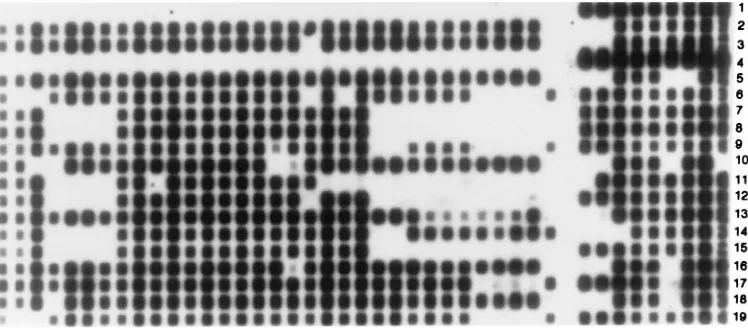
ml of 23SSPE (supplemented with 0.1% SDS) was heat denatured for 10 min at 1008C. The slots of the miniblotter were filled with 140ml of denatured and diluted PCR products, and the PCR products were hybridized for 60 min at 558C. The filter was removed and washed with 23SSPE (supplemented with 0.5% SDS) for 10 min at 558C and was then incubated in streptavidin-peroxidase conjugate (2.5ml of streptavidin-peroxidase conjugate [Boehringer Mannheim Biochemicals, East Sussex, United Kingdom] in 10 ml of 23SSPE supplemented with 0.5% SDS) for 45 min at 428C. The membrane was washed, and hybridized DNA was detected by using enhanced chemiluminescence detection liquid (Am-ersham International plc, Buckinghamshire, United Kingdom) and exposing the membrane for 5 min to X-ray film (Hyperfilm; Amersham International plc) (Fig. 1). The membrane was stripped by washing two times for 30 min each time in 1% SDS at 808C, followed by 15 min of incubation at 20 mM EDTA (pH 8) at room temperature.
Data analysis.The hybridization patterns were analyzed by using a word processor (see Fig. 2).
DNA fingerprinting.Genomic DNA was digested with restriction endonucle-asePvuII (New England Biolabs), electrophoresed through a 0.8% agarose gel, and vacuum blotted onto a nylon membrane (13). The molecular size standard used was a mixture ofPstI- andEcoRI-digested fragments of bacteriophagel
DNA (5 ng of each digest). After blotting, the membranes were hybridized with a digoxigenin-labelled IS6110probe. The IS6110probe was synthesized in digoxi-genin-labelled form by gene amplification with primers INS1 and INS2 in the presence of 35mM digoxigenin dUTP, 65mM dTTP, and 100mM (each) dATP, dCTP, and dUTP (12). Hybridization and colorimetric detection were performed in hybridization bottles (Hybaid Ltd., London, United Kingdom). The restriction fragment patterns revealed with IS6110were captured on 256 grey scale tagged image file format images at 150 dots per square inch by flatbed scanning. The images were compared by using the computer program Gel-Compar (Applied Maths). Matching coefficient matrices were used to produce dendrograms show-ing the relationship between strains by unweighted paired group method aver-ages. Strains with identical band patterns and those with single band difference were considered indistinguishable.
RESULTS
Patient characteristics.M. tuberculosisisolates from 167 pa-tients were analyzed. Of the 167 papa-tients, 87 were known to be male and 56 were known to be female. Twelve patients con-tributed more than one sample, and in total, 184 isolates ofM.
tuberculosiswere available for the study. In addition fourM.
aviumisolates were included as controls in a blinded fashion.
Spoligotyping. A total of 184 isolates from 167 individual patients were analyzed by spoligotyping. For each of the 12 patients who contributed more than one sample, serial isolates generated identical spoligotypes. The PCR products obtained from four M. avium isolates did not hybridize with the M.
tuberculosis-specific oligonucleotides, confirming the specificity
of the method forM. tuberculosiscomplex organisms. Figure 2 illustrates the spoligotype patterns obtained for the 167 iso-lates. A total of 88 distinct patterns were observed. Of these, 67 were represented by a unique isolate, with the remaining 100 isolates being grouped in 21 clusters, with each cluster varying in size from 2 to 20 isolates.
IS6110-RFLP analysis.As observed by spoligotyping, serial isolates from individual patients generated identical patterns by IS6110-RFLP analysis. From the 167 patients, 20 isolates had only one copy of IS6110and could be grouped into three clusters on the basis of the size of the single restriction frag-ment. Among the remaining isolates, 125 patterns were ob-served, with 111 of the patterns each represented by a single isolate, and 36 isolates grouped into 14 separate clusters. The results of cluster analysis with the two systems are summarized in Table 1.
Clusters identified by both spoligotyping and IS6110-RFLP analysis.Eleven clusters, comprising isolates from 32 patients, were defined on the basis of isolates sharing identical spoligo-types as well as IS6110-RFLP patterns. The evidence from epidemiological analysis was consistent with transmission be-tween individuals of isolates in eight of these clusters (Table 2). Patients in these clusters either were the members of the same family (clusters 2, 3, 5, and 6), they shared living accommoda-tions (clusters 1, 7, and 8), or they had contact through their social network (cluster 4). Patients in the other three clusters had no known epidemiological links.
Spoligotype clusters subdivided by IS6110-RFLP analysis.
Spoligotype analysis resulted in the grouping of a much larger number of isolates within apparently identical clusters than was the case for IS6110-RFLP typing. In total, 58 isolates included within spoligotype clusters were identified as unique strains by IS6110-RFLP analysis. The large spoligotype cluster represented by isolates C19 to C38 in Fig. 2, for example, was subdivided into 18 IS6110-RFLP types.
[image:2.612.121.495.71.235.2]IS6110-RFLP clusters subdivided by spoligotyping.The 20 isolates with only one copy of IS6110could be grouped into three clusters on the basis of the size of the single fragment. Spoligotype analysis of these isolates identified 15 distinct pat-terns. There were no known epidemiological links established among these 20 isolates from 20 different patients. A further six isolates clustered by IS6110-RFLP analysis, but with no FIG. 1. Spoligotyping patterns of some of the isolates ofM. tuberculosisanalyzed in this study. The PCR products of different isolates were hybridized to 43 spacer sequences which are found interspersed between the DRs in theMycobacteriumgenome. The black dot represents the presence of the spacer, and the blank space indicates the absence of the particular spacer within the amplified DNA from the mycobacterium. The numbers indicate the isolate number.
on May 15, 2020 by guest
http://jcm.asm.org/
known epidemiological link, could also be distinguished from each other by spoligotyping.
DISCUSSION
This study found that of the 184 isolates from the 167 pa-tients which were studied by DNA fingerprinting by RFLP analysis, 125 different types were identified, whereas only 88 distinct types were observed by spoligotyping. M. tuberculosis
isolates obtained from 32 patients were placed in 11 clusters by both methods.
The results of this direct comparison demonstrate that IS6110-RFLP analysis is more effective than spoligotyping in FIG. 2. Schematic representation obtained by using Microsoft Word of the spoligotyping patterns of all 167 isolates ofM. tuberculosisfrom different patients in the study arranged in order to show the similarities between the spoligotype patterns. The black rectangles represent positive signals, and the dots indicate a lack of hybridization.
on May 15, 2020 by guest
http://jcm.asm.org/
discriminating individual M. tuberculosisisolates. An analysis of the present panel of samples based solely on spoligotyping would have resulted in a significant number of isolates with unique IS6110-RFLP patterns being mistakenly included within clusters. An advantage is apparent, however, in the combined use of both systems. Particularly for isolates with a low copy number of IS6110, spoligotyping provides a powerful additional tool for strain differentiation. The discriminatory power of spoligotyping is a function of the diversity of the oligonucleotide sequences included in the hybridization mem-brane. In its present format, the spacer sequences included are derived from M. tuberculosis H37Rv (37 sequences) and M.
bovis BCG (6 sequences). We are analyzing sequences from
the DR region of isolates with unique IS6110 patterns but placed in clusters by spoligotyping. By using this information to design additional oligonucleotides for inclusion in subsequent membranes, it may be possible to produce improved
spoligo-typing protocols with an enhanced ability to differentiate strains in comparison to the ability of the present format.
RFLP analysis for IS6110has been used widely for retro-spective epidemiological studies (5, 7, 9, 10). However, the time required for culture will limit its use in the management of patients with acute clinical cases of infection or in rapidly responding to an outbreak. Spoligotyping is a PCR-based method and has a clear technical advantage over IS6110-RFLP analysis. The ability to perform the analysis directly with clin-ical samples will make the spoligotyping system particularly useful in cases in which, for example, nosocomial transmission of a particular strain of M. tuberculosisis suspected. Such a mode of transmission has played an important role in out-breaks of multidrug-resistant tuberculosis (2, 3, 11), and spo-ligotyping provides an important tool that can be used to assist in the rapid detection and containment of such incidents. The ease of analysis and storage of spoligotype patterns represents an additional technical advantage. In contrast to the require-ment for relatively sophisticated software for analysis of IS6110-RFLP gels, spoligotype patterns can readily be re-corded by hand or with any simple word processing program. The spoligotyping method is also economical since in the re-verse line blot hybridization assay used, filters with the multiple covalently linked spacer oligonucleotides are used to type as many as 43 specimens at one time, and these filters can be reused at least 20 times.
In summary, while the overall ability of spoligotyping to distinguish between individual isolates ofM. tuberculosisis less than that of IS6110-RFLP analysis, it has a particular applica-tion as a first-line typing technique because it can be per-TABLE 1. Cluster analysis by using two techniques
for strain typinga
Technique uniqueNo. of strains
No. of strains in clus-ters
No. of clusters
Spoligotyping 67 100 21
IS6110-RFLP analysis 111 56 17
[image:4.612.57.300.90.163.2]Spoligotyping1IS6110-RFLP analysis 135 32 11 aA total of 167 strains were tested.
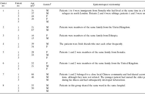
TABLE 2. Sociodemographic data for patients infected with strains exhibiting similar patterns by spoligotyping and IS6110-RFLP analysis Cluster
no. Patientno. (yr)Agea Genderb Epidemiological relationship
1 1 31 M Patients 1 to 4 were immigrants from Somalia who had lived at the same time in a hotel for refugees in north London. Patients 2 and 4 were siblings; patients 1 and 3 were unrelated.
2 37 M
3 44 F
4 26 F
5 M
2 1 21 M Patients were members of the same family from the United Kingdom.
2 67 M
3 1 16 F Patients were members of the same family from Ethiopia.
2 15 M
4 1 39 M The patients were Irish friends who met each other frequently.
2 34 F
5 1 28 F Patients 1 and 2 were members of the same family from Somalia.
2 33 M
3 F
6 1 52 F Patients 1 and 2 were members of the same family from the United Kingdom.
2 21 M
3 F
7 1 66 M Patients 1 and 2 belonged to a close local Chinese community and had shared accommoda-tions, although they were not related. The younger patient had nursed the older patient during his illness and had subsequently developed tuberculosis.
2 46 M
3 M
8 1 55 M Patients in this group shared the same ward in the same hospital.
2 50 M
3 M
aBlank spaces indicate that the patient’s age was not known. bM, male; F, female.
on May 15, 2020 by guest
http://jcm.asm.org/
[image:4.612.59.529.406.711.2]formed with extracts of sputum or bronchoalveolar lavage sam-ples. This provides the clinician with information on whether the patient has been infected by nosocomial transmission of a particular strain or strains known to be drug resistant. The data in the present study suggest that spoligotyping is most useful when different patterns refute the possibility of an epidemio-logical link between patients. When isolates give an identical spoligotyping pattern, our data suggest that it is necessary to conduct a second-line typing technique, such as IS6110-RFLP analysis, following organism culture, as was done in the present study. Confirmation of the strain type by the two approaches used together has greater discriminatory ability than that by either test alone. Spoligotyping has a particular advantage in distinguishing M. tuberculosis strains with low IS6110 copy numbers.
ACKNOWLEDGMENT
This work was supported by the British Lung Foundation.
REFERENCES
1.Alland, D., G. E. Kalkut, A. R. Moss, R. A. McAdam, J. A. Hahn, W. Bosworth, E. Drucker, and B. R. Bloom.1994. Transmission of tuberculosis in New York City, an analysis by DNA fingerprinting and conventional epidemiological methods. N. Engl. J. Med.330:1710–1716.
2.Dooley, S. W., M. E. Villarino, M. Lawrence, L. Salinas, S. Amil, J. V. Rullan, W. R. Jarvis, A. B. Bloch, and G. M. Cauthen.1992. Nosocomial transmis-sion of tuberculosis in a hospital unit for HIV-infected patients. JAMA
267:2632–2634.
3.Edlin, B. R., J. Y. Tokars, M. H. Grieco, J. T. Crawford, J. Williams, E. M. Sordillo, K. R. Ong, J. O. Kilburn, S. W. Dooley, K. G. Castro, W. R. Jarvis, and S. D. Holmberg.1992. An outbreak of multidrug resistant tuberculosis among hospitalized patients with the acquired immunodeficiency syndrome. N. Engl. J. Med.326:1514–1521.
4.Godfrey-Fausset, P., P. R. Mortimer, P. A. Jenkins, and N. G. Stoker.1992. Evidence of transmission of tuberculosis by DNA fingerprinting. Br. Med. J.
305:221–223.
5.Goyal, M., L. P. Ormerod, and R. J. Shaw.1994. Epidemiology of an
outbreak of drug-resistant tuberculosis in the UK using restriction fragment length polymorphism. Clin. Sci.86:749–751.
6.Hermans, P. W. M., D. van Soolingen, E. M. Bik, P. E. W. De Haas, J. W. Dale, and J. D. A. van Embden.1991. Insertion element IS987from Myco-bacterium bovisBCG is located in a hot-spot integration region for insertion elements inMycobacterium tuberculosiscomplex strains. Infect. Immun.59:
2695–2705.
7.Hermans, P. W. M., D. van Soolingen, J. W. Dale, A. R. J. Schuitema, R. A. McAdam, D. Catty, and J. D. A. van Embden.1990. Insertion element IS986 fromMycobacterium tuberculosis: a useful tool for diagnosis and epidemiol-ogy of tuberculosis. J. Clin. Microbiol.28:2051–2058.
8.Kamerbeek, J., L. Schouls, A. Kolk, M. van Agterveld, D. van Soolingen, S. Kuijper, A. Bunschoten, H. Molhuizen, R. Shaw, M. Goyal, and J. van Embden.Simultaneous detection and strain differentiation of Mycobacte-rium tuberculosis for diagnosis and epidemiology. Submitted for publication. 9.Mazurek, G. H., M. D. Cave, K. D. Eisenach, R. J. Wallace, Jr., J. H. Bates, and J. T. Crawford.1991. Chromosomal DNA fingerprint patterns produced with IS6110as strain-specific markers for epidemiologic study of tuberculo-sis. J. Clin. Microbiol.29:2030–2033.
10. Otal, I., C. Martı´n, V. Vincent-Levy-Frebault, D. Thierry, and B. Gicquel.
1991. Restriction fragment length polymorphism analysis using IS6110as an epidemiological marker in tuberculosis. J. Clin. Microbiol.29:1252–1254. 11. Pearson, M. L., J. A. Jereb, T. R. Frieden, et al.1992. Nosocomial
transmis-sion of multidrug-resistant Mycobacterium tuberculosis. A risk to patients and health care workers. Ann. Intern. Med.117:191–196.
12. van Embden, J. D. A., M. D. Cave, J. T. Crawford, J. W. Dale, K. D. Eisenach, B. Gicquel, P. W. M. Hermans, C. Martin, R. Mcadam, T. M. Shinnick, and P. M. Small.1993. Strain identification of Mycobacterium tuberculosis by DNA fingerprinting: recommendations for a standardized methodology. J. Clin. Microbiol.31:406–409.
13. van Soolingen, D., P. W. M. Hermans, P. E. W. de Haas, D. Soll, and J. D. A. van Embden.1991. Occurrence and stability of insertion sequences in My-cobacterium tuberculosiscomplex strains: evaluation of an insertion se-quence-dependent DNA polymorphism as a tool in the epidemiology of tuberculosis. J. Clin. Microbiol.29:2578–2586.
14. Yang, Z. H., P. E. W. de Haas, D. van Soolingen, J. D. A. van Embden, and A. B. Andersen.1994. Restriction fragment length polymorphism of Myco-bacterium tuberculosisstrains isolated from Greenland during 1992: evidence of tuberculosis transmission between Greenland and Denmark. J. Clin. Mi-crobiol.32:3018–3025.
15. Yuen, L. K., B. C. Ross, K. M. Jackson, and B. Dwyer.1992. Characterization ofMycobacterium tuberculosisstrains from Vietnamese patients by Southern blot hybridization. J. Clin. Microbiol.31:1615–1618.