Glucocorticoids decrease tissue mast cell
number by reducing the production of the c-kit
ligand, stem cell factor, by resident cells: in
vitro and in vivo evidence in murine systems.
S Finotto, … , Y A Mekori, D D Metcalfe
J Clin Invest.
1997;
99(7)
:1721-1728.
https://doi.org/10.1172/JCI119336
.
The local delivery of glucocorticoids to tissues significantly decreases mast cell number.
This pharmacologic effect of glucocorticoids is believed to be one of the mechanisms by
which glucocorticoids regulate allergic inflammation. To determine the mechanism by which
glucocorticoids are able to exert this effect, we first applied the glucocorticoid fluocinonide to
mouse dermis and observed that the decrease in mast cell number was associated with an
increase in mast cell apoptosis. This did not appear to be due to a direct effect of the
glucocorticoid on mast cells, as the addition of 0.01-1.0 microM of the glucocorticoid
dexamethasone into stem cell factor (SCF)-dependent mast cell cultures did not enhance
mast cell death. However, addition of dexamethasone to cultured fibroblasts did result in a
downregulation of SCF mRNA and a significant decrease in SCF protein production.
Similarly, immunohistochemistry performed on fluocinonide-treated mouse dermis revealed
a decrease in immunoreactive SCF. Administration of SCF at sites of fluocinonide
administration to the dermis abolished the mast cell-depleting effect of this glucocorticoid.
Thus, glucocorticoids decrease tissue mast cell number by downregulating tissue SCF
production required for the survival of local mast cells. This observation may be applicable
to the design of improved strategies to treat mast cell-mediated disorders.
Research Article
Find the latest version:
The Journal of Clinical Investigation Volume 99, Number 7, April 1997, 1721–1728
Glucocorticoids Decrease Tissue Mast Cell Number by Reducing the Production
of the c-kit Ligand, Stem Cell Factor, by Resident Cells
In Vitro and In Vivo Evidence in Murine Systems
Susetta Finotto,* Yoseph A. Mekori,‡ and Dean D. Metcalfe*
*Laboratory of Allergic Diseases, National Institute of Allergy and Infectious Diseases, National Institutes of Health, Bethesda, Maryland 20892; and ‡Department of Medicine, Sackler School of Medicine, Tel Aviv University, Israel
Abstract
The local delivery of glucocorticoids to tissues significantly
decreases mast cell number. This pharmacologic effect of
glucocorticoids is believed to be one of the mechanisms by
which glucocorticoids regulate allergic inflammation. To
determine the mechanism by which glucocorticoids are able
to exert this effect, we first applied the glucocorticoid
fluoci-nonide to mouse dermis and observed that the decrease in
mast cell number was associated with an increase in mast
cell apoptosis. This did not appear to be due to a direct
ef-fect of the glucocorticoid on mast cells, as the addition of
0.01–1.0
m
M of the glucocorticoid dexamethasone into stem
cell factor (SCF)-dependent mast cell cultures did not
en-hance mast cell death. However, addition of dexamethasone
to cultured fibroblasts did result in a downregulation of
SCF mRNA and a significant decrease in SCF protein
pro-duction. Similarly, immunohistochemistry performed on
fluocinonide-treated mouse dermis revealed a decrease in
immunoreactive SCF. Administration of SCF at sites of
flu-ocinonide administration to the dermis abolished the mast
cell–depleting effect of this glucocorticoid. Thus,
glucocorti-coids decrease tissue mast cell number by downregulating
tissue SCF production required for the survival of local
mast cells. This observation may be applicable to the design
of improved strategies to treat mast cell–mediated
disor-ders. (
J. Clin. Invest.
1997. 99:1721–1728.) Key words:
glu-cocorticoids
•stem cell factor
•apoptosis
•mast cells
•fibro-blasts
Introduction
Mast cells resident in tissues are capable of inducing or potenti-ating local inflammation after activation by a variety of stimuli including interactions between antigen and membrane-bound IgE or IgG; and exposure to C3a or C5a (1–7). Glucocorticoids delivered at potential or on-going sites of tissue inflammation decrease the number of resident tissue mast cells, an effect that contributes to the antiinflammatory properties of these agents. Thus, glucocorticoids applied to the skin (8, 9), lung (10), and
intestine (11) result in a significant reduction in mast cells. However, the mechanism by which glucocorticoids reduce tis-sue mast cell number is not known. Understanding the way in which glucocorticoids decrease mast cell number could lead to improved strategies to interrupt mast cell–mediated inflamma-tion while limiting the side effects of locally administered ste-roids.
The number of tissue mast cells is believed to be regulated in the microenvironment principally by the c-kit ligand, stem cell factor (SCF)1 (12–14) which is produced by resident cells
such as fibroblasts (15–18). Withdrawal of SCF from SCF-dependent mast cell cultures in vitro results in apoptosis (19, 20), and administration of SCF in vivo in both murine and hu-man studies leads to both local and systemic mast cell hyper-plasia (21–23). Therefore, we hypothesized that glucocorti-coids affect mast cell number in tissues by regulating SCF production by cells in the microenvironment.
To explore this hypothesis, we performed a variety of in vitro and in vivo experiments. As will be shown, glucocorti-coids do not appear to directly alter mast cell number by acting on mast cells themselves. Rather, glucocorticoids decrease the production of SCF by fibroblasts in vitro and resident connec-tive tissue cells in vivo; and the mast cell–depleting effects of glucocorticoids in vivo are reversed by the local administration of SCF.
Methods
Materials.DME, glutamine, penicillin, and streptomycin (Gibco Laboratories, Grand Island, NY); FBS (Bioproducts for Science, Inc., Indianapolis, IN); water-soluble dexamethasone (Sigma Chemical Co., St. Louis, MO); Lidex (fluocinonide) ointment 0.05% (Syntex Laboratories, Inc., Palo Alto, CA); recombinant murine SCF (rm-SCF) expressed in Escherichia coli (R & D Systems, Minneapolis, MN); polyclonal rabbit anti–mouse SCF (Genzyme Corp., Cam-bridge, MA); normal goat serum (BioSource, International, Cama-rillo, CA); normal rabbit serum (Dako Corp., Carpinteria, CA); Vectastain ABC-AP kit and alkaline phosphatase substrate kit (Vec-tor Labs, Inc., Burlingame, CA); Levamisole (Sigma Chemical Co.); biotinylated anti–rabbit IgG (Vector Labs, Inc.); Cy3-conjugated streptavidin (Jackson ImmunoResearch Laboratories, Inc., West Grove, PA); and ApoTag Plus in situ apoptosis detection kit (On-cor Inc., Gaithersburg, MD) were obtained from the manufacturers.
Cell cultures. Human fetal skin and NIH 3T3 fibroblasts (passage 5, CRL 1475-CCD27SK) were purchased from American Type Cul-ture Collection (Rockville, MD). The cell culCul-tures were maintained in DME with 10% FBS, 2 mM glutamine, 100 U/ml penicillin, and 50
mg/ml streptomycin. MCP5 mast cells of Balb/c origin (19) were maintained in RPMI 1640 with 10% heat-inactivated FBS, 50 mM Address correspondence to Dean Metcalfe, M.D., NIH/NIAID/
LAD, Building 10 Room 11C205, 10 Center Drive MSC1881, Be-thesda, MD 20892-1881. Phone: 301-496-2165; FAX: 301-480-8384; E-mail: [email protected]
Received for publication 11 April 1996 and accepted in revised form 24 January 1997.
b-mercaptoethanol, 2 mM l-glutamine, 25 mM Hepes, 1 mM sodium
pyruvate, nonessential amino acids, 100 U/ml penicillin, 50 mg/ml streptomycin, and 10% (vol/vol) WEHI-3 conditioned medium as a source of IL-3. rmSCF was added (70 ng/ml) to some of the cell sus-pensions where indicated.
RNA samples and reverse transcription. Human fetal skin fibro-blasts in subconfluent cultures were washed with PBS and incubated three times for 24 h with or without specified amounts of dexametha-sone. At the end of each experiment, adherent fibroblasts were de-tached with trypsin/versene (Biofluids, Inc., Rockville, MD), counted, washed twice in PBS, lysed in 4 M guanidinium isothiocyanate and to-tal RNA isolated using guanidine thiocyanate/phenol-chloroform ex-traction as described (24).
cDNAs were synthesized from total RNA with avian myeloblast virus reverse transcriptase and random hexamer primers (cDNA Cy-cle™ kit for RT-PCR; Invitrogen Corp., San Diego, CA). Two
oligo-nucleotides were synthesized (Operon Technologies, Inc., Alameda, CA) that correspond to the sequence in cDNA for human (h)SCF (oligonucleotide I: 59-CTTCAACATTAAGTCCTGAG-39 and oli-gonucleotide II: 59-CAGTGTTGATACAAGCCACA-39). These oli-gonucleotides amplify a 359-bp and a 275-bp product of SCF mRNA when exon 6 is present or deleted by alternative splicing, respectively (25, 26). cDNA amplification was performed at 40 cycles each of de-naturation at 958C for 1 min, annealing at 558C for 1 min, and exten-sion at 728C for 2 min.
Probe construction and labeling. The 359-bp PCR product of SCF mRNA obtained as described above was cloned in a pBluescript II SK(1/2) vector at the EcoRV site of the polylinker using blunt end ligation. The new construct was then sequenced according to the dideoxy-chain-termination method with a Sequenase kit, version 2.0 (United States Biochemical Corp., Cleveland, OH). The plasmid was then linearized by restriction digestion with XhOI. The linearized DNA template was in vitro transcribed with the MAXIscript™ system
(Ambion Inc., Austin, TX) using T3 RNA polymerase to obtain an antisense probe for detecting SCF mRNAs. In vitro transcribed anti-sense RNA was uniformly labeled with [a-32P]uridine triphosphate
(Amersham, Arlington Heights, IL). After in vitro transcription, the DNA template was digested with RNase-free DNase I. The probe was then purified with a 7 mol/liter urea/6% polyacrylamide gel (Na-tional Diagnostics, Inc., Atlanta, GA). The full-length probe (483 nu-cleotides [nt]) was subsequently used in a ribonuclease protection as-say (RPA). Hybridization of this transcript to human total RNA protects a 359 nt fragment of SCF which extends from nucleotide 679 to 1038 of the cDNA sequence (GenBank accession No. MC59964) (26). The pTRI-b-actin-human antisense control template contains a 245-bp fragment of the human cytoplasmic b-actin gene which ex-tends from codon 220 to 303 (nucleotides 704–947 of cDNA se-quence, GenBank accession No. X00351). The b-actin fragment was inserted into the kpnI-EcoRI sites of a pTRIPLEscript vector (Am-bion Inc.). The plasmid was linearized with XbaI and HindIII, and the linearized DNA template was in vitro transcribed as above using T7 RNA polymerase, gel purified to obtain a fragment of 304 nt, and then used in the RPAs. Hybridization of the transcript to human total RNA protects a 245 nt fragment of b-actin mRNA.
RPA. Preliminary studies demonstrated that SCF mRNA levels in fibroblasts were at the lower limits of detection of standard North-ern blots, therefore the RPA was utilized employing a commercially available kit (RPA II; Ambion Inc.). Each 10 mg of total RNA was hybridized overnight at 438C to 50,000 cpm of gel purified antisense probe, followed by RNase T1 (100 U/ml) digestion for 30 min at 378C. RNase digestion was terminated by inactivation of the enzyme fol-lowed by ethanol precipitation. Digestion products were subjected to gel electrophoresis using a 7 mol/liter urea/6% polyacrylamide gel (Sequagel; National Diagnostics, Inc.). As control, yeast RNA was hybridized to each probe and then treated with or without RNase T1. In addition, for each experiment the DNA molecular weight marker was labeled with [a-32P]uridine triphosphate and subjected to
electro-phoresis along with the other samples. Next, Kodak x-ray film
(East-man Kodak, Rochester, NY) was exposed to dried gel at 2708C with intensifying screens. Densitometric analysis was performed using a Molecular Dynamics Scanner (Molecular Dynamics, Sunnyvale, CA). Two separate experiments in duplicate were performed.
SCF ELISA. Fibroblasts were cultured in DME with 10% FBS with and without dexamethasone at 1.00, 0.100, and 0.010 mM for 72 h with medium and dexamethasone replaced at 24 and 48 h. Superna-tants corresponding to the last 24 h were collected, concentrated by ultracentrifugation through an anisotrope membrane (Centricon 10; Amicon, Inc., Beverly, MA), and stored at 2808C until analyzed by ELISA. Fibroblasts were then detached with trypsin/versene solution and viability was assessed by trypan blue dye exclusion. Soluble SCF was measured in the concentrated supernatants using a Quantikine™
ELISA kit (R & D Systems). Standards for soluble SCF were pre-pared using culture medium as diluent. Under these experimental conditions, the minimum detectable dose of SCF is 3.0 pg/ml. SCF levels were corrected to cell number and expressed as pg of SCF/106
cells.
SCF detection in 3T3 fibroblasts by immunohistochemistry. 3T3 fi-broblasts were cultured on 12-mm-diameter coverslips in 24-well plates in duplicate in DME with 10% FBS with and without 1 mM
2208C. dexamethasone for 72 h with medium and dexamethasone re-placed at 24 and 48 h. At the end of the culture, wells were washed in PBS, coverslips were removed from the wells, air dried, and fixed in methanol for 5 min at 2208C. Coverslips were rehydrated in three washes of PBS and incubated for 1 h with 100% heat-inactivated nor-mal goat serum to prevent nonspecific binding. Coverslips were then incubated with rabbit anti–mouse SCF (10 mg/ml) in PBS with 10% normal goat serum overnight at 48C. The control for nonspecific bind-ing was performed usbind-ing nonimmune rabbit serum at an equivalent concentration, instead of the primary antibody. The coverslips were washed in PBS, and specific binding was detected with a biotinylated anti–rabbit IgG antibody (1:100) for 1 h at room temperature fol-lowed by incubation with Cy3™-streptavidin conjugated antibody (1:
500) for 1 h at room temperature. After three washes in PBS, cover-slips were rinsed in distilled water and mounted with an aqueous mounting medium. Three experiments in duplicate were performed.
FACS® analysis. At time 0 and at specified time points, cells were analyzed by flow cytometric analysis and by using propidium iodide (PI) (Sigma Chemical Co.) uptake to assess cell viability after growth factor deprivation and dexamethasone treatment. Dexamethasone was added at the beginning of the cultures at the indicated concentra-tions to cells placed in 96-well plates at 7 3 104 cells/ml. Cells were
then collected and suspended at 106 cells/ml in PBS containing 0.1%
BSA and PI added at a final concentration of 2.5 mg/ml between 5 and 15 min before analysis (FACS 440; Becton Dickinson, Mountain View, CA).
In vivo studies. Groups of three 5–6-mo-old female Balb/c mice (Jackson Laboratory, Bar Harbor, ME) were lightly anesthetized by Methophane inhalation before injection. Intradermal injection of an ear was performed three times a week for 2 wk with vehicle alone (sterile saline with 0.1% BSA, fraction V; Sigma Chemical Co.) or ve-hicle containing rmSCF (30 mg rmSCF/Kg in 20 ml of sterile saline containing 0.1% BSA). A third group received concomitant daily treatment with fluocinonide (0.05 mg/treatment) at the site of rmSCF injection. An additional control group was treated with fluocinonide alone. In additional experiments, the left ear was treated with fluoci-nonide and the right ear was not treated or treated with an ointment control cream (Aquaphor®; Beiersdorf Inc., Norwalk, CT). Mast cell
number per mm2 was determined as described (27) with
modifica-tions. Briefly, both ears from each of three mice per condition were removed after death by cervical dislocation, fixed in 10% neutral buffered formalin for 24 h, immersed in 70% ethanol, and embedded in paraffin. Three tissue sections (4 mm) from each ear, unless other-wise stated, were placed on polylysine-coated slides and stained with Lennert’s Giemsa at pH 0.5 (28). Each section was examined at 4003
number of mast cells per mm2 in each ear was calculated by averaging
the results obtained from each section. This final determination was considered as one observation. Mast cells as percentage of control was determined by comparing mast cell number under each condition and time point to the mean number of mast cells per mm2 in control
ears.
Immunohistochemistry. For immunostaining, 4-mm tissue sections were floated onto polylysine-coated slides, dewaxed through xylene, rehydrated through an ethanol series, washed in PBS, and incubated in 100% heat-inactivated normal goat serum to prevent nonspecific binding. The slides were then subjected to staining for SCF protein using rabbit anti–mouse SCF (10 mg/ml) in PBS with 0.1% BSA and 10% normal goat serum overnight at 48C. The control for nonspecific binding was performed by omitting the primary antibody and by us-ing nonimmune rabbit serum at an equivalent concentration instead of the primary antibody. The sections were washed with PBS. Specific binding was detected by the Vectastain ABC-AP and alkaline phos-phatase substrate kits according to the manufacturer’s instructions. Endogenous alkaline phosphatase was blocked using 1 mM Levami-sole in the substrate solution. Slides were counterstained with hema-toxylin solution, Gill No. 2 (Sigma Chemical Co.), dehydrated, and mounted in Permount (Fisher Scientific, Pittsburgh, PA). Three mice for each group and six sections for each sample were stained and sub-sequently analyzed using a light microscope at a magnification of 400. In situ apoptosis (TUNEL assay). For detection of apoptotic mast cells, ear samples were obtained at specified time points, fixed in 10% buffered formalin at 48C, embedded in paraffin, and 4-mm sections were mounted on polylysine-coated slides. Next, slides were deparaf-finized in xylene, hydrated through an ethanol series, washed in PBS, and subjected to proteinase treatment (20 mg/ml) (Proteinase K; GIBCO BRL, Gaithersburg, MD) for 15 min at room temperature. Endogenous peroxidase was inhibited by 2.5% hydrogen peroxide treatment. Incubation with a digoxigenin-nucleotide triphosphate tar-gets catalytically the 39-OH ends of fragmented DNA through the en-zyme terminal deoxynucleotidyl transferase. As a negative control, water was added instead of terminal deoxynucleotidyl transferase en-zyme to the reaction buffer. This treatment was followed by incuba-tion with antidigoxigenin antibody peroxidase conjugate and detec-tion was performed with a peroxidase substrate in accordance with the manufacturer’s instructions (ApopTag™ in situ apoptosis
detec-tion kit). Slides were briefly rinsed in acid water (pH 0.4) and stained with alcian blue, at pH 0.5, to visualize mast cells. Next, samples were dehydrated through an ethanol series followed by a xylene wash and then mounted in Permount. Three mice per group and 200 alcian blue positive mast cells per mouse were analyzed for each sample at 1,0003. Results are reported as percent apoptotic mast cells.
Results
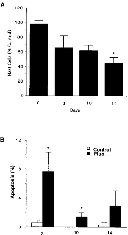
Depletion of tissue mast cells. It has been reported that corti-costeroids applied to the skin result in a decrease in mast cell number (8, 9). To examine this phenomenon, we first applied the glucocorticoid fluocinonide topically to mouse skin and de-termined the number of cutaneous mast cells over 14 d. As can be seen in Figs. 1 A and 2, A and C, the number of mast cells in
the cutaneous tissues of mouse ears decreased by z 35% by
day 3 and 56% by day 14 (P, 0.05, compared with day 0), sim-ilar to published reports (8, 9).
To further examine this mast cell depletion, we investi-gated whether the decrease in mast cell number was accompa-nied by an increase in the number of mast cells undergoing apoptosis. In fact, fluocinonide treatment was accompanied by an increase in the number of mast cells undergoing apoptosis (Figs. 1 B and 2, B and D). The number of mast cells
undergo-ing apoptosis was maximum at day 3 (7.6262.7). An increase
in mast cells undergoing apoptosis above control continued to
be observed through day 14 of the study and was statistically significant (P, 0.05) through day 10.
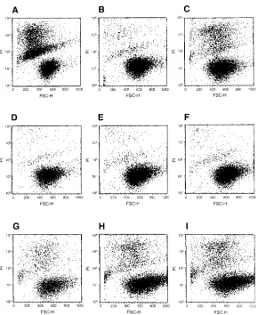
Effect of glucocorticoid on mast cell survival in vitro. The observation that the decrease of mast cell number in vivo is ac-companied by mast cell apoptosis suggested that glucocorti-coids could either act by decreasing the production of SCF by tissue stromal cells which is required to maintain mast cell via-bility, or alternatively by blocking the ability of SCF to main-tain mast cell viability by acting directly on mast cells. To ex-amine the latter possibility, we investigated the ability of dexamethasone to decrease the survival of mast cells in vitro in the presence of SCF (19). As can be seen in Fig. 3 and Table I, mast cells cultured in IL-3 remained viable while mast cells
[image:4.612.315.531.57.454.2]cultured without IL-3 exhibited apoptosis (compare Fig. 3, A
Figure 1. Effect of fluocinonide on mast cell number and number of mast cells in apoptosis in mouse ear dermis. Fluocinonide-induced decrease in mast cell number as percent control is shown in A, and the percentage of mast cells undergoing apoptosis over 14 d is shown in B. Mast cell numbers were determined as described in Methods. The average number of mast cells in untreated mouse ears was 13868.7 per mm2. Data are presented as mean6SEM (n5 3).
with B) as evident by increased PI uptake (19). IL-3–depen-dent mast cells removed from IL-3 and placed in SCF showed a decrease in PI uptake, although this decrease was less than that observed in growth factor–deprived mast cells (compare Fig. 3, C and A). The addition of dexamethasone from 0.01 to 1.00 mM did not alter the ability of SCF (or IL-3) to maintain mast cell viability through 96 h (Fig. 3, D–I) (Table I). Thus, dexamethasone did not block the effect of SCF in promoting mast cell viability.
Effect of dexamethasone on SCF production by fibro-blasts. To examine whether glucocorticoids decrease mast cell number in tissues by downregulating the production of SCF by stromal cells, we first investigated the in vitro effect of dexa-methasone on the production of SCF mRNA by human fetal skin fibroblasts. As shown in Fig. 4, when human fetal skin fi-broblasts are cultured in dexamethasone (0.1 and 1.0 mM) the amount of SCF mRNA is decreased as assessed by RPA (Fig. 4, A and B) in a dose–response manner (Fig. 4 C).
To verify that the decrease in SCF mRNA is accompanied by a decrease in SCF protein, we added increasing amounts of dexamethasone to human fetal fibroblasts in culture. This re-sulted in a significant decrease in the amount of SCF protein in the culture supernatants, again in a dose–response manner (Fig. 5 A). Thus, 1 mM dexamethasone decreased SCF protein production from 311.0650.0 to 146.4621.3 pg 106 cells. To rule
out the possibility of a cytotoxic effect on human fetal skin fi-broblasts from dexamethasone, we measured cell viability by trypan blue dye exclusion at the end of each treatment. No change in fibroblast viability was observed under all
experi-mental conditions (98.7561.03% and 98.9661.03% after no
treatment or 1.0 mM dexamethasone, respectively; n5 3 in
triplicate).
Furthermore, to establish that glucocorticoids can decrease SCF in mouse NIH 3T3 fibroblasts as well as in human fibro-blasts, we cultured 3T3/NIH mouse fibroblasts on cover slips
[image:5.612.58.434.59.510.2]for 72 h with and without 1 mM dexamethasone. This resulted
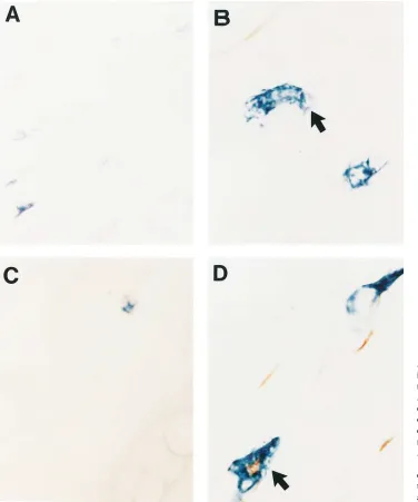
Figure 2. Light microscopy show-ing mast cells in control and fluoci-nonide-treated mouse ear dermis. Alcian blue–stained mast cells are easily seen in an untreated section of dermis (3400) (A), while mast cells in fluocinonide-treated der-mis are decreased in number (C). A normal mast cell (arrow) at
in a visible decrease in cell-associated SCF after dexametha-sone treatment (Fig. 5, B and C).
In vivo treatment with fluocinonide decreases SCF protein at the treated sites. To confirm the in vitro observation in vivo that glucocorticoids downregulate SCF production by fibro-blasts, we next examined normal and fluocinonide-treated mouse skin for the presence of SCF protein using
immunohis-tochemistry. SCF protein was detected in untreated mouse skin within stromal cells and in the epidermis (Fig. 6 A) as has been reported in human skin (25). The amount of protein asso-ciated with stromal cells appeared to decrease after application of fluocinonide (Fig. 6 B). Thus, glucocorticoids decrease SCF production by fibroblasts in vitro and similarly by stromal cells in vivo.
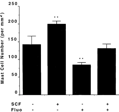
SCF injection in vivo. The above data are consistent with the hypothesis that application of glucocorticoids to the dermis depletes mast cells by decreasing the amount of SCF produced by stromal cells, and this in turn leads to mast cell apoptosis. If this hypothesis is correct, it should be possible to reverse the effect of fluocinonide by the introduction of SCF at the site of application of the glucocorticoid. As can be seen in Fig. 7, in-troduction of SCF into mouse skin in the absence of fluocino-nide increases mast cell number as reported (20); and as previ-ously noted, application of fluocinonide decreases mast cell number. This depletion of mast cells induced by fluocinonide was reversed with local introduction of SCF (P, 0.01), consis-tent with the hypothesis that the mast cell–depleting effect of glucocorticoids is the result of a downregulation of local SCF production.
Discussion
[image:6.612.60.350.55.409.2]Local contact of glucocorticoids with dermal, respiratory, and intestinal tissues is reported to be associated with a decrease in
Figure 3. Analysis of PI uptake by flow cytometry in cultured mast cells. IL-3–dependent MCP5 cells were in-cubated for 48 h without IL-3 or SCF (A) or in media containing IL-3 (B, D, E, F) or SCF (70 ng/ml) (C, G, H, I), containing 0.01 mM (D and G), 0.1 mM (E and H), and 1.00 mM (F and I) dexamethasone before anal-ysis. For details, see Methods.
Table I. Analysis of Mast Cell Survival (%) with Dexamethasone (DEX) Treatment
DEX
Growth factor (h) 0.01 mM 0.10 mM 1.00 mM
IL-3 (48) 101.2860.35 101.0160.68 100.0660.88 (72) 105.6362.22 103.6260.66 103.9460.48 (96) 106.0963.33 104.3062.33 104.6562.15 SCF (48) 99.3263.42 101.3461.22 99.6161.69 (72) 99.1462.15 104.5864.65 104.3962.14 (96) 96.7764.82 94.8163.66 93.4466.79
[image:6.612.56.299.558.673.2]mast cell number within these tissues (8–11, 29, 30). This de-crease in mast cell number takes place over days and generally requires repeated application of glucocorticoids (8). This bio-logic effect of a glucocorticoid would, through the diminution of mast cell number, be expected to downregulate allergic in-flammation. The mechanism by which this downregulation of mast cell number occurs is unknown.
It was our hypothesis that glucocorticoids decrease mast cell number either by modulating the synthesis of SCF within tissues, or the ability of SCF to maintain the viability of mast cells themselves, as it is known that mature mast cells require SCF for survival (19, 31). In examining the latter possibility, we found that the addition of the glucocorticoid dexametha-sone to SCF-dependent mast cell cultures did not interfere with the ability of SCF to maintain the viability of these cells (Fig. 3). That is, dexamethasone did not induce mast cell apop-tosis in the presence of SCF. However, our in vitro studies
re-producing the reported decrease in mast cell number after the application of a topical glucocorticoid, in this case fluocinonide (Fig. 1 A), revealed the apoptosis of mast cells within fluocino-nide-treated tissue (Figs. 1 B and 2, C and D). Assuming that this apoptosis was associated with a deprivation of SCF, it was then logical to hypothesize that glucocorticoids have the ability to downregulate the production of SCF from stromal cells, such as fibroblasts, which are known to produce SCF in tissues (15–18, 32). In vitro studies first revealed that dexamethasone-treated fibroblasts produced less SCF mRNA (Fig. 4) in a dose–response fashion. Furthermore, the amount of SCF pro-tein produced by dexamethasone-treated human and mouse fibroblasts was also decreased in vitro (Fig. 5). Immuno-histochemistry for SCF within fluocinonide-treated dermis similarly was interpreted as showing that fluocinonide appears to downregulate SCF within tissue (Fig. 6). Thus, both in vitro and in vivo evidence was consistent in showing that glucocorti-coids downregulate SCF production. This finding is not unex-pected. Glucocorticoids have been reported to decrease the production of cytokines including IL-1 and TNF (33–36).
If glucocorticoids applied to the dermis induce mast cell apoptosis through a downregulation of local SCF production by resident cells, then the administration of SCF at sites under-going fluocinonide treatment should compensate for the de-crease in local synthesis of SCF. The net result would be a nor-malization of tissue mast cell number. Indeed, this was the case (Fig. 7), as the administration of exogenous SCF maintained mast cell number in fluocinonide-treated dermis (Fig. 7).
[image:7.612.59.301.57.528.2]In summary, these data demonstrate that the decrease in tissue mast cell number which follows the application of gluco-corticoids is associated with apoptosis of mast cells. Further-more, this apoptosis is shown to be the result of a glucocorti-coid-dependent decrease in local SCF production. From these data it would be anticipated that injection of glucocorticoids
Figure 4. Effect of dexamethasone on SCF mRNA levels in fibro-blasts. Autoradiogram of electrophoretic anal-ysis of RNase protec-tion products for SCF (A) and b-actin (B) from fibroblasts treated for 72 h with dexameth-asone and separated on 6% polyacrylamide gels in 7 M urea (see Methods). 10 mg of total RNA from control (lane 1) and dexameth-asone-treated fibro-blasts (0.100 and 1.00
mM, lanes 2 and 3, re-spectively) was hybrid-ized with 32P-labeled
hu-man SCF probe (A) where the full-length protected product of SCF is 359 nt (the sec-ond protected form of SCF of 275 nt is seen only in the control sam-ple), or a 32P-labeled
human b-actin probe (B) where the full-length product (304 nt) and the protected prod-uct (245 nt) are visible. Densitometric analysis of SCF mRNA normal-ized to human b-actin mRNA in fibroblasts exposed to the indi-cated doses of dexa-methasone is also shown (C).
[image:7.612.318.552.461.681.2]Figure 5. Inhibition of SCF protein production in fibroblasts by dexa-methasone. (A) Human fetal skin fibroblasts were treated for 72 h with dif-ferent concentrations of dexamethasone or with medium alone. Soluble SCF was measured in cell free supernatants with an ELISA assay (see Methods), data are shown as the mean6SEM (n5 3, each experiment performed in triplicate). Statistical analysis was performed using Student’s t test (two-tailed) where *P, 0.05; **P, 0.01; ***P, 0.001. (B and C) Immunohistochemical detection of SCF protein in 3T3/NIH mouse fibro-blasts (3400) after culture for 72 h without (B) or with (C) dexametha-sone. Coverslips of fibroblast cultures were stained with a polyclonal anti-body against SCF (10 mg/ml).
into inflamed tissues would result in a more dramatic reduc-tion in mast cell number, as has been reported in synovial tis-sue after injection of glucocorticoids (37).
These data suggest that strategies based upon these obser-vations could be developed and directed at a more efficient de-livery of glucocorticoids to target stromal cells that produce SCF. This would be expected to minimize the side effect of these drugs or to maximize their ability to decrease mast cell number. Such strategies would be particularly helpful in de-signing new approaches to treat allergic diseases and disorders associated with mast cell hyperplasia.
References
1. Gordon, J.R., and S.J. Galli. 1991. Release of both preformed and newly synthesized tumor necrosis factor a (TNF-a)/cachectin by mouse mast cells stimulated via the FceRI. A mechanism for sustained action of mast cell derived TNF-a during IgE-dependent biological responses. J. Exp. Med. 174:103–107.
2. Mican, J.M., N. Arora, P.R. Burd, and D.D. Metcalfe. 1992. Passive cuta-neous anaphylaxis in mouse skin is associated with local accumulation of inter-leukin-6 (IL-6) mRNA and immunoreactive IL-6 protein. J. Allergy Clin. Im-munol. 90:815–824.
3. Plaut, M., J.H. Pierce, C.J. Watson, J. Hanley-Hyde, R.P. Nordan, and W.E. Paul. 1989. Mast cell lines produce lymphokines in response to cross-link-age of FceRI or to calcium ionophore. Nature (Lond.). 339:64–67.
4. Haak-Frendscho, M., J. Ridgway, R. Shields, K. Robbins, C. Gorman, and P. Jardieu. 1993. Human IgE receptor alpha-chain IgG chimera blocks pas-sive cutaneous anaphylaxis reaction in vivo. J. Immunol. 151:351–358.
5. Daeron, M., O. Malbec, S. Latour, M. Arock, and W.H. Fridman. 1995. Regulation of high affinity IgE receptor–mediated mast cell activation by mu-rine low-affinity IgG receptors. J. Clin. Invest. 95:577–585.
6. Johnson, A.R., T.E. Hugli, and H.J. Muller-Eberhard. 1975. Release of histamine from rat mast cells by the complement peptides C3a and C5a. Immu-nology. 28:1067–1080.
7. Ramos, B.F., Y. Zhang, and B.A. Jakschik. 1994. Neutrophil elicitation in the reverse passive Arthus reaction. Complement-dependent and independent mast cell involvement. J. Immunol. 152:1380–1384.
8. Lavker, R.M., and N.M. Schechter. 1985. Cutaneous mast cell depletion results from topical corticosteroids usage. J. Immunol. 135:2368–2373.
9. Pipkorn, U., A. Hammarlund, and L. Enerback. 1989. Prolonged treat-ment with topical glucocorticoids results in an inhibition of the allergen-induced weal-and-flare response and a reduction in skin mast cell numbers and hista-mine content. Clin. Exp. Allergy. 19:19–25.
10. Salvato, G. 1959. Mast cells in bronchial connective tissue of man. Im-portance of such cells in allergic tissue injury. Experientia (Basel). 15:308–309.
11. Goldsmith, P., B. McGarity, A.F. Walls, M.K. Church, G.H. Milward-Sadler, and D.A. Robertson. 1990. Corticosteroid treatment reduces mast cell numbers in inflammatory bowel disease. Dig. Dis. Sci. 35:1409–1413.
12. Godin, I., R. Deed, J. Cooke, K.M. Zsebo, M. Dexter, and C.C. Wylie. 1991. Effects of the steel gene product on mouse primordial germ cells in cul-ture. Nature (Lond.). 352:807–809.
13. Anderson, D.M., S.D. Lyman, A. Baird, J.M. Wignall, J. Eisenman, C. Rauch, C.J. March, H.S. Boswell, S.D. Gimpel, D. Cosman, and D.E. Williams. 1990. Molecular cloning of mast cell growth factor, a hematopoietin that is ac-tive in both membrane bound and soluble forms. Cell. 63:235–243.
14. Zsebo, K.M, M.J. Wypychn, I.K. McNiece, H.S. Lu, K.A. Smith, S.B. Karkare, R.K. Sachdev, V.N. Yuschenkoff, N.C. Birkett, and L.R. Williams. 1990. Identification, purification, and biological characterization of hematopoi-etic stem cell factor from buffalo rat liver-conditioned medium. Cell. 63:195– 201.
15. Zsebo, K.M., D.A. Williams, E.N. Geissler, V.C. Broudy, F.H. Martin, H.L. Atkins, R.Y. Hsu, N.C. Birkett, K.H. Okino, D.C. Murdock, et al. 1990. Stem cell factor (SCF) is encoded at the Sl locus of the mouse and is the ligand for the c-kit tyrosine kinase receptor. Cell. 63:213–224.
16. Flanagan, J.G., D.C. Chan, and P. Leder. 1991. Transmembrane form of the kit-ligand growth factor is determined by alternative splicing and is missing
in the Sld mutant. Cell. 64:1025–1035.
17. Flanagan, J.G., and P. Leder. 1990. The kit-ligand: a cell surface mole-cule altered in steel mutant fibroblasts. Cell. 63:185–194.
18. Aye, M.T., S. Hashemi, B. Leclair, A. Zeibdawi, E. Trudel, M. Hal-penny, V. Fuller, and G. Cheng. 1992. Expression of stem cell factor and c-kit mRNA in cultured endothelial cells, monocytes, and cloned human bone mar-row stromal cells (CFU-RF). Exp. Hematol. 20:523–527.
19. Mekori, Y.A., C.K. Oh, and D.D. Metcalfe. 1993. IL-3-dependent mu-rine mast cells undergo apoptosis on removal of IL-3. Prevention of apoptosis by c-kit ligand. J. Immunol. 151:3775–3784.
20. Inemura, A., A.A. Mtsai, A. Ando, B.K. Wershil, and S.J. Galli. 1994. The c-kit ligand, stem cell factor, promotes mast cell survival by suppressing apoptosis. Am. J. Pathol. 144:321–328.
21. Costa, J.J., G.D. Demetri, D.F. Hayes, E.A. Merica, D.M. Menchaca, and S.J. Galli. 1993. Increased skin mast cells and urine methyl histamine in pa-tients receiving recombinant methionyl human stem cell factor. Proc. Am. As-soc. Cancer Res. 34:211.
22. Tsai, M., L.S. Shih, G.F.J. Newlands, T. Takeishi, K.E. Langley, K.M. Zsebo, H.R.P. Miller, E.N. Geissler, and S.J. Galli. 1991. The rat c-kit ligand, stem cell factor, induces development of connective tissue-type and mucosal mast cells in vivo: analysis by anatomical distribution, histochemistry and pro-tease phenotype. J. Exp. Med. 174:125–131.
23. Galli, S.J., A. Iemura, D.S. Garlick, C. Gamba-Vitalo, K.M. Zsebo, and R.G. Andrews. 1993. Reversible expansion of primate mast cell populations in vivo by stem cell factor. J. Clin. Invest. 91:148–152.
24. Chomczynsky, P., and N. Sacchi. 1987. Single-step method of RNA iso-lation by acid guanidinium thiocyanate-phenol-chloroform extraction. Anal. Biochem. 162:156–159.
25. Longley, B.J., G.S. Morganroth, L. Tyrrell, T.G. Ding, D.M. Anderson, D.E. Williams, and R. Halaban. 1993. Altered metabolism of mast-cell growth factor (c-kit ligand) in cutaneous mastocytosis. N. Engl. J. Med. 328:1302–1307. 26. Martin, F.H., S.V. Suggs, K.E. Langley, H.S. Lu, J. Ting, K.H. Okino, C.F. Morris, I.K. McNiece, F.W. Jacobsen, E.A. Mendiaz, et al. 1991. Primary structure and functional expression of rat and human stem cell factor DNAs. Cell. 63:203–210.
27. Galli, S.J., N. Arizono, T. Murakami, A.M. Dvorak, and J.G. Fox. 1987. Development of large numbers of mast cells at sites of idiopathic chronic der-matitis in genetically mast cell-deficient WBB6F1-W/Wv mice. Blood. 69:1661– 1666.
28. Lennert, K. 1978. Staining and cytochemical reactions for the diagnosis of lymphomas. In Malignant Lymphomas. K. Lennert, editor. Springer-Verlag, New York. 77–79.
29. Schleimer, R.P. 1989. The effects of glucocorticoids on mast cells and basophils. In Anti-Inflammatory Steroids Action: Basic and Clinical Aspects. R.P. Schleimer, H.N. Claman, and A. Oronsky, editors. Academic Press, San Diego. 226–258.
30. King, S.J., H.R.P. Miller, G.F.J. Newlands, and R.G. Woodbury. 1986. Depletion of mucosal mast cell protease by corticosteroids: effect on intestinal anaphylaxis in the rat. Proc. Natl. Acad. Sci. USA. 82:1214–1218.
31. Yee, N.S., I. Paek, and P. Besmer. 1994. Role of kit-ligand in prolifera-tion and suppression of apoptosis in mast cells. Basis for radiosensitivity of white spotting and steel mutant mice. J. Exp. Med. 179:1777–1787.
32. Heinrich, M.C., D.C. Dooley, A.C. Freed, L. Band, M.E. Hoatlin, W.W. Keeble, S.T. Peters, K.V. Silvey, E.S. Ey, D. Kabat, et al. 1993. Constitutive ex-pression of steel factor gene by human stromal cells. Blood. 82:771–783.
33. Knudsen, P.J., C.A. Dinarello, and T.B. Strom. 1987. Glucocorticoids inhibit transcriptional and post-transcriptional expression of interleukin-1 in U937 cells. J. Immunol. 139:4129–4134.
34. Beutler, B., N. Krochin, I.W. Milsark, C. Leudke, and A. Cerami. 1986. Control of cachectin (tumor necrosis factor) synthesis: mechanism of endotoxin resistance. Science (Wash. DC). 232:977–980.
35. Scheinman, R.I., P.C. Cogswell, A.K. Lofquist, and A.S. Baldwin. 1995. Role of transcriptional activation of IkBa in mediation of immunosuppression by glucocorticoids. Science (Wash. DC). 270:283–286.
36. Auphan, N., J.A. DiDonato, C. Rosette, A. Helmberg, and M. Karin. 1995. Immunosuppression by glucocorticoids: inhibition of NF-kB activity through induction of IkB synthesis. Science (Wash. DC). 270:286–290.