R E S E A R C H A R T I C L E
Open Access
Reproducibility of range of motion and muscle
strength measurements in patients with hip
osteoarthritis
–
an inter-rater study
Erik Poulsen
1,2*, Henrik Wulff Christensen
2, Jeannette Østergaard Penny
3,4, Søren Overgaard
3,4, Werner Vach
2,5and Jan Hartvigsen
1,2Abstract
Background:Assessment of range of motion (ROM) and muscle strength is fundamental in the clinical diagnosis of hip osteoarthritis (OA) but reproducibility of these measurements has mostly involved clinicians from secondary care and has rarely reported agreement parameters. Therefore, the primary objective of the study was to determine the inter-rater reproducibility of ROM and muscle strength measurements. Furthermore, the reliability of the overall assessment of clinical hip OA was evaluated. Reporting is in accordance with proposed guidelines for the reporting of reliability and agreement studies (GRRAS).
Methods:In a university hospital, four blinded raters independently examined patients with unilateral hip OA; two hospital orthopaedists independently examined 48 (24 men) patients and two primary care chiropractors examined 61 patients (29 men). ROM was measured in degrees (deg.) with a standard two-arm goniometer and muscle strength in Newton (N) using a hand-held dynamometer. Reproducibility is reported as agreement and reliability between paired raters of the same profession. Agreement is reported as limits of agreement (LoA) and reliability is reported with intraclass correlation coefficients (ICC). Reliability of the overall assessment of clinical OA is reported as weighted kappa.
Results:Between orthopaedists, agreement for ROM ranged from LoA [-28–12 deg.] for internal rotation to [-8–13 deg.] for extension. ICC ranged between 0.53 and 0.73, highest for flexion. For muscle strength between orthopaedists, LoA ranged from [-65–47N] for external rotation to [-10–59N] for flexion. ICC ranged between 0.52 and 0.85, highest for abduction. Between chiropractors, agreement for ROM ranged from LoA [-25–30 deg.] for internal rotation to [-13–21 deg.] for flexion. ICC ranged between 0.14 and 0.79, highest for flexion. For muscle strength between chiropractors, LoA ranged between [-80–20N] for external rotation to [-146–55N] for abduction. ICC ranged between 0.38 and 0.81, highest for flexion. Weighted kappa for the overall assessment of clinical hip OA was 0.52 between orthopaedists and 0.65 between chiropractors.
Conclusions:Reproducibility of goniometric and dynamometric measurements of ROM and muscle strength in patients with hip OA is poor between experienced orthopaedists and between experienced chiropractors. Orthopaedists and chiropractors can to a moderate degree differentiate between hips with or without osteoarthritis.
Keywords:Hip, Examination, Inter-observer, Reliability, Osteoarthritis, Hip
* Correspondence:e.poulsen@nikkb.dk
1Institute of Sports Science and Clinical Biomechanics, University of Southern Denmark, Odense, Denmark
2Nordic Institute of Chiropractic and Clinical Biomechanics, Odense, Denmark Full list of author information is available at the end of the article
Background
In primary care, when patients over 40 years of age present with hip pain, the most common diagnosis is osteoarthritis (OA) [1,2]. A combination of radiographic signs and clinical findings is usually recommended for confirming the diagnosis. But although approximately half demonstrate definite radiological signs of OA [1], radio-graphs are not recommended solely for just confirming the diagnosis. Thus, the clinical exam is of key importance [3]. Clinical practice guidelines recommend assessment of range of motion (ROM) and muscle strength when adult patients present with hip pain [4] and the two clinical signs documented to correlate with hip OA besides pain are reduced ROM [5-8] and muscle strength [5,8-11]. Reduced ROM is further documented as a clinical pre-dictor for hip OA [2,12] and in patients with mild symp-tomatic hip OA, specific ranges of reduced ROM are correlated with radiographic signs [13].
A number of studies have evaluated the reliability of ROM and muscle strength measurements in patients with hip OA and reported moderate to excellent reliability [6,7,14-19]. But the presence of methodological issues raises questions about the external validity of these results. Equipment ill-suited for clinical practice has been used [7,18] or the number of study subjects has been small, limiting the between-subject variation [6,14,16,17]. In-appropriate correlation coefficients have been reported [14,15] or reliability coefficients have been reported alone, ignoring agreement parameters [15,17,19]. Reliability
coef-ficients indicate the procedure’s ability to discriminate
be-tween patients, whereas agreement parameters reflect error between repeated measurements [16,17]. So, when measurements are used to assess change over time, agree-ment parameters should be reported [20].
Intra-rater reproducibility is commonly found to be more reliable than inter-rater reproducibility because between-rater variability is eliminated [21-23]. In clinical or research settings, intra-rater reproducibility could be adequate where only one rater performs the measure-ments, whereas inter-rater reproducibility is essential for clinicians when follow-up consultations on the same pa-tient are performed by different clinicians or when clini-cians have to agree on a diagnosis. Three studies have examined inter-rater reliability of ROM measurements on hip OA patients but none reported agreement para-meters [16,17,24]. One study reported inter-rater reli-ability on muscle strength measurements in hip OA patients but agreement parameters were not reported [17]. Only one study evaluating reproducibility among primary care clinicians has been identified [16].
Therefore, the primary purpose of this study was to as-sess the inter-rater reproducibility of passive ROM and muscle strength measurements in patients with unilat-eral hip OA among clinicians in both primary care and
hospital secondary care. The secondary purpose was to assess the inter-rater reliability of the degree of clinical hip OA among the same clinicians based on findings of ROM and strength measurements.
Methods Participants
The study participants took part in a randomised clinical trial described elsewhere [25]. Recruitment of the parti-cipants is illustrated in Figure 1. Inclusion criteria included unilateral hip pain >3 months and unilateral radiographic hip OA on the painful side. The complete lists of inclusion and exclusion criteria are presented in Table 1. Prior to examination, each participant com-pleted a questionnaire with details on age, gender, height, weight, side of hip pain, duration of complaint and pain severity. The participant reported average pain experienced during the previous week and worst pain experienced during the previous week.
Prior to their involvement, each participant received verbal and written information about the study and signed a written consent form. The study was granted approval by the Regional Ethics Committee of Southern Denmark, approval number S-20080027 and was regis-tered and approved by the Danish Data Protection Agency, J.nr. 2008-41-1910.
Raters
Four raters participated. There were two medical doctors from hospital care: one male senior orthopaedic surgeon specialising in hip surgery with clinical experience of >20 years and one female first year resident in
ortho-paedic surgery with 4 years’experience. And there were
two male chiropractors working in primary care, both with clinical experience of >20 years: one with 8 years of clinical interest in specific hip conditions and one with no specific interest or clinical experience with hip condi-tions. At the time of examination, these raters were aware of the inclusion- and exclusion criteria but had no prior knowledge of which side of the body involved the hip condition and they were blind to the radiographic findings.
Setting and equipment
care. It was decided to test them on raters with minimal protocol standardisation and without rigorous training.
Procedures
The protocol for the examination procedures is attached as an appendix [see Additional file 1]. The aim of the
protocol was to resemble test procedures used in daily practice and it was created by consensus between the raters.
A day was scheduled to familiarise raters with the use of the equipment and rehearse individual examination procedures. Two university students acted as study
Patients referred from primary care general practitioners or chiropractors and suspected of
n = 331
Clinical and radiological examination, n = 143
Day of randomisation, n = 118 Invited to participate in the reproducibility study
n = 67 Not meeting inclusion criteria, n = 15
Meeting exclusion criteria, n = 148 Not wanting to participate, n = 7
Other reasons, n = 18
Not meeting inclusion criteria, n = 22 Meeting exclusion criteria, n = 0
Other reasons, n = 3
Participating in inter-rater reproducibility study n = 61
n = 6
having clinical and/or radiological hip osteoarthritis,
[image:3.595.58.540.88.345.2]Excluded from inter-rater reproducibility study,
Figure 1Flow chart of participants included in the study.
Table 1 Inclusion and exclusion criteria for participants
Inclusion criteria Exclusion criteria
−40-80 years of age −Bilateral hip pain
−Unilateral hip pain >3 months −Previous hip or knee joint replacement surgery
−Radiographic hip osteoarthritis (OA) defined by joint space width measurements <2.00 mm or a side difference >10%
−Indication for hip joint replacement surgery within the next 6 months
−Ability to speak and read Danish −Hip OA due to hip fracture or infection
−Rating of worst hip pain during the last week as≤2 on an 11-box numerical rating scale
−Manual therapy for the hip within the last 12 months
−Hip dysplasia; Center Edge angle <25 degrees and Acetabular index Angle >10 degrees
−Local knee pain originating from the knee on the same side as the hip OA
−Low back pain dominating over hip symptoms
−Inflammatory joint disease
−Polyarthritis defined as >3 regional sites
−Cerebrovascular disease
−Polyneuropathy or neuromuscular disease
−Malignant disease
−Conditions other than hip OA appearing to be the cause of the patient’s symptoms
[image:3.595.55.541.451.732.2]subjects. Initially, measurements for ROM and strength were included for all six directions of movement, i.e. extension, flexion, abduction, adduction, internal and external rotation. Strength testing in adduction was excluded due to consensus on issues concerning practi-cality and interpretation when examining this patient group. The procedure requires stability of the pelvis and opposite leg during testing and HHD placement includes lower leg strength. In order to detect differences in max-imum strength in patients with early to mild hip osteo-arthritis, it was decided to use a break test and not an isometric test [26]. The protocol was revised and a train-ing day was scheduled with eight patients with hip pain and radiographic hip OA. Following the training session, corrections were made regarding the positioning of parti-cipants. The final protocol was approved by all raters. Measurements were performed on both hips.
On the days of data collection, four separate cubicles were created by room dividers with identical examin-ation tables. Four participants were asked to each enter a cubicle, undress to their underwear and wait for a rater. Each participant was then examined by the four raters in turn, randomly rearranging the sequence of raters after each examination to minimise any possible learning effect. Raters were free to determine which hip to examine first. Communication between rater and par-ticipants regarding examination procedures was allowed
but information pertaining to the participant’s case
his-tory was not. No communication between raters was allowed in between sessions. An assistant was assigned to each rater to record the result of the examination findings on a standardised form and to assist holding the goniometer during ROM in extension. ROM was mea-sured once and muscle strength meamea-sured twice.
Following completion of all measurements, each rater independently assessed each hip for the degree of clin-ical hip OA and assigned it to one of three categories: no hip OA, mild hip OA or severe hip OA. The decision of the category was based on the opinion of each rater.
For generalisability and to obtain a representative study sample it was decided to include a minimum of 60 participants.
Statistical analysis
Double data entry was performed by a person not involved in the study. Descriptive statistics are presented for participant characteristics. For the continuous vari-ables of hip ROM and muscle strength, means and standard deviations (SDs) for each rater are reported, and since we were interested in the reproducibility be-tween raters of the same profession, i.e. orthopaedists and chiropractors, pair-wise mean differences and SDs between raters of the same profession are reported. The value reported for muscle strength is an average of two
measurements. Bland and Altman plots were inspected visually for indication of heteroscedasticity. Measure-ment error is reported as standard error of the measure-ment (SEMagreemeasure-ment) described by de Vet et al. and is reported for the purpose of comparison with other
stud-ies [20]. SEMagreement incorporates measurement error
between raters and error from interaction between raters and participants.
Agreement between raters is reported as 95% limits of agreement (LoA) as described by Bland and Altman where the clinical interpretation is based on the 95% range [27]. So, if the systematic rater error between two raters is zero, half the range can be considered the smal-lest detectable change (within 95% confidence). Percent agreements between raters are reported for ROM as agreement within 10 deg. for flexion and 5 deg. for all other ROMs. Ten deg. for flexion was chosen since the range in flexion is considerably larger. Clinically
accept-able percent agreement between clinicians was seta priori
to 75%. Reliability is reported with the intraclass cor-relation coefficient (ICC2.1) including 95% confidence intervals and is reported within raters of the same pro-fession. Interpretation of ICC is according to the clas-sification: < 0.69, poor; 0.70-0.79, fair; 0.80-0.89, good; 0.90-1.00, excellent [28]. Acceptable reliability was set a prioriat≥0.70 [29]. ICC2.1was used in order to gener-alise the to a wider population of raters [30]. The reli-ability of the overall assessment of clinical hip OA is
reported with Cohen’s weighted kappa. The
interpret-ation of Cohen’s weighted kappa is according to the
classification by Landis and Koch [31]: <0.00, poor; 0.00-0.20, slight; 0.21-0.40, fair; 0.41-0.60, moderate; 0.61-0.80, substantial, 0.81-1.00, almost perfect. Kappa is weighted as 1.0 / 0.5 / 0.0. Acceptable kappa values
were set a priori at ≥0.60. Analysis was performed
using Stata 10 software (StataCorp, Texas, USA).
Results
SEMagreement, percent agreement for ROM, LoA and ICC for the pair-wise comparison are also listed in Table 3.
Statistically significant differences (p<0.05) were found in general between all pair-wise measurements. But spe-cific patterns for ROM measurements were not noted for the pair-wise comparisons. One chiropractor demon-strated systematically higher values for all hip muscle strength measurements. The systematic difference for the individual measurements is further reflected in the LoA with the upper and lower limits deviating non-symmetrically from zero. Visual inspection of the Bland and Altman plots did not indicate heteroscedasticity.
Percent agreement for ROM between orthopaedists ranged between 42 and 79%. Between chiropractors,
the range was 31 – 83%. Between orthopaedists, LoA
for ROM ranged from [-8-13 deg.] for extension to [-28-11 deg.] for internal rotation and between chiro-practors the range was from [-13-21 deg.] for flexion to [-25-30 deg.] for internal rotation. LoA for internal rota-tion between orthopaedists are illustrated in Figure 2 and between chiropractors in Figure 3. Reliability for ROM between orthopaedists ranged from 0.53 (95% CI 0.26-0.72) for external rotation to 0.73 (0.38-0.87) for flexion. Between chiropractors, the range was 0.14 (-0.09-0.36) for internal rotation to 0.79 (0.63-0.88) for flexion.
For muscle strength, LoA between orthopaedists ran-ged from [-65-47N] for external rotation to [-101-59N] for flexion and between chiropractors, the range was from [-80-20N] for external rotation to [-146-55N] for abduction. LoA for abduction between orthopaedists are illustrated in Figure 4 and between chiropractors in Figure 5. ICC for orthopaedists ranged from 0.52 (0.29-0.70) for internal rotation to 0.85 (0.29-0.70) for abduction. For chiropractors, the ICC ranged from 0.38 (0.00-0.64) for abduction to 0.81 (0.69-0.88) for flexion.
Between orthopaedists, reliability (weighted kappa) for the degree of clinical hip OA based on ROM and muscle strength assessment was 0.52 and between chiropractors, 0.65.
Discussion
To our knowledge, this is the first study involving clini-cians from both primary care (chiropractors) and hos-pital secondary care (orthopaedists). We found generally poor to moderate inter-rater reproducibility for all ROM and muscle strength measurements both between ortho-paedists and between chiropractors. Acceptable reprodu-cibility was found only for hip ROM in flexion, both between orthopaedists and between chiropractors. Reli-ability for the assessment of clinical hip OA is moderate both between orthopaedists and between chiropractors.
Clinical interpretation
When incorporating the measurement error into a clin-ical context, the wide limits of all LoAs for ROM for both orthopaedists and chiropractors indicate that an ef-fect following intervention should be a minimum of 17 deg. for flexion, 10 for extension, 15 for abduction, 12 for adduction and 20 for internal rotation and 17 for ex-ternal rotation before it with (95% confidence) can be distinguished from random fluctuations due to measure-ment error, if measured by two different raters. Consid-ering the normal range for flexion and abduction, this is possible but unlikely for extension, adduction and in-ternal and exin-ternal rotation. Interpretation of the results for flexion and abduction though must be done with
care as Müller and Büttner argue the ICC is“dependent
on the range of the measuring scale” [32]. So the larger
the scale, the higher the coefficient and the range for flexion and abduction is considerably larger than the other ROMs of the hip. The clinical interpretation of re-liability must involve the lower 95% CIs which further reflect the poor to moderate findings [33]. Only muscle strength for abduction between orthopaedists demon-strated acceptable lower 95% CI of 0.74 and between chiropractors for flexion with lower 95% CI of 0.69.
For hip muscle strength, the same interpretation of LoA is not possible as muscle strength diminishes with each decade and is up to 50% higher in males [26]. Fur-ther, variation in force applied between raters can be sig-nificantly different and between raters of opposite sex [34,35]. The latter was not apparent between the ortho-paedists as mean flexion and external rotation was sig-nificantly higher for the female orthopaedist.
[image:5.595.55.289.98.208.2]Observing the results between the two orthopaedists and the two chiropractors did not give any indication of one group of professionals producing more reliable surements than the other. However, the reliability mea-sures between chiropractors were lower when assessing both ROM and muscle strength and could reflect that their clinical practice clientele are typically not solely hip pain patients. The variation between the orthopaedic surgeon and the first year intern probably reflects the difference in experience.
Table 2 Characteristics of participants
Involved side, right / left (n) 36 / 25
Gender, men / women (n) 29 / 32
Mean age in years (SD) 65.6 (8.0)
Mean body mass index in kg/m2(SD) 26.8 (3.4) Mean duration of symptoms in months (SD) 37 (32) Mean average level of pain during the last week* (SD) 4.7 (1.8) Mean worst level of experienced pain during the last
week* (SD)
5.7 (2.0)
The level of standardisation and minimal training is likely to have influenced the systematic differences seen in almost all individual measurements. As differences were not systematically higher for one specific rater across indi-vidual ROMs, indiindi-vidual habits such as placement of the
instrument and rater’s force are likely to be the cause.
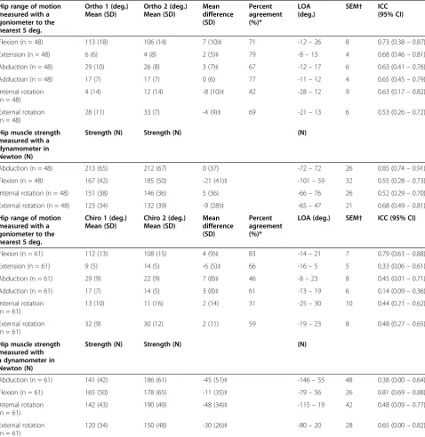
[image:6.595.59.538.111.603.2]The poor results of ROM in internal and external rota-tion could reflect participants being posirota-tioned supine and not sitting, as position is known to influence the pre-cision of individual measurements [17]. One chiropractor Table 3 Inter-rater reproducibility of hip range of motion (deg.) and muscle strength (N) for 2 orthopaedists and 2 chiropractors
Hip range of motion measured with a goniometer to the nearest 5 deg.
Ortho 1 (deg.) Mean (SD)
Ortho 2 (deg.) Mean (SD)
Mean difference (SD)
Percent agreement (%)*
LOA (deg.)
SEM† ICC (95% CI)
Flexion (n = 48) 113 (18) 106 (14) 7 (10)‡ 71 -12–26 8 0.73 (0.38–0.87)
Extension (n = 48) 6 (6) 4 (8) 2 (5)‡ 79 -8–13 4 0.68 (0.46–0.81)
Abduction (n = 48) 29 (10) 26 (8) 3 (7)‡ 67 -12–17 6 0.63 (0.41–0.76)
Adduction (n = 48) 17 (7) 17 (7) 0 (6) 77 -11–12 4 0.65 (0.45–0.79)
Internal rotation (n = 48)
4 (14) 12 (14) -8 (10)‡ 42 -28–12 9 0.63 (0.17–0.82)
External rotation (n = 48)
28 (11) 33 (7) -4 (9)‡ 69 -21–13 6 0.53 (0.26–0.72)
Hip muscle strength measured with a dynamometer in Newton (N)
Strength (N) Strength (N) (N)
Abduction (n = 48) 213 (65) 212 (67) 0 (37) -72–72 26 0.85 (0.74–0.91)
Flexion (n = 48) 167 (42) 185 (50) -21 (41)‡ -101–59 32 0.55 (0.28–0.73)
Internal rotation (n = 48) 151 (38) 146 (36) 5 (36) -66–76 26 0.52 (0.29–0.70)
External rotation (n = 48) 123 (34) 132 (39) -9 (28)‡ -65–47 21 0.68 (0.49–0.81)
Hip range of motion measured with a goniometer to the nearest 5 deg.
Chiro 1 (deg.) Mean (SD)
Chiro 2 (deg.) Mean (SD)
Mean difference (SD)
Percent agreement (%)*
LOA (deg.) SEM† ICC (95% CI)
Flexion (n = 61) 112 (13) 108 (15) 4 (9)‡ 83 -14–21 7 0.79 (0.63–0.88)
Extension (n = 61) 9 (5) 14 (5) -6 (5)‡ 66 -16–5 5 0.33 (0.06–0.61)
Abduction (n = 61) 29 (9) 22 (9) 7 (8)‡ 46 -8–23 8 0.45 (0.01–0.71)
Adduction (n = 61) 17 (7) 14 (5) 3 (8)‡ 61 -13–19 6 0.14 (0.09–0.36)
Internal rotation (n = 61)
13 (10) 11 (16) 2 (14) 31 -25–30 10 0.44 (0.21–0.62)
External rotation (n = 61)
32 (9) 30 (12) 2 (11) 59 -19–23 8 0.48 (0.27–0.65)
Hip muscle strength measured with a dynamometer in Newton (N)
Strength (N) Strength (N) (N)
Abduction (n = 61) 141 (42) 186 (61) -45 (51)‡ -146–55 48 0.38 (0.00–0.64)
Flexion (n = 61) 165 (50) 178 (65) -11 (35)‡ -79–56 26 0.81 (0.69–0.88)
Internal rotation (n = 61)
142 (43) 190 (49) -48 (34)‡ -115–19 42 0.48 (0.09–0.77)
External rotation (n = 61)
120 (34) 150 (48) -30 (26)‡ -80–20 28 0.65 (0.00–0.82)
deg., degrees; N, newton; SD, standard deviation; LOA, limits of agreement; SEM, standard error of the measurement; ICC, intraclass correlation coeffficient; CI, confidence interval.
*Percent agreement. Difference≤5obetween examiners except for flexion≤10o.
†SEMagreement.
had higher measurements for all strength tests, which is likely to be attributed to the force generated during the break test and in inter-rater variability interpreting when the break test is accomplished. The recorded variation in muscle strength could be due to fatigue from repetitive testing as participants were examined four times. We consider this effect minimal, as examinations were scheduled with a 15-minute interval and each session of strength testing lasted no more than 5 minutes. This allowed time for the ROM examination, a resting period for the participant and a change-over of raters. The results are also likely to be influenced by the orthopae-dists or chiropractors having limited experience with the
HHD. The procedures were tested in a validation study as part of the randomised clinical trial mentioned earlier (data not published). The rater tested had similar experi-ence with the HHD and demonstrated similar levels of intra-rater reliability but with much narrower LoA inter-vals. For ROM measurements, the rater demonstrated clinically acceptable intra-rater reproducibility without routine use of a goniometer in practice.
Comparison with other studies
Several studies have documented from poor to excellent inter-rater reliability of ROM in patients with hip OA using a goniometer. Sutlive et al. found fair to good
-30
-20
-10
10
20
0
Difference between ortho 1 and 2 (degrees)
-40 -20 0 20 40
Mean of ortho 1 and 2 (degrees)
observed average agreement 95% limits of agreement
[image:7.595.58.541.88.307.2]y=0 is line of perfect average agreement
Figure 2Limits of agreement between two orthopaedists for hip internal rotation range of motion (degrees).
-20
20
40
60
0
Difference between chiro 1 and 2 (degrees)
-20 0 20 40
Mean of chiro 1 and 2 (degrees)
observed average agreement 95% limits of agreement
y=0 is line of perfect average agreement
[image:7.595.58.541.501.715.2]reliability but agreement parameters were not reported [19]. Holm et al. studied teams of raters but results for mean measurements of each ROM were combined from all raters [14]. Cibere et al. found clinically acceptable re-liability both before and after standardisation of ROM and muscle strength measurements but they did not in-corporate variance components from the patients or random error and agreement parameters were not reported [17]. Theiler et al. reported reliability
coeffi-cients similar to those in our study but used Pearson’s
correlation coefficient which does not incorporate sys-tematic differences between raters [15]. For hip muscle
strength, Arnold et al. found excellent inter-rater reli-ability using a different HHD model but subjects were a mix of patients with both hip and knee OA [36]. Studies have documented good to excellent intra- and inter-rater reliability on healthy subjects using goniometer and HHD but they are not comparable to hip OA patients as age and disease characteristics influence the variation between subjects [21-23,35,37,38].
Study limitations
There are a number of limitations associated with this
study. First, raters were aware of the participant’
-200
-150
-100
-50
50
0
Difference between chiro 1 and 2 (N)
100 150 200 250 300
Mean of chiro 1 and 2 (Newton)
observed average agreement 95% limits of agreement
[image:8.595.62.539.88.306.2]y=0 is line of perfect average agreement
Figure 5Limits of agreement between two chiropractors for abduction hip strength (Newton).
-100
-50
50
100
0
Diff. between ortho 1 and 2 (N)
100 150 200 250 300 350
Mean of ortho 1 and 2 (Newton)
observed average agreement 95% limits of agreement
[image:8.595.60.540.503.712.2]y=0 is line of perfect average agreement
inclusion criterion of unilateral clinical and radiographic hip OA, so in the context of the clinical setting, no other hip conditions had to be considered. Second, the study did not involve rigorous training of the raters; however, we were interested in results reflecting current clinical practice. Several studies have reported on the added ef-fect of protocol standardisation and rigorous training in musculoskeletal medicine [17,39,40] and such training could potentially result in better agreement. Third, the raters had prior knowledge of patients having unilateral clinical and radiographic hip OA which could inflate re-liability coefficients. When one hip was examined, the rater would know if the other hip would be affected by OA or not. Fourth, the orthopaedic surgeon was not available for one of the examination sessions, so only 48 participants were included in the analysis between orthopaedists, instead of the 61 originally recruited. Fifth, the assessment of clinical hip OA was based solely on ROM and muscle strength evaluation. In clinical practice, a more extensive list of individual tests is used
as well as information from the patient’s case history. It
is further possible that the overall assessment was influ-enced by indications of a procedure being painful, to which the raters were not blinded. Sixth, we decided to omit adductor strength testing even though adductor strength has been documented to be reduced in patients with hip OA [5,9]. But measurement equipment has not been suitable for the clinical setting and in this patient group we concluded on the training day that stability of the pelvis and opposite leg were insufficient. We are aware that reproducibility of adductor strength testing by HHD on young healthy subjects has been reported as clinically acceptable [41]. Last, differentiation between levels of clinical hip OA following the overall assessment was only made from mild to severe hip OA. In the as-sessment of radiographic hip OA, it is common to cat-egorise into none, mild, moderate and severe.
The literature on reproducibility of the clinical hip examination in patients with hip OA is limited and het-erogeneous but recently the first set of guidelines on the reporting of reliability and agreement studies was pub-lished [33]. As patient characteristics differ in symptom and disease severity in primary and hospital care, future studies should take place in the setting where patient populations are examined and managed and involve clinicians from the same setting. To improve external validity, more than two clinicians should be included and selected randomly from an appropriate population of clinicians.
Conclusions
When using goniometry for the assessment of hip range of motion and hand-held dynamometry for hip muscle strength in patients with hip osteoarthritis, reproducibility
of individual measurements was generally poor between a pair of orthopaedists and a pair of chiropractors, indi-cating standardisation and rigorous training would be essential if this were to be improved. Both orthopaedists and chiropractors have a moderate ability to differentiate between hips without clinical osteoarthritis and hips assessed as having either mild or severe clinical osteoarthritis.
Additional file
Additional file 1:Examination protocol.
Abbreviations
OA: Osteoarthritis; ROM: Range of motion; Deg: Degrees; HHD: Hand-held dynamometer; N: Newton; ICC: Intraclass correlation coefficient; SD: Standard deviation; CI: Confidence interval; LoA: Limits of agreement.
Competing interests
All authors declare that they have no competing interests.
Authors’contributions
EP, HWC, SO and JH contributed to the conception and design of the study. EP, HWC, JØP and SO participated in the data collection. EP drafted the manuscript and performed the statistical analysis. EP, HWC, SO, WV and JH participated in the interpretation of the data. All authors participated in the critical revision of the article and made important contributions to the content. All authors read and approved the final manuscript.
Acknowledgements
We would like to thank research secretary Jytte Johannesen for designing the recording forms, Suzanne Capell for proof reading the manuscript and project nurse Annie Gam-Pedersen for the organisation of patients and clinicians on the days of examination.
Author details
1Institute of Sports Science and Clinical Biomechanics, University of Southern Denmark, Odense, Denmark.2Nordic Institute of Chiropractic and Clinical Biomechanics, Odense, Denmark.3Department of Orthopaedic Surgery and Traumatology, Odense University Hospital, Odense, Denmark.4Institute of Clinical Research, University of Southern Denmark, Odense, Denmark. 5Institute of Medical Biometry and Medical Informatics, University of Freiburg, Freiburg, Germany.
Received: 14 March 2012 Accepted: 30 November 2012 Published: 6 December 2012
References
1. Birrell F, Croft P, Cooper C, Hosie G, Macfarlane GJ, Silman A:Radiographic change is common in new presenters in primary care with hip pain. PCR Hip Study Group.Rheumatology (Oxford)2000,39:772–775.
2. Bierma-Zeinstra SM, Oster JD, Bernsen RM, Verhaar JA, Ginai AZ, Bohnen AM:Joint space narrowing and relationship with symptoms and signs in adults consulting for hip pain in primary care.J Rheumatol2002, 29:1713–1718.
3. Cibere J:Do we need radiographs to diagnose osteoarthritis?Best Pract Res Clin Rheumatol2006,20:27–38.
4. Cibulka MT, White DM, Woehrle J, Harris-Hayes M, Enseki K, Fagerson TL,et al:Hip pain and mobility deficits–hip osteoarthritis: clinical practice guidelines linked to the international classification of functioning, disability, and health from the orthopaedic section of the American Physical Therapy Association.J Orthop Sports Phys Ther2009,39:A1–A25. 5. Arokoski MH, Haara M, Helminen HJ, Arokoski JP:Physical function in men
with and without hip osteoarthritis.Arch Phys Med Rehabil2004, 85:574–581.
7. Pua YH, Wrigley TV, Cowan SM, Bennell KL:Intrarater test-retest reliability of hip range of motion and hip muscle strength measurements in persons with hip osteoarthritis.Arch Phys Med Rehabil2008,89:1146–1154. 8. Rydevik K, Fernandes L, Nordsletten L, Risberg MA:Functioning and
disability in patients with hip osteoarthritis with mild to moderate pain.
J Orthop Sports Phys Ther2010,40:616–624.
9. Rasch A, Bystrom AH, Dalen N, Berg HE:Reduced muscle radiological density, cross-sectional area, and strength of major hip and knee muscles in 22 patients with hip osteoarthritis.Acta Orthop2007, 78:505–510.
10. Pua YH, Wrigley TV, Collins M, Cowan SM, Bennell KL:Association of physical performance with muscle strength and hip range of motion in hip osteoarthritis.Arthritis Rheum2009,61:442–450.
11. Suetta C, Aagaard P, Magnusson SP, Andersen LL, Sipila S, Rosted A,et al: Muscle size, neuromuscular activation, and rapid force characteristics in elderly men and women: effects of unilateral long-term disuse due to hip-osteoarthritis.J Appl Physiol2007,102:942–948.
12. Birrell F, Croft P, Cooper C, Hosie G, Macfarlane G, Silman A:Predicting radiographic hip osteoarthritis from range of movement.Rheumatology (Oxford)2001,40:506–512.
13. Holla JF, Steultjens MP, van der Leeden M, Roorda LD, Bierma-Zeinstra SM, den Broeder AA,et al:Determinants of range of joint motion in patients with early symptomatic osteoarthritis of the hip and/or knee: an exploratory study in the CHECK cohort.Osteoarthr Cartil2011,19:411–419. 14. Holm I, Bolstad B, Lutken T, Ervik A, Rokkum M, Steen H:Reliability of
goniometric measurements and visual estimates of hip ROM in patients with osteoarthrosis.Physiother Res Int2000,5:241–248.
15. Theiler R, Stucki G, Schutz R, Hofer H, Seifert B, Tyndall A,et al:Parametric and non-parametric measures in the assessment of knee and hip osteoarthritis: interobserver reliability and correlation with radiology.
Osteoarthr Cartil1996,4:35–42.
16. Croft PR, Nahit ES, Macfarlane GJ, Silman AJ:Interobserver reliability in measuring flexion, internal rotation, and external rotation of the hip using a plurimeter.Ann Rheum Dis1996,55:320–323.
17. Cibere J, Thorne A, Bellamy N, Greidanus N, Chalmers A, Mahomed N,et al: Reliability of the hip examination in osteoarthritis: effect of
standardization.Arthritis Rheum2008,59:373–381.
18. Rasch A, Dalen N, Berg HE:Test methods to detect hip and knee muscle weakness and gait disturbance in patients with hip osteoarthritis.Arch Phys Med Rehabil2005,86:2371–2376.
19. Sutlive TG, Lopez HP, Schnitker DE, Yawn SE, Halle RJ, Mansfield LT,et al: Development of a clinical prediction rule for diagnosing hip osteoarthritis in individuals with unilateral hip pain.J Orthop Sports Phys Ther2008,38:542–550.
20. de Vet HC, Terwee CB, Knol DL, Bouter LM:When to use agreement versus reliability measures.J Clin Epidemiol2006,59:1033–1039.
21. Aalto TJ, Airaksinen O, Harkonen TM, Arokoski JP:Effect of passive stretch on reproducibility of hip range of motion measurements.Arch Phys Med Rehabil2005,86:549–557.
22. Prather H, Harris-Hayes M, Hunt DM, Steger-May K, Mathew V, Clohisy JC: Reliability and agreement of hip range of motion and provocative physical examination tests in asymptomatic volunteers.PM R2010, 2:888–895.
23. Bierma-Zeinstra SM, Bohnen AM, Ramlal R, Ridderikhoff J, Verhaar JA, Prins A:Comparison between two devices for measuring hip joint motions.
Clin Rehabil1998,12:497–505.
24. Bodenheimer T, Lorig K, Holman H, Grumbach K:Patient self-management of chronic disease in primary care.JAMA2002,288:2469–2475. 25. Poulsen E, Christensen HW, Roos EM, Vach W, Overgaard S, Hartvigsen J:
Non-surgical treatment of hip osteoarthritis. Hip school, with or without the addition of manual therapy, in comparison to a minimal control intervention: Protocol for a three-armed randomized clinical trial.BMC Musculoskelet Disord2011,12:88.
26. Andrews AW, Thomas MW, Bohannon RW:Normative values for isometric muscle force measurements obtained with hand-held dynamometers.
Phys Ther1996,76:248–259.
27. Bland JM, Altman DG:Statistical methods for assessing agreement between two methods of clinical measurement.Lancet1986,1:307–310. 28. Currier DP:Elements of Reseach in Physical Therapy. 3rd edition. Baltimore:
Williams & Wilkins; 1990.
29. Aaronson N, Alonso J, Burnam A, Lohr KN, Patrick DL, Perrin E,et al: Assessing health status and quality-of-life instruments: attributes and review criteria.Qual Life Res2002,11:193–205.
30. Weir JP:Quantifying test-retest reliability using the intraclass correlation coefficient and the SEM.J Strength Cond Res2005,19:231–240. 31. Landis JR, Koch GG:The measurement of observer agreement for
categorical data.Biometrics1977,33:159–174.
32. Muller R, Buttner P:A critical discussion of intraclass correlation coefficients.Stat Med1994,13:2465–2476.
33. Kottner J, Audige L, Brorson S, Donner A, Gajewski BJ, Hrobjartsson A,et al: Guidelines for Reporting Reliability and Agreement Studies (GRRAS) were proposed.J Clin Epidemiol2011,64:96–106.
34. Wikholm JB, Bohannon RW:Hand-held Dynamometer Measurements: Tester Strength Makes a Difference.J Orthop Sports Phys Ther1991, 13:191–198.
35. Thorborg K, Bandholm T, Schick M, Jensen J, Holmich P:Hip strength assessment using handheld dynamometry is subject to intertester bias when testers are of different sex and strength.Scand J Med Sci Sports
2011,Oct 28:. Epub ahead of print.
36. Arnold CM, Warkentin KD, Chilibeck PD, Magnus CR:The reliability and validity of handheld dynamometry for the measurement of lower-extremity muscle strength in older adults.J Strength Cond Res2010, 24:815–824.
37. Lu YM, Lin JH, Hsiao SF, Liu MF, Chen SM, Lue YJ:The relative and absolute reliability of leg muscle strength testing by a handheld dynamometer.J Strength Cond Res2011,25:1065–1071.
38. Fulcher ML, Hanna CM, Raina EC:Reliability of handheld dynamometry in assessment of hip strength in adult male football players.J Sci Med Sport
2010,13:80–84.
39. Cibere J, Bellamy N, Thorne A, Esdaile JM, McGorm KJ, Chalmers A,et al: Reliability of the knee examination in osteoarthritis: effect of standardization.Arthritis Rheum2004,50:458–468.
40. Brunse MH, Stochkendahl MJ, Vach W, Kongsted A, Poulsen E, Hartvigsen J,
et al:Examination of musculoskeletal chest pain - an inter-observer reliability study.Man Ther2010,15:167–172.
41. Thorborg K, Petersen J, Magnusson SP, Holmich P:Clinical assessment of hip strength using a hand-held dynamometer is reliable.Scand J Med Sci Sports2010,20:493–501.
doi:10.1186/1471-2474-13-242
Cite this article as:Poulsenet al.:Reproducibility of range of motion and muscle strength measurements in patients with hip osteoarthritis– an inter-rater study.BMC Musculoskeletal Disorders201213:242.
Submit your next manuscript to BioMed Central and take full advantage of:
• Convenient online submission
• Thorough peer review
• No space constraints or color figure charges
• Immediate publication on acceptance
• Inclusion in PubMed, CAS, Scopus and Google Scholar
• Research which is freely available for redistribution