http://dx.doi.org/10.4236/ijcm.2013.412A1006
Do Tumor Characteristics and Pre-Transplant
Locoregional Therapy Predict Survival after OLT in
Patients with Hepatocellular Carcinoma?
Mohamed Kohla1,2,3*, Richard Shaw3, Garret Hisatake2, Robert Osorio2, Maurizio Bonacini2
1Department of Hepatology, National Liver Institute, Menoufiya University, Shebeen El-Kom, Egypt; 2Department of Transplantation,
California Pacific Medical Center, San Francisco, USA; 3Research Institute, California Pacific Medical Center, San Francisco, USA.
Email: *dr_mohamedsamy@yahoo.com
Received October 30th, 2013; revised November 19th, 2013; accepted December 15th, 2013
Copyright © 2013 Mohamed Kohla et al. This is an open access article distributed under the Creative Commons Attribution License,
which permits unrestricted use, distribution, and reproduction in any medium, provided the original work is properly cited. In accor-dance of the Creative Commons Attribution License all Copyrights © 2013 are reserved for SCIRP and the owner of the intellectual property Mohamed Kohla et al. All Copyright © 2013 are guarded by law and by SCIRP as a guardian.
ABSTRACT
HCC prognosis after OLT is associated with criteria related to the number and size. However, the degree of
differentia-tion and efficacy of locoregional therapies may also influence outcome. Aim: Characterize patients with and without
HCC and compare outcomes according to tumor characteristics. Methods: Retrospective query of an electronic medical
record of 328 patients transplanted at California Pacific Medical Center (CPMC) in 2001-2007. HCC was defined by pre-OLT listing data as well as the finding of a tumor consistent with HCC at liver explant. Milan and UCSF criteria were applied to the lesions as described by pathology upon explant examination. Results: 328 patients were evaluated,
with 109 liver malignancies, 103 females (26 (25%) HCC) and 225 males (83 (37%) HCC p = 0.04). HCC patients were
older (56 ± 7.2 yr) than non HCC patients (51 ± 9.2, p < 0.001). The age of the donor and cold ischemia time was not different in the 2 groups. Survival was shorter in HCC (mean 984 ± 599 days) vs. non HCC (1103 ± 642) but not statis-tically significant (p = 0.10). Kaplan Meier survivals were superposable when comparing patients with or without ma-lignancy and when patients with low (≤22) vs. high MELD (>22) were compared. Survival curves in patients that ful-filled Milan vs. UCSF criteria were identical. However, more patients outside Milan died of metastatic disease (5/6,
83%) vs. within Milan (6/14, 43%, p = 0.01). Cox proportional hazards regression showed that MELD, but not
malig-nancy, differentiation or necrosis, was associated with mortality; HR = 6% (95% C.I. 1% - 10%) per additional MELD
point (p = 0.02). 69 pts had TACE pre-OLT, 17 had RFA ± any other modality. There was no difference in survivals in
pts who received any locoregional therapy vs. those who did not (p = 0.5). Deaths occurred in 20 (18%) HCC vs. 43
(19%) non HCC pts. Causes of mortality were different: of 20 HCC patients, 11(55%) died of HCC/metastatic disease vs. 2 (5%) in 41 non HCC deaths (p < 0.0001). Conclusion: In our cohort, survival of HCC patients was comparable to that of non HCC patients. However, mortality from metastatic disease was higher, particularly in those outside Milan. Overall mortality was associated with higher MELD scores, but not with tumor necrosis, the degree of differentiation at explant or locoregional therapy.
Keywords: Tumor Characteristics; Locoregional Therapy; Hepatolcellular Carcinoma; Liver Transplantation; OLT;
Survival
1. Introduction
Hepatocellualr carcinoma is the most common primary malignant tumor of the liver. Traditionally, the primary therapeutic modality for HCC has been surgical excision [1]. Optimal candidates for surgical resection show a
(a theoretical concern, deserving attention). Efforts to prevent tumor recurrence have involved administration of retinoids and intra-arterial I131 labeled lipiodol [5,6]. In Western countries, where liver cancer typically develops in the setting of well-established cirrhosis, fewer than 5% of patients are ideal candidates for hepatic resection [7], on the other hand, in Asian countries, the applicability of resection is higher, reflecting a less advanced liver dis- ease related to chronic hepatitis B virus infection [8-10]. Collectively, no more than 25% of patients are candi- dates for surgical resection due to tumor size or location, multifocal disease, and poor hepatic reserve. In cirrhotic cases, OLT represents the only chance for curative ther- apy [1], because OLT has been claimed to simultane- ously cure the malignant disease and replace the prema- lignant cirrhotic liver.
Early series of OLT for HCC yielded poor outcomes [3,11-20].
In those series, 3- and 5-year survival after OLT ranged 15% - 67% and 15% - 48%, respectively. These inferior results reflected the inclusion of patients with advanced HCC. Subsequently, patients with confined HCC (solitary lesion ≤ 5 cm or ≤ 3 lesions with diameter
≤ 3 cm), no major vessel invasion and no extra hepatic
involvement and Milan criteria, were reported to show an excellent long-term outcome with a 5-year survival rate of 70% and a recurrence rate below 15% [21]. Based on pathologic review, modestly expanded selection criteria (solitary lesion ≤ 6.5 cm, or ≤ 3 lesions with the largest one ≤ 4.5 cm and a total tumor diameter ≤ 8 cm), UCSF criteria were suggested to offer an excellent outcome with a 1-year and 5-year survival rates of 90% and 75.2% respectively[22]. In clinical practice, however, the Milan criteria based on pre-OLT radiological findings, could be more useful and a more widely accepted selection criteria than the UCSF criteria based on post-OLT pathologic findings [23]. Generally, authors report overall patient survival rates of 35% to 58 % at 5 years follow-up [3,13-16] with HCC recurrence associated with the poorest survival rates. In the pre-1990 era, HCC recur-rence following OLT was reported as high as 84% [17]. The most important factors that have been described af-fecting OLT survival in patients with HCC include: tu-mor size, vascular invasion [18], degree of tutu-mor differ-entiation [19], extra hepatic disease, and lymph node metastases [2]. Compared with the results before 1990, OLT in selected patients with HCC has seen significant improvements in patient and graft survival [20].
In order to decrease the waitlist dropout, various treat-ment modalities including resection, radiofrequency ab-lation (RFA), percutaneous ethanol injection (PEI), and transarterial chemoembolization (TACE) have been used to prevent HCC progression. It remains uncertain whe- ther excellent outcomes can be obtained in HCC patients
who previously underwent locoregional treatments (down staged) and met the Milan criteria at the time of OLT [23].
Taking into consideration the high dropout rate for pa-tients with HCC awaiting OLT [24], an adjustment to the MELD score was implemented to give greater priority for organ allocation for patients with a solitary tumor of 2 cm or greater and those with two or three lesions, each not exceeding 3 cm (UNOS stage II criteria) [24]. These patients are assigned a MELD score of 29, equivalent to a 30% 3-month mortality rate, and also are entitled to an additional increase in MELD score by 2 points for every 3 months on the waiting list without exceeding UNOS stage II criteria [24].
By reducing the waiting time for OLT for patients with HCC, this new scheme of organ allocation also may po-tentially justify a modest expansion of tumor size criteria OLT [25], and may improve intention-to-treat survival for HCC.
Tumor recurrence post transplant might be related to higher doses of immunosuppressives, and the latter are
known to represent a significant risk factor for tumor
growth, as shown in some experimental and clinical stud-ies [26-28], in one of which [29], tumor recurrence was related to the dosage of the immunosuppression given in the first postoperative year when most of the recurrences developed. A high dosage of cyclosporin administered during postoperative months 3 to 12 was significantly related to a low recurrence-free survival. This observa- tion suggested that the clinical studies carried out in the early 1990s, when the criteria for transplantation of heap- tocellular carcinoma were introduced, and might have been influenced by immunosuppressive regimen adopted at that time [29].
2. Aim of the Work
To study the impact of hepatocellular carcinoma, pre- OLT locoregional therapy, and tumor characteristics on the outcome after OLT in patients who received a liver transplant (OLT) at California Pacific Medical Center.
3. Methods
A retrospective query of the electronic medical records at California Pacific Medical Center (OTTR), San Fran-cisco, USA, was run for patients transplanted from 2001 till 2007.
Factors affecting the outcome after liver transplanta-tion were studied.
Dependent variable
• Patient survival (time from OLT to death)
Independent variables Donor variables:
Net-work for Organ Sharing (UNOS) database.
• Donor race.
• Donor age.
• Donor gender.
• Donor HB core Ab status.
• Donor CMV status.
• Donor BMI.
• Cold ischemia time.
• Warm ischemia time.
Recipient variables:
• Recipient race.
• Recipient age.
• Recipient gender.
• Etiology of liver disease.
• Presence of HCC.
• MELD at OLT.
• Bilirubin at OLT.
• Albumin at OLT.
• Sodium at OLT.
• Hemoglobin at OLT.
• Alcohol, assessed by social worker, Abuse/depend-
ence vs. not.
• Diabetes pre-OLT and or post-OLT, HbA1c was eva-
luated just pre-OLT and at last post-OLT follow up.
• Number of immunosuppressive drugs at 1 year post
transplant and at the last follows up.
• Estimated iron at explant pathology (0, trace, 1 - 4+).
• HCC by native liver pathology.
• HCC subcategories (number of nodules, largest
nod-ule in cm, lobar involvement, differentiation, vascular invasion, metastases).
• Type of pre-OLT locoregional therapy for HCC
(TACE, RFA, resection, PEI, or combination).
• Effect of locoregional therapy for HCC in terms of
necrosis at explant.
Statistical Analysis
Data were analyzed using the statistical package SPSS version 15 distributed by SPSS incorporator (SPSS Inc., v.15, Chicago, IL).
The following tests were run: 1) Univariate analysis, log rank test.
2) Cox Regression model using variables with p < 0.2. 3) Kaplan Meier curves for actuarial survival.
4. Results
Statistical analysis was run on 328 patients who have been transplanted from the beginning of 2001 till the end of April 2007; the aim of this analysis was study HCC in particular.
4.1. HCC and Overall Survival
Out of 328 patients included in this analysis, 219 did not
have HCC while 109 had HCC.
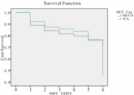
Figure 1 shows a Kaplan Meier survival analysis
com-paring the actuarial survival in patients with HCC (green
legend) versus those without HCC (blue legend); the p
value by Wilcoxon test was 0.445, which was statisti-cally insignificant.
4.2. Milan Criteria for HCC
Patients were categorized into 3 groups for this analysis: First group: 219 patients without HCC (blue legend). Second group: 96 patients within Milan criteria (green legend).
Third group: 7 patients outside Milan criteria (yellow legend).
N.B. 6 patients had incidental HCC on explants.
Figure 2 shows a Kaplan Meier survival analysis
com-paring the actuarial survival in the 3 groups; the p value by Wilcoxon test was 0.549, which was statistically in-significant.
4.3. Pre-OLT Locoregional Therapy
Patients were categorized into 4 groups according to type of pre-OLT locoregional therapy.
First group: 234 patients who did not receive any pre-OLT therapy (non-HCC or incidental, HCC who did not receive treatment, marked with blue legend).
Second group: 69 patients who received transarterial chemoembolization (TACE) alone pre-OLT (green leg-end).
Third group: 17 patients who received radiofrequency ablation pre-OLT (alone or in combination with others, marked with yellow legend).
Fourth group: 8 patients who received any other pre- OLT locoregional therapy (percutaneous ethanol injec-tion, resecinjec-tion, etc, marked with purple legend).
Figure 3 shows a Kaplan Meier survival analysis
com-paring the actuarial survival in the 4 groups, the p value by Wilcoxon test was 0.501, which was statistically in-significant.
4.4. Degree of HCC Differentiation at Explant Pathology
Currently, all OLT recipients at CPMC have their native livers pathologically examined, it was assumed that HCC patients with well differentiated HCC may have a better survival than patients with moderately-poorly differenti-ated.
This analysis was run on 3 groups:
First group: 247 patients with no evidence of HCC at explants (blue legend).
Figure 1. Kaplan Meier survival analysis comparing the actuarial survival in patients with HCC versus those with-out HCC.
Figure 2. Kaplan Meier survival analysis comparing the actuarial survival according to Milan criteria (without HCC, within Milan and outside Milan criteria).
Figure 3. Kaplan Meier survival analysis comparing the actuarial survival according to the type of locoreginal ther-apy (non HCC, TACE only, any RFA and other types of therapy).
[image:4.595.57.288.311.471.2]Third group: 32 patients with moderately-poorly dif-ferentiated HCC (yellow legend).
Figure 4 shows a Kaplan Meier survival analysis
com-paring the actuarial survival in the 3 groups, the p value by Wilcoxon test was 0.178, which was statistically in-significant.
However, when comparing each 2 groups to each other, those with well differentiated HCC showed a trend of better survival than those with moderately-poorly
differ-entiated HCC, and the p value by Wilcoxon test was
0.059.
4.5. Degree of Necrosis at Explants
First group: 247 patients with no HCC at explant (blue legend).
Second group: 35 patients with HCC at explant with-out tumor necrosis (green legend).
Third group: 20 patients with HCC at explants with only partial necrosis (yellow legend).
Fourth group: 26 patients with HCC at explants show-ing total necrosis (purple legend).
Figure 5 shows a Kaplan Meier survival analysis
com-paring the actuarial survival in the 4 groups; the p value by Wilcoxon test was 0.753, which was statistically in-significant.
4.6. Cox Regression Multivariate Analysis
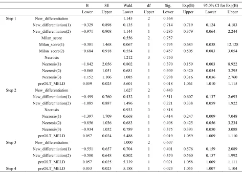
A Cox Regression multivariate analysis was run on the 328 (2001-2007), this is shown in the Table 1.
Pre-OLT MELD was found to be statistically signifi-cant as a predictor of survival on Cox Regression multi-variate analysis with a p value of 0.023.
5. Discussion
[image:4.595.57.286.525.688.2]Most of our HCC patients were transplanted in the
[image:4.595.308.539.542.689.2]Table 1. Cox Regression multivariate analysis for 328 patients.
B SE Wald df Sig. Exp(B) 95.0% CI for Exp(B)
Lower Upper Lower Upper Lower Upper Lower Upper
Step 1 New_differentiation 1.145 2 0.564
New_differentiation(1) −0.329 0.898 0.135 1 0.714 0.719 0.124 4.183
New_differentiation(2) −0.971 0.908 1.144 1 0.285 0.379 0.064 2.244
Milan_score 0.556 2 0.757
Milan_score(1) −0.381 1.468 0.067 1 0.795 0.683 0.038 12.128
Milan_score(2) −0.684 0.918 0.554 1 0.457 0.505 0.083 3.054
Necrosis 1.212 3 0.750
Necrosis(1) −1.842 2.056 0.802 1 0.370 0.159 0.003 8.922
Necrosis(2) −0.868 1.051 0.681 1 0.409 0.420 0.054 3.295
Necrosis(3) −1.152 1.106 1.085 1 0.298 0.316 0.036 2.760
preOLT_MELD 0.059 0.025 5.601 1 0.018 1.061 1.010 1.115
Step 2 New_differentiation 1.627 2 0.443
New_differentiation(1) −0.499 0.760 0.432 1 0.511 0.607 0.137 2.693
New_differentiation(2) −1.085 0.887 1.496 1 0.221 0.338 0.059 1.922
Necrosis 0.933 3 0.818
Necrosis(1) −1.397 1.709 0.668 1 0.414 0.247 0.009 7.048
Necrosis(2) −0.856 1.036 0.683 1 0.408 0.425 0.056 3.234
Necrosis(3) −0.934 1.052 0.789 1 0.375 0.393 0.050 3.088
preOLT_MELD 0.057 0.024 5.488 1 0.019 1.059 1.009 1.110
Step 3 New_differentiation 1.000 2 0.607
New_differentiation(1) −0.551 0.657 0.704 1 0.401 0.576 0.159 2.089
New_differentiation(2) −0.580 0.648 0.802 1 0.370 0.560 0.157 1.992
preOLT_MELD 0.057 0.025 5.339 1 0.021 1.058 1.009 1.111
Step 4 preOLT_MELD 0.053 0.023 5.188 1 0.023 1.055 1.007 1.104
Only Pre-OLT MELD was found to be statistically significant as a predictor of survival on Cox Regression multivariate analysis with a p value of 0.023.
Figure 5. Kaplan Meier survival analysis comparing the actuarial survival according to necrosis at explant pathol-ogy (non HCC, no necrosis, partial necrosis, and total ne-crosis).
MELD era. Those patients are typically transplanted at significantly lower medical MELD scores compared to patients with end stage liver disease without HCC; and the reason for this is to decrease the possibility of HCC patients to be delisted because of tumor progression; ac-cordingly, the sickest patients are characterized by high
mortality both on the waiting list and after liver trans-plantation. Patients with HCC are transplanted in better condition compared to patients without HCC; thus a similar survival is expected.
Many HCC patients received locoregional therapy as a bridge to OLT to minimize the probability of dropping out from the list due to tumor progression. Typically, locoregional therapy was anticipated once the diagnosis of HCC was established based on characteristic findings on imaging, even for tumors of 2 cm in diameter or less. The vast majority of our patients had TACE pre-OLT and a minority had RFA with or without TACE.
In our database, we found:
• The whole HCC cohort had no survival disadvantage
compared to those without HCC.
• Pre-OLT locoregional therapy did not show any
sur-vival advantage, though it led to more necrosis of the tumor at the explant for those who had RFA and sig-nificant reduction in tumor size for those who had TACE.
[image:5.595.58.286.472.605.2]tu-mor size did not influence survival post-OLT in patients within Milan criteria [30] compared to those who did not have TACE.
However, these findings were not shown to be signifi-cant on univariate analysis, probably because the sample size was too small. Accordingly, we do not have enough numbers of patients who survived long enough to show any statistically significant difference.
Tumor burden and biological behavior:
We assumed that those having well differentiated HCC might have a better outcome than those having poorly or moderately differentiated HCC. This was based on a dif-ferent biological behavior and a tendency to metastasize.
In the univariate analysis, those who had complete ne-crosis by locoregional therapy at explant were not in-cluded in this analysis, because the pathologist could not determine the degree of differentiation. The group having well differentiated HCC had a trend for better survival. This variable needs to be studied on a larger number of patients with longer follow-up periods using death from metastatic HCC as an end point, excluding all other caus- es of mortality to minimize confounding factors. As pre- viously stated in this context, identification of the degree of differentiation of HCC at the explant is liable to inter- personal and even intrapersonal variations. This is a po- tential weakness in all retrospective studies having ex- plant specimens examined by more than one pathologist over a relatively long duration of time.
When we tested those having complete necrosis at ex- plant independently in univariate analysis having the same assumption of probable better survival by decreas- ing tumor burden, we did not find any statistically sig- nificant difference. There was no way to make certain whether all of this necrotic tissue was tumor tissue, or some of the surrounding liver tissue which was acciden-tally targeted by less selective locoregional techniques.
REFERENCES
[1] M. Shimoda, R. M. Ghobrial, I. C. Carmody, et al., “Pre-
dictors of Survival after Liver Transplantation for Hepa- tocellualr Carcinoma Associated with Hepatitis C,” Liver Transplantation, Vol. 10, No. 12, 2004, pp. 1478-1486.
http://dx.doi.org/10.1002/lt.20303
[2] B. Ringe, R. Pichlmayr, C. Wittekind, et al., “Surgical
Treatment of Hepatocellular Carcinoma: Experience with Liver Resection and Transplantation in 198 Patients,”
World Journal of Surgery, Vol. 15, No. 2, 1991, pp. 270- 285. http://dx.doi.org/10.1007/BF01659064
[3] S. Iwatsuki, T. E. Starzl, D. G. Sheahan, et al., “Hepatic Resection versus Transplantation for Hepatocellular Car- cinoma,” Annals of Surgery, Vol. 214, No. 3, 1991, pp.
221-228.
http://dx.doi.org/10.1097/00000658-199109000-00005 [4] H. Bismuth, L. Chiche, R. Adam, et al., “Surgical Treat-
ment of Hepatocellular Carcinoma in Cirrhosis: Liver Resection or Transplantation?”Transplantation Proceed- ings, Vol. 25, No. 1, 1993, pp. 1066-1067.
[5] Y. Muto, H. Moriwaki, M. Ninomaya, et al., “Prevention
of Second Primary Tumors by an Acyclic Retinoid, Poly- prenoic Acid, in Patients with Hepatocellular Carci- noma,” The New England Journal of Medicine, Vol. 334,
No. 24, 1996, pp. 1561-1567.
http://dx.doi.org/10.1056/NEJM199606133342402 [6] W. Y. Lau, T. W. Leung, S. K. Ho, et al., “Adjuvant
in-tra-Arterial Iodine-131-Labelled Lipiodol for Respectable hepatocellular Carcinoma: A Prospective Randomized Trial,” Lancet, Vol. 353, No. 9155, 1999, pp. 797-801. http://dx.doi.org/10.1016/S0140-6736(98)06475-7 [7] J. M. Llovet, J. Fuster and J. Bruix, “Intention-to-Treat
Analysis of Surgical Treatment for Early Carcinoma: Re- section versus Transplantation,” Hepatology, Vol. 30, No.
6, 1999, pp. 1434-1440.
http://dx.doi.org/10.1002/hep.510300629
[8] Z. Y. Tang, Y. Q. Yu and X. D. Zhou, “Evolution of Sur- gery in the Treatment of Hepatocellular Carcinoma from the 1950s to the 1990s,” Seminars in Surgical Oncology,
Vol. 9, No. 4, 1993, pp. 293-297. http://dx.doi.org/10.1002/ssu.2980090403
[9] M. Makuuchi, “Surgical Management for Hepatocellular Carcinoma,” In: V. Arroyo, J. Bosch and J. Rodēs, Eds.,
Treatments in Hepatology, Barcelona, Masson, Vol. 1995,
pp. 341-352.
[10] The Liver Cancer Study Group of Japan, “Predictive Fac- tors for Long Term Prognosis after Partial Hepatectomy for Patients with Hepatocellular Carcinoma in Japan,”
Cancer, Vol. 74, No. 10, 1994, pp. 2772-2780. http://dx.doi.org/10.1002/1097-0142(19941115)74:10<27 72::AID-CNCR2820741006>3.0.CO;2-V
[11] B. Ringe, R. Pichlmyar, C. Wittekind, et al., “Surgical
Treatment of Hepatocellular Carcinoma: Experience with Liver Resection and Transplantation in 198 Patients,”
World Journal of Surgery, Vol. 15, No. 2, 1991, pp.
270-285. http://dx.doi.org/10.1007/BF01659064
[12] P. Moreno, E. Jaurrieta, J. Figueras, et al., “Orthotopic Liver Transplantation: Treatment of Choice in Cirrhotic Patients with Hepatocellular Carcinoma?” Transplanta-tion Proceedings, Vol. 27, No. 4, 1995, pp. 2296-2298.
[13] H. Bismuth, L. Chiche, R. Adam, et al., “Liver Resection versus Transplantation for Hepatocellular Carcinoma in Cirrhotic Patients,” Annals of Surgery, Vol. 218, No. 2,
1993, pp. 145-151.
http://dx.doi.org/10.1097/00000658-199308000-00005 [14] D. G. Farmer, M. H. Rosove, A. Shaked, et al., “Current
Treatment Modalities for Hepatocellular Carcinoma,”
Annals of Surgery, Vol. 219, No. 3, 1994, pp. 236-247.
http://dx.doi.org/10.1097/00000658-199403000-00003 [15] H. Bismuth, P. E. Majno and R. Adam, “Liver
Trans-plantation for Hepatocelluar Carcinoma,” Seminars in Liver Disease, Vol. 19, No. 3, 1999, pp. 311-322.
http://dx.doi.org/10.1055/s-2007-1007120
Tu-mor Characteristics on Outcome,” Annals of Surgery, Vol. 228, No. 4, 1998, pp. 479-490.
http://dx.doi.org/10.1097/00000658-199810000-00005 [17] I. Penn, “Hepatic Transplantation for Primary and Metas-
tatic Cancers of the Liver,” Surgery, Vol. 110, No. 4,
1991, pp. 726-734.
[18] I. Yokoyama, S. Todo, S. Iwatsuki, et al., “Liver Trans-
plantation in the Treatment of Primary Liver Cancer,”
Hepatogastroenterology, Vol. 37, No. 2, 1990, pp. 188- 193.
[19] J. G. O’Grady, R. J. Polson, K. Rolles, et al., “Liver
Transplantion for Malignant Disease. Results in 93 con-secutive patients,” Annals of Surgery, Vol. 207, No. 4,
1988, pp. 373-379.
http://dx.doi.org/10.1097/00000658-198804000-00002 [20] H. Matsunami, Y. Shimizu, S. V. Lynch, et al., “Liver
Transplantation as a Therapeutic Option for Hepatocellu- lar Carcinoma,” Oncology, Vol. 62, Suppl. 1, 2002, pp.
82-86. http://dx.doi.org/10.1159/000048281
[21] V. Mazzaferro, E. Regalia, R. Doci, et al., “Liver Trans-
plantation for the Treatment of Small Hepatocellular Car- cinomas in Patients with Cirrhosis,” The New England Journal of Medicine, Vol. 334, No. 11, 1996, pp. 693-699.
http://dx.doi.org/10.1056/NEJM199603143341104 [22] F. Y. Yao, L. Ferrell, N. M. Bass, et al., “Liver Trans-
plantation for Hepatocellular Carcinoma: Expansion of the Tumor Size Limits Does Not Adversely Impact Sur- vival,” Hepatology, Vol. 33, No. 6, 2001, pp. 1394-1403.
http://dx.doi.org/10.1053/jhep.2001.24563
[23] D. Y. Kim, M. S. Choi, J. H. Lee, et al., “Milan Criteria
Are Useful Predictors for Favorable Outcomes in Hepa- tocellular Carcinoma Patients Undergoing Liver Trans- plantation after Chemoembolization,” World Journal of Gastroenterology, Vol. 12, No. 43, 2006, pp. 6992-6997.
[24] “United Network for Organ Sharing,” 2002. http://www.unos.org
[25] F. Y. Yao, N. M. Bass, B. Nikolai, et al., “Liver Trans-
plantation for Hepatocelluar Carcinoma: Analysis of Sur- vival According to the Intention-to-Treat Principle and Dropout from the Waiting List,” Liver Transplantation,
Vol. 8, No. 10, 2002, pp. 873-883. http://dx.doi.org/10.1053/jlts.2002.34923
[26] I. Yokoyama, B. Carr, H. Saitu, et al., “Accelerated
Growth Rate of Recurrent Hepatocellular Carcinoma after Liver Transplantation,” Cancer, Vol. 68, No. 10, 1991, pp. 2095-2100.
http://dx.doi.org/10.1002/1097-0142(19911115)68:10<20 95::AID-CNCR2820681002>3.0.CO;2-Y
[27] I. Penn, “The Effect of Immunosuppression on Pre-Ex- isting Cancers,” Transplantation, Vol. 55, No. 4, 1993, pp. 742-747.
http://dx.doi.org/10.1097/00007890-199304000-00011 [28] C. E. Freise, L. Ferrell, T. Liu, et al., “Effect of Systemic
Cyclosporin on Tumor Recurrence after Liver Transplan- tation in a Model of Hepatocellular Carcinoma,” Trans- plantation, Vol. 67, No. 4, 1999, pp. 510-513.
http://dx.doi.org/10.1097/00007890-199902270-00003 [29] V. Marco, B. Roberto, C. Alessandro, et al., “Low Re-
currence Rate of Hepatocellular Carcinoma after Liver Transplantation: Better Patient Selection or Lower Im- munosuppression?” Transplantation, Vol. 74, No. 12,
2002, pp. 1746-1751.
[30] M. Amer, M. Yousri, F. Barakat, et al., “Pretransplant Chemoembolization for Hepatocellular Carcinoma ≤ cm Does Not Influence the Outcome after Liver Transplanta-tion,” 58th annual meeting of the AASLD, November