RESEARCH ARTICLE
Disruption of the ERK/MAPK pathway in neural crest cells as a
potential cause of Pierre Robin sequence
Carolina Parada1, Dong Han1, Alexandre Grimaldi1, Patricia Sarrión1, Shery S. Park1, Richard Pelikan1, Pedro A. Sanchez-Lara1,2,3and Yang Chai1,*
ABSTRACT
Disrupted ERK1/2 signaling is associated with several developmental syndromes in humans. To understand the function of ERK2 (MAPK1) in the postmigratory neural crest populating the craniofacial region, we studied two mouse models:Wnt1-Cre;Erk2fl/flandOsr2-Cre;Erk2fl/fl.
Wnt1-Cre;Erk2fl/fl mice exhibited cleft palate, malformed tongue,
micrognathia and mandibular asymmetry. Cleft palate in these mice was associated with delay/failure of palatal shelf elevation caused by tongue malposition and micrognathia. Osr2-Cre;Erk2fl/fl mice, in which theErk2deletion is restricted to the palatal mesenchyme, did not display cleft palate, suggesting that palatal clefting inWnt1-Cre; Erk2fl/flmice is a secondary defect. Tongues inWnt1-Cre;Erk2fl/flmice
exhibited microglossia, malposition, disruption of the muscle patterning and compromised tendon development. The tongue phenotype was extensively rescued after culture in isolation, indicating that it might also be a secondary defect. The primary malformations in Wnt1-Cre;Erk2fl/fl mice, namely micrognathia and mandibular asymmetry, are linked to an early osteogenic differentiation defect. Collectively, our study demonstrates that mutation ofErk2in neural crest derivatives phenocopies the human Pierre Robin sequence and highlights the interconnection of palate, tongue and mandible development. Because the ERK pathway serves as a crucial point of convergence for multiple signaling pathways, our study will facilitate a better understanding of the molecular regulatory mechanisms of craniofacial development.
KEY WORDS: ERK pathway, Cleft palate, Glossoptosis, Micrognathia, Mandibular asymmetry, Pierre Robin sequence, Neural crest cells
INTRODUCTION
The ERK transduction pathway is one of the signaling cascades that facilitates the transmission of extracellular signals to cytoplasmic and nuclear effectors (Pearson et al., 2001) via the protein kinase cascade comprising MAPKKK (RAF), MAPKK (MEK1/2) and MAPK (ERK). Activated ERK dimers regulate targets in the cytosol and nucleus, including the serum response factor (SRF) (Pearson et al., 2001; Shaw and Saxton, 2003). Newbern and colleagues generated mouse models of inactivation ofERK2(MAPK1) as well as of upstream components of the ERK cascade [B-Raf(BRAF) and C-Raf(RAF1),MEK1(MAP2K1) andMEK2(MAP2K2)] in neural crest derivatives, which resulted in analogous craniofacial
malformations to those in human, including maxillary hypoplasia, cleft palate, malformed or absent tongue, and micrognathia (Newbern et al., 2008). Neural crest-specific inactivation of Srf caused fully penetrant mandibular hypoplasia, but the maxillary prominences and tongue were unaffected (Newbern et al., 2008). These findings suggest that the mechanisms controlling mandibular development are different from those of maxilla, palate and tongue. Although the ERK pathway mediates BMP, TGFβ, FGF and EGF signals, which contribute significantly to palatogenesis (Bush and Jiang, 2012), the function of this pathway in the postmigratory neural crest populating the developing palate is still unclear.
The development of the secondary palate begins with the appearance of the palatal primordia from the maxillary process; the primordia are composed of a core of neural crest-derived mesenchyme covered by ectoderm-derived epithelium (Chai and Maxson, 2006). The palatal shelves grow vertically along the two sides of the tongue. As the mandible starts growing and the tongue descends, the palatal shelves reorient to acquire a horizontal position. This movement is known as palatal shelf elevation. Once in a horizontal position, the bilateral palatal shelves grow toward each other, establish contact along the midline, and fuse (Bush and Jiang, 2012; Ferguson, 1977, 1988; Iwata et al., 2011; Yu and Ornitz, 2011). Alterations in any of these steps may lead to a cleft palate, which is one of the most common human birth defects. The elevation process is not well understood, but may involve a heterogeneous mechanism along the anteroposterior (AP) axis (Yu and Ornitz, 2011). The anterior portions of the palatal shelves seem to elevate by rotating, whereas the middle and posterior portions change their orientation via a remodeling mechanism (Bush and Jiang, 2012). Numerous theories regarding palatal shelf elevation have been proposed and can be grouped into two categories: those positing that the shelves are elevated as a consequence of the influence of extrinsic structures; and those positing that the shelves themselves play an active role in the process (Ferguson, 1977). In humans, cleft palate due to mechanical interference with palatal shelf elevation by a malpositioned tongue and small jaw is clinically classified as Pierre Robin sequence or syndrome (PRS) (Rangeeth et al., 2011). Studies of human patients have shown that mutations inSATB2,SOX9,BMP2and the collagens lead to PRS-like clefting (Melkoniemi et al., 2003; Tan et al., 2013). The etiology of PRS has also been associated with a range of syndromes and chromosomal anomalies plus extrinsic fetal deformational forces, but the developmental mechanisms causing this condition are unclear, in part because of the lack of an animal model (Tan et al., 2013).
In this study, we show that disruption of the ERK pathway in neural crest derivatives in a murine model causes a primary defect affecting mandibular development, which is associated with a disturbance of the osteogenic differentiation program. In these mice, micrognathia and mandibular asymmetry result in secondary tongue malformations and cleft palate. This sequence of events during Received 11 April 2015; Accepted 2 September 2015
1
Center for Craniofacial Molecular Biology, University of Southern California, Los
Angeles, CA 90033, USA.2Department of Pathology & Pediatrics, Keck School of
Medicine, University of Southern California, Los Angeles, CA 90033, USA.
3
Children’s Hospital Los Angeles, Los Angeles, CA 90027, USA.
*Author for correspondence ([email protected])
DEVEL
O
mouse development is consistent with the phenotype of human PRS. Our study highlights how defects in one craniofacial structure can adversely affect the development of others and ultimately lead to complex craniofacial malformations.
RESULTS
Domains of ERK pathway activation during craniofacial development
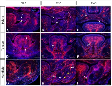
Activation of the ERK pathway occurs as early as E10.5 in the frontonasal region and the branchial arches (Corson et al., 2003). To investigate the roles of the ERK signaling pathway, we analyzed the localization of active, phosphorylated (P) ERK1/2 (Mapk3/1) in the palate, tongue and mandibular primordia. In the palatal shelves, P-ERK1/2 was detectable in both the epithelium and mesenchyme from E12.5 to E14.5 (Fig. 1A-C). In the developing tongue, P-ERK1/2 was detectable in the neural crest-derived mesenchyme as well as in myogenic progenitors and muscle fibers (Fig. 1D-F). By E14.5, P-ERK1/2 was restricted to the muscular component of the tongue, with only a few cells displaying positive signal in the neural crest-derived mesenchyme. In the mandible at E12.5, P-ERK1/2 was detectable in the chondrocytes of the Meckel’s cartilage located peripherally and in the neighboring condensed mesenchyme (Fig. 1G). At 13.5, when osteogenic differentiation begins in the mandible, P-ERK1/2 was restricted to the osteogenic front, the undifferentiated condensed mesenchyme surrounding the differentiated area, and the peripheral chondrocytes in the Meckel’s cartilage (Fig. 1H). At E14.5, P-ERK1/2 expression persisted in the undifferentiated mesenchyme, whereas expression was undetectable in differentiated cells. Moreover, the number of positive cells in the Meckel’s cartilage was reduced compared with E12.5 and E13.5 (Fig. 1I). These results suggest that the ERK pathway is active in the mesenchymal progenitors that have entered the osteogenic differentiation program and becomes inactive as differentiation progresses.
Wnt1-Cre;Erk2fl/flmice exhibit severe craniofacial defects
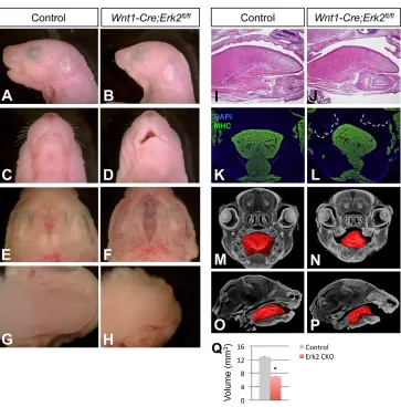
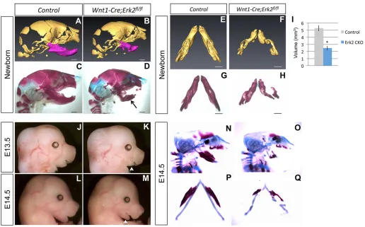
Previously, Newbern and colleagues generatedWnt1-Cre;Erk2fl/fl mice and reported that they exhibited severe craniofacial malformations (Newbern et al., 2008). In order to analyze the molecular and cellular mechanisms underlying these craniofacial defects, we generatedWnt1-Cre;Erk2fl/flmice using a pure C57BL/ 6 background. The craniofacial phenotype in our mice was less severe than that reported previously (Newbern et al., 2008). Our Wnt1-Cre;Erk2fl/fl mice died at birth and exhibited multiple craniofacial malformations including maxillary hypoplasia, complete cleft palate, tongue defects, micrognathia, and non-preferential mandibular asymmetry (Fig. 2).
[image:2.612.49.422.451.738.2]Palatal shelf elevation was compromised in Wnt1-Cre;Erk2fl/fl mice. Because elevation may occur via different mechanisms in the anterior versus posterior regions of the palate (reviewed by Bush and Jiang, 2012), we analyzed microCT scan sections along the AP axis at the newborn stage, but found that all regions were affected similarly (n=3; Fig. S1A-J). The elevation defect was also detectable along the AP axis at E14.5 and E15.5 (data not shown). We also found thatErk2mutant mice displayed microglossia, a tongue defect not previously described (Fig. 2G-N). 3D reconstruction of microCT scans showed a∼45% reduction in the tongue volume of Wnt1-Cre;Erk2fl/fl mice (Fig. 2M-Q). In addition, Wnt1-Cre; Erk2fl/fl tongues exhibited malposition and disruption of muscle patterning. Although no significant difference was detectable in HE-stained sagittal sections, malpositioning of the tongue inWnt1-Cre; Erk2fl/flmice was evident after myosin heavy chain immunostaining of coronal sections (Fig. 2K,L). The organization of the fibers in both the intrinsic and extrinsic muscles of the tongue was altered in Wnt1-Cre;Erk2fl/flmice, resulting in a gross disruption of the muscle pattern and position. Typically, one half of the tongue was able to descend whereas the other side remained high, blocking the elevation of one palatal shelf (Fig. 2K,L). This finding was confirmed by microCT scan reconstructions (Fig. 2M,N) and is
Fig. 1. Specific domains of ERK pathway activation in the craniofacial region.P-ERK1/2 immunostaining (red) in the palatal shelves (A-C), tongue primordium (D-F) and mandible (G-I) of control mice at E12.5, E13.5 and E14.5. In A-C, arrowheads indicate signals in the epithelium whereas arrows indicate signals in the mesenchyme. In D-F, arrowhead indicates signals in the neural crest-derived mesenchyme whereas arrows indicate signals in the muscle. In G-I, arrowheads indicate signals in the
chondroblasts of the Meckel’s cartilage
whereas arrows indicate signals in the osteogenic progenitors. Yellow arrows indicate the location of terminally
differentiated osteoblasts.n=3 for each
stage. Scale bars: 100 µm.
DEVEL
O
consistent with the mandibular phenotype. The mandibles of Wnt1-Cre;Erk2fl/flmice also exhibited a severe disruption in bone development. As reported previously,Erk2mutant mandibles were significantly smaller than those of controls. Additionally, we observed a dramatic asymmetry in most of the mandibles (Table S1), which was associated with the asymmetry of the tongue and the elevation of a single palatal shelf. At the newborn stage, the more severely affected side of the mandible corresponded to the side lacking palatal shelf elevation (n=3; Fig. S1). In Wnt1-Cre;Erk2fl/fl newborns in which the micrognathia affected both sides equally, the tongue was symmetric and positioned in a high location and neither palatal shelf elevated (n=2; data not shown).
Cleft palate inWnt1-Cre;Erk2fl/flmice is a consequence of tongue malposition and micrognathia
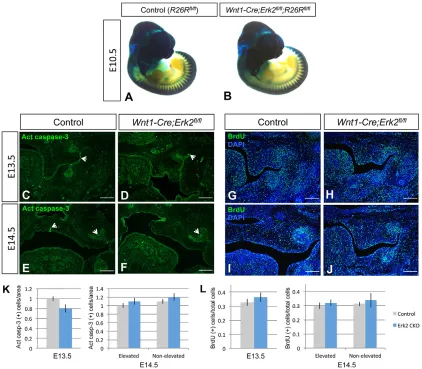
Because the ERK transduction pathway controls diverse cellular activities, including cell migration, survival and proliferation (Pearson et al., 2001), we investigated whether any of these processes was altered in Wnt1-Cre;Erk2fl/fl palatal shelves. To analyze neural crest cell migration, we performedlacZstaining of whole-mount Wnt1-Cre;R26R (control) and Wnt1-Cre;Erk2fl/fl; R26Rembryos and frozen sections. We detected no difference at E10.5 or E11.5 (Fig. 3A,B, Fig. S2A-D), demonstrating that the availability of mesenchymal progenitors was unaffected. Next, we examined mesenchymal cell survival and proliferation at E12.5-E14.5. For analysis at E14.5, the elevated and non-elevated palatal shelves were analyzed independently in the asymmetric cases
(Fig. 3L). We found no statistically significant difference in the number of apoptotic or proliferating cells at any stage (Fig. 3C-L, Fig. S2E-H). These results indicate thatWnt1-Cre;Erk2fl/fl palatal shelves did not have any intrinsic defect.
Because mutations affecting the mesenchyme can affect epithelial cells in a paracrine manner, we also checked whether the fate of the midline edge epithelium was compromised in Wnt1-Cre;Erk2fl/fl embryos. We cultured E13.5 palatal explants with the midlines placed in contact. After a 3-day incubation period, palatal fusion was complete and no remaining midline epithelial cells were detectable in either control orWnt1-Cre;Erk2fl/flexplants (Fig. S3A,B). We also analyzed the AP patterning of the palatal epithelium byin situhybridization ofShh, a marker that labels the rugae. The number and localization of rugae along the AP axis of the palates in E14.5 Wnt1-Cre;Erk2fl/fl mice were comparable to those of controls (Fig. S3C,D). These findings suggest that ERK pathway activation in the mesenchyme is not necessary for either the establishment of AP patterning or midline epithelial cell fate in the palate.
[image:3.612.51.414.53.421.2]In order to understand the potential molecular causes underlying the palatal clefting in Wnt1-Cre;Erk2fl/fl mice, we carried out RNA microarray analysis of E13.5 and E14.5 palatal shelves, including both epithelium and mesenchyme. Despite the fact that ERK2 mediates a wide variety of extracellular signals, only a few genes were differentially expressed in the palates of mutants compared with those of controls at either stage (Tables S2 and S3). These genes do not appear to have any known functions in palatogenesis.
Fig. 2.Wnt1-Cre;Erk2fl/flmice exhibit severe craniofacial malformations.
(A-D) Sagittal (A,B) and ventral (C,D)
views of newborn control andWnt1-Cre;
Erk2fl/flheads. (E-H) Intraoral views of the palates (E,F) and tongues (G,H) of control
andWnt1-Cre;Erk2fl/flnewborns. (I,J) HE
staining of sagittal sections of control
andWnt1-Cre;Erk2fl/flnewborn tongues.
(K,L) Myosin heavy chain (MHC; green) immunostaining of coronal sections of
control andWnt1-Cre;Erk2fl/flnewborn
heads. Dashed lines delineate the malformed and unfused palatal shelves in
Wnt1-Cre;Erk2fl/flmice. (M-Q) 3D
reconstructions of control andWnt1-Cre;
Erk2fl/fltongues from microCT scans.
Tongue volumes of control andWnt1-Cre;
Erk2fl/fl(Erk2 CKO) samples are quantified
in Q. *P<0.05.n=3.
DEVEL
O
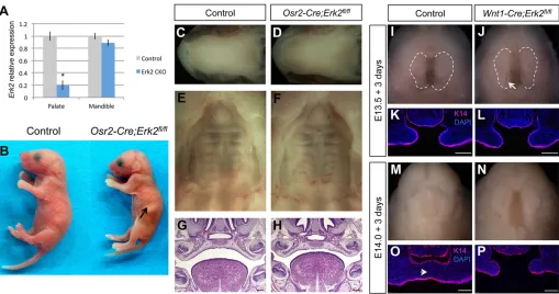
Given the lack of significant molecular and cellular changes in Wnt1-Cre;Erk2fl/flpalates and the tight relationship between the phenotypes of palate, tongue and mandible, we hypothesized that the palatal clefting in these mice is a consequence of the malposition of the tongue and secondary to the mandibular defects. To test this hypothesis, we used bothin vivoandin vitro approaches. First, we generatedOsr2-Cre;Erk2fl/flmice because Osr2is expressed in the palatal shelves from E12.5 to newborn stage but not in the mandible at any developmental stage (Parada et al., 2013; data not shown). Indeed, the expression ofErk2was reduced in the palatal shelves of Osr2-Cre;Erk2fl/fl mice, as indicated by qPCR analysis, whereas expression in the mandible was unaffected (Fig. 4A). Palatal clefting in these mice would suggest that ERK2 plays an intrinsic role in the elevation of the palatal shelves. However, Osr2-Cre;Erk2fl/fl newborns did not exhibit cleft palate (n=16; Fig. 4B-H). The tongues and mandibles in these mice were also unaffected (Fig. 4B-D).
Second, we performed in vitro experiments using a rotational culture system. The mandible and tongue were dissected at E13.5, before palatal shelf elevation, and at E14.0, immediately after elevation but before the palatal shelves make contact along the midline. After 3 days of culture, the palatal shelves from E13.5 control andErk2mice were elevated although not fused, owing to
technical limitations (n=4; Fig. 4I-L). Immunostaining for K14 and counterstaining with DAPI were performed to show the palatal epithelium and the integrity of the mesenchymal tissue. When dissected at E14.0 and cultured for 3 days, the palatal shelves of the control samples were fused at the midline, with no epithelial cells remaining in this region (n=3; Fig. 4M,O). Although Wnt1-Cre; Erk2fl/fl palatal shelves did not fuse, removing the tongue and mandible was sufficient to rescue the elevation defect (Fig. 4N,P). It is likely that fusion did not occur because of the previous delay in the elevation process. Taken together, these findings strongly suggest that the palatal shelf elevation defect inErk2mutant mice is the result of a primary malformation in the tongue and/or mandible.
Proliferation and differentiation are unaffected in tongues of Wnt1-Cre;Erk2fl/flmice
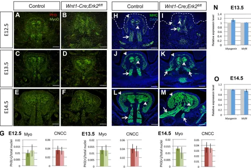
[image:4.612.96.518.57.426.2]Tongues ofWnt1-Cre;Erk2fl/flnewborn mice exhibited microglossia, disruption of muscle patterning and malposition. We carefully examined cell survival, proliferation and differentiation in the tongue at different key stages. We found that cell survival was not affected inWnt1-Cre;Erk2fl/fltongues at E12.5 through E14.5 (Fig. S4). We evaluated cell proliferation separately in the muscular and neural crest-derived components using double immunostaining for Myod1, a marker of myogenic determination, and phosphohistone Fig. 3. Neural crest cell migration, cell survival, and proliferation of the palatal mesenchyme are unaffected inWnt1-Cre;Erk2fl/flmice.(A,B)lacZstaining
of whole-mount E10.5Wnt1-Cre;R26Rfl/fl(control) andWnt1-Cre;Erk2fl/fl;R26Rfl/flembryos. (C-F) Active caspase 3 immunostaining (green) of E13.5 and
E14.5 control andWnt1-Cre;Erk2fl/flpalates. Arrowheads indicate positive signal. (G-J) BrdU staining (green) of E13.5 and E14.5 control andWnt1-Cre;Erk2fl/fl
palates. (K,L) Quantification of apoptotic and proliferating cells in C-F and G-J, respectively.n=3. Scale bars: 100 µm.
DEVEL
O
H3 (PH3), a marker of proliferation. Myod1-negative cells in the tongue correspond to the neural crest-derived mesenchyme. At E12.5 through E14.5, the numbers of proliferating cells in both the muscular and the neural crest components of the tongue were comparable inWnt1-Cre;Erk2fl/fland control embryos (Fig. 5A-G). Next, we examined myogenic differentiation by analyzing the expression of myosin heavy chain (MHC), which labels mature muscle fibers. We detected no significant differences in the intensity of the signal between control andErk2mutant tongues from E12.5 to E14.5 (Fig. 5H-M). Quantitative analysis of myogenin (Myog) and Mrf4 (Myf6) expression at E13.5 and E14.5 also confirmed that muscle differentiation was unaffected in the tongues of Wnt1-Cre;Erk2fl/fl embryos (Fig. 5N,O). Although no myogenic differentiation defects were detectable in Erk2 mutant tongues, muscle patterning and organization were altered. Both microglossia and asymmetry in the tongue muscles, mainly affecting the extrinsic muscles, were detectable immediately before palatal shelf elevation at E13.5 (Fig. 5C,D,J,K, Fig. S5A,B). As development progressed, these phenotypes worsened and the patterns of both intrinsic and extrinsic muscles were severely disrupted by E14.5 (Fig. 5E,F,L,M, Fig. S5C,D). The stage of onset of the tongue extrinsic muscle asymmetry is consistent with the timing of the failure of palatal shelf elevation.
Next, we checked whether differentiation of neural crest-derived mesenchyme was affected in the tongues ofWnt1-Cre;Erk2fl/flmice by analyzing the expression of scleraxis (Scx).Scxis a marker for tendons, which are derived from neural crest cells in the craniofacial region. In control mice,Scxwas expressed in the tongue septum and tendons of the extrinsic muscles at both E13.5 and E15.5. At E13.5,
Scx expression was downregulated in Wnt1-Cre;Erk2fl/fl tongues (Fig. 6A,B). At E15.5,Scxexpression was completely undetectable in both the septum and tendons ofErk2mutant tongues (Fig. 6C,D). Type 1 collagen (Col1a1) was also undetectable in the tendons of the extrinsic muscles at this stage (data not shown). Because of the absence of tendons in Wnt1-Cre;Erk2fl/fl tongues, we evaluated whether the relationship between the extrinsic muscles of the tongue and the mandibular bone primordium was affected by examining the expression of Sp7 (osterix), a marker for osteogenic progenitors, and MHC. At E13.5, Sp7 expression was significantly downregulated in Wnt1-Cre;Erk2fl/fl mandibles (Fig. 6E,F, and see next section), which does not allow a proper comparison. At E15.5, the extrinsic muscles in control mice were not in contact with the Meckel’s cartilage or the osteogenic progenitors, whereas they were attached directly to the cartilage and in close vicinity to the bone primordium in Wnt1-Cre;Erk2fl/fl mice (Fig. 6G,H). We speculate that this abnormal relationship between muscles, cartilage and bone contributes to a mechanical disruption of tongue muscle development.
[image:5.612.53.562.56.324.2]Based on the lack of intrinsic cellular defects in the tongue muscles and the correlation between the asymmetry and size of the mandible and tongue inWnt1-Cre;Erk2fl/flmice, we hypothesized that the tongue malformation is a secondary defect. To address this issue, we performedin vitro experiments in which mandibles of E12.5 mice were removed and the tongue was cultured in isolation for 7 days. After this incubation period, macroscopic examination indicated that the sizes of control andWnt1-Cre;Erk2fl/fl tongues were similar (Fig. 7A). Next, we analyzed the volumes of cultured control and Erk2 mutant tongues quantitatively using BioVis3D Fig. 4. Cleft palate is a consequence of tongue and mandible malformations inWnt1-Cre;Erk2fl/flmice.(A) Quantitative analysis ofErk2expression in the
palatal shelves and mandible of control andOsr2-Cre;Erk2fl/flmice at E13.5. *P<0.05. (B) Macroscopic views of control andOsr2-Cre;Erk2fl/flnewborns. Arrow
indicates milk in the stomach of theOsr2-Cre;Erk2fl/flmouse. (C-F) Intraoral views of the tongues, mandibles and palates of control andOsr2-Cre;Erk2fl/fl
newborns. (G,H) HE staining of coronal sections of E16.5 control andOsr2-Cre;Erk2fl/flmice. (I,J) Intraoral views of heads from E13.5 control andWnt1-Cre;Erk2fl/fl
embryos from which the mandible and tongue were removed and cultured for 3 days (n=4). Dashed lines delineate the palatal shelves. (K,L) K14 immunostaining
(red) of coronal sections of E13.5 cultured explants. (M,N). Intraoral views of heads from E14.0 control andWnt1-Cre;Erk2fl/flembryos from which the mandible
and tongue were removed and cultured for 3 days (n=3). (O,P) K14 immunostaining (red) of coronal sections of E14.0 cultured explants. Arrowhead in O points to
the midline of the control sample, where palatal shelves are fused and no epithelial cells remain. Scale bars: 100 µm.
DEVEL
O
software (n=4 and 3, respectively). Although the tongues of Wnt1-Cre;Erk2fl/fl mice were 16% smaller than those of controls after culture, statistical analysis showed that the difference was not significant (P=0.282; Fig. 7B). These findings suggest that the microglossia was considerably rescued upon removal of the mandible. Additionally, histological analyses and MHC immunostaining showed that the orientation and organization of muscle fibers of the intrinsic muscles of control and Wnt1-Cre; Erk2fl/fl tongues were comparable (Fig. 7C-F). The pattern and organization of the extrinsic muscles could not be evaluated using this approach. These results suggest that most of the phenotypes in Wnt1-Cre;Erk2fl/fl tongues were the consequence of a primary defect in the mandible.
Mandibular osteogenic differentiation is severely compromised inErk2mutant mice
Based on our results, we suggest thatWnt1-Cre;Erk2fl/flmice mimic human PRS, in which micrognathia leads to malposition of the tongue (glossoptosis), which blocks the elevation of the palatal shelves, eventually causing cleft palate (Movie 1). In order to study the primary defect inWnt1-Cre;Erk2fl/flmice, we performed a series of analyses of mandibles. 3D reconstruction of microCT scans at the newborn stage showed a∼50% reduction in the mandibular volume of Wnt1-Cre;Erk2fl/fl mice, as well as obvious mandibular
asymmetry (Fig. 8A,B,E,F,I). These findings were confirmed using skeletal staining. Remarkably, the proximal region of the mandible was severely affected whereas the defect in the distal region was mild. Specifically, the condyle was greatly reduced in size; the angle and the coronoid process were severely disrupted, or completely absent in some cases (Fig. 8A-H; data not shown). We collected embryos from E12.5 through E16.5 to determine the onset time of the mandibular malformations in Wnt1-Cre;Erk2fl/fl newborns. Micrognathia was detectable by E13.5 (Fig. 8J,K), which coincides with the appearance of the tongue defects. At E14.5, the mandibular phenotype was more severe (Fig. 8L,M), consistent with the increase in severity of the tongue defects. Moreover, the morphology of the Meckel’s cartilage was disrupted at E14.5, with a reduction in size and a complete discontinuity on one side. The volume of mandibular bone was reduced, although the severity of this trait was not always correlated with the severity of the malformation of the Meckel’s cartilage (Fig. 8N-Q).
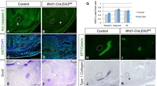
[image:6.612.51.561.57.399.2]In order to determine the cause(s) of the mandibular defects in Wnt1-Cre;Erk2fl/fl mice, we analyzed potential cellular activities that could be affected from E12.5 to E15.5. Cell proliferation and survival in the mandibles of Wnt1-Cre;Erk2fl/fl mice were indistinguishable from controls at all time points (Fig. 9A-D; data not shown). Because the morphology of the Meckel’s cartilage was affected, we examined the expression of several chondrogenesis Fig. 5. Proliferation and differentiation are unaffected inWnt1-Cre;Erk2fl/fltongues.(A-F) PH3 (red) and Myod1 (green) double immunostaining of coronal
sections of E12.5-E14.5 control andWnt1-Cre;Erk2fl/fltongues. (G) Quantification of proliferation (PH3+) in positive myogenic cells (Myo) and
Myod1-negative cranial neural crest cells (CNCC) from A-F. Dark bars, control tongues; light bars,Wnt1-Cre;Erk2fl/fltongues. (H-M) MHC immunostaining (green) of
coronal sections of E12.5-E14.5 control andWnt1-Cre;Erk2fl/fltongues. Arrows point to the extrinsic muscles and arrowheads point to the intrinsic muscles.
(N,O) Quantitation of real-time qPCR ofMyogandMrf4in control (gray bars) andWnt1-Cre;Erk2fl/fl(blue bars) tongues at E13.5 and E14.5.n=3 for each stage
and experiment. Scale bars: 100 µm.
DEVEL
O
markers. Expression ofSox9and Link protein is detectable in most chondroblasts in the Meckel’s cartilage of control embryos (Fig. 9E, Fig. S6A,E; data not shown). Surprisingly, in Wnt1-Cre;Erk2fl/fl mandibles at E13.5 and E14.5, the expression of these markers was comparable to that of controls in both intensity and pattern, although there was a significant difference in shape and size of the cartilage (Fig. 9E,F, Fig. S6A,B,E,F). Similarly,Ctgfwas expressed in the peripheral chondroblasts of the Meckel’s cartilage of both control andWnt1-Cre;Erk2fl/flmice (Fig. S6C,D,G,H).
Next, we examined the expression of two osteogenic differentiation markers. Sp7 is a marker for osteogenic progenitors and type 1 collagen (Col1a1) is a marker for terminally differentiated osteoblasts. We first analyzed embryos at E13.5, which corresponds to the onset of the mandibular defects in Wnt1-Cre;Erk2fl/fl mice. At this stage, when deposition of osteogenic matrix is first detectable in control mandibles, Sp7 expression was dramatically downregulated and Col1a1 expression was undetectable in the mandibular primordium of Wnt1-Cre;Erk2fl/fl mice (Fig. 9H-K). We also checked other developmental stages, before and after the beginning of mandibular osteogenic differentiation. At E12.5, Col1a1 was not expressed in controls (data not shown) and Sp7 was detectable in the condensed mesenchyme neighboring the Meckel’s cartilage. In Wnt1-Cre; Erk2fl/fl mice, the number of Sp7-positive cells was reduced, suggesting that the availability of osteogenic progenitors was compromised (Fig. S7A,B). At E14.5 and E15.5, the domains expressing either Sp7 or Col1a1were reduced in an asymmetric pattern inWnt1-Cre;Erk2fl/flmice (Fig. S7C-J). These data indicate that there is an initial decrease in the pool of osteogenic progenitors followed by a significant delay in the osteogenic process, which results in a small, asymmetric mandible inWnt1-Cre;Erk2fl/flmice.
DISCUSSION
In this work we have investigated the function of the ERK/MAPK transduction pathway in the postmigratory neural crest cells populating the craniofacial region.Wnt1-Cre;Erk2fl/flmice exhibit severe craniofacial malformations that mimic human PRS, which is characterized by a primary mandibular defect that causes
malposition of the tongue and consequently cleft palate. Similarly, the cleft palate in Wnt1-Cre;Erk2fl/fl mice is not the primary defect, but is instead the result of defects in the mandible that affect the tongue and eventually the formation of the palate. Accordingly, the ERK/MAPK pathway is active in mesenchymal progenitors committed to the osteogenic mesenchyme and regulates early steps in the osteogenic differentiation program in the mandibular primordium, which appears to be essential for the proper size and morphology of the mandible.
Failure of palatal shelf elevation and expression of PRS phenotypes inWnt1-Cre;Erk2fl/flmice
Elevation of the palatal shelves is a complex process that depends on both intrinsic and extrinsic factors. Our results highlight the relevance of extrinsic influences for palatal shelf elevation. Among them, the cranial base and mandible have been the focus of numerous studies. As the palatal shelves elevate, the cranial base cartilage straightens significantly. One theory speculates that this straightening generates forces in the midline that are transmitted to the alar regions of the sphenoid and then to the palatal shelves during elevation (Brinkley and Vickerman, 1978). In Wnt1-Cre;Erk2fl/fl mice the cranial base is not affected (data not shown), but mandibular morphology is severely disrupted. In wild-type mice, the mandibular primordium grows downward and lengthens from E12.5 to newborn stage (Ramaesh and Bard, 2003). This growth pattern seems to provide the tongue with physical space to move downward, and this movement closely coincides with the reorientation of the palatal shelves from a vertical to a horizontal position. The lack of mandibular growth inWnt1-Cre;Erk2fl/flmice diminishes the space available for the tongue to descend. Subsequently, the high position of the tongue (glossoptosis) blocks palatal shelf elevation.
[image:7.612.49.563.57.258.2]Loss ofErk2in the neural crest-derived mesenchyme also affects the size of the tongue. Similar cases of microglossia occur in other mouse models with mesenchymal mutations, such as Wnt1-Cre; Tgfbr2fl/fl(Hosokawa et al., 2010; Iwata et al., 2013) andWnt1-Cre; Alk5fl/fl(Han et al., 2014) mice. However, in those cases, changes in molecules secreted by the mesenchymal cells directly affect the tongue myogenic progenitors, resulting in altered myogenic Fig. 6. Cranial neural crest cell-derived tendons are abnormal inWnt1-Cre;Erk2fl/fltongues.(A-D)In situhybridization of scleraxis (Scx) in coronal sections
of E13.5 and E15.5 control andWnt1-Cre;Erk2fl/flheads. Arrows indicateScxexpression. (E-H) Double immunostaining of Sp7 (red) and MHC (green) in
coronal sections of E13.5 and E15.5 control andWnt1-Cre;Erk2fl/flheads. Dashed lines delineate the Meckel’s cartilage. The arrowhead in H indicates the
abnormally close proximity of the extrinsic muscles of the tongue to the Meckel’s cartilage and osteogenic progenitors.n=3 for each stage and analysis.
Scale bars: 100 µm.
DEVEL
O
proliferation and/or differentiation. InWnt1-Cre;Erk2fl/flmice, the proliferation, survival and differentiation of the myogenic precursors are unaffected, suggesting that mechanical restriction produced by the small mandible plays a major role in the tongue phenotype. The misorientation of the tongue muscles inWnt1-Cre; Erk2fl/flmice is consistent with the asymmetry of the mandible, and the onsets of the macroscopic defects in the tongue and mandible coincide. Furthermore, the septum and tendons for the extrinsic muscles of the tongue are affected in Wnt1-Cre;Erk2fl/fl mice; consequently, the insertion of these muscles into the mandible is abnormal. This feature may be independent of the defect in mandibular osteogenesis or linked to it, but either way it might alter the distribution of mechanical forces and contribute to the malposition of the tongue. Unfortunately, it is not possible to assess its influence with the techniques currently available. To examine the intrinsic function of the ERK pathway in the muscular component of the tongue, we also generated Myf5-Cre;Erk2fl/fl mice. These mice did not display any malformation affecting the tongue (data not shown), which highlights the importance of the ERK pathway in neural crest-derived structures for the correct patterning and organization of tongue muscles.
Several previous studies have reported the failure of palate elevation due to physical obstruction by the tongue in mice (Huang et al., 2008; Song et al., 2013). However, only mutation ofPrdm16 causes failed palate elevation associated with a highly positioned tongue and smaller mandibular bone, mimicking human PRS (Bjork et al., 2010) and similar to Wnt1-Cre;Erk2fl/fl mice. Still, there is no direct evidence to show that the cleft palate inPrdm16 mutant mice is due to the mandibular malformation. ActRcII
-deficient mice, which lack activin type II receptor (Acvr2), also show skeletal and facial abnormalities reminiscent of PRS but with very low penetrance (Matzuk et al., 1995). Here we present an animal model with 100% phenotype penetrance, ideal for the analysis of this developmental disorder with the ultimate goal of elucidating its pathogenesis and generating preventive approaches and/or early and more effective therapies. This is relevant from a clinical perspective because PRS in humans has a relatively high incidence of 1:8500-14,000 live births and it frequently leads to life-threatening obstructive apnea and feeding difficulties during the neonatal period (Tan et al., 2013).
The role of ERK/MAPK in mandibular osteogenesis
[image:8.612.49.352.55.403.2]A number of human syndromes result from mutations in diverse members of the ERK/MAPK cascade (Aoki et al., 2008; Narumi et al., 2007; Newbern et al., 2008; Roberts et al., 2006). All of these syndromes display various skeletal manifestations, including short stature and craniofacial and limb abnormalities (Aoki et al., 2008; Narumi et al., 2007; Newbern et al., 2008; Roberts et al., 2006). In this work, we have shown that ablation of the ERK pathway in neural crest derivatives leads to a severe defect in osteogenic differentiation in the mandible. Although our results suggest that the ERK pathway is active in specific domains of the palate, tongue and mandible at different developmental stages, the phenotype of Wnt1-Cre;Erk2fl/fl mice indicates a differential requirement for this pathway in these structures. Similarly,Srfmutant mice display fully penetrant mandibular hypoplasia but their maxillary hypoplasia is less severe than that ofWnt1-Cre;Erk2fl/flmice and the tongue is not affected (Newbern et al., 2008). Srf is a downstream target of the
Fig. 7. Tongue defects inWnt1-Cre;Erk2fl/flmice are secondary to micrognathia.(A) Macroscopic view of tongue
explants from control andWnt1-Cre;Erk2fl/flembryos after 7
days of culture. Dashed lines delineate the tongue. (B) Volume
analyses of cultured control andWnt1-Cre;Erk2fl/fltongues
(n=4 andn=3, respectively). (C-F) HE staining (C,D) and MHC
immunostaining (E,F; green) of coronal sections of tongue
explants cultured for 7 days.n=4. Scale bars: 200 µm.
DEVEL
O
ERK pathway, which acts through the ternary complex factors (Dalton et al., 1993). Therefore, we speculate that the high sensitivity of the mandible to loss of Erk2 is due to a lack of functional redundancy with other members of the pathway and/or to the expression of a different set of transcriptional downstream targets compared with the palate or tongue.
Mandibular bone formation occurs via intramembranous ossification, in which mesenchymal cells differentiate directly into osteoblasts (Orliaguet et al., 1993). Previousin vitroandin vivo studies have shown that the ERK pathway is involved in the regulation of both endochondral and intramembranous ossification processes (Chen et al., 2014; Kyono et al., 2012). For example, Prx1-Cre;Erk1−/−;Erk2fl/flmice exhibit defective bone formation in their limbs and calvaria, which is mainly caused by a disruption of the osteoblast differentiation program after Runx2, Sp7 and Atf4 expression and before osteocalcin (Bglap) expression. In addition, ectopic cartilage was formed in these mice (Matsushita et al., 2009). Prx1-Cre;Erk1−/−;Erk2fl/fl mice exhibit no mandibular defects, probably because only a subset of neural crest cells was affected. In Wnt1-Cre;Erk2fl/fl mice, the expression of master transcription factors such as Sp7 and Runx2 was severely downregulated in the mandibular primordium (data not shown), suggesting that the ERK pathway is involved in the regulation of early steps in the osteogenic differentiation program. Specifically, ERK activation might define the pool of osteogenic progenitors in the mandible that will differentiate into osteoblasts but is unlikely to be involved at later
stages of osteoblast differentiation. This model is based on the activation pattern of the ERK pathway, in which P-ERK is detectable in mesenchymal cells that are committed to the osteogenic lineage but disappears at later stages in differentiated Col1a1-positive osteoblasts. In contrast to our data, late effects of the ERK pathway on osteoblast differentiation have been reported in limbs and calvarial bone (Ge et al., 2007; Kyono et al., 2012). Further analyses of downstream molecular events in Wnt1-Cre; Erk2fl/flmice are needed to understand the functional mechanism of the ERK pathway during mandible bone development.
MATERIALS AND METHODS
Generation of transgenic mice
Mating Wnt1-Cre;Erk2fl/+ mice with Erk2fl/fl or Erk2fl/fl;R26Rfl/fl mice generated Wnt1-Cre;Erk2fl/fl and Wnt1-Cre;Erk2fl/fl;R26Rfl/+ mice, respectively. Osr2-Cre;Erk2fl/+mice were crossed withErk2fl/flmice to generate Osr2-Cre;Erk2fl/fl mice. Animal usage was approved by the Institutional Animal Care and Use Committee (IACUC) at the University of Southern California.
MicroCT scanning and 3D reconstruction
[image:9.612.49.565.55.376.2]Control andWnt1-Cre;Erk2fl/fl newborn mice were sacrificed and heads were fixed in 4% paraformaldehyde (PFA). The skulls were imaged using a microCT system (Scanco Medical_V1.2a) as previously described (Parada et al., 2013). Visualization and 3D microCT reconstruction of the skull were performed using Isosurface parameters in Avizo 7 (Visualization Sciences Group).
Fig. 8. The onset of mandibular defects precedes palatal shelf elevation.(A-D) Sagittal views of 3D reconstructions from microCT scans (A,B) and Alizarin
Red/Alcian Blue staining (C,D) of newborn control andWnt1-Cre;Erk2fl/flskulls. Arrow in D points to the malformed mandible inErk2mutant mice. (E-H) Intraoral
views of 3D reconstructions from microCT scans (E,F) and Alizarin Red/Alcian Blue staining (G,H) of newborn control andWnt1-Cre;Erk2fl/flmandibles.
(I) Quantification of the volume of control andWnt1-Cre;Erk2fl/flmandibles from 3D reconstructions. *P<0.05. (J-M) Macroscopic sagittal views of E13.5 and E14.5
control andWnt1-Cre;Erk2fl/flembryos. Arrowheads indicate malformed mandibles. (N-Q) Skeletal staining of skulls and mandibles from E14.5 control and
Wnt1-Cre;Erk2fl/flembryos.n=3 for each stage. Scale bars: 1 mm.
DEVEL
O
Skeletal staining
Skeletal staining of the skull ofWnt1-Cre;Erk2fl/fland control mice was performed using a modified Alcian Blue-Alizarin Red S staining protocol. Briefly, newborns were fixed in 4% PFA, followed by a 1 h wash in double-distilled (dd) H2O and post-fixation in 70% ethanol.
The skin and internal organs were removed. The skeletons were stained with 0.02% Alcian Blue 8GX for 2 days. The samples were washed with ethanol/glacial acetic acid (7:3) for 1 h. Then, they were soaked in 100% ethanol overnight and then in ddH2O for 1 day. Once the
cartilage was detectable, Alizarin Red staining was performed overnight. Finally, the samples were treated with a KOH/glycerol series and stored in glycerol.
Histological analysis
For general morphology, deparaffinized sections were stained with Hematoxylin and Eosin (HE) using standard procedures. X-gal staining and detection of β-galactosidase (β-gal) activity in whole-mount embryos and tissue sections were performed as previously described (Chai et al., 2000).
Organ culture of palates
Timed-pregnant mice were sacrificed at E13.5. The palatal shelves of the embryos were microdissected and cultured in serum-free chemically defined medium as previously described (Ito et al., 2003). To test epithelial degeneration and fusion, palatal shelves were placed in contact at the midline and cultured for 3 days. Then, they were fixed in 4% PFA and prepared for histology and immunostaining.
Organ culture of tongues and volumetric analysis
Timed-pregnant mice were sacrificed at E12.5. The tongues of the embryos were microdissected and cultured in serum-free chemically defined medium as previously described for the culture of palatal
explants (Ito et al., 2003). Tongues were cultured for 7 days. Then, they were fixed in 4% PFA and prepared for histology and immunostaining. 3D reconstruction and volumetric analyses were performed using BioVis3D software.
Rotational explant culture
Timed-pregnant mice were sacrificed at E13.5 and decapitated in PBS. The mandibles and tongues were removed from the embryos and each explant including the upper two-thirds of the head was placed in a 50-ml Falcon tube containing 5 ml BGJb (Gibco, 12591) supplemented with 50% fetal bovine serum and antibiotics. Tubes were placed in a rotary apparatus rotating at 50 rpm in an incubator at 37°C and 5% CO2. After 3 days in culture, the
explants were fixed in 4% PFA and processed.
BrdU incorporation and immunohistochemistry
[image:10.612.52.566.57.339.2]Cell proliferation within the palate was monitored by intraperitoneal 5-bromo-2′-deoxyuridine (BrdU, Sigma) injection (100 µg/g body weight) at E12.5, E13.5 and E14.5. Two hours after injection, mice were sacrificed and embryos were fixed in 4% PFA and processed for immunohistochemistry. Detection of BrdU-labeled cells in E13.5 and E14.5 embryos was carried out using an antibody to BrdU followed by incubation with a fluorescent antibody. The BrdU Labeling and Detection Kit (Boehringer Mannheim) was used to process E12.5 samples. Other antibodies used for immunohistochemistry included myosin heavy chain (MHC; DSHB; 1:10), active caspase 3 (Abcam, ab2302; 1:100), K14 (Santa Cruz Biotechnology, sc-17104; 1:25), Ki67 (Abcam, ab15580; 1:100), Link protein (DSHB; 1:25), Myod1 (Abcam, ab203383; 1:50), phosphohistone H3 (PH3; Millipore, 06-570; 1:100), SP7/osterix (Abcam, ab22552; 1:100) and P-ERK1/2 (Cell Signaling Technology, 4370; 1:25). Alexa Fluor 488 and 594 fluorescent secondary antibodies (Invitrogen Life Technologies; 1:400) were used. Sections were counterstained with DAPI and imaged by fluorescence microscopy.
Fig. 9. Osteogenic differentiation is compromised inWnt1-Cre;Erk2fl/flmandibles.(A-D) Active caspase 3 (A,B; green) and Ki67 (C,D; green)
immunostaining of control andWnt1-Cre;Erk2fl/flmandibles. Arrowheads indicate apoptotic cells. (E,F)Sox9 in situhybridization of control andWnt1-Cre;Erk2fl/fl
embryos. (G) Quantification from C,D of proliferating cells in three different areas of the mandible: Meckel’s cartilage, adjacent mesenchyme, and the osteogenic
front (OF). (H-K) Sp7 immunostaining and type 1 collagen (Col1a1)in situhybridization of control andWnt1-Cre;Erk2fl/flmandibles. Arrowhead indicates weak
expression ofCol1a1. Dashed lines delineate the Meckel’s cartilage.n=3 for each analysis. Scale bars: 200 µm.
DEVEL
O
In situhybridization
The expression patterns ofShh,Sox9andCol1a1were examined byin situ hybridization using digoxigenin-labeled antisense probes following standard procedures (Wilkinson, 1992; Xu et al., 2005). Paraffinized sections or dissected palatal shelves were used.
Microarray analysis
Total RNA samples (1 µg per sample) were converted into biotin-labeled cRNA using the GeneChip IVT Labeling Kit and standard protocols recommended by Affymetrix. Fragmented cDNA was applied to GeneChip Mouse Genome 430 2.0 Arrays (Affymetrix) that contain probe sets designed to detect over 45,000 transcripts. Microarrays were hybridized, processed and scanned as previously described using the manufacturer’s recommended conditions. The R/Bioconductor software suite was used to generate scaled log2transformed gene expression values using the GCRMA
algorithm. Differential expression of transcripts was evaluated using a moderated t-test and P-values calculated by the limma Bioconductor package (Smyth et al., 2005). The false discovery rate (FDR) was estimated using the SPLOSH (spacings LOESS histogram) method. Transcripts showing >1.5-fold differential expression with a <5% FDR were identified as differentially expressed. All scaled gene expression scores and .cel files are available at the National Center for Biotechnology Information (NCBI) Gene Expression Omnibus (GEO) repository under series accession number GSE67087.
Quantitative (q) PCR analysis
Total RNA was isolated from dissected tissues using the RNeasy Mini Kit (Qiagen). The QuantiTect Reverse Transcription Kit (Qiagen) was used for cDNA synthesis. qPCR was carried out on an iCycler (Bio-Rad) with gene-specific primers and SYBR Green (Bio-Rad). Values were normalized to Gapdh.
Statistical analysis
Two-tailed Student’s t-tests were applied for statistical analysis. For all graphs, data are represented as mean±s.d. P<0.05 was considered statistically significant.
Acknowledgements
We thank Drs Julie Mayo and Bridget Samuels for critical reading of the manuscript and Dr Gary E. Landreth for providingErk2fl/flmice.
Competing interests
The authors declare no competing or financial interests.
Author contributions
C.P. and Y.C. designed experiments and analyzed data. C.P., D.H., A.G. and P.S. performed the experiments. R.P. analyzed the microarray data. S.S.P. prepared the microCT images and 3D reconstructions. P.A.S-L. contributed to the discussion of the results. C.P. and Y.C. wrote the paper.
Funding
This study was supported by grants from the National Institute of Dental and Craniofacial Research, National Institutes of Health [R01DE012711 and U01DE020065] to Y.C. Deposited in PMC for release after 12 months.
Supplementary information
Supplementary information available online at
http://dev.biologists.org/lookup/suppl/doi:10.1242/dev.125328/-/DC1
References
Aoki, Y., Niihori, T., Narumi, Y., Kure, S. and Matsubara, Y.(2008). The RAS/ MAPK syndromes: novel roles of the RAS pathway in human genetic disorders.
Hum. Mutat.29, 992-1006.
Bjork, B. C., Turbe-Doan, A., Prysak, M., Herron, B. J. and Beier, D. R.(2010). Prdm16 is required for normal palatogenesis in mice. Hum. Mol. Genet.19, 774-789.
Brinkley, L. L. and Vickerman, M. M.(1978). The mechanical role of the cranial base in palatal shelf movement: an experimental re-examination. J. Embryol. Exp. Morphol.48, 93-100.
Bush, J. O. and Jiang, R.(2012). Palatogenesis: morphogenetic and molecular mechanisms of secondary palate development.Development139, 231-243.
Chai, Y. and Maxson, R. E., Jr. (2006). Recent advances in craniofacial morphogenesis.Dev. Dyn.235, 2353-2375.
Chai, Y., Jiang, X., Ito, Y., Bringas, P., Jr, Han, J., Rowitch, D. H., Soriano, P., McMahon, A. P. and Sucov, H. M.(2000). Fate of the mammalian cranial neural crest during tooth and mandibular morphogenesis.Development 127, 1671-1679.
Chen, Z., Yue, S. X., Zhou, G., Greenfield, E. M. and Murakami, S.(2014). ERK1 and ERK2 regulate chondrocyte terminal differentiation during endochondral bone formation.J. Bone Miner. Res.30, 765-774.
Corson, L. B., Yamanaka, Y., Lai, K.-M. and Rossant, J.(2003). Spatial and temporal patterns of ERK signaling during mouse embryogenesis.Development 130, 4527-4537.
Dalton, S., Marais, R., Wynne, J. and Treisman, R. (1993). Isolation and characterization of SRF accessory proteins.Philos. Trans. R. Soc. Lond. B Biol. Sci.340, 325-332.
Ferguson, M. W. J.(1977). The mechanism of palatal shelf elevation and the pathogenesis of cleft palate.Virchows Arch. A Pathol. Anat. Histol.375, 97-113.
Ferguson, M. W.(1988). Palate development.Development103Suppl, 41-60.
Ge, C., Xiao, G., Jiang, D. and Franceschi, R. T.(2007). Critical role of the extracellular signal-regulated kinase-MAPK pathway in osteoblast differentiation and skeletal development.J. Cell Biol.176, 709-718.
Han, A., Zhao, H., Li, J., Pelikan, R. and Chai, Y. (2014). ALK5-mediated transforming growth factor β signaling in neural crest cells controls craniofacial muscle development via tissue-tissue interactions.Mol. Cell. Biol. 34, 3120-3131.
Hosokawa, R., Oka, K., Yamaza, T., Iwata, J., Urata, M., Xu, X., Bringas, P., Jr, Nonaka, K. and Chai, Y.(2010). TGF-beta mediated FGF10 signaling in cranial neural crest cells controls development of myogenic progenitor cells through tissue–tissue interactions during tongue morphogenesis. Dev. Biol. 341, 186-195.
Huang, X., Goudy, S. L., Ketova, T., Litingtung, Y. and Chiang, C.(2008). Gli3-deficient mice exhibit cleft palate associated with abnormal tongue development.
Dev. Dyn.237, 3079-3087.
Ito, Y., Yeo, J. Y., Chytil, A., Han, J., Bringas, P., Jr, Nakajima, A., Shuler, C. F., Moses, H. L. and Chai, Y.(2003). Conditional inactivation of Tgfbr2 in cranial neural crest causes cleft palate and calvaria defects. Development 130, 5269-5280.
Iwata, J., Parada, C. and Chai, Y.(2011). The mechanism of TGF-βsignaling during palate development.Oral Dis.17, 733-744.
Iwata, J.-i., Suzuki, A., Pelikan, R. C., Ho, T.-V. and Chai, Y.(2013). Noncanonical transforming growth factorβ(TGFβ) signaling in cranial neural crest cells causes tongue muscle developmental defects.J. Biol. Chem.288, 29760-29770.
Kyono, A., Avishai, N., Ouyang, Z., Landreth, G. E. and Murakami, S.(2012). FGF and ERK signaling coordinately regulate mineralization-related genes and play essential roles in osteocyte differentiation.J. Bone Miner. Metab.30, 19-30.
Matsushita, T., Chan, Y. Y., Kawanami, A., Balmes, G., Landreth, G. E. and Murakami, S.(2009). Extracellular signal-regulated kinase 1 (ERK1) and ERK2 play essential roles in osteoblast differentiation and in supporting osteoclastogenesis.Mol. Cell. Biol.29, 5843-5857.
Matzuk, M. M., Kumar, T. R. and Bradley, A.(1995). Different phenotypes for mice deficient in either activins or activin receptor type II.Nature374, 356-360.
Melkoniemi, M., Koillinen, H., Mannikko, M., Warman, M. L., Pihlajamaa, T., Kääriäinen, H., Rautio, J., Hukki, J., Stofko, J. A., Cisneros, G. J. et al.(2003). Collagen XI sequence variations in nonsyndromic cleft palate, Robin sequence and micrognathia.Eur. J. Hum. Genet.11, 265-270.
Narumi, Y., Aoki, Y., Niihori, T., Neri, G., Cavé, H., Verloes, A., Nava, C., Kavamura, M. I., Okamoto, N., Kurosawa, K. et al.(2007). Molecular and clinical characterization of cardio-facio-cutaneous (CFC) syndrome: overlapping clinical manifestations with Costello syndrome. Am. J. Med. Genet. A143A, 799-807.
Newbern, J., Zhong, J., Wickramasinghe, R. S., Li, X., Wu, Y., Samuels, I., Cherosky, N., Karlo, J. C., O’Loughlin, B., Wikenheiser, J. et al.(2008). Mouse and human phenotypes indicate a critical conserved role for ERK2 signaling in neural crest development.Proc. Natl. Acad. Sci. USA105, 17115-17120.
Orliaguet, T., Dechelotte, P., Scheye, T. and Vanneuville, G. (1993). The relationship between Meckel’s cartilage and the development of the human fetal mandible.Surg. Radiol. Anat.15, 113-118.
Parada, C., Li, J., Iwata, J., Suzuki, A. and Chai, Y.(2013). CTGF mediates Smad-dependent transforming growth factorβsignaling to regulate mesenchymal cell proliferation during palate development.Mol. Cell. Biol.33, 3482-3493.
Pearson, G., Robinson, F., Beers Gibson, T., Xu, B. E., Karandikar, M., Berman, K. and Cobb, M. H.(2001). Mitogen-activated protein (MAP) kinase pathways: regulation and physiological functions.Endocr. Rev.22, 153-183.
Ramaesh, T. and Bard, J. B. L.(2003). The growth and morphogenesis of the early mouse mandible: a quantitative analysis.J. Anat.203, 213-222.
Rangeeth, B. N., Reddy, N. V. and Moses, J.(2011). Pierre robin sequence and the pediatric dentist.Contemp. Clin. Dent.2, 222-225.
Roberts, A., Allanson, J., Jadico, S. K., Kavamura, M. I., Noonan, J., Opitz, J. M., Young, T. and Neri, G.(2006). The cardiofaciocutaneous syndrome.J. Med.
Genet.43, 833-842.
DEVEL
O
Shaw, P. E. and Saxton, J. (2003).Ternary complex factors: prime nuclear targets for mitogen-activated protein kinases. Int. J. Biochem. Cell Biol. 35, 1210-1226.
Smyth, G. K., Michaud, J. and Scott, H. S.(2005). Use of within-array replicate spots for assessing differential expression in microarray experiments.
Bioinformatics21, 2067-2075.
Song, Z., Liu, C., Iwata, J., Gu, S., Suzuki, A., Sun, C., He, W., Shu, R., Li, L., Chai, Y. et al.(2013). Mice with Tak1 deficiency in neural crest lineage exhibit cleft palate associated with abnormal tongue development. J. Biol. Chem. 288, 10440-10450.
Tan, T. Y., Kilpatrick, N. and Farlie, P. G.(2013). Developmental and genetic perspectives on Pierre Robin sequence.Am. J. Med. Genet. C Semin. Med. Genet.163, 295-305.
Wilkinson, D. G.(1992). Whole mount in situ hybridization of vertebrate embryos. In
In Situ Hybridization(ed. D. D. Wilkinson), pp. 75-83. Oxford: IRL Press.
Xu, X., Bringas, P., Soriano, P. and Chai, Y.(2005). PDGFR-αsignaling is critical for tooth cusp and palate morphogenesis.Dev. Dyn.232, 75-84.
Yu, K. and Ornitz, D. M.(2011). Histomorphological study of palatal shelf elevation during murine secondary palate formation.Dev. Dyn.240, 1737-1744.