inflammation in response to Th2 cytokines
Miho Masuoka, … , Yutaka Narisawa, Kenji Izuhara
J Clin Invest. 2012;
122(7)
:2590-2600.
https://doi.org/10.1172/JCI58978
.
Allergic inflammation triggered by exposure of an allergen frequently leads to the onset of
chronic inflammatory diseases such as atopic dermatitis (AD) and bronchial asthma. The
mechanisms underlying chronicity in allergic inflammation remain unresolved. Periostin, a
recently characterized matricellular protein, interacts with several cell surface integrin
molecules, providing signals for tissue development and remodeling. Here we show that
periostin is a critical mediator for the amplification and persistence of allergic inflammation
using a mouse model of skin inflammation. Th2 cytokines IL-4 and IL-13 stimulated
fibroblasts to produce periostin, which interacted with
a
vintegrin, a functional periostin
receptor on keratinocytes, inducing production of proinflammatory cytokines, which
consequently accelerated Th2-type immune responses. Accordingly, inhibition of periostin
or
a
vintegrin prevented the development or progression of allergen-induced skin
inflammation. Thus, periostin sets up a vicious circle that links Th2-type immune responses
to keratinocyte activation and plays a critical role in the amplification and chronicity of
allergic skin inflammation.
Research Article
Immunology
Find the latest version:
Periostin promotes chronic allergic
inflammation in response to Th2 cytokines
Miho Masuoka,1 Hiroshi Shiraishi,1 Shoichiro Ohta,2 Shoichi Suzuki,1 Kazuhiko Arima,1Shigehisa Aoki,3 Shuji Toda,3 Naoki Inagaki,4 Yuichi Kurihara,5 Sayaka Hayashida,5
Satoshi Takeuchi,5 Kenta Koike,6 Junya Ono,7 Hirokazu Noshiro,6 Masutaka Furue,5
Simon J. Conway,8 Yutaka Narisawa,9 and Kenji Izuhara1,2
1Division of Medical Biochemistry, Department of Biomolecular Sciences, 2Department of Laboratory Medicine, and 3Department of Pathology and Biodefense,
Saga Medical School, Saga, Japan. 4Department of Pharmacology, Gifu Pharmaceutical University, Gifu, Japan. 5Department of Dermatology,
Kyushu University, Fukuoka, Japan. 6Department of Surgery, Saga Medical School, Saga, Japan. 7Shino-Test Corporation, Sagamihara, Japan. 8Developmental Biology and Neonatal Medicine Program, HB Wells Center for Pediatric Research, Indiana University School of Medicine,
Indianapolis, Indiana, USA. 9Division of Dermatology, Department of Internal Medicine, Saga Medical School, Saga, Japan.
Allergic inflammation triggered by exposure of an allergen frequently leads to the onset of chronic
inflamma-tory diseases such as atopic dermatitis (AD) and bronchial asthma. The mechanisms underlying chronicity in
allergic inflammation remain unresolved. Periostin, a recently characterized matricellular protein, interacts
with several cell surface integrin molecules, providing signals for tissue development and remodeling. Here we
show that periostin is a critical mediator for the amplification and persistence of allergic inflammation using a
mouse model of skin inflammation. Th2 cytokines IL-4 and IL-13 stimulated fibroblasts to produce periostin,
which interacted with
α
vintegrin, a functional periostin receptor on keratinocytes, inducing production of
proinflammatory cytokines, which consequently accelerated Th2-type immune responses. Accordingly,
inhibi-tion of periostin or
α
vintegrin prevented the development or progression of allergen-induced skin
inflamma-tion. Thus, periostin sets up a vicious circle that links Th2-type immune responses to keratinocyte activation
and plays a critical role in the amplification and chronicity of allergic skin inflammation.
Introduction
Inflammatory responses are triggered by insults on hosts, such as invasion of microbes or tissue damage, that the immune sys-tem senses and then eliminates or repairs (1). When a triggering insult is eliminated, the acute inflammatory responses are nor-mally terminated, followed by transition to the homeostatic state, whereas failure to eliminate the inflammatory trigger (or some other reason) causes transition to a chronic inflammatory state. An allergen exposure can trigger allergic inflammation, leading to the onset of allergic diseases such as atopic dermatitis (AD) and bronchial asthma (2–4). The triggering allergens are recognized by an innate immune system consisting of DCs, basophils, or epi-thelial cells, followed by activation of acquired immunity in which Th2-type responses are dominant. It is generally accepted that persistent exposure to extrinsic allergens causes allergic inflam-matory diseases to be chronic. However, there is no satisfactory explanation of why this should be so.
Fibrosis is a chronic pathological consequence of inflamma-tory responses (5). Often occurring as the end result of Th2-type responses, it leads to distortion of tissues and organs, sometimes impairing their functions (6). Fibrosis is characterized by de novo production and deposition of ECM proteins, such as collagens, fibronectin, elastin, proteoglycans, and laminin. Although main-tenance of tissue structure is the main function of ECM proteins, some of them, such as osteopontin and tenascin-C, modulate immune responses by interacting with immune or nonimmune cells; these are termed matricellular proteins (7, 8).
Periostin (encoded by POSTN) is a recently characterized matri-cellular protein belonging to the fasciclin family (9, 10). Periostin interacts with several integrin molecules — αvβ1, αvβ3, and αvβ5
— on cell surfaces, providing signals for tissue development and remodeling. Analyses of Postn–/– mice have shown that periostin is
important in the development of bone, tooth, and heart valves, as it acts on mesenchymal cells (11, 12). Periostin induces proliferation of cardiomyocytes, modulating the healing process of myocardial infarction (13, 14). We and others have recently shown that periostin has another physiological role in cutaneous wound repair (15–17). Furthermore, periostin is involved in the development of various tumors via the integrin/PI3K/Akt pathway (10). We previously found that periostin is a highly inducible product of IL-4 or IL-13, signature cytokines of Th2-type immune responses, in lung fibro-blasts and is involved in fibrosis of bronchial asthma (18, 19), which suggested the involvement of periostin in allergic inflammation. Periostin enhances fibrosis via binding to other ECM proteins — col-lagen I, fibronectin, and tenascin-C — and colcol-lagen fibrillogenesis via activating lysyl oxidase (LOX), a catalytic enzyme for intra- and intermolecular cross-linking of collagen (19–21). Furthermore, sev-eral lines of evidence have suggested the importance of periostin as a matricellular protein in accelerating lung inflammation by enhanc-ing chemokine production in fibroblasts, eosinophil recruitment, or TGF-β activation in airway epithelial cells (22–24).
AD is a chronic, relapsing, highly pruritic inflammatory skin disease (3, 4). It is caused by the interaction of skin barrier dam-age and allergic skin inflammation in which the Th2-type immune response is dominant, particularly in the acute lesions. The patho-genesis of allergic skin inflammation in AD has been well charac-terized using various mouse models (4). The importance of Th2 cytokines, such as IL-4, IL-5, or IL-13, in allergic skin inflamma-tion has been suggested by analyses based on genetically engi-Authorship note: Miho Masuoka and Hiroshi Shiraishi contributed equally
to this work.
Conflict of interest: The authors have declared that no conflict of interest exists.
neered mice and allergen- or hapten-treated mice. Furthermore, activation of keratinocytes is a hallmark of the pathogenesis of AD (3, 4). Activated keratinocytes in AD produce various proinflam-matory cytokines and chemokines, among which thymic stromal lymphopoietin (TSLP), IL-25, and IL-33 are important in the initi-ation or amplificiniti-ation of Th2 responses (2, 25). In particular, TSLP has a critical role in initiation of the inflammatory cascade of AD at the interface between keratinocytes and DCs (26). However, it is not fully understood how Th2-type dominant inflammation and keratinocyte activation are linked in AD.
In this study, we showed the involvement of periostin in chronic allergic inflammation using AD patients as well as a mouse model of allergic skin inflammation induced by epicutaneous sensitiza-tion with house dust mite extract (HDM). The requirement of periostin for the appearance of allergic skin inflammation was confirmed by its inhibition by genetic Postn deficiency or by block-age of its interaction with the receptor αv integrin. We furthermore
demonstrated, using a coculture system using keratinocytes and fibroblasts, that periostin produced by fibroblasts acted on kera-tinocytes, inducing production of proinflammatory cytokines — including TSLP — for Th2 skewing. These results suggest a unique role for periostin as an intrinsic mediator for amplifying and maintaining allergic skin inflammation by linking Th2-type inflammation and keratinocyte activation.
Results
Epicutaneous sensitization with HDM induces allergic skin inflammation in a Stat6-dependent manner in mice. To investigate the chronicity of allergic inflammation, we adopted a mouse model of allergic skin inflammation by epicutaneous sensitization with HDM, a com-mon allergen associated with human AD (4). It has previously been reported that this mouse model exhibits phenotypes remi-niscent of both acute and chronic AD lesions, including fibrosis, epidermal hyperplasia, spongiosis, lichenification, excoriation, and neurogenic inflammation (27, 28). We confirmed that mice epicutaneously sensitized with HDM exhibited ear swelling and redness, hyperplasia and dysregulated differentiation of the epi-dermis, and dermal fibrosis (Figure 1, A–C, and Supplemental Figure 1, A–D; supplemental material available online with this article; doi:10.1172/JCI58978DS1). Filaggrin, a related protein with skin barrier function, was prematurely expressed in the mid-spinous layer, as shown by staining that was broadened and reduced in intensity in HDM-treated WT mice, (Supplemental Fig-ure 1C). This histological change was coincident with the impair-ment of skin barrier function, as observed in AD patients (29) and reported to be caused by IL-4 or IL-13 (30). The inflamed skin of these mice exhibited CD4+ T cell–dominant infiltration together
with enhanced infiltration of eosinophils and mast cells (Supple-mental Figure 2, A–D). Th2-type cytokines and chemokines were upregulated in ear skin lesions and draining lymph nodes (Fig-ure 1D and Supplemental Fig(Fig-ure 2E), although upregulation of IFN-γ, a clinical feature of chronic AD skin lesions (3, 4), was not observed (data not shown). Systemic responses to HDM (serum IgE and splenic T cell proliferation) were elevated as well (Supple-mental Figure 2F and Supple(Supple-mental Figure 3).
To examine whether allergic skin inflammation in HDM-sen-sitized mice depends on IL-4 and IL-13 signals, we applied epicu-taneous HDM sensitization to mice deficient in Stat6, a common transcription factor of IL-4 and IL-13 signaling pathways (31). All morphologic changes, Th2-type inflammation, and skin barrier
dysfunction induced by HDM sensitization disappeared in Stat6–/–
mice (Figure 1 and Supplemental Figures 1 and 2). These results demonstrated that IL-4/IL-13 signals were critical for allergic skin inflammation in this mouse model.
Periostin is critical for induction of allergic skin inflammation in HDM-sensitized mice. To gain insight into how IL-4/IL-13 signals can lead to allergic skin inflammation, we focused on periostin, an IL-4/ IL-13–inducible molecule (18, 19, 32). We confirmed that IL-4 or IL-13 induced expression of periostin in both human and mouse dermal fibroblasts in vitro (Supplemental Figure 4). Induction of periostin was either little or not observed in other skin tissue cells, including keratinocytes (data not shown). Recurrent sensitization with HDM induced accumulation of periostin in the dermis of ear skin tissues, but this was not observed in Stat6–/– mice (Figure 1,
C–E). Accumulation of periostin was observed in other mouse models of allergic skin inflammation (Supplemental Figure 5), using haptens (4-dinitrofluorobenzene [DNFB] and 2,4,6-trini-trochlorobenzene [TNCB]) or different mouse strains (C57BL/6 and NC/Nga), which strongly supports it is a common feature in mouse models of allergic skin inflammation. These results suggest that periostin is expressed in the skin tissues of HDM-sensitized mice downstream of IL-4/IL-13 signals.
To test the functional significance of periostin in the pathogene-sis of allergic skin inflammation, we sought to induce allergic skin inflammation in Postn–/– mice with HDM. Periostin was expected
to contribute to fibrosis formation by binding to other ECMs and inducing collagen formation in the chronic phase of allergic skin inflammation (19–21). Accordingly, in Postn–/– mice, both ear
swelling and fibrosis induced by recurrent application of HDM were significantly suppressed (Figure 1, A and C, and Supplemen-tal Figure 1D). Surprisingly, Postn deficiency caused impairment of Th2-type responses together with morphologic changes of epi-dermis, even with recurrent HDM stimuli (Figure 1D and Supple-mental Figure 2). Downregulation of allergic skin inflammation was observed even in Postn–/– mice on the C57BL/6 background (H.
Shiraishi, unpublished observations). These results suggest that periostin is a critical molecule of allergic skin inflammation.
Periostin enhances proliferation and differentiation of keratinocytes. Acanthosis — hyperproliferation and dysregulated differentiation of keratinocytes, a typical histological feature of AD (3, 4) — was observed in the HDM-sensitized mouse model (Figure 1C). To examine the role of periostin in acanthosis, we reconstituted skin tissues in vitro with a 3-dimensional organotypic coculture sys-tem using keratinocytes and fibroblasts (Figure 2A and ref. 33). We used WT or Postn–/– fibroblasts in this system to examine the
effects of periostin from fibroblasts on keratinocytes. We mim-icked the AD skin lesions by adding IL-13 in the culture medi-um. Keratinocytes cultured with WT fibroblasts in the presence of IL-13 showed hyperplasia with increased numbers of PCNA-positive cells, particularly in basal cells (Figure 2, B and C, and Supplemental Figure 6). These keratinocytes enhanced stratifi-cation and expression of CK14 and CK10, markers of basal and suprabasal layers, respectively, which excludes the possibility that epidermal hyperplasia is caused by impaired terminal differentia-tion. In contrast, Postn–/– fibroblasts did not induce proliferation
the proliferation and survival of keratinocytes. Accordingly, we found abundant phosphorylation of Akt, which is important for survival, proliferation, and differentiation of keratinocytes (34), in the suprabasal keratinocytes cultured in the presence of periostin (Figure 2C and Supplemental Figure 6). Thus, periostin enhanced proliferation and differentiation of keratinocytes, which suggests a crucial role of periostin in acanthosis.
Activation of keratinocytes by periostin enhances Th2 inflammation. We then examined the effects of periostin on expression of kerati-nocyte-derived cytokines known to be important in skin inflam-mation: TSLP, TNF-α, GM-CSF, IL-1α, IL-25, and IL-33 (2–4, 25). Coculture of keratinocytes and control fibroblasts in the presence
of IL-13 induced expression of TSLP, TNF-α, GM-CSF, and IL-1α, but not IL-25 or IL-33, whereas their production was markedly suppressed by Postn–/– fibroblasts (Figure 2D and data not shown).
We assumed that inflammatory cytokines and chemokines derived from keratinocytes act on various immune cells, including APCs. We analyzed the effects of the conditioned medium of keratino-cytes cocultured with fibroblasts (KCM) on DC function as a rep-resentative of the interaction between these cytokines/chemokines and immune cells, because TSLP, TNF-α, GM-CSF, and IL-1α
[image:4.585.42.408.79.588.2]activate or differentiate DCs to induce Th2 skewing (26, 35, 36). We tested whether KCM induced Th2 cells via activation of DCs (Figure 2E). DCs stimulated by IL-13–treated KCM induced more
Figure 1
Allergic skin inflammation is STAT6/periostin dependent in the HDM mouse model. (A) Ear thick-ness of HDM-painted mice at the indicated times. n = 4 (vehicle-treated WT and HDM-painted
Stat6–/–); 11 (HDM-painted WT and Postn–/–). (B–E) Mice were sacrificed at day 50, and ear skin tissues were prepared. Experi-ments were done at least 3 times.
(B) Epidermal thickness,
Figure 2
proliferation of CD4+ T cells and augmented expression of IL-13
and IL-4, but not IFN-γ, compared with untreated KCM (Figure 2F and Supplemental Figure 7). Th2 skewing by IL-13–treated KCM was partially inhibited by addition of neutralizing Abs against TSLP (Supplemental Figure 8), suggestive of TSLP involvement in this event. Interestingly, IL-17A, a signature cytokine of Th17, was also induced in this condition. However, DCs stimulated by IL-13–treated KCM of Postn–/– fibroblasts impaired the
prolifera-tion of T cells and the inducprolifera-tion of IL-13, IL-4, and IL-17A from T cells. Indeed, the migration of Langerhans cells and dermal DCs to draining lymph nodes and the expression level of MHC class II in skin DCs were reduced in Postn–/– versus littermate WT mice
(Supplemental Figure 9). These results demonstrated that peri-ostin enhanced Th2 skewing through activation of keratinocytes.
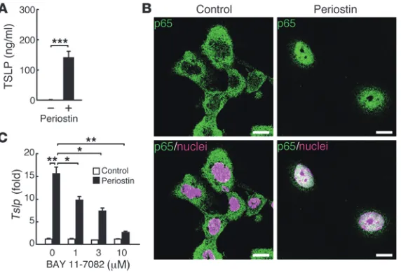
Periostin directly induces TSLP production via NF-κB activation in keratinocytes. We then examined the direct effects of periostin on production of TSLP, a key molecule for initiation of the cascade in allergic skin inflammation (26), in keratinocytes. When cultured on periostin-coated plates, keratinocytes produced significant amounts of TSLP, an effect that was enhanced by the addition of IL-13 (Figure 3A and data not shown). Interestingly, we did not observe the significant effect of periostin for proliferation of kera-tinocytes in monolayered culture condition (data not shown). It has previously been shown that TSLP production is NF-κB depen-dent (26), which raises the possibility that periostin activates the NF-κB pathway in keratinocytes, followed by production of TSLP. Keratinocytes cultured on periostin-coated plates displayed the nuclear translocation of p65 (Figure 3B). Furthermore, BAY 11-7082, an NF-κB inhibitor targeting IκBα phosphorylation, inhibited periostin-induced TSLP expression in a dose-dependent manner in keratinocytes (Figure 3C). These results demonstrated that direct interaction of periostin with keratinocytes induced TSLP production via the NF-κB pathway in keratinocytes.
Interaction of periostin with αv integrin is critical for the appearance of
allergic skin inflammation. Periostin acts as a matricellular protein by binding to several integrin molecules — such as αvβ1, αvβ3, and
αvβ5 — on cardiocytes, endothelial cells, and malignant cells, which
enhances these cells’ proliferation (9, 10, 37). Among these integ-rins, αvβ3 was expressed predominantly on the suprabasal
kerati-nocytes in the epidermis of normal donors and more broadly in
AD patients (Supplemental Figure 10). This finding, taken togeth-er with the importance of fibroblast-dtogeth-erived ptogeth-eriostin for ktogeth-eratino- keratino-cyte activation shown by the 3-dimensional organotypic coculture system, strongly suggests that the interaction of periostin with αv
integrin on cell surface evokes allergic skin inflammation. In the 3-dimensional organotypic coculture system and the monolayered culture system, hyperproliferation, dysregulated differentiation, and production of proinflammatory cytokines in keratinocytes were completely inhibited by neutralizing Abs against αv
inte-grin, and to a lesser extent by neutralizing Abs against β3
integ-rin (Figure 4, A and B, and Supplemental Figure 11). To examine whether binding of periostin to integrin is important for allergic skin inflammation in HDM-sensitized mice, we blocked integrin signals by subcutaneously administering neutralizing anti–αv
inte-grin Abs concomitantly with HDM sensitization; the phenotypic changes induced by HDM were completely suppressed, similar to the observations in Stat6–/– and Postn–/– mice (Figure 4, C–F, and
Supplemental Figure 12). Furthermore, administration of neu-tralizing anti–αv integrin Abs was effective even in established
AD, stopping and improving the progression of skin inflamma-tion, including ear thickness, infiltration of inflammatory cells, epidermal thickness, and IgE production (Figure 5). These results support the notion that the interaction between periostin and αv
integrin is critical for generation of allergic skin inflammation in HDM-sensitized mice.
[image:6.585.48.334.84.279.2]Periostin is highly expressed in the skin tissues of AD patients and is cor-related with disease severity. Finally, we examined whether the find-ings obtained from HDM-sensitized mice and in vitro experiments reflected the pathogenesis of AD patients. Expression of perios-tin was observed only slightly in the dermis on the border of the epidermis in normal skin tissues (n = 4), whereas all AD samples investigated (n = 27) and the HDM-sensitized mice showed signifi-cantly elevated expressions of periostin in the dermis, with a range of expression levels (Figure 6A). We grouped periostin expression levels into mild (n = 3), moderate (n = 11), and marked (n = 13). Inflammation severity, acute change (lymphocyte infiltration in dermis), and chronic changes (epidermal thickness and eosinophil infiltration in dermis) were well correlated with periostin expres-sion levels (Figure 6B and Supplemental Figure 13). Furthermore, serum levels of periostin were significantly elevated in AD patients
Figure 3
(n = 29) compared with healthy volunteers (n = 66, Figure 6C). These results provided strong evidence in support of the involve-ment of periostin in AD pathogenesis.
Discussion
AD is a complex disease caused by the interaction of skin barrier damage and allergic skin inflammation (3, 4). It is well known that Th2-type inflammation is dominant in allergic skin inflammation in AD, based on analyses of AD patients and the HDM-sensitized mouse model. Furthermore, activation of keratinocytes producing various proinflammatory cytokines and chemokines contribute to the initiation or amplification of Th2 responses (2, 25). However, it remained unclear how Th2-type inflammation and activation of keratinocytes interact, leading to amplification and chronicity of AD inflammation. In this study, we demonstrated that perios-tin, a recently characterized matricellular protein, represents a key mediator for amplification and chronicity of allergic skin inflam-mation, as it linked Th2 responses and keratinocyte activation.
Deficiency of Postn or inhibition of αv integrin prevented
devel-opment and/or progression of HDM-induced skin inflammation in our mouse model (Figures 1, 4, and 5 and Supplemental Figures 1, 2, and 12). We confirmed that Postn–/– mice did not have
endog-enous hematopoietic or immune component defects and showed normal distribution and function of dermal DCs and Langer-hans cells (Supplemental Table 1, Supplemental Figure 14, and ref. 11), excluding the possibility that primary immunodeficiency
causes unresponsiveness to the allergen treatment in Postn–/– mice.
Th2-type inflammation initiated by allergen exposure induced production of periostin in fibroblasts through IL-4 and IL-13. Accumulated periostin directly acted on keratinocytes to produce proinflammatory cytokines, including TSLP, via αv integrin. The
proinflammatory cytokines from keratinocytes then amplified the Th2 immune response. Considering all results from this study, we propose a model in which accumulation of periostin constitutes a pathogenic vicious circle that amplifies Th2-type inflammation by binding to αv integrin on keratinocytes (Figure 7). This model
suggests that amplification of allergic skin inflammation requires 2 pathways: exposure of APCs, including DCs, to allergens; and enhancement of immune responses, including activation of APCs, by keratinocyte-derived proinflammatory cytokines induced by periostin. The impaired induction of allergic skin inflammation observed in HDM-treated Postn–/– mice (Figure 1) indicates that
even recurrent stimulation by allergens is not enough to estab-lish allergic skin inflammation without the latter pathway. These effects may be essential in order for APCs to respond efficiently to exogenous allergens in allergic skin inflammation.
[image:7.585.41.407.81.439.2]The roles of matricellular proteins in modulating immune and nonimmune cells in inflamed tissues have been recently eluci-dated. An intracellular isoform of osteopontin in DCs repressed Th17 cell differentiation in a type I, IFN receptor–dependent manner (8). Tenascin-C induces proinflammatory cytokines via TLR4 in macrophages and fibroblasts derived from rheumatoid
Figure 4
Requirement of the integrin/perios-tin interaction on keraintegrin/perios-tinocytes for generation of allergic skin inflam-mation. (A) H&E staining of kerati-nocytes cocultured with WT mouse– derived fibroblasts (2 × 10⁵ cells) in the presence of 10 ng/ml IL-13 and control, anti–αv integrin, and/or anti–
arthritis patients (7). In this study, we demonstrated the signifi-cance of periostin on keratinocytes; however, we cannot exclude the possibility that periostin directly acts on immune cells or non-immune cells other than keratinocytes, modulating their func-tions. Actually, it has been shown that periostin acts on eosino-phils, enhancing their transmigration, chemotaxis, and adhesion (23). Furthermore, periostin activates TGF-β via epithelial cells, followed by induction of collagen production (24). Immune cells and parenchymal/stromal cells activated by periostin together could orchestrate AD inflammation.
Several papers have recently shown the role of periostin in aller-gic airway inflammation. Sehra et al. and Gordon et al. showed that periostin protects mice from allergic airway inflamma-tion (38, 39), whereas Blanchard et al. have shown that periostin accelerates allergen-induced eosinophil recruitment in lung and esophagus (23). Almost the same protocol using intranasal admin-istration of Aspergillus fumigatus caused the different outcomes (23, 39), which suggests that the role of periostin in allergic airway inflammation remains unclear. We here established a persistent Th2-type skin inflammation model, and the outcomes using
Postn–/– mice were different from those of the airway model. This
may be explained by the apparent persistence of Th2-type inflam-mation in the skin model, in contrast to the airway model, which is generally self-limiting in the mouse and resolves spontaneously over time. These results suggest that the skin model is more rel-evant than the airway model to analyze the chronicity of Th2-type inflammation and that the different contribution of periostin to chronic or acute allergic inflammation may explain the different outcomes in the skin and airway models.
HDM is a common allergen associated with human AD and bronchial asthma (4, 40). In the present study, we demonstrated that HDM-treated mice exhibited phenotypes reminiscent of both acute and chronic AD lesions (Figure 1 and Supplemental Figures 1 and 2), as previously reported (27, 28). In light of these prior
reports, we here adopted HDM-sensitized mice as a representative mouse model of allergic skin inflammation. HDM, a mixture of various components including proteases and lipid-binding pro-teins, causes Th2 responses by its potential to activate TLR4 signal-ing or its protease activity via DCs, epithelial cells, and basophils (40, 41). Furthermore, the mice epicutaneously sensitized or caused to inhale HDM showed phenotypes reminiscent of AD or asthma patients in this and other studies (Figure 1, Supplemental Figure 1, and refs. 27, 28, 41). The observations that chronic skin lesions of AD patients showed upregulation of IFN-γ and IL-12 and that IFN-γ deficiency partially improved ovalbumin-sensitized allergic skin inflammation in mice (42–44) are indicative of the involvement of Th1 responses in addition to Th2 responses. However, in our HDM-sensitized mouse model, enhancement of IFN-γ expression in skin tissues was not observed, despite the appearance of other features of chronic AD skin lesions (Figure 1 and data not shown). Huang et al. have also reported that skin lesions in HDM-treated mice do not show IFN-γ expression, whereas splenocytes responsive to allergen stimulation can induce IFN-γ production (28). Expo-sure to microbial products may make the difference between mouse models and patients (4). Furthermore, Th17 responses have been implicated in the acute phase of AD pathogenesis, although this is still controversial (4, 45). Interestingly, DCs stimulated with KCM in the presence of IL-13 induced IL-17–producing T cells as well as IL-4/IL-13–producing T cells (Figure 2F). In spite of complicated and diverse phenotypes, our present study suggests that the induc-tion of these phenotypes completely depended on STAT6, a critical transcriptional factor of Th2 cytokines (Figure 1 and Supplemental Figure 1). Furthermore, periostin induction may be a common fea-ture in mouse models of allergic skin inflammation, because induc-tion of periostin was observed not only in HDM-treated mice, but also in hapten-treated mice (Supplemental Figure 5).
[image:8.585.68.517.82.286.2]Among the proinflammatory cytokines derived from activated AD keratinocytes, TSLP is critical in AD inflammation (26). TSLP
Figure 5
is produced mainly from epithelial cells together with other cells, such as mast cells, creating a Th2-permissive microenvironment via DCs (26, 46). Keratinocyte-specific overexpression of TSLP induces allergic skin inflammation in mice (47). Accordingly, pro-duction of TSLP is upregulated in keratinocytes derived from AD patients (46). In this study, we showed that KCM in the presence of periostin containing high amounts of TSLP induced Th2 skewing via DCs (Figure 2). Furthermore, recombinant periostin directly induced production of TSLP in keratinocytes (Figure 3). Addi-tion of neutralizing Abs against TSLP into the IL-13–treated KCM partially inhibited Th2 skewing (Supplemental Figure 8). This result supports the involvement of TSLP in this event, although factors other than TSLP may also contribute to the Th2 skew-ing. Furthermore, the observation that neutralizing Abs against TSLP slightly downregulated Th2 skewing, even in the PBS-treated KCM, indicated that basal production of periostin from untreated fibroblasts can cause induction of TSLP in keratinocytes, followed by Th2 skewing via DCs to a small extent. It has been reported that synergistic stimuli of Th2 cytokines (IL-4 or IL-13) and pro-inflammatory cytokines (TNF-α or IL-1α), or extrinsic stimuli via several TLRs, induce TSLP production in keratinocytes (48–50). In addition to these factors, we demonstrated that periostin was an intrinsic inducer of TSLP from keratinocytes, a finding we believe to be novel. It is known that signals via αvβ3 integrin lead to
activa-tion of NF-κB, which is important for TSLP expression (51, 52). Indeed, TSLP production by periostin was dependent on αvβ3
inte-grin/NF-κB pathways (Figure 3 and Figure 4B). These results are consistent with a previous report that mice defective in IκBα, an inhibitor of the NF-κB pathway, develop inflammatory changes (53), showing the importance of NF-κB activation in keratinocytes in allergic skin inflammation.
Hyperplasia and dysregulated differentiation of the epidermis (i.e., acanthosis) are typical features of chronic inflammatory skin diseases, including AD and psoriasis (3, 4, 54). Several cytokines in addition to Th2 cytokines — IL-1α, IL-21, IL-22, and IFN-γ — are reported to be correlated with the generation of acanthosis (42, 45, 55, 56). In this study, we demonstrated that periostin acted as a critical player for acanthosis downstream of IL-4 or IL-13 signals. Furthermore, we showed that periostin induced activation of the Akt pathway through integrins (Supplemental Figure 11), and the Akt pathway is known to be strongly associated with keratinocyte survival and differentiation (34). Together with the NF-κB path-way, another important signal for proliferation of keratinocytes (57), the Akt pathway could be responsible for periostin-induced acanthosis. The pathophysiological role of acanthosis in chronic inflammation remains unknown. We demonstrated that periostin-stimulated keratinocytes enhanced production of TSLP (Figure 3) and that production of proinflammatory cytokines was further enhanced by the increased number of activated keratinocytes (Fig-ure 2). Overall, we assume that acanthosis in AD provides a basis for supplying more massive proinflammatory mediators from keratinocytes. Furthermore, based on the observation that Th2-type responses functioned as an immune system against parasite infection, periostin may contribute to enforcement of this defense mechanism by activation of keratinocytes.
We showed that administration of neutralizing anti–αv integrin
Abs completely suppressed HDM-induced phenotypic changes (Fig-ure 4). Because it is known that several ECM proteins — vitronectin, osteopontin, and thrombospondin — bind to αv integrin (58), we
cannot exclude the possibility that the effects of anti–αv integrin Abs
were the consequence of the inhibition of all functions of αv
[image:9.585.42.543.82.381.2]integ-rin ligands, including periostin. However, we observed that TSLP
Figure 6
Periostin is highly expressed in skin tissues of AD patients, correlated with disease sever-ity. (A) Skin tissues stained with H&E and anti-periostin Ab of a control donor and an AD patient with marked periostin expression. Scale bars: 200
μm. (B) Inflammation score, lymphocyte number in the der-mis, and epidermal thickness in healthy controls and AD patients with periostin expres-sion classified as mild (n = 3), moderate (n = 11), or marked (n = 13). Lymphocyte numbers were counted and epidermal thickness measured in 10 ×400 fields for each sample. (C) Peri-ostin levels in sera obtained from control donors (n = 66) and AD patients (n = 29) using ELISA. Red lines in B and C
production was induced on periostin-coated, but not vitronectin-coated, plates (data not shown). These results suggest that each of the αv integrin ligands transduces different signals and that some
receptors other than αv integrin may be involved in these ligands.
It has been reported that allergen avoidance has limited efficacy (59, 60). Given that the intrinsic amplification pathway is impor-tant for chronicity of AD, periostin and its functional receptor, αv
integrin, can be a good target to develop therapeutic agents for AD. The blockage of αv integrin was effective even in the
HDM-sensi-tized mice (Figure 5). These results support our model and suggest that an agent blocking the interaction between periostin and αv
integrin might act as a not only preventive, but also therapeutic, drug. Antagonists against αvβ3 integrin have been tested in
clini-cal use for tumors, rheumatoid arthritis, and osteoporosis (61–63). However, it is assumed that blockage of periostin has an advantage compared with blockage of αv integrin, because it has been shown
that most αv integrin–deficient mice die before birth (64), so that
blockage of αv integrin will give more severe adverse effects than
blockage of periostin. However, we must be careful concerning the possible risk of adverse effects to block the action of periostin on cardiac development/remodeling or cutaneous wound repair, because periostin plays important roles in these events (11, 12, 15–17). The present findings provide a molecular basis for consid-ering periostin as a potential therapeutic target for AD in humans.
Methods
Mice. 8-week-old BALB/c mice, C57BL/6 mice, NC/Nga mice (all Japan SLC), and Stat6–/– mice (gift from T. Hoshino, Kurume University School of Medicine, Kurume, Japan) were used. Postn–/– mice, prepared as previ-ously described (129SJv;C57BL/6 background; ref. 11), were backcrossed to BALB/c mice, and F4–F8 mice were used. Postn–/– mice were always evalu-ated together with their WT littermates.
HDM preparation. Dermatophagoides farinae (gift of Torii Pharmaceuti-cal) was defatted with ethyl ether, then extracted in 50% glycerin and 5% NaCl buffer at 4°C for 48 hours under constant stirring. After filtration, D. farinae extract was used as HDM.
Allergic skin inflammation model. The mouse model of allergic skin inflam-mation was generated as previously described (27), with minor modifica-tions. Both surfaces of the ear lobes were stripped 3 times using cellophane
tape (Kyowa), and 25-μl HDM (10 mg/ml) solution was painted onto both surfaces of the earlobes. This procedure was performed once per week for 7 weeks. Ear swelling was measured before each HDM painting with a micrometer (Ozaki MFG). The mouse ear tissues, cervical lymph nodes, and serum were collected 24 hours after the last HDM painting. 10 μl anti–αv
integrin Ab (1 mg/ml; BioLegend) or rat IgG1 isotype control Ab (1 mg/ ml; R&D Systems) was administered subcutaneously to ear tissues 6 hours before and 3 days after all HDM paintings (prevention protocol), or from the fourth HDM painting until day 21 (therapeutic protocol).
Histology. Ear tissues were subjected to histological staining with H&E, Masson trichrome, and toluidine blue. Immunohistochemical staining was performed as previously described (19); see Supplemental Methods for Abs. Apoptotic cells were detected by TUNEL assay (Promega). Epidermal thickness was measured, and the number of lymphocytes, eosinophils, and mast cells counted, in 10 ×400 fields for each mouse or human sample, and the averages values were calculated.
IgE. Duplicate serum samples were analyzed using an ELISA kit for total IgE (Bethyl Laboratories).
Cells. See Supplemental Methods for preparation of mouse keratino-cytes and dermal fibroblasts. Mouse embryonic fibroblasts were obtained from E14.5 embryos of Postn+/– or Postn–/– mice. For stimulation, either human or mouse dermal fibroblasts were stimulated by 10–50 ng/ml IL-4 or IL-13 (PeproTech).
RT-PCR. RT-PCR for Postn and Gapdh was performed as previously described (19). See Supplemental Methods for details.
Western blotting. Western blotting for periostin and GAPDH was per-formed as previously described (19).
3-dimensional organotypic coculture of keratinocytes with fibroblasts. Mouse keratinocytes were cultured using a 3-dimensional system (Figure 3A) mod-ified from a previously reported system (33). 2 ml type I collagen gel solu-tion (Nitta Gelatin) containing 2 × 10⁵ mouse embryonic fibroblasts was
poured into an inner dish with a bottom made of nitrocellulose membrane (Millicell-CM). Next, 2 × 10⁶ keratinocytes in total were overlaid on the gel,
and DMEM was poured into the outer dish with the surface level lower than that of the gel, to let the keratinocytes face the air. The culture medium was replaced every 2 days. In some experiments, 10 ng/ml mouse IL-13, 10 μg/ ml anti–αv integrin Ab, or 10 μg/ml anti–β3 integrin Ab (BD Biosciences)
[image:10.585.57.270.82.286.2]was added. In some experiments, the NF-κB inhibitor BAY 11-7082 (con-centrations as indicated; Sigma-Aldrich) was added into the medium. Figure 7
Recombinant proteins. The recombinant protein coded by pMT/Bip/ V5-HisA-periostin was prepared as previously described (19).
Quantitative analysis of cytokines. Amounts of TSLP, TNF-α, and GM-CSF in the culture medium were quantified with ELISA kits for mouse TSLP (R&D Systems), TNF-α, and GM-CSF (eBioscience).
Mixed lymphocyte reaction. Bone marrow–derived DCs were prepared from a bone marrow suspension obtained from the femurs and tibias of C57BL/6 mice. Cells were cultured with 20 ng/ml murine GM-CSF (Pepro-Tech) for 6 days. CD4+ T cells were prepared from spleen and lymph node
cells of BALB/c mice using anti-CD4–coated MACS magnetic beads (Milt-enyi Biotec, Germany). In total, 2 × 10⁵ CD4+ T cells were cocultured with
KCM-treated bone marrow–derived DCs. Cells were harvested after 3 or 5 days for [3H]thymidine incorporation assay or quantitative RT-PCR
analy-sis, respectively. Quantitative RT-PCR analysis was performed after restimu-lation of the cells with anti-CD3 Ab and anti-CD28 Ab for 24 hours.
Confocal microscopy. Keratinocytes were fixed by paraformaldehyde. After blocking with 2% BSA, sections were stained by anti–NF-κB p65 Ab (Santa Cruz Biotechnology), followed by Alexa Fluor 488–labeled anti-rabbit IgG Ab (Invitrogen), or TO-PRO-3 (Invitrogen) for the nucleus. Sections were mounted by Dako Fluorescent Mounting Medium (Dako) and then exam-ined by LSM5 PASCAL G/B (Carl Zeiss).
Human skin tissue specimens. Skin tissues examined for histology were derived from 4 normal donors and 27 AD patients, and serum samples were derived from 66 normal donors and 29 AD patients. The 27 AD patients for histology were aged 13–54 years; 2 had received oral predniso-lone, and 19 had received topical steroid ointments. The 29 AD patients for serum were aged 13–53 years; 3 had received oral prednisolone, and 27 had received topical steroid ointments (1 patient had received both). Patients received steroid treatment at the time of sample collection. No AD patient had bronchial asthma or rhinosinusitis. We scored the severity of skin lesions on a 0–4 scale according to the guidelines established by the Japanese Dermatological Association (65). We grouped the patient sample
into 3 types, according to the degree and pattern of periostin expression: mild (mild expression on the border of the epidermis), moderate (strong expression on the border of the epidermis), and marked (strong and broad increase in the dermis).
Measurement of serum periostin. We performed a human periostin ELISA assay using 2 rat anti-human periostin mAbs (clones no. SS18A and SS17B), as previously reported (66). See Supplemental Methods for details.
Statistics. Results are presented as means + SD in all experiments. Analysis was carried out using 2-sided, unpaired Student’s t test or 2-sided Welch test. A P value less than 0.05 was considered significant.
Study approval. Animal experiments were undertaken following the guidelines for care and use of experimental animals of the Japanese Asso-ciation for Laboratory Animals Science (1987) and were approved by the Saga University Animal Care and Use Committee (Saga, Japan). Human study protocols were approved by the Ethics Committees of Kyushu Uni-versity and Saga Medical School. Informed consent was obtained from all patients and healthy volunteers.
Acknowledgments
We thank Koichi Akashi, Dovie R. Wylie, Hiroyuki Ideguchi, and Yumiko Ohishi for critical review of this manuscript and technical assistance. This work was supported in part by Grants-in-Aid for Scientific Research from the Japan Society for the Promotion of Science.
Received for publication May 12, 2011, and accepted in revised form May 2, 2012.
Address correspondence to: Kenji Izuhara, Division of Medical Biochemistry, Department of Biomolecular Sciences, Saga Medi-cal School, 5-1-1, Nabeshima, Saga 849-8501, Japan. Phone: 81.952. 34.2261; Fax: 81.952.34.2058; E-mail: kizuhara@cc.saga-u.ac.jp.
1. Medzhitov R. Inflammation 2010: new adventures of an old flame. Cell. 2010;140(6):771–776. 2. Kaiko GE, Foster PS. New insights into the
genera-tion of Th2 immunity and potential therapeutic targets for the treatment of asthma. Curr Opin Allergy Clin Immunol. 2011;11(1):39–45.
3. Leung DY, Boguniewicz M, Howell MD, Nomura I, Hamid QA. New insights into atopic dermatitis.
J Clin Invest. 2004;113(5):651–657.
4. Oyoshi MK, He R, Kumar L, Yoon J, Geha RS. Cel-lular and molecular mechanisms in atopic derma-titis. Adv Immunol. 2009;102:135–226.
5. Meneghin A, Hogaboam CM. Infectious disease, the innate immune response, and fibrosis. J Clin Invest. 2007;117(3):530–538.
6. Wynn TA. Fibrotic disease and the TH1/TH2
para-digm. Nat Rev Immunol. 2004;4(8):583–594. 7. Midwood K, et al. Tenascin-C is an endogenous
activator of Toll-like receptor 4 that is essential for maintaining inflammation in arthritic joint dis-ease. Nat Med. 2009;15(7):774–780.
8. Shinohara ML, Kim JH, Garcia VA, Cantor H. Engagement of the type I interferon receptor on dendritic cells inhibits T helper 17 cell development: role of intracellular osteopontin. Immunity. 2008; 29(1):68–78.
9. Hamilton DW. Functional role of periostin in devel-opment and wound repair: implications for con-nective tissue disease. J Cell Commun Signal. 2008; 2(1–2):9–17.
10. Ruan K, Bao S, Ouyang G. The multifaceted role of periostin in tumorigenesis. Cell Mol Life Sci. 2009; 66(14):2219–2230.
11. Rios H, et al. periostin null mice exhibit dwarfism, incisor enamel defects, and an early-onset
peri-odontal disease-like phenotype. Mol Cell Biol. 2005; 25(24):11131–11144.
12. Snider P, et al. Periostin is required for maturation and extracellular matrix stabilization of noncar-diomyocyte lineages of the heart. Circ Res. 2008; 102(7):752–760.
13. Kühn B, et al. Periostin induces proliferation of dif-ferentiated cardiomyocytes and promotes cardiac repair. Nat Med. 2007;13(8):962–969.
14. Shimazaki M, et al. Periostin is essential for cardiac healing after acute myocardial infarction. J Exp Med. 2008;205(2):295–303.
15. Nishiyama T, et al. Delayed re-epithelialization in periostin-deficient mice during cutaneous wound healing. PLoS One. 2011;6(4):e18410.
16. Elliott CG, et al. Periostin modulates myofibroblast differentiation during full-thickness cutaneous wound repair. J Cell Sci. 2012;125(pt 1):121–132. 17. Ontsuka K, et al. Periostin, a matricellular protein,
accelerates cutaneous wound repair by activating der-mal fibroblasts. Exp Dermatol. 2012;21(5):331–336. 18. Hayashi N, Yoshimoto T, Izuhara K, Matsui K,
Tanaka T, Nakanishi K. T helper 1 cells stimulated with ovalbumin and IL-18 induce airway hyper-responsiveness and lung fibrosis by IFN-γ and IL-13 production. Proc Natl Acad Sci U S A. 2007; 104(37):14765–14770.
19. Takayama G, et al. Periostin: a novel component of subepithelial fibrosis of bronchial asthma downstream of IL-4 and IL-13 signals. J Allergy Clin Immunol. 2006;118(1):98–104.
20. Kii I, et al. Incorporation of tenascin-C into the extra cellular matrix by periostin underlies an extra-cellular meshwork architecture. J Biol Chem. 2010; 285(3):2028–2039.
21. Maruhashi T, Kii I, Saito M, Kudo A. Interaction between periostin and BMP-1 promotes proteo-lytic activation of lysyl oxidase. J Biol Chem. 2010; 285(17):13294–13303.
22. Uchida M, et al. Periostin, a matricellular pro-tein, plays a role in the induction of chemokines in pulmonary fibrosis. Am J Respir Cell Mol Biol. 2012;46(5):677–686.
23. Blanchard C, et al. Periostin facilitates eosinophil tissue infiltration in allergic lung and esophageal responses. Mucosal Immunol. 2008;1(4):289–296. 24. Sidhu SS, et al. Roles of epithelial cell-derived
peri-ostin in TGF-β activation, collagen production, and collagen gel elasticity in asthma. Proc Natl Acad Sci U S A. 2010;107(32):14170–14175.
25. Carmi-Levy I, Homey B, Soumelis V. A modular view of cytokine networks in atopic dermatitis. Clin Rev Allergy Immunol. 2011;41(3):245–253. 26. Liu YJ, et al. TSLP: an epithelial cell cytokine
that regulates T cell differentiation by condition-ing dendritic cell maturation. Annu Rev Immunol. 2007;25:193–219.
27. Gao XK, Nakamura N, Fuseda K, Tanaka H, Inaga-ki N, Nagai H. Establishment of allergic dermatitis in NC/Nga mice as a model for severe atopic der-matitis. Biol Pharm Bull. 2004;27(9):1376–1381. 28. Huang CH, Kuo IC, Xu H, Lee YS, Chua KY. Mite
allergen induces allergic dermatitis with concomi-tant neurogenic inflammation in mouse. J Invest Dermatol. 2003;121(2):289–293.
29. Jensen JM, et al. Impaired sphingomyelinase activ-ity and epidermal differentiation in atopic derma-titis. J Invest Dermatol. 2004;122(6):1423–1431. 30. Howell MD, et al. Cytokine modulation of atopic
Immunol. 2007;120(1):150–155.
31. Izuhara K, Arima K, Kanaji S, Ohta S, Kanaji T. IL-13: a promising therapeutic target for bronchial asthma. Curr Med Chem. 2006;13(19):2291–2298. 32. Yuyama N, et al. Analysis of novel
disease-relat-ed genes in bronchial asthma. Cytokine. 2002; 19(6):287–296.
33. Aoki S, Toda S, Ando T, Sugihara H. Bone marrow stromal cells, preadipocytes, and dermal fibroblasts promote epidermal regeneration in their distinc-tive fashions. Mol Biol Cell. 2004;15(10):4647–4657. 34. Muller EJ, Williamson L, Kolly C, Suter MM. Outside-in signalOutside-ing through Outside-integrOutside-ins and cadherOutside-ins: a cen-tral mechanism to control epidermal growth and dif-ferentiation? J Invest Dermatol. 2008;128(3):501–516. 35. Griffiths CE, Dearman RJ, Cumberbatch M,
Kim-ber I. Cytokines and Langerhans cell mobilisation in mouse and man. Cytokine. 2005;32(2):67–70. 36. Zou GM, Tam YK. Cytokines in the generation and
maturation of dendritic cells: recent advances. Eur Cytokine Netw. 2002;13(2):186–199.
37. Norris RA, Moreno–Rodriguez R, Hoffman S, Markwald RR. The many facets of the matricel-luar protein periostin during cardiac development, remodeling, and pathophysiology. J Cell Commun Signal. 2009;3(3–4):275–286.
38. Sehra S, et al. Periostin regulates goblet cell meta-plasia in a model of allergic airway inflammation.
J Immunol. 2011;186(8):4959–4966.
39. Gordon ED, et al. A protective role for periostin and TGF-β in IgE-mediated allergy and airway hyperre-sponsiveness. Clin Exp Allergy. 2012;42(1):144–155. 40. Arlian LG, Platts-Mills TA. The biology of dust
mites and the remediation of mite allergens in allergic disease. J Allergy Clin Immunol. 2001; 107(3 suppl):S406–S413.
41. Hammad H, et al. Inflammatory dendritic cells— not basophils—are necessary and sufficient for induction of Th2 immunity to inhaled house dust mite allergen. J Exp Med. 2010;207(10):2097–2111. 42. Spergel JM, Mizoguchi E, Oettgen H, Bhan AK,
Geha RS. Roles of TH1 and TH2 cytokines in a
murine model of allergic dermatitis. J Clin Invest. 1999;103(8):1103–1111.
43. Grewe M, Gyufko K, Schopf E, Krutmann J. Lesion-al expression of interferon-gamma in atopic ecze-ma. Lancet. 1994;343(8888):25–26.
44. Hamid Q, Naseer T, Minshall EM, Song YL, Boguni-ewicz M, Leung DY. In vivo expression of IL-12 and IL-13 in atopic dermatitis. J Allergy Clin Immunol. 1996;98(1):225–231.
45. Souwer Y, Szegedi K, Kapsenberg ML, de Jong EC. IL-17 and IL-22 in atopic allergic disease. Curr Opin Immunol. 2010;22(6):821–826.
46. Soumelis V, et al. Human epithelial cells trigger dendritic cell mediated allergic inflammation by producing TSLP. Nat Immunol. 2002;3(7):673–680. 47. Yoo J, et al. Spontaneous atopic dermatitis in mice
expressing an inducible thymic stromal lympho-poietin transgene specifically in the skin. J Exp Med. 2005;202(4):541–549.
48. Bogiatzi SI, et al. Proinflammatory and Th2 cytokines synergize to induce thymic stromal lym-phopoietin production by human skin keratino-cytes. J Immunol. 2007;178(6):3373–3377. 49. Kinoshita H, et al. Cytokine milieu modulates
release of thymic stromal lymphopoietin from human keratinocytes stimulated with double-stranded RNA. J Allergy Clin Immunol. 2009; 123(1):179–186.
50. Vu AT, et al. Staphylococcus aureus membrane and diacylated lipopeptide induce thymic stromal lym-phopoietin in keratinocytes through the Toll-like receptor 2-Toll-like receptor 6 pathway. J Allergy Clin Immunol. 2010;126(5):985–993
51. Lee HC, Ziegler SF. Inducible expression of the pro-allergic cytokine thymic stromal lymphopoietin in airway epithelial cells is controlled by NFκB. Proc Natl Acad Sci U S A. 2007;104(3):914–919. 52. Scatena M, Almeida M, Chaisson ML, Fausto N,
Nicosia RF, Giachelli CM. NF-κB mediates αvβ3
integrin-induced endothelial cell survival. J Cell Biol. 1998;141(4):1083–1093.
53. Rebholz B, et al. Crosstalk between keratinocytes
and adaptive immune cells in an IκBα protein-mediated inflammatory disease of the skin. Immu-nity. 2007;27(2):296–307.
54. Korn T, Bettelli E, Oukka M, Kuchroo VK. IL-17 and Th17 cells. Annu Rev Immunol. 2009;27:485–517. 55. Caruso R, et al. Involvement of interleukin-21 in the
epidermal hyperplasia of psoriasis. Nat Med. 2009; 15(9):1013–1015.
56. Szabowski A, et al. c-Jun and JunB antagonistically control cytokine-regulated mesenchymal-epider-mal interaction in skin. Cell. 2000;103(5):745–755. 57. Karin M. Nuclear factor-κB in cancer development
and progression. Nature. 2006;441(7092):431–436. 58. Clemmons DR, Maile LA, Ling Y, Yarber J, Busby WH. Role of the integrin αvβ3 in mediating
increased smooth muscle cell responsiveness to IGF-I in response to hyperglycemic stress. Growth Horm IGF Res. 2007;17(4):265–270.
59. Romei I, Boner AL. Possible reasons for lack of effect of allergen avoidance in atopy-prone infants and sensitive asthmatic patients. Clin Rev Allergy Immunol. 2005;28(1):59–71.
60. Simpson EL. Atopic dermatitis prevention. Derma-tol Ther. 2006;19(2):108–117.
61. Cacciari B, Spalluto G. Non peptidic αvβ3
antago-nists: recent developments. Curr Med Chem. 2005; 12(1):51–70.
62. Jin H, Varner J. Integrins: roles in cancer develop-ment and as treatdevelop-ment targets. Br J Cancer. 2004; 90(3):561–565.
63. Lainer-Carr D, Brahn E. Angiogenesis inhibition as a therapeutic approach for inflammatory synovitis.
Nat Clin Pract Rheumatol. 2007;3(8):434–442. 64. Bader BL, Rayburn H, Crowley D, Hynes RO.
Extensive vasculogenesis, angiogenesis, and organ-ogenesis precede lethality in mice lacking all αv integrins. Cell. 1998;95(4):507–519.
65. Furue M, et al. Guidelines for management of atop-ic dermatitis. Jpn J Dermatol. 2008;118:325–342. 66. Okamoto M, et al. Periostin, a matrix protein, is a