0095-1137/07/$08.00
⫹
0
doi:10.1128/JCM.00280-07
Copyright © 2007, American Society for Microbiology. All Rights Reserved.
Rapid Multiplex Nested PCR for Detection of Respiratory Viruses
䌤
W. Y. Lam,
1Apple C. M. Yeung,
1Julian W. Tang,
1Margaret Ip,
1Edward W. C. Chan,
1Mamie Hui,
1and Paul K. S. Chan
1,2*
Department of Microbiology,
1and Stanley Ho Centre for Emerging Infectious Diseases of the School of Public Health,
2Faculty of
Medicine, The Chinese University of Hong Kong, Prince of Wales Hospital, Shatin, New Territories, Hong Kong
Received 4 February 2007/Returned for modification 11 May 2007/Accepted 19 August 2007
Respiratory tract infections can be caused by a heterogeneous group of viruses and bacteria that produce
similar clinical presentations. Specific diagnosis therefore relies on laboratory investigation. This study
developed and evaluated five groups of multiplex nested PCR assays that could simultaneously detect 21
different respiratory pathogens: influenza A virus (H1N1, H3N2, and H5N1); influenza B virus; parainfluenza
virus types 1, 2, 3, 4a, and 4b; respiratory syncytial virus A and B; human rhinoviruses; human enteroviruses;
human coronaviruses OC43 and 229E; severe acute respiratory syndrome coronavirus; human
metapneumo-viruses;
Mycoplasma pneumoniae
;
Chlamydophila pneumoniae
;
Legionella pneumophila
; and adenoviruses (A to
F). These multiplex nested PCRs adopted fast PCR technology. The high speed of fast PCR (within 35 min)
greatly improved the efficiency of these assays. The results show that these multiplex nested PCR assays are
specific and more sensitive (100- to 1,000-fold) than conventional methods. Among the 303 clinical specimens
tested, the multiplex nested PCR achieved an overall positive rate of 48.5% (95% confidence interval [CI], 42.9
to 54.1%), which was significantly higher than that of virus isolation (20.1% [95% CI, 15.6 to 24.6%]) and that
of direct detection by immunofluorescence assay (13.5% [95% CI, 9.7 to 17.4%]). The improved sensitivity was
partly due to the higher sensitivity of multiplex nested PCR than that of conventional methods in detecting
cultivatable viruses. Moreover, the ability of the multiplex nested PCR to detect noncultivatable viruses,
particularly rhinoviruses, coronavirus OC43, and metapneumoviruses, contributed a major gain (15.6%) in the
overall positive rate. In conclusion, rapid multiplex nested PCR assays can improve the diagnostic yield for
respiratory infections to allow prompt interventive actions to be taken.
Respiratory tract infection is a major cause of
hospitaliza-tion. The 2003 outbreaks of severe acute respiratory syndrome
(SARS) and the more recent human avian H5N1 influenza
virus cases underscore the importance of a rapid and accurate
laboratory diagnosis to investigate infections associated with
severe individual or public health consequences (1).
Respiratory tract infections in humans can be a result of
infection caused by a heterogeneous group of viruses and
bac-teria that produce similar clinical presentations (10, 18, 30).
Specific diagnosis therefore relies almost entirely on laboratory
investigation. Rapid antigen detection assays are now widely
used in routine diagnostic laboratories, but these assays have
been shown to be inferior in sensitivity and specificity to assays
based on PCR (5, 12), which can also be designed to screen for
a wider range of pathogens. Numerous studies have developed
and evaluated multiplex nested PCR, reverse transcription
(RT)-PCR, or real-time PCR for the detection of respiratory
viruses (4, 11, 21, 24, 27). However, these studies are still
limited by their turnaround time and/or the range of viruses
being detected.
This study developed and evaluated five groups of multiplex
nested PCR assays that included an RT step where necessary.
These assays could simultaneously detect 21 different
respira-tory pathogens including influenza virus group A (FluA)
(sub-types H1, H3, and H5), influenza virus group B (FluB),
para-influenza virus type 1 (PIV-1), PIV-2, PIV-3, PIV-4a, PIV-4b,
human respiratory syncytial viruses (hRSV), all serotypes of
human adenoviruses (ADVs) (A to F), human
metapneumo-viruses (hMPV), human coronametapneumo-viruses (HCoVs)
(HCoV-229E, HCoV-OC43, and SARS coronavirus [SARS-CoV]), all
serotypes of human enteroviruses (hEVs), and all serotypes of
human rhinoviruses (hRVs) as well as the fastidious
respira-tory bacteria
Chlamydophila pneumoniae
,
Mycoplasma
pneu-moniae
, and
Legionella pneumophila
.
MATERIALS AND METHODS
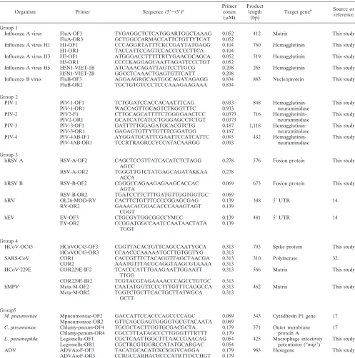
Primer design and preparation.The primers used in this study were either modified from previously published primer sequences (2–4, 7–9, 11, 17, 19, 23, 24, 26, 28, 29, 31–34) or designed from consensus genome regions obtained from GenBank (http://www.ncbi.nlm.nih.gov/). Typically, sequences of 10 to 20 rep-resentative strains of each pathogen were downloaded. The sequences were aligned using Clustal X (http://bips.u-strasbg.fr/en/Documentation/ClustalX/) (25). The program GeneTool Lite 1.0 (BioTools Inc., Edmonton, Alberta, Can-ada) was used to predict the compatibility of primer pairs and to estimate the optimal annealing temperatures. Primer pairs were selected to ensure that the sizes of the amplicons of different pathogens could be easily differentiated by agarose gel electrophoresis. The primers used were 20 to 30 bp in length and had G⫹C contents less than or equal to 70%, thus having an annealing temperature of 50 to 66°C.
Multiplex PCR primer grouping.Five groups of multiplex nested PCR assays, targeting 21 respiratory viruses and bacteria, were developed. Each multiplex nested PCR detected four to five viruses and/or bacteria: group 1 was comprised of FluA and FluB group-specific and subtype H1-, H3-, and H5-specific primers. Group 2 was comprised of PIV-1, PIV-2, PIV-3, PIV-4a, and PIV-4b. Group 3 was comprised of hRSV A and B, hRV, and hEV. Group 4 was comprised of HCoV (HCoV-OC43, HCoV-229E, and SARS-CoV) and hMPV. Group 5 was comprised ofMycoplasma pneumoniae,Chlamydophila pneumoniae,Legionella
* Corresponding author. Mailing address: Department of
Microbi-ology, The Chinese University of Hong Kong, 1/F Clinical Science
Building, Prince of Wales Hospital, Shatin, New Territories, Hong
Kong. Phone: (852) 2632 3333. Fax: (852) 2647 3227. E-mail:
[email protected].
䌤
Published ahead of print on 5 September 2007.
3631
on May 16, 2020 by guest
http://jcm.asm.org/
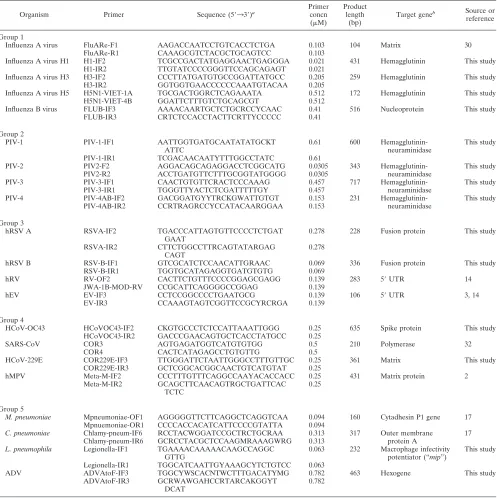
pneumophila, and ADV. The sequences and amplicon sizes of the outer and inner sets of primers are given in Tables 1 and 2.
Nucleic acid extraction and cDNA synthesis. Total RNA and DNA were extracted together by using the QIAamp MinElute Virus Spin kit (QIAGEN, Valencia, CA), and 60l of nucleic acids was eluted according to the manufac-turer’s recommendations.
The RNA template (8l) was mixed with 1l of random primers (2.5 ng/l) and 1l of deoxynucleoside triphosphates (0.5 mM each) in a final volume of 10
l for incubation at 65°C for 5 min. The solution was equilibrated at 4°C and completed with 2 units of RNaseOUT, 4l of 5⫻first-strand buffer, 0.5 mM dithiothreitol, and 10 U Superscript III reverse transcriptase (Invitrogen,
Carls-bad, CA) in a final volume of 20l. RT was performed for 50 min at 50°C and then stopped by heating for 15 min at 70°C. The resulting cDNA products were used immediately for PCR.
[image:2.585.44.541.79.581.2]Fast PCR conditions.In order to provide the shortest possible turnaround time, the recently available “fast” thermal cycler (fast PCR machine; Applied Biosystems, Foster City, CA) was used. When coupled with the DNA polymerase contained in the fast PCR master mix (GeneAmp; Applied Biosystems, Foster City, CA), a 35-cycle PCR assay could be completed within 35 min, compared to the⬃180 min that would normally be required for standard thermocyclers. All multiplex nested PCR assays were optimized to fulfill the manufacturer’s rec-ommendation that two-step cycling with annealing at 64°C was used. Both the
TABLE 1. Primers used in the first round of multiplex nested PCR
Organism Primer Sequence (5⬘33⬘)a
Primer concn (M)
Product length
(bp)
Target geneb Source or
reference
Group 1
Influenza A virus FluA-OF3 TYGAGGCTCTCATGGARTGGCTAAAG 0.052 412 Matrix This study FluA-OR3 GCTGGCCARMACCATTCTGTTYTCAT 0.052
Influenza A virus H1 H1-OF1 CCCAGGRTATTTCKCCGAYTATGAGG 0.104 760 Hemagglutinin This study
H1-OR1 TACCATTCCAGTCCACCCCCCTTCA 0.104
Influenza A virus H3 H3-OF1 ATGGGACCTTTTTRTYGAACGCAGCA 0.052 519 Hemagglutinin This study H3-OR1 CCCCKAGGAGCAATTAGATTCCCTGT 0.052
Influenza A virus H5 H5N1-VIET-1B ATCAAACAGATTAGTCCTTGCG 0.208 265 Hemagglutinin This study H5N1-VIET-2B GGCCTCAAACTGAGTGTTCATT 0.208
Influenza B virus FluB-OF3 AGGAAGRGCAATGGCAGAYAGAGG 0.834 883 Nucleoprotein This study FluB-OR2 TGCTGTGTCCCTCCCAAAGAAGAAA 0.834
Group 2
PIV-1 PIV-1-OF1 TCTGGATCCACCACAATTTCAG 0.933 848 Hemagglutinin- This study
PIV-1-OR1 WACCAGTTGCAGTCTKGGTTTC 0.933 neuraminidase
PIV-2 PIV2-F1 CTTGCAGCATTTTCTGGGGAACTCC 0.0373 716 Hemagglutinin- This study
PIV2-OR1 GCATCATCATCCTGGGAGCCTCTGT 0.0373 neuraminidase
PIV-3 PIV-3-OF1 GATTTTTGGAGATGCACGTCTG 0.187 1,118 Hemagglutinin- This study
PIV-3-OR1 GAGAGTGTTYTGTTTCGGATGG 0.187 neuraminidase
PIV-4 PIV-4AB-IF1 AYGGATGCATTCGAATTCCATCATTC 0.093 432 Hemagglutinin- This study
PIV-4AB-OR1 TCCRTRAGRCCYCCATACAARGG 0.093 neuraminidase
Group 3
hRSV A RSV-A-OF2 CAGCTCCGTTATCACATCTCTAGG
AGCC
0.278 576 Fusion protein This study
RSV-A-OR2 TGGGTTGTCTATGAGCAGATAKKAA ACCA
0.278
hRSV B RSV-B-OF2 CGGGCCAGAAGAGAAGCACCAC
AGTA
0.069 673 Fusion protein This study
RSV-B-OR2 TGATCCTTCTTTGATGTTGGTGGTGC 0.069
hRV OL26-MOD-RV CACTTCTGTTTCCCCGGAGCGAG 0.139 388 5⬘UTR 14
RV-OR2 GAAACACGGACACCCAAAGTAGT
CGGT
0.139
hEV EV-OF3 CTGCGYTGGCGGCCYMCC 0.139 481 5⬘UTR 14
EV-OR2 CCGGATGGCCAATCCAATAACTATA
TGGT
0.139
Group 4
HCoV-OC43 HCoVOC43-OF3 CGGTTACACTGTTCAGCCAATYGCA 0.313 793 Spike protein This study HCoVOC43-OR3 CCAACCCAAAAATGCTTGTGGTYG 0.313
SARS-CoV COR1 CACCGTTTCTACAGGTTAGCTAACGA 0.313 310 Polymerase 32
COR2 AAATGTTTACGCAGGTAAGCGTAAAA 0.313
HCoV-229E COR229E-IF2 TCACCCATTTGAAGAATTGGAATT TTGG
0.313 566 Matrix This study
COR229E-IR2 TCGTACGTAGAAAACCCAGCCTGTGC 0.313
hMPV Meta-M-OF2 CAATATGGTTCCCTTTGTTTCAGGCCA 0.313 462 Matrix This study
Meta-M-OR2 TGGTCTGCTTCACTGCTTATWGCA GCTT
0.313
Group5
M. pneumoniae Mpneumoniae-OF2 GACCATTCCACCCAGCCCCAGC 0.089 343 Cytadhesin P1 gene 17
Mpneumoniae-OR2 GTTCAGCGAGTGGGGTGCGTACAATA 0.089
C. pneumoniae Chlamy-pneum-OF4 TGCGCTACTTGGTGCGACGCTA 0.179 571 Outer membrane 17
Chlamy-penum-OR4 CGCCTTTATAGCCCTTGGGTTTRTTT 0.179 protein A
L. pneumophila Legionella-OF1 CGCTCAATTGGCTTTAACCGAACAG 0.054 425 Macrophage infectivity This study Legionella-OR1 CGCTRCGTGGRCCATATGCARGAC 0.054 potentiator (“mip”)
ADV ADVAtoF-OF3 TACATGCACATCKCSGGVCAGGA 0.179 983 Hexogene This study
ADVAtoF-OR3 CCRGCCARHACHCCCATRTTDCCHGT 0.179
aDegenerate primer abbreviations are as follows: M, A/C; R, A/G; W, A/T; S, C/G; Y, C/T; K, G/T; V, A/C/G; H, A/C/T; D, A/G/T; N, A/C/G/T. bUTR, untranslated region.
on May 16, 2020 by guest
http://jcm.asm.org/
first and second round of PCRs were conducted in a 20-l reaction mixture. Two microliters of the cDNA preparation was used as the template for the first round of PCR for groups 1 to 4, whereas 8l of the extracted preparation was used for group 5, which was comprised of bacteria and a DNA virus. In the second round of PCR, a 0.2-l aliquot of the first-round PCR product was used as a template. The final concentration of each primer present in the reaction mixture is shown in Tables 1 and 2. The cycling conditions for the first and second round of PCRs were an initial denaturation step at 95°C for 10 s and then 30 cycles of denatur-ation at 95°C for 1 s and annealing/extension at 64°C for 40 s, followed by a final extension step at 72°C for 10 s. The cycling conditions were the same for groups 1 to 4, whereas 35 cycles of denaturation at 95°C for 5 s was used instead for group 5.
The PCR products were identified by electrophoresis in 2% agarose gels and stained by ethidium bromide.
Preparation of controls.Cultured stocks of the target pathogens were used as positive controls for the corresponding sets of the multiplex nested PCR assays. For noncultivatable pathogens, clinical specimens known to contain the target agents were used. For enteroviruses, the primers were designed to detect all serotypes of enteroviruses. In this study, the most commonly encountered sero-types including coxsackievirus serosero-types A9, B1, B2, B3, and B5; echovirus serotypes 3, 7, 11, and 30; enterovirus serotype 71; and poliovirus type 1 (vaccine strain) were selected for the evaluation process.
[image:3.585.45.542.81.579.2]Prevention of PCR contamination.Precautions were taken to prevent cross-contamination. The preparation of reagents, processing of samples, and nested
TABLE 2. Primers used in the second round of multiplex nested PCR
Organism Primer Sequence (5⬘33⬘)a
Primer concn (M)
Product length
(bp)
Target geneb Source or
reference
Group 1
Influenza A virus FluARe-F1 AAGACCAATCCTGTCACCTCTGA 0.103 104 Matrix 30
FluARe-R1 CAAAGCGTCTACGCTGCAGTCC 0.103
Influenza A virus H1 H1-IF2 TCGCCGACTATGAGGAACTGAGGGA 0.021 431 Hemagglutinin This study
H1-IR2 TTGTATCCCCGGGTTCCAGCAGAGT 0.021
Influenza A virus H3 H3-IF2 CCCTTATGATGTGCCGGATTATGCC 0.205 259 Hemagglutinin This study
H3-IR2 GGTGGTGAACCCCCCAAATGTACAA 0.205
Influenza A virus H5 H5N1-VIET-1A TGCGACTGGRCTCAGAAATA 0.512 172 Hemagglutinin This study H5N1-VIET-4B GGATTCTTTGTCTGCAGCGT 0.512
Influenza B virus FLUB-IF3 AAAACAARTGCTCTGCRCCYCAAC 0.41 516 Nucleoprotein This study FLUB-IR3 CRTCTCCACCTACTTCRTTYCCCCC 0.41
Group 2
PIV-1 PIV-1-IF1 AATTGGTGATGCAATATATGCKT
ATTC
0.61 600 Hemagglutinin-neuraminidase
This study
PIV-1-IR1 TCGACAACAATYTTTGGCCTATC 0.61
PIV-2 PIV2-F2 AGGACAGCAGAGGACCTCGGCATG 0.0305 343 Hemagglutinin- This study
PIV2-R2 ACCTGATGTTCTTTGCGGTATGGGG 0.0305 neuraminidase
PIV-3 PIV-3-IF1 CAACTGTGTTCRACTCCCAAAG 0.457 717 Hemagglutinin- This study
PIV-3-IR1 TGGGTTYACTCTCGATTTTTGY 0.457 neuraminidase
PIV-4 PIV-4AB-IF2 GACGGATGYYTRCKGWATTGTGT 0.153 231 Hemagglutinin- This study
PIV-4AB-IR2 CCRTRAGRCCYCCATACAARGGAA 0.153 neuraminidase
Group 3
hRSV A RSVA-IF2 TGACCCATTAGTGTTCCCCTCTGAT
GAAT
0.278 228 Fusion protein This study
RSVA-IR2 CTTCTGGCCTTRCAGTATARGAG CAGT
0.278
hRSV B RSV-B-IF1 GTCGCATCTCCAACATTGRAAC 0.069 336 Fusion protein This study
RSV-B-IR1 TGGTGCATAGAGGTGATGTGTG 0.069
hRV RV-OF2 CACTTCTGTTTCCCCGGAGCGAGG 0.139 283 5⬘UTR 14
JWA-1B-MOD-RV CCGCATTCAGGGGCCGGAG 0.139
hEV EV-IF3 CCTCCGGCCCCTGAATGCG 0.139 106 5⬘UTR 3, 14
EV-IR3 CCAAAGTAGTCGGTTCCGCYRCRGA 0.139
Group 4
HCoV-OC43 HCoVOC43-IF2 CKGTGCCCTCTCCATTAAATTGGG 0.25 635 Spike protein This study HCoVOC43-IR2 GACCCGAACAGTGCTCACCTATGCC 0.25
SARS-CoV COR3 AGTGAGATGGTCATGTGTGG 0.5 210 Polymerase 32
COR4 CACTCATAGAGCCTGTGTTG 0.5
HCoV-229E COR229E-IF3 TTGGGATTCTAATTGGGCCTTTGTTGC 0.25 361 Matrix This study
COR229E-IR3 GCTCGGCACGGCAACTGTCATGTAT 0.25
hMPV Meta-M-IF2 CCCTTTGTTTCAGGCCAAYACACCACC 0.25 431 Matrix protein 2
Meta-M-IR2 GCAGCTTCAACAGTRGCTGATTCAC TCTC
0.25
Group 5
M. pneumoniae Mpneumoniae-OF1 AGGGGGTTCTTCAGGCTCAGGTCAA 0.094 160 Cytadhesin P1 gene 17
Mpnuemoniae-OR1 CCCCACCACATCATTCCCCGTATTA 0.094
C. pneumoniae Chlamy-pneum-IF6 RCCTACWGGATCCGCTRCTGCRAA 0.313 317 Outer membrane 17
Chlamy-pneum-IR6 GCRCCTACGCTCCAAGMRAAAGWRG 0.313 protein A
L. pneumophila Legionella-IF1 TGAAAACAAAAACAAGCCAGGC GTTG
0.063 232 Macrophage infectivity potentiator (“mip”)
This study
Legionella-IR1 TGGCATCAATTGYAAAGCYTCTGTCC 0.063
ADV ADVAtoF-IF3 TGGCYWSCACNTWCTTTGACATYMG 0.782 463 Hexogene This study
ADVAtoF-IR3 GCRWAWGAHCCRTARCAKGGYT DCAT
0.782
aDegenerate primer abbreviations are as follows: M, A/C; R, A/G; W, A/T; S, C/G; Y, C/T; K, G/T; V, A/C/G; H, A/C/T; D, A/G/T; N, A/C/G/T. bUTR, untranslated region.
on May 16, 2020 by guest
http://jcm.asm.org/
PCR assays were carried out in separate rooms away from the area where amplified products were analyzed. Filtered pipette tips were used throughout the experiments.
Specificity of the assay.The ability of the multiplex nested PCR assays to detect the presence of more than one pathogen in the same specimen was assessed by use of simulated specimens spiked with two or more pathogens.
After the initial primer selection and optimization using known positive sam-ples, the specificity of each multiplex nested PCR assay was further evaluated by running the assays on 50 clinical specimens known to contain respiratory patho-gens other than the intended targets. This was to reconfirm that the primer sets did not produce false-positive or nonspecific results.
Sensitivity of the assay.For viruses with an RNA genome, the assay sensitivity was determined by using synthetic RNA standards. Synthetic RNA target stan-dards were generated using T7 polymerase (Ambion, Austin, TX) with primers incorporating a T7 promoter sequence. The copy number of synthetic RNA molecules was determined by UV spectrometry and serially diluted in 2.5g/ml yeast tRNA (Ambion). Eight microliters of diluted RNA was reversely tran-scribed with random hexamers using Superscript III reverse transcriptase (In-vitrogen) according to the above-mentioned protocol, and the output cDNA was used as a template for the multiplex nested PCR assays. For DNA pathogens, specific cDNA targets were also quantified by UV spectrometry. The DNA targets with known copy numbers were then serially diluted to serve as templates for the multiplex nested PCR assays.
Furthermore, for cultivable viruses, the method of limiting dilution was used to compare the analytical sensitivity of the multiplex nested PCR assays with that
of virus isolation, and in the case of bacterial pathogens, the detection limit of the multiplex nested PCR assays was expressed as CFU/milliliter.
Evaluation of clinical specimens. A total of 303 nasopharyngeal aspirate (NPA) specimens were collected for this study. These NPA samples were taken from patients who were admitted to the Prince of Wales Hospital for suspected respiratory tract infections. The specimens were kept at 4 to 10°C during trans-portation and temporary storage and were processed on the same day of collec-tion. Two hundred thirty-five specimens were obtained from pediatric patients aged 1 month to 5 years (mean, 2 years of age). The other 68 specimens were obtained from elderly patients aged 65 to 107 years (mean, 65 years of age). Upon receipt, the specimens were separated into two halves, with one half submitted to routine virus isolation and antigen detection by immunofluores-cence assay (IFA) and the other half used for the multiplex nested PCR.
Direct immunofluorescent test (IFA).Specimens were tested for influenza A virus, influenza B virus, PIV (types 1 to 3), respiratory syncytial virus, and ADV by use of a direct immunofluorescence test to screen for the presence of respi-ratory viruses. Briefly, the respirespi-ratory specimens were washed with phosphate-buffered saline. One drop of cell suspension was coated onto a 12-well slide and allowed to dry. The slide was fixed in 100% acetone and incubated with virus-specific mouse fluorescein isothiocyanate-conjugated monoclonal antibodies (Chemicon, Temecula, CA) for 30 min at 37°C. Slides were washed with phos-phate-buffered saline and read using a fluorescence microscope. The presence of bright green fluorescence within intact cells was considered to be a positive result. The results were confirmed by two experienced technicians.
FIG. 1. Agarose gel (2%) electrophoresis showing the first and second rounds of multiplex nested PCR products. M, marker (100-bp ladder);
RSV, hRSV; RV, hRV; EV, hEV; MPV, hMPV; Chl,
C. pneumoniae
; Leg,
L. pneumophila
; Myc,
M. pneumoniae
.
on May 16, 2020 by guest
http://jcm.asm.org/
Virus isolation.A 200-l aliquot of specimen was inoculated onto HEp-2, MDCK, and LLC-MK2cell monolayers. After 1 h of adsorption at 37°C,
main-tenance medium was added, and the tubes were incubated at 37°C for HEp-2 cells and at 33°C for MDCK and LLC-MK2cells. The HEp-2 tubes were
incu-bated for 14 days and examined daily for a viral cytopathic effect. The hemad-sorption assay was performed on day 10 for MDCK and LLC-MK2cells. The
positive growth of viruses was confirmed by IFA using virus-specific antibodies. All specimens were tested for FluA, FluB, PIV (types 1 to 3), hRSV, and ADV using IFA as previously described (13). Cells were collected and stained by use of standard methods (16). The results were confirmed by two experienced tech-nicians.
RESULTS
All five multiplex nested PCR assays produced amplification
products with the expected sizes, which were clearly
distin-guishable by agarose gel electrophoresis (Fig. 1). The ability of
the assays to detect multiple infections is shown in Fig. 2. It was
found that multiple infections did not reduce the sensitivity
of the assays. Testing of the 50 clinical specimens known to
contain pathogens did not reveal any nonspecific
cross-am-plification.
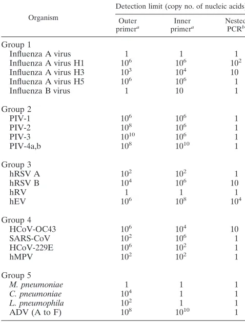
The analyses of analytic sensitivity showed that the multiplex
nested PCR assays were highly sensitive, with a low detection
limit of less than 10 copies of the target nucleic acids, with the
exception of enteroviruses that could still reach a detection
limit of 10
4copies of nucleic acids (Fig. 3 and Table 3).
The sensitivity of multiplex nested PCR assays to detect
cultivable viruses was found to be 100- to 1,000-fold more
sensitive than virus isolation by cell culture. The detection limit
of group 5 multiplex nested PCR for
Legionella pneumophila
and
Mycoplasma pneumoniae
was 1,000 to 10,000
CFU/mil-liliter.
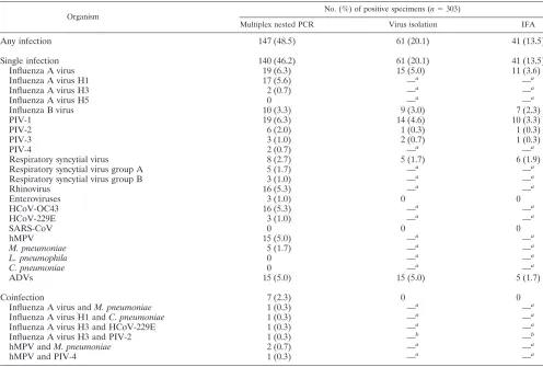
A total of 303 NPA specimens were tested using virus
iso-lation, IFA, and multiplex nested PCR. Altogether, 61
speci-mens were positive by virus isolation, and 41 specispeci-mens were
positive by IFA. All these isolation- or IFA-positive specimens
were also found to be positive by multiplex nested PCR, with
the same corresponding viruses detected (Table 4). The overall
positive rate as determined by multiplex nested PCR was
48.5% (95% confidence interval [CI], 42.9 to 54.1%), which
was significantly higher than those of virus isolation (20.1%
[95% CI, 15.6 to 24.6%]) and IFA (13.5% [95% CI, 9.7 to
17.4%]). The positive rates for each pathogen with respect to
detection method are shown in Table 4.
A subgroup analysis was performed on viruses that were
detected by virus isolation. All these cultivatable viruses
showed a higher positive rate by multiplex nested PCR than by
virus isolation, except for ADV. However, the differences were
not statistically significant (the positive rate [95% CI] for FluA
by PCR versus isolation was 6.3% [3.5 to 9.0%] versus 5.0%
[2.5 to 7.4%], respectively; the positive rate for FluB by PCR
versus isolation was 3.3% [1.3 to 5.3%] versus 3.0% [1.1 to
FIG. 2. Agarose gel (2%) electrophoresis showing the first and second rounds of the group 1 to group 5 (Gp1 to Gp5) multiplex nested PCR
products using a mixture of pathogens as a template. M, marker (100-bp ladder); RSV, hRSV; RV, hRV; EV, hEV; MPV, hMPV; Chl,
C.
pneumoniae
; Leg,
L. pneumophila
; Myc,
M. pneumoniae
.
on May 16, 2020 by guest
http://jcm.asm.org/
4.9%], respectively; the positive rate for PIV-1 by PCR versus
isolation was 6.3% [3.5 to 9.0%] versus 4.6% [2.3 to 7.0%],
respectively; the positive rate for PIV-2 by PCR versus
isola-tion was 2.0% [0.4 to 3.5%] versus 0.3% [0 to 1.0%],
respec-tively; the positive rate for PIV-3 by PCR versus isolation was
1.0% [0 to 2.1%] versus 0.7% [0 to 1.6%], respectively; the
positive rate for hRSV by PCR versus isolation was 2.7% [0.8
to 4.4%] versus 1.7% [0.2 to 3.1%], respectively; the positive
rate for hEV by PCR versus isolation was 1.0% [0 to 2.1%]
versus 0% [0%], respectively; and the positive rate for ADV by
PCR versus isolation was 5.0% [2.5 to 7.4%] versus 5.0% [2.5
to 7.4%], respectively). Compared to virus isolation, the overall
gain in the positive rate for this group of cultivatable viruses as
achieved by multiplex nested PCR was an increase from 20.1%
to 29.7%.
[image:6.585.82.497.66.507.2]When the group of viruses that can be diagnosed by direct
detection using IFA was compared, the positive rate obtained
by multiplex nested PCR was higher than that of IFA for all
the viruses. However, again, the differences were not
statisti-cally significant. The positive rate (95% CI) for FluA by PCR
versus IFA was 6.3% (3.5 to 9.0%) versus 3.6% (1.5 to 5.7%),
respectively; that for FluB by PCR versus IFA was 3.3% (1.3 to
5.3%) versus 2.3% (0.6 to 4.0%), respectively; that for PIV-1
by PCR versus IFA was 6.3% (3.5 to 9.0%) versus 3.3% (1.3 to
5.3%), respectively; that for PIV-2 by PCR versus IFA was
2.0% (0.4 to 3.5%) versus 0.3% (0 to 1.0%), respectively; that
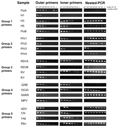
FIG. 3. Agarose gel (2%) electrophoresis showing the performance of the five outer and inner primer groups as well as nested PCR of the
multiplex assays. A single template was added to each corresponding group of primers to test the sensitivity of the assay in detecting that pathogen.
For the nested PCR limit detection test, the PCR products from the first-round PCR are used as a template for the second-round PCR (copy
number of nucleic acids
⫺
copy number of RNA or DNA template in the original samples). RSV, hRSV; RV, hRV; EV, hEV; MPV, hMPV; Chl,
C. pneumoniae
; Leg,
L. pneumophila
; Myc,
M. pneumoniae
.
on May 16, 2020 by guest
http://jcm.asm.org/
for PIV-3 by PCR versus IFA was 1.0% (0 to 2.1%) versus
0.3% (0 to 1.0%), respectively; that for hRSV by PCR versus
IFA was 2.7% (0.8 to 4.4%) versus 2.0% (0.4 to 3.5%),
respec-tively; that for hEV by PCR versus IFA was 1.0% (0 to 2.1%)
versus 0%, respectively; and that for ADV by PCR versus IFA
was 5.0% (2.5 to 7.4%) versus 1.7% (0.2 to 3.1%), respectively.
The overall gain in the positive rate achieved by multiplex
nested PCR for this group of viruses was an increase from
13.5% to 27.7%.
Of the 21 pathogens included in the study, 7 were not
de-tectable by isolation or IFA. Within this group, three were
commonly found in our study samples, including rhinovirus,
with a positive rate of 5.3%, HCoV-OC43 (5.3%), and hMPV
(5.0%). Overall, these three viruses contributed 34.0% of the
PCR-positive cases.
Multiple respiratory viruses were observed in 7 of the 303
(2.3%) specimens (Table 4). None of these coinfections were
detected by virus isolation or IFA, as the majority of them
contained a noncultivatable organism. These cases contributed
4.8% of PCR-positive cases.
Another advantage of this multiplex nested PCR was that it
could be used to subtype pathogens in the same testing cycle.
For the 19 FluA cases detected in multiplex nested PCR, 17
were H1 infections, and 2 were H3 infections. For the eight
cases of hRSV identified, five were hRSV A infections and
three were hRSV B infections. These subtyping results could
be obtained directly by agarose gel electrophoresis without
further testing.
DISCUSSION
Respiratory tract infection accounts for a majority of the
admissions in acute care hospitals. While it has long been
recognized that viruses contribute to a significant proportion of
these cases, the urgency for laboratory diagnosis remains
par-adoxically low in most settings. One of the main reasons is the
long turnaround time of conventional virus detection methods
and their inability to detect fastidious viruses. The lack of
specific treatment for most viral infections is another practical
consideration when prioritizing laboratory resources. The
out-break of SARS and the threat of avian influenza virus
reawak-ened the need for rapid diagnosis to enable the prompt and
accurate diagnosis of index cases.
In this study, we sought to develop and evaluate multiplex
nested PCR assays for the rapid and accurate diagnosis of
respiratory tract infections. The rationale for primer groupings
was as follows. Firstly, RNA pathogens and DNA pathogens
were separated, i.e., groups 1 to 4 for RNA pathogens and
group 5 for DNA pathogens. Secondly, pathogens of the same
or similar family were grouped together, for example, FluA
(H1 to H5) and FluB were grouped together into group 1.
PIVs were grouped into group 2. In this way, each family
member amplified within the same PCR could be easily
differ-entiated. Thirdly, PCR product size was another factor
affect-ing multiplex groupaffect-ing. For example, the primers designed for
hRSVA, hRSVB, hEV, and hRV were compatible to form a
multiplex. Fourthly, only four pathogens were included in each
group because, on one hand, the size of the PCR products
being amplified would be very suitable for visual differentiation
on agarose gel and, on the other hand, the amplification
effi-ciency for each PCR would not be jeopardized too much by
multiplexing. For example, if too many pathogens were
in-cluded in a single multiplex reaction, in order to have sufficient
visual differentiation of PCR products on an agarose gel, some
of the PCR products would need to be very large, and that
might lower the sensitivity of the pathogens being amplified.
Molecular techniques have increased the speed and
sensi-tivity with which such pathogens can be detected and allow
laboratories to identify organisms that do not grow or grow
slowly in conventional viral culture. However, the gain in
an-alytical sensitivity may not necessarily be reflected in clinical
situations. For instance, in settings where clinical specimens
are collected and maintained in good quality, the amount of
virus present may well be enough for detection by the “less
sensitive” conventional methods (culture and IFA). In fact, our
data are in line with this. When cultivatable viruses were
com-pared, despite the finding that a higher sensitivity for multiplex
nested PCR was observed, the differences were not statistically
significant. In particular, we observed the same positive rate
for ADV using both PCR and conventional virus isolation
methods. In a previous study, a discrepancy between direct
detection and RT-PCR for ADV was also reported (19).
Al-though the lack of a statistically significant improvement in a
positive detection rate could be due to a low general
preva-TABLE 3. Performance of individual and nested primer pairs in
the corresponding multiplex nested PCR assay group
Organism
Detection limit (copy no. of nucleic acids)
Outer
primera Inner
primera Nested
PCRb
Group 1
Influenza A virus
1
1
1
Influenza A virus H1
10
610
610
2Influenza A virus H3
10
310
410
Influenza A virus H5
10
610
61
Influenza B virus
1
10
1
Group 2
PIV-1
10
610
61
PIV-2
10
810
61
PIV-3
10
1010
61
PIV-4a,b
10
810
101
Group 3
hRSV A
10
210
21
hRSV B
10
410
610
hRV
1
1
1
hEV
10
610
810
4Group 4
HCoV-OC43
10
610
410
SARS-CoV
10
210
61
HCoV-229E
10
610
21
hMPV
10
210
21
Group 5
M. pneumoniae
1
1
1
C. pneumoniae
10
41
1
L. pneumophila
10
21
1
ADV (A to F)
10
810
101
aThe detection limits of outer and inner primers were tested in the form of
multiplex primer mix.
bThe PCR products from the first-round PCR are used as a template for the
second-round PCR in the nested PCR limit detection test.
on May 16, 2020 by guest
http://jcm.asm.org/
[image:7.585.42.281.91.406.2]lence of the individual organism, the overall results indicate
that the main impact of this multiplex nested PCR was its
broader spectrum of detection. Expanding the detection
spec-trum has also been the main focus of previous studies, and as
many as nine different respiratory pathogens have been
tar-geted (11, 15, 22, 27). In the current study, we included 21
respiratory pathogens and provided the widest spectrum ever
reported. We found that the gain in the overall positive
detec-tion rate from clinical specimens was attributed mainly to the
inclusion of hRV, HCoV-OC43, and hMPV detection. All
these viruses are not detectable by conventional cell culture
isolation or direct detection using IFA. The improvement in
the diagnostic yield by adding hRV was also reported
previ-ously by Gruteke et al. (14). Given that these “trivial”
respi-ratory viruses can cause severe illnesses (6, 20), they should be
included in the development of multiplex assays. Another
ad-vantage of multiplex nested PCR as demonstrated in this study
is the ability to detect coinfections, although the overall
im-provement in the positive rate was not substantial due to the
relatively few instances of coinfection in our study cohort.
While multiplex nested PCR assays may be more economical
due to the fact that multiple pathogens can be detected in a
single assay without a proportional increase in reagent costs,
they have their drawbacks. First, their detection sensitivities
are often lower than those of equivalent monoplex PCR assays.
In this study, only about 30% of the positive specimens showed
a positive result from the first round of PCR. This finding
indicates the need for a nested PCR, which may be associated
with a higher risk of cross-contamination. Second, the presence
of several pairs of primers in a PCR increases the probabilities
of mispairing and nonspecific amplification, particularly the
formation of primer-dimers.
In group 1 multiplex nested PCR, we incorporated specific
primers for influenza A virus subtypes H1, H3, and H5. This is
important in the context where rapid differentiation between
H5 and non-H5 influenza virus is necessary. The group 1
mul-tiplex nested PCR assay was also comprised of primer pairs
targeting the consensus region of influenza A virus. This would
allow the detection of non-H1/H3/H5 subtypes, which may
occasionally cause human infections (e.g., H7 and H9 influenza
viruses).
A nucleic acid extraction kit that can extract both viral DNA
and RNA simultaneously was used in our study. This can
minimize the amount of samples required for the detection of
both DNA and RNA viruses. At the same time, this can
min-imize the time, labor, and materials involved in nucleic acid
extraction.
[image:8.585.43.539.80.415.2]Also, a newly available fast thermal cycler was used in our
TABLE 4. Performance of multiplex nested PCR assays compared to conventional methods
Organism
No. (%) of positive specimens (n⫽303)
Multiplex nested PCR Virus isolation IFA
Any infection
147 (48.5)
61 (20.1)
41 (13.5)
Single infection
140 (46.2)
61 (20.1)
41 (13.5)
Influenza A virus
19 (6.3)
15 (5.0)
11 (3.6)
Influenza A virus H1
17 (5.6)
—
a—
aInfluenza A virus H3
2 (0.7)
—
a—
aInfluenza A virus H5
0
—
a—
aInfluenza B virus
10 (3.3)
9 (3.0)
7 (2.3)
PIV-1
19 (6.3)
14 (4.6)
10 (3.3)
PIV-2
6 (2.0)
1 (0.3)
1 (0.3)
PIV-3
3 (1.0)
2 (0.7)
1 (0.3)
PIV-4
2 (0.7)
—
a—
aRespiratory syncytial virus
8 (2.7)
5 (1.7)
6 (1.9)
Respiratory syncytial virus group A
5 (1.7)
—
a—
aRespiratory syncytial virus group B
3 (1.0)
—
a—
aRhinovirus
16 (5.3)
—
a—
aEnteroviruses
3 (1.0)
0
0
HCoV-OC43
16 (5.3)
—
a—
aHCoV-229E
3 (1.0)
—
a—
aSARS-CoV
0
0
0
hMPV
15 (5.0)
—
a—
aM. pneumoniae
5 (1.7)
—
a—
aL. pneumophila
0
—
a—
aC. pneumoniae
0
—
a—
aADVs
15 (5.0)
15 (5.0)
5 (1.7)
Coinfection
7 (2.3)
0
0
Influenza A virus and
M. pneumoniae
1 (0.3)
—
a—
aInfluenza A virus H1 and
C. pneumoniae
1 (0.3)
—
a—
aInfluenza A virus H3 and HCoV-229E
1 (0.3)
—
a—
aInfluenza A virus H3 and PIV-2
1 (0.3)
—
b—
bhMPV and
M. pneumoniae
2 (0.7)
—
a—
ahMPV and PIV-4
1 (0.3)
—
a—
aaOrganisms not isolated/differentiated by virus isolation. bPIV-2 was isolated.
on May 16, 2020 by guest
http://jcm.asm.org/
study, which allowed rapid cycling, shortening the time
re-quired to complete the PCR. The thermal cycler’s patented
sample temperature control provided a quick and uniform
thermal response. Therefore, the cycling times for the
first-and second-round PCRs were considerably reduced.
Fur-thermore, the GeneAmp fast PCR master mix allows a
two-step PCR (same temperature holding for the annealing and
extension steps) instead of the more conventional three-step
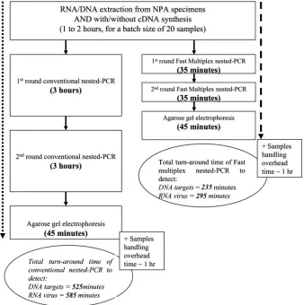
PCR. With the use of this fast PCR system, the time
re-quired for a single round of PCR was reduced from 3 h to 35
min, i.e., saving a total of about 300 min in a nested PCR
assay (Fig. 4). Therefore, the whole testing process can be
completed within 1 day. This rapid turnaround not only is
critical in urgent outbreak investigations but may potentially
decrease the overall costs for the hospital, as has been
shown in previous studies (1, 30). However, one
disadvan-tage of using this fast PCR system is that primers used in
standard multiplex PCR assays need to be redesigned with
higher annealing temperatures.
The multiplex nested PCR assays developed in this study
improved the diagnostic yield in terms of the overall
sensi-tivity as well as the spectrum of coverage for respiratory
infections. Furthermore, the assay provided a rapid
turn-around time, with results being available within the same
day of specimen collection. The overall cost reduction may
justify the routine use of these broader-spectrum, rapid
mo-lecular diagnostic assays.
ACKNOWLEDGMENTS
This study was supported by the Research Fund for the Control of
Infectious Diseases from the Health, Welfare, and Food Bureau of the
Hong Kong Special Administrative Region Government (project no.
01030782).
Part of the work was performed at the Lo Kwee Cheong Research
Laboratory.
REFERENCES
1.Adcock, P. M., G. G. Stout, M. A. Hauck, and G. S. Marshall.1997. Effect of rapid viral diagnosis on the management of children hospitalized with lower respiratory tract infection. Pediatr. Infect. Dis. J.16:842–846. 2.Bellau-Pujol, S., A. Vabret, L. Legrand, J. Dina, S. Gouarin, J.
Petitjean-Lecherbonnier, B. Pozzetto, C. Ginevra, and F. Freymuth.2005. Develop-ment of three multiplex RT-PCR assays for the detection of 12 respiratory RNA viruses. J. Virol. Methods126:53–63.
3.Billaud, G., S. Peny, V. Legay, B. Lina, and M. Valette.2003. Detection of rhinovirus and enterovirus in upper respiratory tract samples using a multi-plex nested PCR. J. Virol. Methods108:223–228.
4.Boivin, G., S. Cote, P. Dery, G. De Serres, and M. G. Bergeron.2004. Multiplex real-time PCR assay for detection of influenza and human respi-ratory syncytial viruses. J. Clin. Microbiol.42:45–51.
5.Casiano-Colon, A. E., B. B. Hulbert, T. K. Mayer, E. E. Walsh, and A. R. Falsey.2003. Lack of sensitivity of rapid antigen tests for the diagnosis of respiratory syncytial virus infection in adults. J. Clin. Virol.28:169–174. 6.Chidekel, A. S., C. L. Rosen, and A. R. Bazzy.1997. Rhinovirus infection
[image:9.585.124.462.69.409.2]associated with serious lower respiratory illness in patients with bronchopul-monary dysplasia. Pediatr. Infect. Dis. J.16:43–47.
FIG. 4. Estimated turnaround times for the fast multiplex nested PCR protocol and conventional nested PCR protocol.
on May 16, 2020 by guest
http://jcm.asm.org/
7.Coiras, M. T., P. Perez-Brena, M. L. Garcia, and I. Casas.2003. Simulta-neous detection of influenza A, B, and C viruses, respiratory syncytial virus, and adenoviruses in clinical samples by multiplex reverse transcription nested-PCR assay. J. Med. Virol.69:132–144.
8.Coiras, M. T., J. C. Aguilar, M. L. Garcia, I. Casas, and P. Perez-Brena. 2004. Simultaneous detection of fourteen respiratory viruses in clinical spec-imens by two multiplex reverse transcription nested-PCR assays. J. Med. Virol.72:484–495.
9.Cote, S., Y. Abed, and G. Boivin.2003. Comparative evaluation of real-time PCR assays for detection of the human metapneumovirus. J. Clin. Microbiol. 41:3631–3635.
10.Debbia, E. A., G. C. Schito, A. Zoratti, L. Gualco, E. Tonoli, and A. Marchese. 2001. Epidemiology of major respiratory pathogens. J. Che-mother.13:205–210.
11.Fan, J., K. J. Henrickson, and L. L. Savatski.1998. Rapid simultaneous diagnosis of infections with respiratory syncytial viruses A and B, influenza viruses A and B, and human parainfluenza virus types 1, 2, and 3 by multiplex quantitative reverse transcription-polymerase chain reaction-enzyme hybrid-ization assay (Hexaplex). Clin. Infect. Dis.26:1397–1402.
12.Fernandez-Sabe, N., J. Carratala, B. Roson, J. Dorca, R. Verdaguer, F. Manresa, and F. Gudiol. 2003. Community-acquired pneumonia in very elderly patients: causative organisms, clinical characteristics, and outcomes. Medicine (Baltimore)82:159–169.
13.Freymuth, F., M. Quibriac, J. Petitjean, F. Daon, and M. L. Amiel.1987. Viruses responsible for respiratory infections in pediatrics. Evaluation of 3,480 nasal aspirates performed in children over a 6-year period. Ann. Pediatr. (Paris)34:493–501. (In French.)
14.Grondahl, B., W. Puppe, A. Hoppe, I. Kuhne, J. A. Weigl, and H. J. Schmitt. 1999. Rapid identification of nine microorganisms causing acute respiratory tract infections by single-tube multiplex reverse transcription-PCR: feasibil-ity study. J. Clin. Microbiol.37:1–7.
15.Gruteke, P., A. S. Glas, M. Dierdorp, W. B. Vreede, J. W. Pilon, and S. M. Bruisten.2004. Practical implementation of a multiplex PCR for acute respiratory tract infections in children. J. Clin. Microbiol.42:5596–5603. 16.Kehl, S. C., K. J. Henrickson, W. Hua, and J. Fan.2001. Evaluation of the
Hexaplex assay for detection of respiratory viruses in children. J. Clin. Mi-crobiol.39:1696–1701.
17.Kendal, A. P., M. S. Pereira, and J. J. Skehel.1982. Concepts and procedures for laboratory-based influenza surveillance. WHO Collaborating Centers for Reference and Research on Influenza, Geneva, Switzerland.
18.McDonough, E. A., C. P. Barrozo, K. L. Russell, and D. Metzgar.2005. A multiplex PCR for detection of Mycoplasma pneumoniae, Chlamydophila pneumoniae, Legionella pneumophila, and Bordetella pertussis in clinical specimens. Mol. Cell. Probes19:314–322.
19.Monto, A. S.2002. Epidemiology of viral respiratory infections. Am. J. Med. 112(Suppl. 6A):4S–12S.
20.Osiowy, C.1998. Direct detection of respiratory syncytial virus, parainfluenza virus, and adenovirus in clinical respiratory specimens by a multiplex reverse transcription-PCR assay. J. Clin. Microbiol.36:3149–3154.
21.Papadopoulos, N. G., P. J. Bates, P. G. Bardin, A. Papi, S. H. Leir, D. J. Fraenkel, J. Meyer, P. M. Lackie, G. Sanderson, S. T. Holgate, and S. L. Johnston.2000. Rhinoviruses infect the lower airways. J. Infect. Dis.181: 1875–1884.
22.Santti, J., T. Hyypia, and P. Halonen.1997. Comparison of PCR primer pairs in the detection of human rhinoviruses in nasopharyngeal aspirates. J. Virol. Methods66:139–147.
23.Stockton, J., J. S. Ellis, M. Saville, J. P. Clewley, and M. C. Zambon.1998. Multiplex PCR for typing and subtyping influenza and respiratory syncytial viruses. J. Clin. Microbiol.36:2990–2995.
24.Stralin, K., A. Backman, H. Holmberg, H. Fredlund, and P. Olcen.2005. Design of a multiplex PCR for Streptococcus pneumoniae, Haemophilus influenzae, Mycoplasma pneumoniae and Chlamydophila pneumoniae to be used on sputum samples. APMIS113:99–111.
25.Templeton, K. E., S. A. Scheltinga, M. F. Beersma, A. C. Kroes, and E. C. Claas.2004. Rapid and sensitive method using multiplex real-time PCR for diagnosis of infections by influenza a and influenza B viruses, respiratory syncytial virus, and parainfluenza viruses 1, 2, 3, and 4. J. Clin. Microbiol. 42:1564–1569.
26.Thompson, J. D., T. J. Gibson, F. Plewniak, F. Jeanmougin, and D. G. Higgins.1997. The CLUSTAL_X windows interface: flexible strategies for multiple sequence alignment aided by quality analysis tools. Nucleic Acids Res.25:4876–4882.
27.Vabret, A., F. Mouthon, T. Mourez, S. Gouarin, J. Petitjean, and F. Freymuth.2001. Direct diagnosis of human respiratory coronaviruses 229E and OC43 by the polymerase chain reaction. J. Virol. Methods97:59–66. 28.Valassina, M., A. M. Cuppone, M. G. Cusi, and P. E. Valensin.1997. Rapid
detection of different RNA respiratory virus species by multiplex RT-PCR: application to clinical specimens. Clin. Diagn. Virol.8:227–232.
29.Vijgen, L., E. Keyaerts, E. Moes, P. Maes, G. Duson, and M. Van Ranst. 2005. Development of one-step, real-time, quantitative reverse transcriptase PCR assays for absolute quantitation of human coronaviruses OC43 and 229E. J. Clin. Microbiol.43:5452–5456.
30.Ward, C. L., M. H. Dempsey, C. J. Ring, R. E. Kempson, L. Zhang, D. Gor, B. W. Snowden, and M. Tisdale.2004. Design and performance testing of quantitative real time PCR assays for influenza A and B viral load measure-ment. J. Clin. Virol.29:179–188.
31.Woo, P. C., S. S. Chiu, W. H. Seto, and M. Peiris.1997. Cost-effectiveness of rapid diagnosis of viral respiratory tract infections in pediatric patients. J. Clin. Microbiol.35:1579–1581.
32.World Health Organization.5 July 2007, accession date. PCR primers for SARS developed by WHO Network Laboratories. World Health Organiza-tion, Geneva, Switzerland. http://www.who.int/csr/sars/primers/en/print/html. 33.Xu, W., M. C. McDonough, and D. D. Erdman.2000. Species-specific iden-tification of human adenoviruses by a multiplex PCR assay. J. Clin. Micro-biol.38:4114–4120.
34.Xu, W., and D. D. Erdman.2001. Type-specific identification of human adenovirus 3, 7, and 21 by a multiplex PCR assay. J. Med. Virol.64:537–542.