Original Article
miR-599 and miR-185 down-regulate periostin
expression in human lung cancer cells
Yang Wang1, Jianming Zheng1, Lixin Yang2, Hai Jin2, Changjing Zuo3, Jing Sheng4, Wei Chen4, Yiping Han5
Departments of 1Pathology, 2Thoracic Surgery, 3Nuclear Medicine, 4Radiology, 5Respiratory Medicine, Changhai
Hospital, Second Military Medical University, 168 Changhai Road, Shanghai 200433, P. R. China
Received July 15, 2016; Accepted October 28, 2016; Epub February 15, 2017; Published February 28, 2017
Abstract: Background: Prognosis of lung cancer remains poor. The over-expression of periostin, osteoblast-specific
factor, was reported in cancers such as ovarian and breast cancers. However, it is unclear about effects of periostin on the invasion of human lung cancer, and whether the mRNA and protein expression of periostin was regulated by miRNA needs to be explored. Therefore, we explored effects of periostin on the invasion of human lung cancer cells, and how periostin expression was regulated by miRNA. Methods: Human lung cancer cell line A549 cells were treated with different concentrations of periostin protein, and tumor invasion was detected by Transwell assay. A549 and another lung cancer cell line BEAS-2B cells were transfected with mixture of Dicer-siRNA-152 and Drosha-siR-NA-1200, or negative control (NC) siRNA. mRNA expression of periostin were detected by qPCR. Protein expression of periostin were examined by Western Blot. HEK293A cells were transfected with vectors (pMIR/report-3’POSTN and pRL-TK), and miRNA (miR-543, miR-296-3P, miR-599, miR-185, miR-202-3P, and NC miRNA, respectively). In addition, HEK293A cells were transfected with periostin gene mutated at binding sites of miR-599 or miR-185. Expression of periostin was determined by measuring relative light unit in dual-luciferase reporter assay. Results: Periostin protein altered invasion of A549 cells in a dose-dependent manner. mRNA expression of periostin in A549
and BEAS-2B cells increased significantly after miRNA was interrupted. Protein expression of periostin in BEAS-2B
cells increased markedly after miRNA was interrupted. Expression of periostin was lowest when HEK293A cells were transfected with miR-599 and miR-185. Expression of periostin was not altered by miR-599 or miR-185 mimics when HEK293A cells were transfected with periostin gene mutated at binding sites of miR-599 or miR-185. Conclu-sions: Periostin protein alters invasion of human lung cancer cells. miR-599 and miR-185 down-regulate periostin expression in lung cancer cells.
Keywords: Periostin, lung cancer, miR-599, miR-185
Introduction
Human lung cancer is the most common cause of cancer-related death in men and second most common in women after breast cancer worldwide [1]. Common treatments for lung cancer include palliative care, surgery, radia-tion therapy and chemotherapy [2-4]. Neverth- eless, prognosis of lung cancer remains poor,
with five-year survival rate of less than 20% [5].
Periostin, also known as osteoblast-specific
factor, is encoded by the POSTN gene in hu- mans [6]. It functions as a ligand for integrins, and mediates adhesion and migration of epi-thelial cells [7]. As a secreted extracellular ma-
trix protein, periostin was originally identified in
cells from the mesenchymal lineage, including osteoblasts, osteoblast-derived cells, periodon-tal ligament, and periosteum. It was revealed to be associated with the differentiation of mes-enchyme in developing heart, and facilitate the epithelial-mesenchymal transition in endome-trial epithelial cells via through ILK-Akt signaling pathway [8, 9]. Periostin over-expression was reported in several types of cancer, such as ovarian and breast cancers [7, 10]. However, it is unclear about effects of different concentra-tions of periostin on the invasion of human lung cancer.
post-transcriptional regulation of gene expres-sion [11, 12]. miRNAs function through base-pairing with complementary sequences within mRNA molecules [13]. The mRNA molecules are subsequently cleaved into two pieces, and destabilized due to shortening of poly (A) tail. As a result, the translation of mRNA into
pro-teins becomes less efficient [14]. Dysregulation
of miRNA has been associated with diseases such as hereditary progressive hearing loss, heart diseases, mental disorders, and cancer [15-18]. miRNA deregulation was shown to involve in chronic lymphocytic leukemia [19]. Low miR-324a levels were revealed to be an indicator of poor survival in non-small cell lung cancer [20]. In addition, miR-543 was revealed to promote proliferation of gastric cancer cells by targeting SIRT1 [21]. Dysregulation of miR-296/S100A4 axis promoted tumor invasion by inducing epithelial-mesenchymal transition in human ovarian cancer [22]. Hsa-miR-599 was shown to act as a tumor suppressor and it inhibited cells proliferation, migration and inva-sion in hepatocellular carcinoma [23]. High miR-185 correlated with metastasis and poor survival in colorectal cancer [24], and miR-202-3p was reported to be a tumor suppressor in gastric cancer [25]. However, whether the mRNA and protein expression of periostin was regulated by miRNA, and if so which particular types of miRNA are involved need to be ex- plored.
Therefore, we explored effects of periostin on the invasion of human lung cancer cells, and how periostin expression was regulated by miRNA in current study.
Methods
Cells and reagents
Cells: A549 (human lung adenocarcinoma cell line; Novobio Inc., Shanghai, China), BEAS-2B (immortalized human bronchial epithelial cells; Novobio Inc., Shanghai, China), and HEK293A cells (human embryonic kidney cells; Novobio Inc., Shanghai, China) were cultured inRPMI 1640 medium which was supplemented with 100 mL/L fetal bovine serum (FBS), 100 kU/L penicillin and 100 mg/L chloramphenicolin a
cell incubator with 5% CO2 at 37°C. Cells were
subcultured after being digested by 0.25% tryp -sin. A549 and BEAS-2B cells that were trans-fected with the mixture of Dicer-siRNA-152
(Sense: 5’-UGC UUG AAG CAG CUC UGG ATT-3’; Anti-sense: 5’-UCC AGA GCU GCU UCA AGC ATT-3’) and Drosha-siRNA-1200 (Sense: 5’-AAC GAG UAG GCU UCG UGA CUU TT-3’; Anti-sense: 5’-AAG UCA CGA AGC CUA CUC GUU TT-3’), or negative control siRNA were purchased from Novobio Inc. (Shanghai, China). The cells were named A549-152/1200, BEAS-2B-152/1200, A549-NC, and BEAS-2B-NC, respectively. Main reagents: fetal bovine serum (FBS; Gibco Inc., Grand Island, NY, USA); typsin (Gibco Inc., Grand Island, NY, USA); RPMI1640 (GibcoInc., Grand Island, NY, USA); primers and probes (Invitrogen Inc., Grand Island, NY, USA); miR-NAeasy Mini Kit (QIAGEN Inc., Valencia, CA, USA); Trizol reagent (Invitrogen Inc., Grand Island, NY, USA); cDNA synthesis kit (Tiangene Biotech Inc., Beijing, China); chloroform (Sino- pharm Inc., Shanghai, China); isopropanol
(Sinopharm Inc., Shanghai, China); 75% ethanol
(Sinopharm Inc., Shanghai, China); DEPC H2O (Invitrogen Inc., Grand Island, NY, USA); Super- ScriptIII reverse transcriptase (Invitrogen Inc., Grand Island, NY, USA); SYBR Green I (Invitrogen Inc., Grand Island, NY, USA); Rnase Inhibitor (Fermentas Inc., Hanover, MD, USA); Platinum Taq DNA Polymerase (Invitrogen Inc., Grand Island, NY, USA); 100 mM dNTPs (Invitrogen Inc., Grand Island, NY, USA); RIPA buffer (Be- yotime Inc., Shanghai, China); BCA kit (Beyotime
Inc., Shanghai, China); X-ray films (Kodac Inc., Dayton, OH, USA); polyvinylidene fluoride
(PV-DF) membrane (EMD Millipore Inc., Darmstadt, Germany); enhanced chemiluminescence (ECL) solution (Fermentas Inc., Hanover, MD, USA); anti-periostinprimary antibody (Abcam Inc., Canbridge, MA, USA); actin primary anti-body (Novobio Inc., Shanghai, China); second-ary goat anti-rabbit antibody (Novobio Inc., Shanghai, China); Dual-Luciferase® Reporter Assay System (Promega Inc., Fitchburg, USA); transwell palte (Sigma-Aldrich Inc., St. Louis, MI); negative controlmiRNA, miR-543, miR-296- 3P, miR-599, miR-185, and miR-202-3P (Ge- nePharm Inc., Shanghai, China); vectors (pMIR/ report-3’POSTN; pRL-TK; pMIR/report-miR-599- mut; and pMIR/report-miR-185-mut; Novobio Inc., Shanghai, China).
96 Touch™ Real-Time PCR Detection System (Bio-Rad Inc., Hercules, CA, USA); cell incubator
(Thermo Scientific Inc., Waltham, MA); electro -phoresis and transfer system (Tanon Inc., Full- erton CA, USA); Gel-Pro analyzer (Meyer Instru- ments Inc., Houston, TX, USA); Turner BioSys- tems GloRunner (BioSurplus Inc., San Diego, CA, USA).
Transwell invasion assay
A549 cells were digested bytrypsin, and diluted to make concentration of 5×10⁵/mL. The mem-brane of the upper compartment was coated with 50 µL of matrigel (1 g/L), and incubated at 37°C for 1 h in order to reconstruct structure of basal membrane. Eight hundred µL of 1640
medium with 20% fetal bovine serum (FBS),
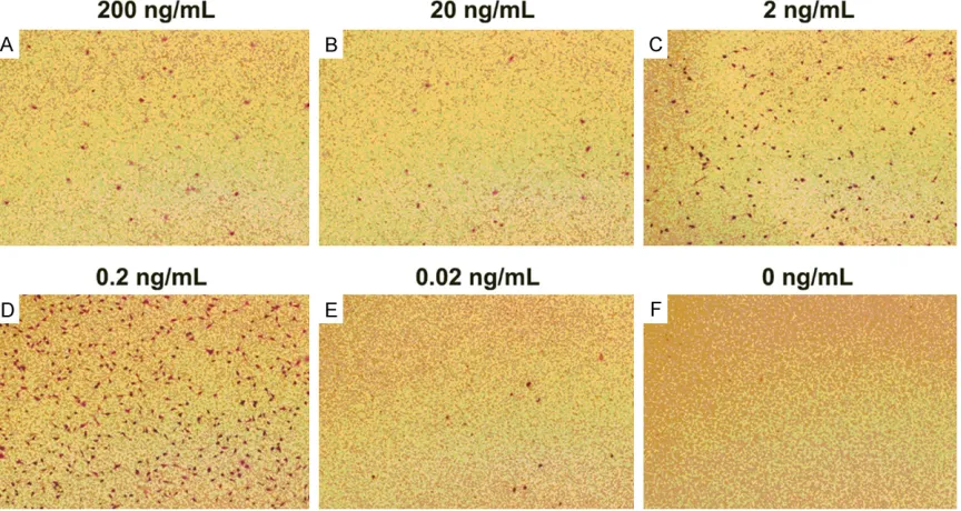
and periostin protein were added into lower compartment. The concentrations of periostin were 200, 20, 2, 0.2, 0.02, and 0 ng/mL during primary screening, and were 0, 0.015, 0.03, 0.06, 0.12, 0.25, 0.5, 1, and 2 ng/mL respec-tively during concentration gradient study. Two hundred µL of A549 cell suspension were incu-bated in upper compartment of Transwell, and cells were cultured at a humid incubator with
5% CO2 at 37°C for 24 h. Transwell was then taken out, and cells on the upper surface of Transwell was erased with cotton swab. Cells on the microporous membrane at lower surface
of Transwell was fixed with 4% polyformalde
-hyde for 30 min, and stained with 1% crystal
violet for 10 min. The microporous membrane was washed with phosphate-buffered solution (PBS) for 3 times, and cells that penetrated the membrane were observed under microscope. Quantitativepolymerase chain reaction (qPCR)
Total RNA of A549-152/1200, A549-NC, A549, 2B-152/1200, 2B-NC, and
BEAS-2B cells was extracted and purified byTrizolfol -lowing manufacturer’s instructions, respective-ly. A universal cDNA synthesis kit was utilized for reverse transcription. Each reaction
con-tained 0.5 μL of random primers (0.2 μg/μL) and 1 μLof SuperScrip III reverse transcriptase (200 U/μL). The specific primer for detection of
periostin gene was F: GCTGCCATCACATCGGAC- AT; R: CCTCCCATAATAGACTCAGAACACT. The pri- mer for detecting β-actin gene was F: AGAAA- ATCTGGCACCACACC; R: AGAGGGTACAGGGATA- GCA. qPCR was performed by utilizing MiRcute miRNA qPCR Detection kit. PCR conditions
were as follows: pre-denaturing at 95°C for 2 min; denaturing at 95°C for 10 s; and annealing and polymerization at 60°C for 30 s, and 70°C for 45 s. There were 40PCR cycles. PCR was performed in CFX96 Touch™ Real-Time PCR Detection System. The expression of periostin was determined as the ratio of relative optical density of target gene to β-actin.
Western blot
Proteinswere extracted from A549-152/1200, A549-NC, A549, 2B-152/1200, BEAS-2B-NC, and BEAS-2B cells, respectively. Then
they were separated in 10%
SDS-polyacrylami-de separating gel by eletrophoresis at 120 V, and signal was transferred to PVDF membranes at 100 V for 120 min. Membranes were blocked
with 5% non-fat milk for 1 h, and incubated with
anti-periostin antibody (1:2000) at 4°C over-night. The membranes were then washed with tris-buffered saline and tween 20 (TBST) for 3 times, and every time lasted for 10 min. Membranes were then incubated with goat anti-rabbit secondary antibody labeled with horseradish peroxidase (HRP) (1/5000) at room temperature for 1 h. Membranes were washed and incubated shortly with ECL solu-tion. Films were exposed in a dark room. Experiments were repeated for 3 times.
Dual-luciferase reporter assay
cells were transfected with pMIR/report-miR-185-mut (miR-pMIR/report-miR-185-mut: periostin gene that has mutation at miR-185 binding site), and pRL-TK; (5) miR-185-mut NC group: cells were trans-fected with pMIR/report-miR-185-mut, pRL-TK, and NC; and (6) miR-185-mut mimics group: cells were transfected with pMIR/report-miR-185-mut, pRL-TK and miR-185 mimic. Cell medium was changed and cells were then cul-tured for another 36 h. Cells were then lysed
with 300 μL of lysis buffer on ice for 10 min,
and centrifuged at 13000 rpm for 5 min. One
hundred μL of supernatant from each group was mixed with 100 μL of firefly detection
reagent, and relative light unit (RLU) was
mea-sured. One hundred μL of renilla detection
reagent was then added, and a second RLU
was measured. The ratio of firefly RLU and renil -la RLU was calcu-lated.
Statistical analysis
Results were shown as mean ± SEM. One-way analysis of variance (ANOVA) was used to com-pare differences among 3 or more groups, fol-lowed by Bonferroni post hoc testing for multi-ple comparisons. P values of 0.05 or less were
regarded significant. Figures and statistical
analysis were made using GraphPad Prism 5.0 software (GraphPad Software Inc., La Jolla, CA).
Results
Periostin protein altered invasion of A549 cells in a dose-dependent manner
Invasion of A549 cells affected by periostin pro-tein was detected by Transwell invasion assay. The concentrations of periostin utilized were 200, 20, 2, 0.2, 0.02, and 0 ng/mL during pri-mary screening, and were 0, 0.015, 0.03, 0.06, 0.12, 0.25, 0.5, 1, and 2 ng/mL respectively during concentration gradient study. During pri-mary screening, the invasion of A549 cells was highest when concentration of periostin protein was 2 ng/mL (Figure 1). In addition, the inva-sion of A549 cells had positive correlation with concentrations of periostin protein between 0 and 0.5 ng/mL, whereas it had negative corre-lation with concentrations of periostin protein between 0.5 and 2 ng/mL (Figure 2).
mRNA and protein expression of periostin in human lung cancer cell line A549 and BEAS-2B increased significantly after miRNA was interrupted
[image:4.612.92.524.77.307.2]Blot, respectively. Relative mRNA expression of periostin in A549 (P < 0.05) and BEAS-2B cells (P < 0.01; Figure 3) increased significantly after
miRNA was interrupted by Dicer- and Drosha-siRNA, as compared to cells treated with nega-tive control siRNA or cells that received no treatment. Similarly, protein expression of peri-ostin in BEAS-2B cells increased markedly after miRNA was interrupted by Dicer- and Drosha-siRNA when compared to negative con-trol and blank groups (P < 0.01; Figure 4). Protein expression of periostin in A549 cells was too low to be detected by Western Blot. Expression of periostin was lowest when HEK293A cells were transfected with miR-599 and miR-185
HEK293A cells were transfected with vectors
(miR-543, miR-296-3P, miR-599, miR-185, miR- 202-3P, and negative control miRNA, respec-tively). Expression of periostin was determined by measuring relative light unit in dual-lucifer-ase reporter assay. Compared to other miRNA, transfection of miR-599 and miR-185 decre- ased expression of periostin dramatically (P < 0.001, Figure 5).
Expression of periostin was not altered by miR-599 or miR-185 mimics when HEK293A cells were transfected with periostin gene mutated at binding sites of miR-599 or miR-185
[image:5.612.89.525.75.428.2]ics group. Expression of periostin was deter-mined by measuring relative light unit in dual-luciferase reporter assay. Expression of periostin was not altered by 599 or miR-185 mimics when HEK293A cells were trans-fected with periostin gene mutated at binding sites of miR-599 or miR-185 (P > 0.05, Figure 6).
Discussion
We have demonstrated that periostin protein alters invasion of human lung cancer cells, and miR-599 and miR-185 down-regulate periostin expression in lung cancer cells.
Periostin was shown to bind to integrins on many cancer cells, and activate Akt/PKB- and FAK-mediated signaling pathways, which result-ed in increasresult-ed cell survival, angiogenesis, metastasis, and the epithelial-mesenchymal transition [26]. In addition, periostin was re- vealed to be highly upregulated in glioblasto-mas. The expression of periostinin gliomas cor-related directly with tumor grade and recur-rence, and inversely with survival [27]. More- over, glioma stem cells in glioblastomas were reported to secrete periostin, which recruited M2 tumor-associated macrophages from pe- ripheral blood to tumor environment through integrin signaling. M2 tumor-associated macro-Figure 3. mRNA expression of periostin in human lung cancer cell line A549 and BEAS-2B increased significantly
after miRNA was interrupted. A549 and BEAS-2B cells that were transfected with the mixture of Dicer-siRNA-152 and Drosha-siRNA-1200, or negative control siRNA were cultured. mRNA expression of periostin were detected by
qPCR. Relative mRNA expression of periostin in A549 and BEAS-2B cells increased significantly after miRNA was
[image:6.612.95.525.74.265.2]interrupted by Dicer- and Drosha-siRNA, as compared to cells treated with negative control siRNA or cells that re-ceived no treatment (mean ± SEM, n = 3/group). *indicates that P < 0.05 as compared to A549-NC or A549 groups. **indicates that P < 0.01 when compared to BEAS-2B-NC and BEAS-2B groups. NC: negative control. 152: Dicer-siRNA-152. 1200: Drosha-siRNA-1200.
[image:6.612.91.290.371.559.2]phages are tumor-supportive and immunosup-pressive. Therefore, through possible recruit-ment mechanism, periostin supported tumor progressionin gliomas [28].
The incidence and mortality of lung cancer are the highest among all cancer types in men worldwide. It has the third highest incidence, and is second after breast cancer in mortality among women [1]. We have unveiled that peri-ostin protein altered invasion of human lung cancer cells in current study. The invasion of A549 cells had positive correlation with con-centrations of periostin protein between 0 and 0.5 ng/mL, whereas it had negative correlation with concentrations of periostin protein bet- ween 0.5 and 2 ng/mL. Future investigations are needed to elucidate dosage responses of perostin protein in animal models. Since we investigated human lung cancer cells alone in current study, chemoattraction of M2 tumor-associated macrophages might not be able to explain the altered tumor invasion. It is possible other molecular signaling pathways in lung can-cer cells are involved, which require further research efforts.
[image:7.612.328.520.74.266.2]Expression levels of miRNA was linked to prog-nosis of various cancers. Either high miR-185 or low miR-133b levels was reported to corre-late with metastasis and poor survival in colorectal cancer [24]. Tumor cell proliferation in hepatocellular carcinoma resulted from the interaction of miR-21 with MAP2K3, a tumor repressor gene [29]. Plasma miR-21, miR-494, and miR-1973 were revealed to be promising biomarkers for Hodgkin lymphoma [30]. In addition, miR-205 was shown to inhibit the metastatic nature of breast cancer [31]. MiRNA-200 family, including miR-MiRNA-200a, miR-MiRNA-200b, Figure 5. Expression of periostin was lowest when
HEK293A cells were transfected with miR-599 and miR-185. HEK293A cells were transfected with vec-tors (pMIR/report-3’POSTN and pRL-TK), and miRNA (miR-543, miR-296-3P, miR-599, miR-185, miR-202-3P, and negative control miRNA, respectively). Ex-pression of periostin was determined by measuring relative light unit in dual-luciferase reporter assay. Compared to other miRNA, transfection of miR-599 and miR-185 decreased expression of periostin dramatically (mean ± SEM, n = 3/group). ***sug-gests that P < 0.001 when compared to blank, NC, miR-543, miR-296-3P, and miR-202-3P groups. NC: negative control.
[image:7.612.92.287.76.239.2]miR-200c, miR-141 and miR-429, were down-regulated in tumor progression of breast can-cer [32]. We revealed in current study that miR-599 and miR-185 down-regulated periostin expression in lung cancer cells, whereas the inhibition was abolished after binding sites of miR-599 or miR-185 were muted. This provides novel approaches of targeting periostin in lung cancer therapy.
In conclusion, we have demonstrated novel data suggesting that periostin protein alters invasion of human lung cancer cells. In addi-tion, miR-599 and miR-185 down-regulate peri-ostin expression in lung cancer cells. Although future research is needed to reveal how to regu-late perostin precisely in animal models, peri-tostin may serve as a promising therapeutic target for human lung cancer.
Acknowledgements
The research was supported by “1255 Plan for Discipline Construction” funds at Changhai Hospital, Second Military Medical University (CH125521106).
Disclosure of conflict of interest
None.
Address correspondence to: Dr. Yiping Han, Depart- ment of Respiratory Medicine, Changhai Hospital, Second Military Medical University, 168 Changhai Road, Shanghai 200433, P. R. China. Tel: (86)-13916418267; E-mail: yphan2006@163.com
References
[1] McGuire S. World cancer report 2014. Geneva, Switzerland: World Health Organization, inter-national agency for research on cancer, WHO press, 2015. Adv Nutr 2016; 7: 418-419. [2] Blum T and Schonfeld N. The lung cancer
pa-tient, the pneumologist and palliative care: a developing alliance. Eur Respir J 2015; 45: 211-226.
[3] Puri V and Robinson CG. In response to treat-ment outcomes in stage I lung cancer: a com-parison of surgery and stereotactic body radia-tion therapy. J Thorac Oncol 2016; 11: e65-66. [4] He J. [Comprehensive treatment for lung can-cer based on minimally invasive thoracic sur-gery]. Zhongguo Fei Ai Za Zhi 2016; 19: 329-331.
[5] Wei X, Li Q, Li Y, Duan W, Huang C, Zheng X, Sun L, Luo J, Wang D, Zhang S, Xin X and Gao
M. Prediction of survival prognosis of non-small cell lung cancer by APE1 through regula-tion of epithelial-mesenchymal transiregula-tion. On-cotarget 2016; 7: 28523-39.
[6] Park SY, Piao Y, Jeong KJ, Dong J and de Groot JF. Periostin (POSTN) regulates tumor resis-tance to antiangiogenic therapy in glioma mod-els. Mol Cancer Ther 2016; 15: 2187-97. [7] Gillan L, Matei D, Fishman DA, Gerbin CS,
Kar-lan BY and Chang DD. Periostin secreted by epithelial ovarian carcinoma is a ligand for alpha(V)beta(3) and alpha(V)beta(5) integrins and promotes cell motility. Cancer Res 2002; 62: 5358-5364.
[8] Lie-Venema H, Eralp I, Markwald RR, van den Akker NM, Wijffels MC, Kolditz DP, van der Laarse A, Schalij MJ, Poelmann RE, Bogers AJ, Gittenberger-de Groot AC. Periostin expression by epicardium-derived cells is involved in the development of the atrioventricular valves and
fibrous heart skeleton. Differentiation 2008;
76: 809-819.
[9] Zheng QM, Lu JJ, Zhao J, Wei X, Wang L and Liu PS. Periostin facilitates the epithelial-mesen-chymal transition of endometrial epithelial cells through ILK-Akt signaling pathway. Biomed Res Int 2016; 2016: 9842619.
[10] Contie S, Voorzanger-Rousselot N, Litvin J, Clezardin P and Garnero P. Increased expres-sion and serum levels of the stromal cell-se-creted protein periostin in breast cancer bone metastases. Int J Cancer 2011; 128: 352-360. [11] Ambros V. The functions of animal microRNAs.
Nature 2004; 431: 350-355.
[12] Bartel DP. MicroRNAs: genomics, biogenesis, mechanism, and function. Cell 2004; 116: 281-297.
[13] Bartel DP. MicroRNAs: target recognition and regulatory functions. Cell 2009; 136: 215-233.
[14] Fabian MR, Sonenberg N and Filipowicz W. Regulation of mRNA translation and stability by microRNAs. Annu Rev Biochem 2010; 79: 351-379.
[15] Mencia A, Modamio-Hoybjor S, Redshaw N, Morin M, Mayo-Merino F, Olavarrieta L, Aguirre LA, del Castillo I, Steel KP, Dalmay T, Moreno F and Moreno-Pelayo MA. Mutations in the seed region of human miR-96 are responsible for nonsyndromic progressive hearing loss. Nat Genet 2009; 41: 609-613.
[17] Hommers LG, Domschke K and Deckert J. Het-erogeneity and individuality: microRNAs in mental disorders. J Neural Transm (Vienna) 2015; 122: 79-97.
[18] Mraz M and Pospisilova S. MicroRNAs in chron-ic lymphocytchron-ic leukemia: from causality to as-sociations and back. Expert Rev Hematol 2012; 5: 579-581.
[19] Musilova K and Mraz M. MicroRNAs in B-cell lymphomas: how a complex biology gets more complex. Leukemia 2015; 29: 1004-1017. [20] Vosa U, Vooder T, Kolde R, Fischer K, Valk K,
Tonisson N, Roosipuu R, Vilo J, Metspalu A and
Annilo T. Identification of miR-374a as a prog -nostic marker for survival in patients with ear-ly-stage nonsmall cell lung cancer. Genes Chromosomes Cancer 2011; 50: 812-822. [21] Li J, Dong G, Wang B, Gao W and Yang Q.
miR-543 promotes gastric cancer cell proliferation by targeting SIRT1. Biochem Biophys Res Com-mun 2016; 469: 15-21.
[22] Yan W, Chen J, Chen Z and Chen H. Deregulat-ed miR-296/S100A4 axis promotes tumor in-vasion by inducing epithelial-mesenchymal transition in human ovarian cancer. Am J Can-cer Res 2016; 6: 260-269.
[23] Tian J, Hu X, Gao W, Zhang J, Chen M, Zhang X,
Ma J and Yuan H. Identification a novel
tumor-suppressive hsa-miR-599 regulates cells pro-liferation, migration and invasion by targeting oncogenic MYC in hepatocellular carcinoma. Am J Transl Res 2016; 8: 2575-2584.
[24] Akcakaya P, Ekelund S, Kolosenko I, Caramuta S, Ozata DM, Xie H, Lindforss U, Olivecrona H and Lui WO. miR-185 and miR-133b deregula-tion is associated with overall survival and me-tastasis in colorectal cancer. Int J Oncol 2011; 39: 311-318.
[25] Zhao Y, Li C, Wang M, Su L, Qu Y, Li J, Yu B, Yan M, Yu Y, Liu B and Zhu Z. Decrease of miR-202-3p expression, a novel tumor suppressor, in gastric cancer. PLoS One 2013; 8: e69756.
[26] Morra L and Moch H. Periostin expression and epithelial-mesenchymal transition in cancer: a review and an update. Virchows Arch 2011; 459: 465-475.
[27] Mikheev AM, Mikheeva SA, Trister AD, Tokita MJ, Emerson SN, Parada CA, Born DE, Car-nemolla B, Frankel S, Kim DH, Oxford RG, Ko-sai Y, Tozer-Fink KR, Manning TC, Silber JR and Rostomily RC. Periostin is a novel therapeutic target that predicts and regulates glioma ma-lignancy. Neuro Oncol 2015; 17: 372-382. [28] Zhou W, Ke SQ, Huang Z, Flavahan W, Fang X,
Paul J, Wu L, Sloan AE, McLendon RE, Li X, Rich JN and Bao S. Periostin secreted by glio-blastoma stem cells recruits M2 tumour-asso-ciated macrophages and promotes malignant growth. Nat Cell Biol 2015; 17: 170-182. [29] Xu G, Zhang Y, Wei J, Jia W, Ge Z, Zhang Z and
Liu X. MicroRNA-21 promotes hepatocellular carcinoma HepG2 cell proliferation through re-pression of mitogen-activated protein kinase-kinase 3. BMC Cancer 2013; 13: 469. [30] Jones K, Nourse JP, Keane C, Bhatnagar A and
Gandhi MK. Plasma microRNA are disease re-sponse biomarkers in classical Hodgkin lym-phoma. Clin Cancer Res 2014; 20: 253-264. [31] Wu H and Mo YY. Targeting miR-205 in breast
cancer. Expert Opin Ther Targets 2009; 13: 1439-1448.