Proceedings of the Second Workshop on Shortcomings in Vision and Language, pages 26–36 26
A Survey on Biomedical Image Captioning
Vasiliki Kougia, John Pavlopoulos, Ion Androutsopoulos
Department of Informatics, Athens University of Economics and Business, Greece
{kouyiav,annis,ion}@aueb.gr
Abstract
Image captioning applied to biomedical im-ages can assist and accelerate the diagnosis process followed by clinicians. This article is the first survey of biomedical image caption-ing, discussing datasets, evaluation measures, and state of the art methods. Additionally, we suggest two baselines, a weak and a stronger one; the latter outperforms all current state of the art systems on one of the datasets.
1 Introduction
Radiologists or other physicians may need to ex-amine many biomedical images daily, e.g.PET/CT
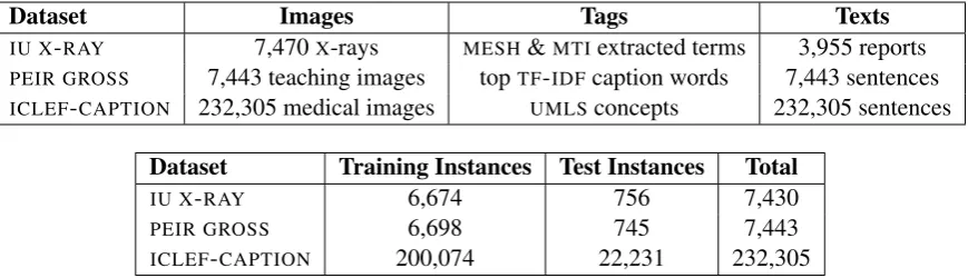
scans or radiology images, and write their findings as medical reports (Figure1b). Methods assisting physicians to focus on interesting image regions (Shin et al., 2016) or to describe findings (Jing et al.,2018) can reduce medical errors (e.g., sug-gesting findings to inexperienced physicians) and benefit medical departments by reducing the cost per exam (Bates et al.,2001;Lee et al.,2017).
Despite the importance of biomedical image captioning, related resources are not easily acces-sible, hindering the emergence of new methods. The publicly available datasets are only three and not always directly available.1 Also, there is cur-rently no assessment of simple baselines to de-termine the lower performance boundary and es-timate the difficulty of the task. By contrast, plex (typically deep learning) systems are com-pared to other complex systems, without estab-lishing if they surpass baselines (Zhang et al., 2017b; Wang et al., 2018). Furthermore, cur-rent evaluation measures are adopted directly from generic image captioning, ignoring the more chal-lenging nature of the biomedical domain (Cohen
1See, for example,http://peir.path.uab.edu/
library/that requires web scrapping.
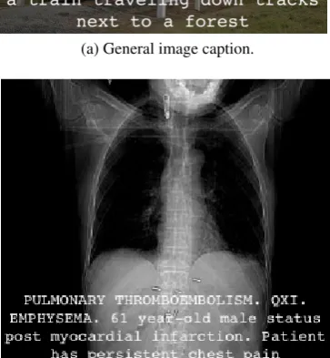
(a) General image caption.
[image:1.595.324.506.306.503.2](b) Biomedical image caption.
Figure 1: Example of a caption produced by the model of Vinyals et al. (2015) for a non-biomedical image (1a), and example of a radiology image with its associ-ated caption (1b) from the Pathology Education Infor-mational Resource (PEIR) Digital Library.
and Demner-Fushman,2014) and thus the poten-tial benefit from employing other measures ( Kil-ickaya et al.,2016). Addressing these limitations is crucial for the fast development of the field.
methods and attempts to compare their results, with the caveat that only two works use the same dataset (Shin et al.,2016;Jing et al.,2018) and can be directly compared. Section4describes evalua-tion measures that have been used and introduces two baselines. The first one is based on word fre-quencies and provides a low performance bound-ary. The second one is based on image retrieval and the assumption that similar images have sim-ilar diagnoses; we show that it is a strong base-line outperforming the state of the art in at least one dataset. Section 7 discusses related (mostly deep learning) biomedical image processing meth-ods for other tasks, such as image classification and segmentation. Section8highlights limitations of our work and proposes future directions.
2 Datasets
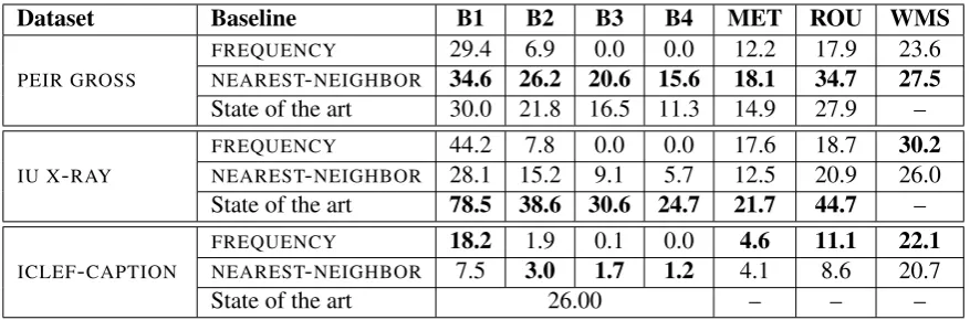
Datasets for biomedical image captioning com-prise medical images and associated texts. Pub-licly available datasets containX-rays (IU X-RAY
in Table1), clinical photographs (PEIR GROSS in
Table1), or a mixture of X-rays and photographs (ICLEF-CAPTIONin Table1). The associated texts may be single sentences describing the images, or longer medical reports based on the images (e.g., as in Figure 1b). Current publicly avail-able datasets are rather small (IU X-RAY, PEIR GROSS) or noisy (e.g.,IMAGE-CLEF, which is the largest dataset, was created by automatic means that introduced a lot of noise). We do not include in Table 1 datasets like the one of Wang et al. (2017), because their medical reports are not pub-licly available.2 Furthermore, we observe that all three publicly available biomedical image caption-ing datasets suffer from two main shortcomcaption-ings:
• There is a great class imbalance, with most images having no reported findings.
• The wide range of diseases leads to very scarce occurrences of disease-related terms, making it difficult for models to generalize.
IU X-RAY
Demner-Fushman et al. (2015) presented an ap-proach for developing a collection of radiology examinations, including images and narrative re-ports by radiologists. The authors suggested an
2See, for example, alsohttps://nihcc.app.box.
com/v/ChestXray-NIHCCwhere only images and text-mined disease labels are released for public use.
accurate anonymization approach for textual radi-ology reports and provided public access to their dataset through the Open Access Biomedical Im-age Search Engine (OpenI).3The images are 7,470 frontal and lateral chestX-rays, and each radiology report consists of four sections. The ‘comparison’ section contains previous information about the patient (e.g., preceding medical exams); the ‘indi-cation’ section contains symptoms (e.g., hypoxia) or reasons of examination (e.g., age); ‘findings’ lists the radiology observations; and ‘impression’ outlines the final diagnosis. A system would ide-ally generate the ‘findings’ and ‘impression’ sec-tions, possibly concatenated (Jing et al.,2018).
The ‘impression’ and ‘findings’ sections of the dataset of Demner-Fushman et al. (2015) were used to manually associate each report with a number of tags (called manual encoding), which were Medical Subject Heading (MESH)4 and RadLex5 terms assigned by two trained coders. Additionally, each report was associated with au-tomatically extracted tags, produced by Medical Text Indexer6 (calledMTI encoding). These tags allow systems to learn to initially generate terms describing the image and then use the image along with the generated terms to produce the caption. Hence, this dataset, which is the only one in the field with manually annotated tags, has an added value. From our processing, we found that 104 re-ports contained no image, 489 were missing ‘find-ings’, 6 were missing ‘impression’, and 25 were missing both ‘findings’ and ‘impression’; the 40 image-caption-tags triplets corresponding to the latter 25 reports were discarded in our later exper-iments. We shuffled the instances of the dataset (image-text-tags triplets) and used 6,674 of them as the training set (images from the 90% of the re-ports), with average caption length 38 words and vocabulary size 2,091. Only 2,745 training cap-tions were unique, because 59% of them were the same in more than one image (e.g., similar images with the same condition). Table1provides more information about the datasets and their splits.
PEIR GROSS
The Pathology Education Informational Resource (PEIR) digital library is a public access image
3https://openi.nlm.nih.gov/ 4https://goo.gl/iDvwj2 5
http://www.radlex.org/
Dataset Images Tags Texts IU X-RAY 7,470X-rays MESH&MTIextracted terms 3,955 reports
PEIR GROSS 7,443 teaching images topTF-IDFcaption words 7,443 sentences
ICLEF-CAPTION 232,305 medical images UMLSconcepts 232,305 sentences
Dataset Training Instances Test Instances Total
IU X-RAY 6,674 756 7,430
PEIR GROSS 6,698 745 7,443
[image:3.595.83.518.62.187.2]ICLEF-CAPTION 200,074 22,231 232,305
Table 1: Biomedical image captioning publicly available datasets. Images are annotated with tags, which may be medical terms (IU X-RAY) or words from the captions (PEIR GROSS) (Jing et al.,2018). A text may be linked to a single image (PEIR GROSS&ICLEF-CAPTION) or multiple ones (IU X-RAY). It may comprise a single sentence (PEIR GROSS) or multiple sentences (ICLEF-CAPTION,IU X-RAY). The lower table shows the number of training and test instances (image-text-tags triples) in each dataset, as used in our experiments. We excluded 40 out of the 7,470IU X-RAYinstances, as discussed in the main text.
database for use in medical education.7 Jing et al. (2018), who were the first to use images from this database, employed 7,442 teaching images of gross lesions (i.e., visible to the naked eye) from 21PEIRpathology sub-categories, along with their
associated captions.8 We developed code that downloads the images for this dataset (calledPEIR GROSS) and preprocesses their respective
cap-tions, which we release for public use.9
The dataset is split to 6,698 train and 745 test in-stances (Table1).10 The vocabulary size from the train captions is 4,051 with average caption length 17 words. From the 6,698 train captions only 632 were duplicates (i.e., the same caption for more than one images), which explains why this dataset has a much larger vocabulary thanIU X-RAY, de-spite the fact that captions are shorter.
ICLEF-CAPTION
This dataset was released in 2017 for the Im-age Concept Detection and Caption Prediction (ICLEF-CAPTION) task (Eickhoff et al., 2017) of
IMAGE-CLEF(de Herrera et al.,2018). The dataset consists of 184,614 biomedical images and their captions, extracted from biomedical articles on PubMed Central (PMC).11 The organizers used an automatic method, based on a biomedical image
7http://peir.path.uab.edu/library/ 8
PEIRpathology contains 23 sub-categories, but only 22 contain a gross sub-collection (7,443 images in total). We observe that one image was not included byJing et al.(2018).
9
Our code is publicly available athttps://github. com/nlpaueb/bio_image_caption.
10
We used 10% of the dataset for testing, as the 1k images used by Jing et al. for validation and testing were not released.
11https://www.ncbi.nlm.nih.gov/pmc/
type hierarchy (M¨uller et al.,2012), to classify the 5.8M extracted images as clinical or not and also discard compound ones (e.g., images consisting of multipleX-rays), but their estimation was that the overall noise in the dataset would be as high as 10% or 20% (Eickhoff et al.,2017).
In 2018, the ICLEF-CAPTION organizers
em-ployed a Convolutional Neural Network (CNN), to classify the same 5.8M images based on their type and to extract the non-compound clinical ones, leading to 232,305 images along with their respec-tive captions (de Herrera et al., 2018). Although they reported that compound images were re-duced, they noted that noise still exists, with non-clinical images present (e.g., images of maps). Additionally, a wide diversity between the types of the images has been reported (Liang et al., 2017). The length of the captions varies from 1 to 816 words (Su et al.,2018;Liang et al.,2017). Only 1.4% of the captions are duplicates (associ-ated with more than one image), probably due to the wide image type diversity. The average cap-tion length is 21 words and the vocabulary size is 157,256. A further 10k instances were used for testing in 2018, but they are not publicly available. Hence, in our experiments we split the 235,305 in-stances into training and test subsets ( Table1).
For tag annotation, the organizers usedQUICK -UMLS (Soldaini and Goharian, 2016) to identify concepts of the Unified Medical Language Sys-tem (UMLS) in the caption text, extracting 111,155
asso-ciated concepts and there are images assoasso-ciated with even thousands of concepts. The organizers observe the existence of noise and note that irrel-evant concepts have been extracted, mainly due to the fully automatic extraction process.
3 Methods
Varges et al. (2012) employed Natural Language Generation to assist medical professionals turn cardiological findings (e.g., from diagnostic imag-ing procedures) into fluent and readable textual de-scriptions. From a different perspective, Schlegl et al. (2015) used both the image and the textual report as input to a CNN, trained to classify im-ages with the help of automatically extracted se-mantic concepts from the textual report. Kisilev et al. (2015a,b) employed a radiologist to mark an image lesion, and a semi-automatic segmenta-tion approach to define the boundaries of that le-sion. Then, they used structured Support Vector Machines (Tsochantaridis et al.,2004) to generate semantic tags, originating from a radiology lexi-con, for each lesion. In subsequent work they used aCNNto rank suspicious regions of diagnostic im-ages and, then, generate tags for the top ranked re-gions, which can be embedded in diagnostic sen-tence templates (Kisilev et al.,2016).
Shin et al.(2016) were the first to apply aCNN
-RNN encoder-decoder approach to generate cap-tions from medical images. They used the IU X
-RAYdataset and a Network in Network (Lin et al., 2013) or GoogLeNet (Szegedy et al., 2015) as the encoder of the images, obtaining better results with GoogLeNet. The encoder was pretrained to predict (from the images) 17 classes, correspond-ing toMESHterms that were frequent in the reports
and did not co-occur frequently with otherMESH
terms. An LSTM (Hochreiter and Schmidhuber, 1997) orGRU (Cho et al., 2014) was used as the RNNdecoder to generate image descriptions from the image encodings. In a second training phase, the mean of theRNNs state vectors (obtained while
describing each image) was used as an improved representation of each training image. The orig-inal 17 classes that had been used to pretrain the
CNN were replaced by 57 finer classes, by apply-ing k-means clusterapply-ing to the improved vector rep-resentations of the training images. TheCNNwas
then retrained to predict the 57 new classes and this led to improvedBLEU(Papineni et al.,2002) scores for the overallCNN-RNNsystem. The
gen-erated descriptions, however, were not evaluated by humans. Furthermore, the generated descrip-tions were up to 5 words long and looked more like bags of terms (e.g., ‘aorta thoracic, tortuous, mild’), rather than fluent coherent reports.
Zhang et al. (2017b) were the first to employ an attention mechanism in biomedical image to text generation, with theirMDNET.12 MDNETused
RESNET (He et al.,2016) for image encoding, but
extending its skip connections to address vanish-ing gradients. The image representation acts as the starting hidden state of a decoder LSTM,
en-hanced with an attention mechanism over the im-age. (During training, this attention mechanism is also employed to detect diagnostic labels.) The de-coder is cloned to generate a fixed number of sen-tences, as many as the symptom descriptions. This model performed slightly better than a state of the art generic image captioning model (Karpathy and Fei-Fei,2015) in most evaluation measures.
Jing et al.(2018) segment each image to equally sized patches and useVGG-19 (Simonyan and Zis-serman, 2014) to separately encode each patch as a ‘visual’ feature vector. A Multi-Layer Per-ceptron (MLP) is then fed with the visual feature vectors of each image (representing its patches) and predicts terms from a pre-determined term vocabulary. The word embeddings of the pre-dicted terms of each image are treated as ‘seman-tic’ feature vectors representing the image. The decoder, which produces the image description, is a hierarchical RNN, consisting of a sentence-levelLSTMand a word-levelLSTM. The
sentence-level LSTM produces a sequence of embeddings, each specifying the information to be expressed by a sentence of the image description (acting as a topic). For each sentence embedding, the word-level LSTM then produces the words of the cor-responding sentence, word by word. More pre-cisely, at each one of its time-steps, the sentence-level LSTM of Jing et al. examines both the vi-sual and the semantic feature vectors of the im-age. Following previous work on image caption-ing, that added attention to encoder-decoder ap-proaches (Xu et al.,2015;You et al.,2016;Zhang et al., 2017b), an attention mechanism (an MLP
fed with the current state of the sentence-level
12
LSTM and each one of the visual feature vectors of the image) assigns attention scores to the vi-sual feature vectors, and the weighted sum of the visual feature vectors (weighted by their attention scores) becomes a visual ‘context’ vector, specify-ing which patches of the image to express by the next sentence. Another attention mechanism (an-otherMLP) assigns attention scores to the seman-tic feature vectors (that represent the terms of the image), and the weighted sum of the semantic fea-ture vectors (weighted by attention) becomes the semantic context vector, specifying which terms of the image to express by the next sentence. At each time-step, the sentence-levelLSTMconsiders the visual and semantic context vectors, produces a sentence embedding and updates its state, until a stop control instructs it to stop. Given the sen-tence embedding, the word-level LSTM produces the words of the corresponding sentence, again un-til a special ‘stop’ token is generated. Jing et al. showed that their model outperforms models cre-ated for general image captioning with visual at-tention (Vinyals et al.,2015;Donahue et al.,2015; Xu et al.,2015;You et al.,2016).
Wang et al.(2018) adopted an approach similar to that ofJing et al.(2018), using aRESNET-based
CNN to encode the images and an LSTM decoder to produce image descriptions, but their LSTM is
flat, as opposed to the hierarchical LSTM ofJing et al. (2018). Wang et al. also demonstrated that it is possible to extract additional image features from the states of the LSTM, much as Jing et al. (2018), but using a more elaborate attention-based mechanism, combining textual and visual infor-mation. Wang et al. experimented with the same OpenI dataset that Shin et al. and Jing et al. used. However, they did not provide evaluation results on OpenI and, hence, no direct comparison can be made against the results of Shin et al. and Jing et al. Nevertheless, focusing on experiments that generated paragraph-sized image descriptions, the results of Wang et al. on the (not publicly avail-able)CHEST X-RAYdataset (e.g.,BLEU-1 0.2860,
BLEU-2 0.1597) are much worse than the OpenI
results of Jing et al. (e.g., BLEU-1 0.517, BLEU-2 0.386), possibly because of the flat (not hierarchi-cal)LSTMdecoder of Wang et al.13
ICLEF-CAPTION run successfully for two con-secutive years (Eickhoff et al., 2017; de Herrera
13
CHEST X-RAY 14 contains 112,120X-ray images with tags (14 disease labels) and medical reports, but only the im-ages and tags (not the reports) are publicly available.
et al., 2018) and stopped in 2019. Participating systems (see Table3) used image similarity to re-trieve images similar to the one to be described, then aggregating the captions of the retrieved im-ages; or they employed an encoder-decoder archi-tecture; or they simply classified each image based on UMLS concepts and then aggregated the
re-spectiveUMLS‘semantic groups’14to form a cap-tion. Liang et al. (2017) used a pre-trained VG
-GNET CNN encoder and an LSTM decoder,
simi-larly toKarpathy and Fei-Fei(2015). They trained three such models on different caption lengths and used an SVM classifier to choose the most
suit-able decoder for the given image. Furthermore, they used a 1-Nearest Neighbor method to retrieve the caption of the most similar image and aggre-gated it with the generated caption. Zhang et al. (2018), who achieved the best results in 2018, used the Lucene Image Retrieval software (LIRE) to
re-trieve images from the training set and then sim-ply concatenated the captions of the top three re-trieved images to obtain the new caption. Abacha et al.(2017) used GoogLeNet to detectUMLS con-cepts and returned the aggregation of their respec-tiveUMLSsemantic groups as a caption. Su et al.
(2018) andRahman(2018) also employed differ-ent encoder-decoder architectures.
Gale et al. (2018) argued that existing biomed-ical image captioning systems fail to produce a satisfactory medical diagnostic report from an im-age, and to provide evidence for a medical deci-sion. They focused on classifying hip fractures in pelvic X-rays, and argued that the diagnostic re-port of such narrow medical tasks could be sim-plified to two sentence templates; one for positive cases, including 5 placeholders to be filled by de-scriptive terms, and a fixed negative one. They used DENSENET (Huang et al., 2017) to get
im-age embeddings and a two-layer LSTM, with at-tention over the image, to generate the constrained textual report. Their results, shown in Table2, are very high, but this is expected due to the extremely simplified and standardized ground truth reports. (Gale et al. report an improvement of more than 50BLEUpoints when employing this assumption.) The reader is also warned that the results of Ta-ble 2 are not directly comparable, since they are obtained from very different datasets.
Method Dataset B1 B2 B3 B4 MET ROU CID
Shin et al. (2016) IU X-RAY 78.5 14.4 4.7 0.0 - -
-Jing et al. (2018) IU X-RAY 51.7 38.6 30.6 24.7 21.7 44.7 32.7
PEIR GROSS 30.0 21.8 16.5 11.3 14.9 27.9 32.9 Wang et al. (2018) CHEST X-RAY14† 28.6 15.9 10.3 7.3 10.7 22.6
-Zhang et al. (2017b) BCIDR† 91.2 82.9 75.0 67.7 39.6 70.1 2.04
Gale et al. (2018) FRONTAL PELVIC X-RAYS† 91.9 83.8 76.1 67.7 - -
-Table 2: Evaluation of biomedical image captioning methods with BLEU-1/-2/-3/-4 (B1, B2, B3, B4), METEOR (MET), ROUGE-L (ROU), and CIDER(CID) percentage scores. Zhang et al.(2017a) andHan et al.(2018) also performed biomedical captioning, but did not provide any evaluation results. Datasets with†are not publicly available;BDIDRconsists of 1,000 pathological bladder cancer images, each with 5 reports;FRONTAL PELVIC X -RAYScomprises 50,363 images, each supplemented with a radiology report, but simplified to a standard template; CHEST X-RAY14 is publicly available, but without its medical reports.
Team Year Approach BLEU
Liang et al. 2017 ED+IR 26.00
Zhang et al. 2018 IR 25.01
Abacha et al. 2017 CLS 22.47
Su et al. 2018 ED 17.99
[image:6.595.75.524.62.161.2]Rahman 2018 ED 17.25
Table 3: Top-5 participating systems at the ICLEF -CAPTIONcompetition, ranked based on averageBLEU (%), the official evaluation measure. Systems used an encoder-decoder (ED), image retrieval (IR), or classi-fiedUMLS concepts (CLS). We exclude 2017 systems employing external resources, which may have seen test data during training (Eickhoff et al.,2017). 2018 models were limited to use only pre-trainedCNNs.
4 Evaluation
The most common evaluation measures in biomedical image captioning are BLEU(Papineni et al., 2002), ROUGE (Lin, 2004) and METEOR
(Banerjee and Lavie,2005), which originate from machine translation and summarization. The more recent CIDER measure (Vedantam et al., 2015), which was designed for general image captioning (Kilickaya et al.,2016), has been used in only two biomedical image captioning works (Zhang et al., 2017b;Jing et al.,2018). SPICE(Anderson et al.,
2016), which was also designed for general image captioning (Kilickaya et al., 2016), has not been used in any biomedical image captioning work we are aware of. Below, we describe each measure separately and discuss its advantages and limita-tions with respect to biomedical image captioning.
BLEU is the most common measure (Papineni et al.,2002). It measures word n-gram overlap be-tween the generated and the ground truth caption.
A brevity penalty is added to penalize short gen-erated captions. BLEU-1 considers unigrams (i.e., words), whileBLEU-2, -3, -4 consider bigrams, tri-grams, and 4-grams respectively. The average of the four variants was used as the official measure inICLEF-CAPTION.
METEOR(Banerjee and Lavie, 2005) extended
BLEU-1 by employing the harmonic mean of pre-cision and recall (F-score), biased towards recall, and by also employing stemming (Porter stemmer) and synonymy (WordNet). To take into account longer subsequences, it includes a penalty of up to 50% when no common n-grams exist between the machine-generated description and the reference.
ROUGE-L (Lin et al., 2013) is the ratio of the length of the longest common subsequence be-tween the machine-generated description and the reference human description, to the size of the ref-erence (ROUGE-L recall); or to the generated
de-scription (ROUGE-L precision); or a combination of the two (ROUGE-L F-measure). We note that severalROUGEvariants exist, based on different
n-gram lengths, stemming, stopword removal, etc., butROUGE-Lis the most commonly used variant in biomedical image captioning so far.
CIDER (Vedantam et al., 2015) measures the cosine similarity between n-gram TF-IDF
repre-sentations of the two captions (words are also stemmed). This is calculated for unigrams to 4-grams and their average is returned as the final evaluation score. The intuition behind using TF
[image:6.595.80.284.267.349.2]gener-ally document collections), which may be mistak-enly penalized. We also noticed that the scores returned by the provided CIDER implementation may exceed 100%.15 We excludeCIDER results, since these issues need to be investigated further.
SPICE (Anderson et al., 2016) extracts tuples
from the two captions (machine-generated, refer-ence), containing objects, attributes and/or rela-tions; e.g., (patient), (has, pain), (male, patient). Precision and recall are computed using WordNet synonym matching between the two sets of tuples, and theF1 score is returned. The creators ofSPICE
report improved results over both METEOR and
CIDER, but it has been noted that results depend on parsing quality (Kilickaya et al.,2016). When ex-perimenting with the provided implementation16 of this measure, we noticed that it failed to parse long texts to evaluate them. Similarly to CIDER, we excludeSPICEfrom further analysis below.
Word Mover’s Distance (WMD) (Kusner et al., 2015) computes the minimum cumulative cost re-quired to move all word embeddings of one tion to aligned word embeddings of the other cap-tion.17 It completely ignores, however, word or-der, and thus readability, which is one of the main assessment dimensions in the biomedical field (Tsatsaronis et al., 2015). Other previously discussed n-gram based measures also largely ig-nore word order, but at least consider local order (inside n-grams). WMDscores are included in Ta-ble4as similarity valuesWMS= (1 +WMD)−1.
5 Baselines
5.1 Frequency Baseline
The first baseline we propose (FREQUENCY) uses the frequency of words in the training captions to always generate the same caption. The most frequent word always becomes the first word of the caption, the next most frequent word always becomes the second word of the caption, etc. The number of words in the generated caption is the average length of training captions. Systems should at least outperform this simplistic baseline and its score should be low across datasets.
15
We used the official evaluation server implementation CIDER-D(Chen et al.,2015).
16https://goo.gl/bo11Bz
17We used Gensim’s implementation of
WMD (https://goo.gl/epzecP) and biomedical word2vec embeddings (https://archive.org/details/ pubmed2018_w2v_200D.tar).
5.2 Nearest Neighbor Baseline
The second baseline (NEAREST-NEIGHBOR) is
based on the intuition that similar biomedical im-ages have similar diagnostic captions; this would also explain why image retrieval systems perform well in biomedical image captioning (Table3). We use RESNET-1818 to encode images, and cosine similarity to retrieve similar training images. The caption of the most similar retrieved image is re-turned as the generated caption of a new image. This baseline can be improved by employing an image encoder trained on biomedical images, such asX-rays (Rajpurkar et al.,2017).
6 Experimental Results
As shown in Table 4, FREQUENCY scores high when evaluated with BLEU-1 and WMS, probably because these measures are based on unigrams.
FREQUENCY, which simply concatenates the most common words of the training captions, is re-warded every time the most common words appear in the reference captions.
To our surprise, NEAREST-NEIGHBOR outper-forms not only FREQUENCY, but also the state of the art in PEIR GROSS, in all evaluation
mea-sures (Table 4). This could be explained by the fact thatPEIR GROSSimages are phototographs of medical conditions, notX-rays, and thus they may
be handled better by the RESNET-18 encoder of
NEAREST-NEIGHBOR. In future work, we intend to experiment with an encoder trained on medical images (e.g.,CHEXNET).19
InIU X-RAY, NEAREST-NEIGHBOR scores low in all measures, possibly because in this case the images are X-rays and the RESNET-18 encoder
fails to handle them properly. Again, by exper-imenting with a different encoder, trained on X -rays, this baseline might be improved.
In ICLEF-CAPTION, both of our baselines
per-form poorly, and much worse than the best sys-tem (cf. Table3), which achieved average BLEU
26%. This is partially explained by the size of this dataset (Section 2), which contains multiple dif-ferent images and captions. Moreover, this dataset was created automatically and includes noise and a great diversity of image types (e.g., irrelevant, generic images such as maps) and captions.
18
https://goo.gl/28K1y2
19https://stanfordmlgroup.github.io/
Dataset Baseline B1 B2 B3 B4 MET ROU WMS
PEIR GROSS
FREQUENCY 29.4 6.9 0.0 0.0 12.2 17.9 23.6
NEAREST-NEIGHBOR 34.6 26.2 20.6 15.6 18.1 34.7 27.5
State of the art 30.0 21.8 16.5 11.3 14.9 27.9 –
IU X-RAY
FREQUENCY 44.2 7.8 0.0 0.0 17.6 18.7 30.2
NEAREST-NEIGHBOR 28.1 15.2 9.1 5.7 12.5 20.9 26.0
State of the art 78.5 38.6 30.6 24.7 21.7 44.7 –
ICLEF-CAPTION
FREQUENCY 18.2 1.9 0.1 0.0 4.6 11.1 22.1
NEAREST-NEIGHBOR 7.5 3.0 1.7 1.2 4.1 8.6 20.7
[image:8.595.80.519.61.206.2]State of the art 26.00 – – –
Table 4: Evaluation ofFREQUENCYandNEAREST-NEIGHBORon all datasets, withBLEU-1/-2/-3/-4 (B1, B2, B3, B4), METEOR (MET), ROUGE(ROU), Word Mover’s Similarity (WMS) percent scores. Best results to date per dataset are also included (state of the art). InICLEF-CAPTION, only the averageBLEUhas been reported (26.00).
7 Related Fields
Deep learning methods have been widely applied to biomedical images and address various biomed-ical imaging tasks (Litjens et al., 2017). Below, we briefly describe the tasks that are most related to biomedical image captioning, namely biomed-ical image classification, detection, segmentation, retrieval, as well as general image captioning.
The most related field is image captioning for general images. This is not a new task (Duygulu et al.,2002), but recent work leverages big datasets and has achieved impressive results on generating natural language captions (Karpathy and Fei-Fei, 2015). The work of Xu et al. (2015) was the first to incorporate attention to the encoder-decoder ar-chitecture for image captioning. Appart from im-proving performance, attention over images helps visualize how the model decides to generate each word and improves interpretability. Image cap-tioning can also be addressed jointly with other tasks, such as video captioning (Donahue et al., 2015) or image tagging (Shin et al.,2016).
Biomedical image classification aims at classi-fying a biomedical image as normal or abnormal, or assigning multiple disease labels (Rajpurkar et al., 2017, 2018). Also, it may refer to classi-fying an abnormality as malignant or benign ( Es-teva et al., 2017), or assigning other labels (e.g, labels showing the severity of a lesion). A related task is biomedical image detection, which is used to localize and highlight organs or wider anatomi-cal regions (de Vos et al.,2016) as well as specific abnormalities (Dou et al.,2016). This task is per-formed as a first step to assist other tasks, such as image classification or segmentation (Bi et al.,
2017;Rajpurkar et al.,2017).
Biomedical image segmentation aims to divide a biomedical image to different regions represent-ing organs or abnormalities, which can be used for further medical analysis, to learn their features, or classification. The most popularCNN-based archi-tecture isU-NET(Ronneberger et al.,2015), a ver-sion of the network ofLong et al.(2015), altered to produce more precise outputs. Later works (O.¨ C¸ ic¸ek et al.,2016;Milletari et al.,2016) extended
U-NETfor 3Dimage segmentation.
Biomedical image retrieval facilitates searching images in large biomedical databases, based on certain features like symptoms, diseases, and med-ical cases in general (Liu et al., 2016). Related tasks are also image registration, which performs a spatial alignment of the images (Miao et al., 2016;Yang et al., 2016), biomedical image gen-eration (Bahrami et al.,2016), and resolution en-hancement of 2Dand 3D biomedical images ( Ok-tay et al.,2016).
8 Limitations and Future Work
evalu-ation measures for biomedical image captioning, and the extent to which evaluation measures from generic image captioning, summarizaton, or ma-chine translation are appropriate.
Secondly, we hope to distill key features from current biomedical image captioning methods (e.g., methods that first tag the images and then generate captions from both the images and their tags vs. methods that directly generate captions; methods that retrieve similar images vs. methods that do not; types of pretraining used in image encoders and text decoders). This will allow us to provide a more structured and coherent presen-tation of current methods and highlight possible choices that have not been explored so far.
Thirdly, we plan to consult physicians (e.g., ra-diologists, nuclear doctors) to obtain a better view of their real-life needs and the degree to which current methods are aligned with their needs. We would also like to contribute to a roadmap of fu-ture activities towards integrating biomedical im-age captioning methods in real-life diagnostic pro-cedures and clinical diagnosis systems.
Acknowledgments
We are grateful to the anonymous reviewers, who suggested several of the future possible improve-ments mentioned above. We also thank Dr. Dim-itrios Papamichail for discussions that motivated us to consider biomedical image captioning.
References
A. B. Abacha, A. Garc´ıa Seco de Herrera, S. Gayen, D. Demner-Fushman, and S. Antani. 2017. NLM at ImageCLEF 2017 caption task. InCLEF CEUR Workshop, Dublin, Ireland.
P. Anderson, B. Fernando, M. Johnson, and S. Gould. 2016. SPICE: Semantic propositional image caption evaluation. InECCV, pages 382–398, Amsterdam, Netherlands.
K. Bahrami, F. Shi, I. Rekik, and D. Shen. 2016. Con-volutional neural network for reconstruction of 7T-like images from 3T MRI using appearance and anatomical features. In Deep Learning and Data Labeling for Medical Applications, pages 39–47, Athens, Greece.
S. Banerjee and A. Lavie. 2005. METEOR: An auto-matic metric for MT evaluation with improved cor-relation with human judgments. InACL Workshop on Intrinsic and Extrinsic Evaluation Measures for Machine Translation and/or Summarization, pages 65–72, Ann Arbor, MI, USA.
D. W. Bates, M. Cohen, L. L. Leape, J. M. Overhage, M. M. Shabot, and T. Sheridan. 2001. Reducing the frequency of errors in medicine using information technology. Journal of the American Medical Infor-matics Association, 8(4):299–308.
L. Bi, J. Kim, A. Kumar, L. Wen, D. Feng, and M. Ful-ham. 2017. Automatic detection and classification of regions of FDG uptake in whole-body PET-CT lymphoma studies. Computerized Medical Imaging and Graphics, 60:3–10.
X. Chen, H. Fang, T.-Y. Lin, R. Vedantam, S. Gupta, P. Dollr, and C. L. Zitnick. 2015. Microsoft COCO captions: Data collection and evaluation server. arXiv:1504.00325.
K. Cho, B. van Merrienboer, C. Gulcehre, D. Bah-danau, F. Bougares, H. Schwenk, and Y. Bengio. 2014. Learning phrase representations using RNN encoder–decoder for statistical machine translation. InEMNLP, pages 1724–1734, Doha, Qatar.
K. B. Cohen and D. Demner-Fushman. 2014. Biomedi-cal Natural Language Processing. John Benjamins.
D. Demner-Fushman, M. D. Kohli, M. B. Rosenman, S. E. Shooshan, L. Rodriguez, S. Antani, G. R. Thoma, and C. J. McDonald. 2015. Preparing a collection of radiology examinations for distribution and retrieval. Journal of the American Medical In-formatics Association, 23(2):304–310.
J. Donahue, L. Anne Hendricks, S. Guadarrama, M. Rohrbach, S. Venugopalan, K. Saenko, and T. Darrell. 2015. Long-term recurrent convolutional networks for visual recognition and description. In CVPR, pages 2625–2634, Boston, MA, USA.
Q. Dou, H. Chen, L. Yu, L. Zhao, J. Qin, D. Wang, V. CT. Mok, L. Shi, and P.-A. Heng. 2016. Au-tomatic detection of cerebral microbleeds from MR images via 3D convolutional neural networks. IEEE Transactions on Medical Imaging, 35(5):1182– 1195.
P. Duygulu, K. Barnard, J. F. G. de Freitas, and D. A. Forsyth. 2002. Object recognition as machine trans-lation: Learning a lexicon for a fixed image vocabu-lary. InECCV, pages 97–112, Florence, Italy.
C. Eickhoff, I. Schwall, A. Garc´ıa Seco de Herrera, and H. M¨uller. 2017. Overview of ImageCLEFcap-tion 2017 - Image capImageCLEFcap-tion predicImageCLEFcap-tion and concept ex-traction tasks to understand biomedical images. In CLEF CEUR Workshop, Dublin, Ireland.
A. Esteva, B. Kuprel, R. A. Novoa, J. Ko, S. M. Swet-ter, H. M. Blau, and S. Thrun. 2017. Dermatologist-level classification of skin cancer with deep neural networks. Nature, 542(7639):115–118.
Z. Han, B. Wei, S. Leung, J. Chung, and S. Li. 2018. Towards automatic report generation in spine radi-ology using weakly supervised framework. In In-ternational Conference on Medical Image Comput-ing and Computer-Assisted Intervention, pages 185– 193, Granada, Spain.
K. He, X. Zhang, S. Ren, and J. Sun. 2016. Deep resid-ual learning for image recognition. InCVPR, pages 770–778, Las Vegas, USA.
A. Garc´ıa Seco de Herrera, C. Eickhoff, V. Andrea-rczyk, and H. M¨uller. 2018. Overview of the Im-ageCLEF 2018 caption prediction tasks. In CLEF CEUR Workshop, Avignon, France.
S. Hochreiter and J. Schmidhuber. 1997. Long short-term memory. Neural Computation, 9(8):1735– 1780.
G. Huang, Z. Liu, L. Van Der Maaten, and K. Q. Wein-berger. 2017. Densely connected convolutional net-works. In CVPR, pages 4700–4708, Hawaii, HI, USA.
B. Jing, P. Xie, and E. Xing. 2018. On the automatic generation of medical imaging reports. In ACL, pages 2577–2586, Melbourne, Australia.
A. Karpathy and L. Fei-Fei. 2015. Deep visual-semantic alignments for generating image descrip-tions. In CVPR, pages 3128–3137, Boston, MA, USA.
M. Kilickaya, A. Erdem, N. Ikizler-Cinbis, and E. Er-dem. 2016. Re-evaluating automatic metrics for im-age captioning. InEACL, pages 199–209, Valencia, Spain.
P. Kisilev, E. Sason, E. Barkan, and S. Hashoul. 2016. Medical image captioning: Learning to describe medical image findings using multi-task-loss CNN. InDeep Learning for Precision Medicine, Riva del Garda, Italy.
P. Kisilev, E. Walach, E. Barkan, B. Ophir, S. Alpert, and S. Y. Hashoul. 2015a. From medical image to automatic medical report generation. IBM Journal of Research and Development, 59(2):1–7.
P. Kisilev, E. Walach, S. Y. Hashoul, E. Barkan, B. Ophir, and S. Alpert. 2015b. Semantic descrip-tion of medical image findings: Structured learning approach. In British Machine Vision Conference, pages 1–11, Swansea, UK.
M. Kusner, Y. Sun, N. Kolkin, and K. Weinberger. 2015. From word embeddings to document dis-tances. InICML, pages 957–966, Lille, France.
J.-G. Lee, S. Jun, Y.-W. Cho, H. Lee, G. B. Kim, J. B. Seo, and N. Kim. 2017. Deep learning in medical imaging: General overview. Korean Journal of Ra-diology, 18(4):570–584.
S. Liang, X. Li, Y. Zhu, X. Li, and S. Jiang. 2017. ISIA at the ImageCLEF 2017 image caption task. InCLEF CEUR Workshop, Dublin, Ireland.
C.-Y. Lin. 2004. ROUGE: A package for auto-matic evaluation of summaries. In Text Summa-rization Branches Out ACL Workshop, pages 74–81, Barcelona, Spain.
M. Lin, Q. Chen, and S. Yan. 2013. Network in net-work. arXiv:1312.4400.
G. Litjens, T. Kooi, B. E. Bejnordi, A. A. A. Setio, F. Ciompi, M. Ghafoorian, J. A.W.M. Van Der Laak, B. Van Ginneken, and C. I. S´anchez. 2017. A survey on deep learning in medical image analysis. Medi-cal Image Analysis, 42:60–88.
X. Liu, H. R. Tizhoosh, and J. Kofman. 2016. Gen-erating binary tags for fast medical image retrieval based on convolutional nets and radon transform. In IJCNN, pages 2872–2878, Vancouver, Canada.
J. Long, E. Shelhamer, and T. Darrell. 2015. Fully con-volutional networks for semantic segmentation. In CVPR, pages 3431–3440, Boston, MA, USA.
S. Miao, Z. J. Wang, and R. Liao. 2016. A CNN re-gression approach for real-time 2D/3D registration. IEEE transactions on medical imaging, 35(5):1352– 1363.
F. Milletari, N. Navab, and S. Ahmadi. 2016. V-Net: Fully convolutional neural networks for volumetric medical image segmentation. InInternational Con-ference on 3D Vision (3DV), pages 565–571, Cali-fornia, CA, USA.
H. M¨uller, J. Kalpathy-Cramer, D. Demner-Fushman, and S. Antani. 2012. Creating a classification of image types in the medical literature for visual cat-egorization. In Medical Imaging 2012: Advanced PACS-based Imaging Informatics and Therapeutic Applications, San Diego, CA, USA.
¨
O. C¸ ic¸ek, A. Abdulkadir, S. S. Lienkamp, T. Brox, and O. Ronneberger. 2016. 3D U-Net: Learning dense volumetric segmentation from sparse annotation. In Medical Image Computing and Computer-Assisted Intervention, pages 424–432, Athens, Greece.
O. Oktay, W. Bai, M. Lee, R. Guerrero, K. Kamnit-sas, J. Caballero, A. de Marvao, S. Cook, D. ORe-gan, and D. Rueckert. 2016. Multi-input cardiac image super-resolution using convolutional neural networks. InInternational Conference on Medical Image Computing and Computer-Assisted Interven-tion, pages 246–254, Athens, Greece.
Md M. Rahman. 2018. A cross modal deep learning based approach for caption prediction and concept detection by CS Morgan State. In CLEF CEUR Workshop, Avignon, France.
P. Rajpurkar, J. Irvin, R. L. Ball, K. Zhu, B. Yang, H. Mehta, et al. 2018. Deep learning for chest ra-diograph diagnosis: A retrospective comparison of the CheXNeXt algorithm to practicing radiologists. PLOS Medicine, 15(11):1–17.
P. Rajpurkar, J. Irvin, K. Zhu, B. Yang, H. Mehta, et al. 2017. CheXNet: Radiologist-level pneumo-nia detection on chestX-rays with deep learning. arXiv:1711.05225.
O. Ronneberger, P. Fischer, and T. Brox. 2015. U-Net: Convolutional networks for biomedical im-age segmentation. In Medical Image Computing and Computer-Assisted Intervention, pages 234– 241, Munich, Germany.
T. Schlegl, S. M. Waldstein, W.-D. Vogl, U. Schmidt-Erfurth, and G. Langs. 2015. Predicting seman-tic descriptions from medical images with convolu-tional neural networks. In Information Processing in Medical Imaging, pages 437–448, Isle of Skye, UK.
H.-C. Shin, K. Roberts, L. Lu, D. Demner-Fushman, J. Yao, and R. M. Summers. 2016. Learning to read chest X-rays: Recurrent neural cascade model for automated image annotation. InCVPR, pages 2497– 2506, Las Vegas, USA.
K. Simonyan and A. Zisserman. 2014. Very deep con-volutional networks for large-scale image recogni-tion. arXiv:1409.1556.
L. Soldaini and N. Goharian. 2016. QuickUMLS: a fast, unsupervised approach for medical concept ex-traction. InMedIR.
Y. Su, F. Liu, and M. P. Rosen. 2018. UMass at Image-CLEF caption prediction 2018 task. InCLEF CEUR Workshop, Avignon, France.
C. Szegedy, W. Liu, Y. Jia, P. Sermanet, S. Reed, D. Anguelov, D. Erhan, V. Vanhoucke, and A. Ra-binovich. 2015. Going deeper with convolutions. In CCVPR, pages 1–9, Boston, MA, USA.
G. Tsatsaronis, G. Balikas, P. Malakasiotis, I. Parta-las, M. Zschunke, M. R. Alvers, D. Weissenborn, A. Krithara, S. Petridis, D. Polychronopoulos, et al. 2015. An overview of the BIOASQ large-scale biomedical semantic indexing and question answer-ing competition. BMC Bioinformatics, 16(1):138.
I. Tsochantaridis, T. Hofmann, T. Joachims, and Y. Al-tun. 2004. Support vector machine learning for interdependent and structured output spaces. In ICML, pages 104–114, Banff, Alberta, Canada,.
S. Varges, H. Bieler, M. Stede, L. C. Faulstich, K. Ir-sig, and M. Atalla. 2012. SemScribe: Natural lan-guage generation for medical reports. In LREC, pages 2674–2681, Istanbul, Turkey.
R. Vedantam, Z. C. L. Zitnick, and D. Parikh. 2015. CIDEr: Consensus-based image description evalua-tion. InCVPR, Boston, MA, USA.
O. Vinyals, A. Toshev, S. Bengio, and D. Erhan. 2015. Show and tell: A neural image caption generator. In CVPR, pages 3156–3164, Boston, MA, USA.
B. D. de Vos, J. M. Wolterink, P. A. de Jong, M. A. Viergever, and I. Iˇsgum. 2016. 2D image classifica-tion for 3D anatomy localizaclassifica-tion: Employing deep convolutional neural networks. InMedical Imaging: Image Processing.
X. Wang, Y. Peng, L. Lu, Z. Lu, M. Bagheri, and R. M. Summers. 2017. ChestX-ray8: Hospital-scale chest X-ray database and benchmarks on weakly-supervised classification and localization of com-mon thorax diseases. InCVPR, pages 2097–2106, Hawaii, HI, USA.
X. Wang, Y. Peng, L. Lu, Z. Lu, and R. M. Summers. 2018. TieNet: Text-image embedding network for common thorax disease classification and reporting in chest X-rays. InCCPVR, pages 9049–9058, Que-bec City, Canada.
K. Xu, J. Ba, R. Kiros, K. Cho, A. Courville, R. Salakhudinov, R. Zemel, and Y. Bengio. 2015. Show, attend and tell: Neural image caption gener-ation with visual attention. InICML, pages 2048– 2057, Lille, France.
X. Yang, R. Kwitt, and M. Niethammer. 2016. Fast predictive image registration. InDeep Learning and Data Labeling for Medical Applications, pages 48– 57, Athens, Greece.
Q. You, H. Jin, Z. Wang, C. Fang, and J. Luo. 2016. Image captioning with semantic attention. InCVPR, pages 4651–4659, Las Vegas, NV, USA.
Y. Zhang, X. Wang, Z. Guo, and J. Li. 2018. ImageSem at ImageCLEF 2018 caption task: Image retrieval and transfer learning. In CLEF CEUR Workshop, Avignon, France.
Z. Zhang, P. Chen, M. Sapkota, and L. Yang. 2017a. TandemNet: Distilling knowledge from medical im-ages using diagnostic reports as optional semantic references. InInternational Conference on Medical Image Computing and Computer Assisted Interven-tion, pages 320–328, Quebec City, Canada.