Original Article
The difference of posterior
slope angle between intra-operation
and post-operation in total knee arthroplasty
Xiaolei Song1, Jiangtao Dong2, Juan Wang2, Xiaozuo Zheng2, Yisheng Wang1
1Department of Orthopaedic Surgery, The First Affiliated Hospital of Zhengzhou University, Zhengzhou 450000,
China; 2Department of Joint, The Third Hospital of Hebei Medical University, Ziqiang Road, Shijiazhuang, Hebei,
China
Received December 11, 2015; Accepted April 9, 2016; Epub February 15, 2017; Published February 28, 2017
Abstract: This study aimed to analyze the difference of posterior slope angle between intra-operation and post-operation during total knee arthroplasty (TKA) to provide a theoretical basis for adjusting the posterior slope angle. Clinical patients (44 cases) with TKA admitted to the Hospital from June 1 to September 2 in 2013 were involved in this study. According to the difference of the prosthesis, patients were divided into two groups: cruciate-retaining group (n=22, 33 knees) and posterior-stabilized group (n=22, 31 knees). Then the difference between posterior slope angle of intra-operation and post-operation were analyzed. In the cruciate-retaining group, the posterior slope angle of post-operation was 5.73°±2.81° greater than that of intra-operation (P=0.00); While in the posterior-stabilized group, the posterior slope angle of post-operation was 1.66°±1.43° greater than that of intra-operation (P=0.00). In the cruciate-retaining group, the posterior slope angle of post-operation is much smaller than the intra operation which indicating that there is no need to adjust the posterior slope angle when it is too small during intra-operation. As for the posterior-stabilized group, there was no statistical difference of posterior slope angle between post-operation and intra-operation, so we could predict the posterior slope angle of post-operation according to that of intra-operation.
Keywords: Arthroplasty, replacement, knee, tibia plateau, posterior slope angl
Introduction
Total knee arthroplasty (total knee arthroplas-ty, TKA) was designed to remove the joint sur-face which had been damaged and could not be repaired to restore knee joint function by
replacing the damaged joints with artificial joint
components [1]. It could correct the deformity effectively, ease pain, enhance the function of the affected limb and maintain the stability of the joint [2]. In total knee arthroplasty, the choice of slope angle posterior (PSA), TKA (PSA)
would affect the flexion and extension of
knee-joint function, knee-joint stability and the life of the prosthesis after post-operation [3]. Therefore,
the influence of PSA on TKA was gradually more
and more important. In current study, the accu-rately select of the inclined angle was a major problem facing the surgeon in operation. In this study, we analyzed the difference on posterior slope angle between intra-operation and
post-aimed to provide a guide for the doctors for the selecting of posterior slope angle between intra-operation and post-operation.
Materials and methods
Materials
All the clinical patients (44 cases) with TKA were admitted to the Hospital from June 1 to September 2 in 2013. Inclusion criteria: (1) All the operation were performed by the same sur-geon (2) All the patients suffered from total
knee arthroplasty for the first time (3) The
(33 knees), male (2 knees), female (31 knees), average age (63.95); In the posterior-stabilized group, there were prosthetic knee replacement (22 cases), total joint replacement (31 knees), male (5 knees), female (26 knees), average age (65.52). The basic data of the CR group and the PS group were compared, including gender, age and preoperative KSS score. The results
showed there were no significant differences
between the two groups. The data were shown in Table 1.
Surgical methods
All cases were completed by the same surge- on. After the preoperative preparation, 15 cm length of incision was performed in the middle of anterior knee. After the opening of skin and subcutaneous tissue, then the synovium and the lower part of the patellar fat pad were cut off. The periosteum was cut at 1 cm of the medial tibia, and the medial articular capsule was initially loosened by the dissection along the periosteum. The eversion of the patella, knee joint, resection of the femoral tibial osteo-phytes, hyperplastic synovial membrane, meni- scus and anterior anterior ligament were per-formed. The tibial plateau was pried out by plate hook, the positioner was setup in the neck bone and the proximal tibial osteotomy device was installed at roximal tibia. The
posi-tion was fixed by vertical angel of tibial glucos -amine line and along the outside. To restore the patient’s own back backward, the cut of poste-rior articular cartilage of the tibial plateau was as a reference and the post dip angle was recorded. The femur was positioned and washed with hydrogen peroxide solution and 0.9% sodium chloride injection before installa-tion of prosthesis. The wound was stitched up
at the neutral position of the knee joint. The posterior slope angle was measured at the pos-terior lateral position by x line. Gemini prosthe-sis and rp-f prostheprosthe-sis were respectively used for CR and PS groups.
The measurement of posterior slope angle
The posterior tilt angle was measured by the matching of the tibial bone marrow locator. The cut of posterior articular cartilage of the tibial plateau was as a reference and the post dip angle was recorded. The posterior slope angle between intra-operation and post-operation was measured at the X line of knee joint. Having no obvious tibial rotation and a clear outline of the tibial plateau prevail were as standards for the selection of X-ray. The posterior angle of the tibia was measured with the axis of the middle and upper end of the tibia. Reference curve was perpendicular to the axis, the angel be- tween the connection line for the posterior horn of the anterior tibial platform and reference curve was recorded as the posterior angle of the tibia (4-8) (Figure 1).
Groups
Based on the type of prosthesis used, the pros-thesis was divided into CR and PS group and the PSA of tibial plateau was recorded before surgery, intraoperative and postoperative. The difference of PSA during intra-operation and post-operation between the groups was
com-pared to confirm whether the PSA was reduced
to the preoperative anatomical PSA. The differ-ence on posterior slope angle between intra-operation and post-intra-operation was analyzed to explore the relationship of PSA between intra-operation and post-intra-operation.
Functional outcome and safety concern be-tween two methods
[image:2.612.92.295.96.215.2]The curative effects of TKA closely correlate to the intra-operative osteotomy and the debond-ing of surrounddebond-ing soft tissues. One of the key points of success is to recover the balance of soft tissues around the knee-joint. Except for the operative techniques of physicians, the prostheses were pretty important in the opera-tions of TKA. The prostheses with minor size may furthest recover the functions of knee-joint. The main basis in choosing thighbone prostheses is keeping the knee-joint stable
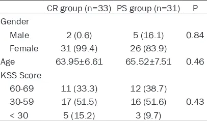
Table 1. Comparison of preoperative basic data between two TKA groups (n, %)
CR group (n=33) PS group (n=31) P Gender
Male 2 (0.6) 5 (16.1) 0.84 Female 31 (99.4) 26 (83.9)
Age 63.95±6.61 65.52±7.51 0.46 KSS Score
60-69 11 (33.3) 12 (38.7)
30-59 17 (51.5) 16 (51.6) 0.43 < 30 5 (15.2) 3 (9.7)
when bend the knees. Only depending on the residuary natural bones of patients is
impracti-cable, or it will make flexion gaps of knee-joint
greater than the Swollen knee clearance thus cuase the dislocation of knee-joint. If the patel-la should be reppatel-laced is mainly on the basis of the lesion of patelia or the state of patients’ knee-joint. Patients with serve regression and partial dislocation or subluxation in patellofem-oral joint are in need of replacing the patella; Patients (especially the elderly people) with thinner patella or slight regression in cartilage are not in need of replacing the patella, on the contrary, the replacement for those patients may cause the lability of the functions of joints
oppress the blood vessels, less limbs’ activities cause the hypercoagulable state, the intracor-poral blood vessels are damaged and the blood
flow rate decends, the coagulation is activated,
the functions of antithrombase recedes and
the endogenous fibrinolysis system is
restrain-ed. Hense, patients with the above-mentioned risk factors should be promptly applied the anti-freezing drugs and antiplatelet drugs after the operations.
Statistical analysis
[image:3.612.90.377.72.266.2]All experiments were independently performed in triplicate. Statistical analyses using analysis Figure 1. Posterior slop angle of post-operation in PS group (left); Posterior
slop angle of post-operation in CR group (right).
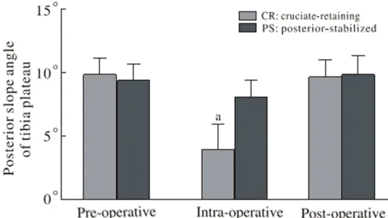
Figure 2. Comparison of posterior slope angle of pre- and postoperative be-tween two groups (aP < 0.05, vs. post-operative).
and poor post-operative eff- ect. The main operating points for replacing the patella are that the thickness of the pa- tella should be greater than 12 Rim after being excised, the thickness of patella re- placed by prosthesis should be the same with the pre-operative thickness of patel-la, the excision line should be parallel with the surface of pa- tella, and the patella should be placed in the inner side as far as possible.
[image:3.612.91.372.322.480.2]of variance (ANOVA) and Duncan’s multiple range test for differences among different groups were performed with SPSS software version 20 (Chicago, IL, USA). Comparisons that
yielded P < 0.05 were considered significant.
Results
In the CR group, the PSA was 9.66°±1.32° and 3.94°±2.24° for post-operation and intra-oper-ation. The PSA was 5.73°±2.81° higher in the post-operation than that in intra-operation; In the PS group, the PSA was 9.81°±1.54° and 8.10°±1.27° for post-operation and intra-oper-ation. The PSA was 1.66°±1.43° higher in the post-operation than that in intra-operation. The
data showed there was significant difference
for the PSA between the post-operation than that in intra-operation in the two groups (CR group: P=0.00; PS group: P=0.00). However,
there was no significant difference between
the posterior tilt angle in post-operation and tibial plateau angle intra-operation (CR group: P=0.57; PS group: P=0.29) which indicated postoperative tibial slope angle was substan-tially reduced to the preoperative anatomic tilt-ing angle. The data was shown in Figure 2.
Discussion
With the increase of China’s aging and osteoar-thritis, joint replacement surgery entered a period of rapid development [9, 10]. Currently, the correct of the power lines and soft tissue surgery balance in knee replacement surgery is
still one of the difficulties [11]. Therefore, the
posterior slope angle of the tibial plateau pros-thesis is gradually being valued by clinicians. In the recent study, a number of domestic and foreign research reports indicate that there was inter-individual difference in PSA. In this study, the PSA measured was 9.85°±1.25°. Kunwano’s study showed that the PSA was 8.1°±4.0° measured by CT 3D reconstruction system [12]. Yoo measured the tibial lateral slice with 5 different reference standards and the average was 10.6°±3.5° [5, 13]. In Khattak’s report, the PSA was 10°±4° [13]. The specimen of adult knee joint, dry tibial and MRI of adult knee joints were used to measure PSA by Huang Wenhua et al [10]. The average value of PSA in the lateral position of the knee was 11.3°±4.4° studied by Wenyan DU [14]. The posterior tibial plateau angle was 15.02°±4.2° in Shudong zhang’s study [15]. In addition,
there was still no uniform standard on the tibial osteotomy in TKA operation [16]. Wang Yehua et al. considered that the difference of the angle between the posterior tibial plateau was larger, and it should be measured before and after the surgery to make the posterior tibial osteotomy individual. Kansara and Markel con-sidered that osteotomy of 0° and 5° had no effect on the activity of the knee joint [2]. How- ever, a small number of cases would appear front dip angle with the 0° osteotomy com-pared with 5° osteotomy, so 5° osteotomy was considered to be safer.
Another study confirmed that the knee joint flexion function was best when the PSA of tibial
prosthesis was 7°~9° after TKA [17]. But in our hospital, it would result in 0 degree posterior tilt and the tibial plateau cartilage could be complete amputated during the knee replace-ment. Moreover, postoperative X-ray measure-ment displayed PSA was still within the normal physiological range. In face of this embarrass-ing situation, the prosthesis of CR (Gemini) and Johnson’s PS (RP-F) were incorporated in this study. It can be seen that both the PS and CR
groups were significantly different on posterior
slope angle between intra-operation and post-operation (CR group: P=0.00; PS group: P= 0.00) which means that the PSA of intra-opera-tion could not be represented by that of the post-operation.
For the prosthesis involved in this study, post-operative tibial slope angle was substantially reduced to the preoperative anatomic tilting angle (CR group: P=0.57; PS group: P=0.29). So, it can be concluded that the PSA is basically accurate. For the case of the small deviation of the measurement, the main reasons are as follows after the analysis of prosthesis and major surgical procedures: (1) The choice of rear angle is not intuitive because of the use of
external fixation of the tibia in the surgical
instruments which resulted the choice error for the PSA; (2) The completely capture of tibial cartilage was as the standard which made
more subjective influence factors for the sur -geon; (3) There were errors in the device, so the measurement of PSA was not actual. Since the minor application of other knee prosthesis, it
was difficult to achieve larger sample size. In
affect the purpose of the present study. At the same time, as a single center prospective study, it also has some limitations such as the
deficiency of specimens. Therefore, it still need
the large sample, multi center, multi prosthesis model for the study of knee replacement sur-gery to provide a strong theoretical support for the select of prosthesis in knee replacement surgery
Disclosure of conflict of interest
None.
Address correspondence to: Yisheng Wang, Depart-
ment of Orthopaedic Surgery, The First Affiliated
Hospital of Zhengzhou University, No.1 of Eastern Jianshe Road, Erqi District, Zhengzhou 450000, Henan Province, China. E-mail: shijungao2015@163. com
References
[1] Marouane H, Shirazi-Adl A and Hashemi J.
Quantification of the role of tibial posterior
slope in knee joint mechanics and ACL force in simulated gait. J Biomech 2015; 48: 1899-1905.
[2] Kansara D and Markel DC. The effect of poste-rior tibial slope on range of motion after total knee arthroplasty. J Arthroplasty 2006; 21: 809-813.
[3] Kuriyama S, Ishikawa M, Nakamura S, Furu M, Ito H and Matsuda S. Posterior tibial slope and femoral sizing affect posterior cruciate liga-ment tension in posterior cruciate-retaining total knee arthroplasty. Clin Biomech 2015; 30: 676-681.
[4] Chiu KY, Zhang SD and Zhang GH. Posterior slope of tibial plateau in Chinese. J Arthroplasty 2000; 15: 224-227.
[5] Yoo JH, Chang CB, Shin KS, Seong SC and Kim TK. Anatomical references to assess the posterior tibial slope in total knee arthroplasty: a comparison of 5 anatomical axes. J Arth- roplasty 2008; 23: 586-592.
[6] Chambers AW, Wood AR, Kosmopoulos V, Sanchez HB and Wagner RA. Effect of
posteri-or tibial slope on flexion and anteriposteri-or-posteriposteri-or
tibial translation in posterior cruciate-retaining total knee arthroplasty. J Arthroplasty 2016; 31: 103-106.
[7] Zhang Y, Wang J, Xiao J, Zhao L, Li ZH, Yan G and Shi ZJ. Measurement and comparison of tibial posterior slope angle in different meth-ods based on three-dimensional reconstruc-tion. Knee 2014; 21: 694-698.
[8] Okazaki K, Tashiro Y, Mizu-uchi H, Hamai S, Doi
T and Iwamoto Y. Influence of the posterior tibial slope on the flexion gap in total knee ar -throplasty. Knee 2014; 21: 806-809.
[9] Ishida H, Suehiro T, Kurozumi C, Ono K, Ando S and Watanabe S. Correlation between neck
slope angle and deep cervical flexor muscle
thickness in healthy participants. J Bodywork Movement Ther 2015; 19: 717-721.
[10] Schlatterer B, Linares JM, Cazal J, Merloz P and Plaweski S. Computer Assisted Orthopedic Surgery-France (CAOS-France). Posterior tibial
slope accuracy with patient-specific cutting
guides during total knee arthroplasty: A pre-liminary study of 50 cases. Orthopaed Trau- matol Surg Res 2015; 101 Suppl: S233-40. [11] Chimento A, Saturnino C, Iacopetta D, Mazzotta
R, Caruso A, Plutino MR, Mariconda A, Ramunno A, Sinicropi MS, Pezzi V and Longo P. Inhibition of human topoisomerase I and II and anti-proliferative effects on MCF-7 cells by new titanocene complexes. Bioorgan Med Chem 2015; 23: 7302-7312.
[12] Kuwano T, Urabe K, Miura H, Nagamine R, Matsuda S, Satomura M, Sasaki T, Sakai S, Honda H and Iwamoto Y. Importance of the lat-eral anatomic tibial slope as a guide to the tibial cut in total knee arthroplasty in Japanese patients. J Orthopaed Sci 2005; 10: 42-47. [13] Khattak MJ, Umer M, Davis ET, Habib M and
Ahmed M. Lower-limb alignment and posterior tibial slope in Pakistanis: a radiographic study. J Orthopaed Surg 2010; 18: 22-25.
[14] Darrah SD, Miller MA, Ren D, Hoh NZ, Scanlon JM, Conley YP and Wagner AK. Genetic vari-ability in glutamic acid decarboxylase genes: associations with post-traumatic seizures after severe TBI. Epilepsy Res 2013; 103: 180-194. [15] Lin L and Sun J. Adaptive conditional feature screening. Comput Statist Data Analys 2016; 94: 287-301.
[16] Goyal N and Stulberg SD. Evaluating the preci-sion of preoperative planning in patient
spe-cific instrumentation: can a single MRI yield
different preoperative plans? J Arthroplasty 2015; 30: 1250-3.