Original Article
Video-assisted thoracoscopic lobectomy versus open
lobectomy for clinical stage I non small cell lung
cancer: a case-control study
Jun Xu1, Xiaoqiang E2, Jun Tian1, Jinglong Yan1
1Department of Surgery, The Second Affiliated Hospital of Harbin Medical Universuity, 246 Xuefu Road, Harbin 150086, Hei Longjiang, People’s Republic of China; 2Department of Surgery, The First Affiliated Hospital of Harbin Medical Universuity, 23 Youzheng Street, Harbin 150086, Hei Longjiang, People’s Republic of China
Received March 23, 2015; Accepted June 3, 2015; Epub February 15, 2016; Published February 29, 2016
Abstract: This study aimed to compare outcomes following video-assisted thoracoscopic lobectomy (VATS) versus open lobectomy for clinical stage I non small cell lung cancer at a China center. Eighty-one consecutive patients who underwent video-assisted thoracoscopic lobectomy from January 2007 to December 2013 were compared with 94 patients undergoing open surgery during the same time period. Patients were matched for age, sex, Ameri-can Society of Anesthesiologists (ASA) class, location of tumor and clinical TNM stage. Endpoints were short- and long-term perioperative outcomes. The VATS approach was associated with longer operative time, less blood loss, similar surgical margin, and decreased duration of narcotic and epidural use, similar complications and decreased length of stay. There was no difference in the number of lymph nodes harvested, 5-year overall survival and 5-year disease-free survival between the VATS group and open group. VATS is safe and effective for clinical stage I non small cell lung cancer.
Keywords: Non-small cell lung cancer, lobectomy, minimally invasive surgery, video-assisted thoracoscopic sur-gery, prognosis
Introduction
Diagnosis of clinical stage I non small cell lung cancer, has been increasing worldwide because of the development of screening methods [1-5]. An aggressive surgical approach of pulmonary resection plus mediastinal lymphadenectomy has long been the standard treatment, even for early stage non small cell lung cancer [6-8]. Non-small cell lung cancer, even at this early stage, tends to metastasize to the mediastinal lymph nodes, and mediastinal lymphadenecto-my is often required [9-11]. Recently, video-assisted thoracoscopic lobectomy (VATS) has been introduced to treat patients with clinical stage I non small cell lung cancer. Previous studies demonstrated that VATS is less inva-sive than open resection, thus leading to faster recovery and better cosmetic outcomes [12-15]. However, the prognosis of patients who underwent VATS for clinical stage I non small cell lung cancer was controversial. The
objec-tive of this study was to compare VATS and open lobectomy for clinical stage I non small cell lung cancer in terms of clinical results and the survival of patients.
Materials and methods
Patient selection
required conversion to an open procedure were also excluded. The most common reasons for conversion in these patients were the direct invasion of adjacent organs, uncontrolled bleeding, and positive proximal margin on fro-zen section analysis. A total of 94 patients who met these criteria above mentioned were iden-tified. These 94 patients were matched to 94 patients who underwent open lobectomy for the same indication during the same time peri-od. Matching variables were age, sex, American Society of Anesthesiologists (ASA) class, loca-tion of tumor and clinical TNM stage. The TNM stage was based on the 7th edition of the TNM classification of lung cancer, which was pro-posed by Union for International Cancer Control (UICC) and International Association for the Study of Lung Cancer (IASLC), and the medias-tinal lymph node staging was based on the newest lymph node map proposed by IASLC [16, 17]. For those of the patients operated before 2010, their staging was recalculated to match the 7th edition of TNM classification of lung cancer proposed by UICC and IASLC.
Operative technique for VATS
VATS was performed as previously described [14]. Only trocar and endsocopic instruments were used in VATS approach and no rib spread-ing was applied. All patients underwent one-lung ventilation and were placed in the lateral decubitus position. Mediastinal lymphadenec-tomy was performed after lobeclymphadenec-tomy. We per-form intra-operative lymph nodes frozen sec-tions. On the left side, lymph node station 5, 6, 7, 8, 9, 10, 11 and 12 were systematic dissect-ed. On the right side, dissection of lymph node station 2R, 4R, 7, 8, 9, 10, 11 and 12 were also performed. Lymph nodes mentioned above were dissected with the integrity of the sur-rounding structure, the anatomical landmarks clear recognition and with no nodal structures. Lymph node station 10, 11, 12 and the affect-ed lobe were systematic dissectaffect-ed and removaffect-ed
en bloc. Briefly, on the right side, when the mediastinal pleura was opened, the dissection of station 2R and 4R was undertaken up to the lowest visible part of the subclavian artery. After the lung was retracted anteriorly, the dis-section of station 7 was performed. Regardless of middle lobectomy and lower lobectomy, sta-tion 8 and 9 was systematically harvested. On the left side, station 5 and 6 was systematically
harvested. Then, dissection of station 7, 8 and 9 was done.
Clinical and follow-up data
Clinical and follow-up data were obtained from a prospectively maintained database. A compli-cation was defined as any unintended event occurring within 30 days of pulmonary resec-tion. Complications were graded in severity from 1 to 5 based on a modified Clavien-Dindo system. The definition of Clavien-Dindo system was as follows: Grade 1: oral medication or bedside medical care required; Grade 2: intra-venous medical therapy required; Grade 3: radiologic, endoscopic, or operative interven-tion required; Grade 4: chronic deficit or disabil-ity associated with the event; and Grade 5: death related to surgical complication. Minor complications were defined as Grades 1-2 and major complications as Grades 3-5 [18]. During the first year after radical resection and/or adjuvant therapy was completed, patients were seen every 3 months at the outpatient depart-ment. In the second year, follow-up took place every 6 months, and then follow-up was per-formed at the end of each year after treatment. During follow-up, diagnostic investigations were performed. All patients received CT scan of chest before discharge and before each follow-up visit. Any post-operative complications and medical conditions requiring hospitalization were reviewed. The follow-up was closed in December 2014.
Statistical analysis
vari-ables related to overall survival and disease-free survival. Univariate variables with probability values less than 0.10 were selected for inclu-sion in the multivariate Cox proportional hazard regression model. Adjusted odds ratios (HR) along with the corresponding 95% confidence intervals (CI) were calculated. P < 0.05 was considered statistically significant. SPSS 13.0 (SPSS Inc., Chicago, IL, USA) was applied. Results
Patient demographics
The VATS and open groups were similar for the matched variables of age, sex, ASA score,
[image:3.612.91.372.83.295.2]loca-There were no differences between the groups with regard to pathologic TNM stage (Table 4). The calculated 5-year overall survival rates for all stages were 73% in the VATS group and 68% in the open group (P = 0.806, Figure 2). There were no differences between the groups with regard to pathologic TNM stage (Table 4). Fifty-three patients in the VATS group devel-oped tumor recurrence, 23 from distant or hematogenous recurrence, and 15 from locore-gional or lymphatic recurrence; the correspond-ing findcorrespond-ings in the open group were 15, and 11, respectively. The pattern of recurrence was similar in the two groups, and no unexpected Table 1. Demographic data
VATS (n = 94) Open (n = 94) P value Age (y) 63 (42-71) 64 (39-70) 0.485 Gender (Male:Female) 58:36 54:40 0.552
Location of tumor 0.857
Right upper lobe 41 38 Right middle lobe 4 7 Right lower lobe 13 14 Left upper lobe 24 21 Left lower lobe 12 14
Clinical stage 0.767
IA 37 39
IB 57 55
ASA score 0.463
I 69 73
II 21 19
III 4 2
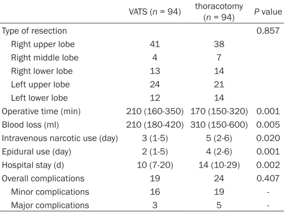
Table 2. Intraoperative and postoperative data
VATS (n = 94) thoracotomy (n = 94) P value
Type of resection 0.857
Right upper lobe 41 38 Right middle lobe 4 7 Right lower lobe 13 14
Left upper lobe 24 21
Left lower lobe 12 14
Operative time (min) 210 (160-350) 170 (150-320) 0.001 Blood loss (ml) 210 (180-420) 310 (150-600) 0.005 Intravenous narcotic use (day) 3 (1-5) 5 (2-6) 0.020 Epidural use (day) 2 (1-5) 4 (2-6) 0.001 Hospital stay (d) 10 (7-20) 14 (10-29) 0.002 Overall complications 19 24 0.407 Minor complications 16 19
Major complications 3 5
-tion of tumor and clinical TNM stage. Demographic variables for all patients are summarized in Table 1.
Operative characteristics and complications
The VATS and open groups were similar for type of resection. The VATS approach was associated with longer operative time, less blood loss, less postoperative pain measured by decreased duration of IV narcotic use and decreased duration of epidural use for patients who had epidur-al anesthesia. Length of stay was shorter in the VATS group. Overall, minor and major compli-cations were similar between the groups (Table 2).
Pathologic characteristics
No patients had grossly positi- ve margins. Tumor pathological TNM stage, histological subtype and lymph node retrieval were similar between the two groups. Pathologic variables for all pa- tients are summarized in Table 3.
Long-term outcomes
[image:3.612.91.369.330.538.2]tumor dissemination or port-site recurrence was observed.
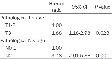
Multivariate analysis identified pathological N stage and pathological venous invasion as the factors with independent effects on disease-free survival (Table 5). The type of operative approach (VATS vs open lobectomy) did not influence the recurrence-free survival. In multi-variate analysis, the pathological T stage and
[image:4.612.91.374.84.281.2]lower risk of postoperative complications fol-lowing VATS versus open lobectomy [6]. While we did not note any overall differences in the incidence of complications among the two groups, we did find that there was a slightly higher incidence of minor complications follow-ing open lobectomy versus VATS. In the current study, we also noted that the median hospital stay following VATS was fewer than the open lobectomy group.
Table 3. Pathological data
VATS
(n = 94) thoracotomy (n = 94) P value
Histological subtype 0.460
Adenocarcinoma 84 87
Squamous cell carcinoma 9 5
Others 1 2
Pathological stage 0.763
IA 31 34
IB 45 41
IIA 8 10
IIB 6 7
IIIA 4 2
Residual tumor (R0/R1/R2) 94/0/0 94/0/0 1.000 Number of harvested lymph nodes 23 (19-31) 24 (19-32) 0.587 Mediastinal lymph nodes dissected 16 (15-24) 17 (16-25) 0.354
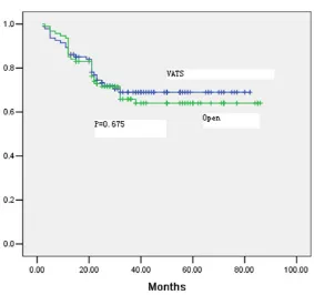
Figure 1. There was no significant difference in disease-free survival be -tween the VATS and open groups (66% vs. 62%) (P = 0.675).
pathological N stage had inde-pendent effects on overall sur-vival (Table 6). The type of oper-ative approach was not im- portant in the multivariate anal-ysis for overall survival.
Discussion
There is a growing interest in video-assisted thoracoscopic surgeries for many different op- erative procedures [19]. While video-assisted thoracoscopic surgery has been widely adopt-ed for many benign diseases, the role of video-assisted thora-coscopic surgery for malignant diseases continues to evolve. The long-term oncologic result is very important to the use of laparoscopic gastrectomy. The video-assisted thoracoscopic surgery has been described for the management of certain malignant indications. However, data on feasibility, safety, and long-term results with video-assisted thoracoscopic surgery for lung neoplasm have not been well defined [20-25]. One of the objectives of the current study was to establish the feasi-bility and safety of VATS for clini-cal stage I non small cell lung cancer.
[image:4.612.90.373.295.561.2]Adequate mediastinal lymphadenectomy is a critical component in radical lobectomy for non small cell lung cancer [24]. Several previous studies have noted no difference in the total number of mediastinal lymph nodes dissected among on small cell lung cancer patients under-going VATS versus open techniques [20-25]. Early study found that fewer mediastinal lymph nodes were generally retrieved with VATS. In our series, we noted that patients treated with VATS had a comparable number of mediastinal lymph nodes retrieved as with an open approach. These data serve to emphasize that adequate mediastinal lymphadenectomy is fea-sible with VATS. In addition, consistent with
pre-tomy. These long-term results of the present data suggest that VATS for clinical stage I non small cell lung cancer is feasible and safe [26-30].
[image:5.612.87.374.78.501.2]There are several limitations that need to be considered when interpreting our data. Limitations of this study include its retrospec-tive nature and limited follow-up period. Despite efforts to control for baseline factors by match-ing, this was not a prospective, randomized trial and the two groups of patients were not the same. Furthermore, the median follow-up is not too long, so later cancer recurrence did not observe.
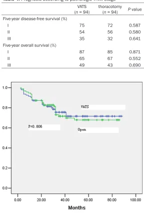
Table 4. Prognosis according to pathologic TNM stage
VATS
(n = 94) thoracotomy (n = 94) P value Five-year disease-free survival (%)
I 75 72 0.587
II 54 56 0.580
III 35 32 0.641
Five-year overall survival (%)
I 87 85 0.871
II 65 67 0.552
[image:5.612.91.374.83.211.2]III 49 43 0.690
Figure 2. There was no significant difference in overall survival between the
VATS and open groups (73% vs. 68%) (P = 0.806).
vious data [20-25], we found that VATS for non small cell lung cancer was very successful in achieving an R0 resection. Ta- ken together, these data dem-onstrate that VATS for non small cell lung cancer can achieve acceptable oncological surgical results.
lobec-In conclusion, VATS to clinical stage I non small cell lung cancer was associated with adequate mediastinal lymph node dissection, a high inci-dence of R0 resection, and comparable long-term oncological outcomes versus open radical lobectomy. As such, in appropriately selected patients, VATS should be considered a surgi-cally and oncologisurgi-cally appropriate approach to patients with clinical stage I non small cell lung cancer.
Acknowledgements
This study was supported by the National Natural Science Foundation of China (NO. 81301530), the scientific research project of Health Department of Heilongjiang Province (2012-602), and the scientific research project of Education Department of Heilongjiang Province (12541531).
Disclosure of conflict of interest
None.
Address correspondence to: Jinglong Yan, Depart-
ment of Surgery, The Second Affiliated Hospital
of Harbin Medical Universuity, 246 Xuefu Road, Harbin 150086, Hei Longjiang, People’s Republic of China. Tel: 86605612; Fax: +86-0451-86605612; E-mail: jinglongyanmed@163.com
a patient with complete situs inversus. Int J Clin Exp Med 2014; 7: 2928-2931.
[4] Siegel R, Naishadham D and Jemal A. Cancer statistics, 2013. CA Cancer J Clin 2013; 63: 11-30.
[5] She J, Yang P, Hong Q and Bai C. Lung cancer in China: challenges and interventions. Chest 2013; 143: 1117-1126.
[6] Tan Q, Huang J, Ding Z, Lin H, Lu S and Luo Q. Meta-analysis for curative effect of lobectomy and segmentectomy on non-small cell lung cancer. Int J Clin Exp Med 2014; 7: 2599-2604.
[7] Zhang X, Yan J, Ren Y, Shen C, Ying X and Pan S. Robot-assisted versus laparoscopic partial nephrectomy for localized renal tumors: a me-ta-analysis. Int J Clin Exp Med 2014; 7: 4770-4779.
[8] Sabuncuoglu MZ, Sabuncuoglu A, Sozen I, Benzin MF, Cakir T and Cetin R. Minimally inva-sive surgery using mini anterior incision for thy-roid diseases: a prospective cohort study. Int J Clin Exp Med 2014; 7: 3404-3409.
[9] Chen D, Cheng P, Ding D and Ke Z. Feasibility and safety of a novel reverse puncture device (RPD) for laparoscopic esophagogastrostomy/ esophagojejunostomy. Int J Clin Exp Med 2014; 7: 2497-2503.
[10] Dómine Gómez M, Morán Bueno T, Artal Cor-tés A, Remon Masip J and Lianes Barragán P. SEOM clinical guidelines for the treatment of small-cell lung cancer 2013. Clin Transl Oncol 2013; 15: 985-990.
[11] Camps C, Felip E, García-Campelo R, Trigo JM and Garrido P. SEOM clinical guidelines for the treatment of non-small cell lung cancer (NSCLC) 2013. Clin Transl Oncol 2013; 15: 977-984.
[12] Zhong C, Fang W, Mao T, Yao F, Chen W and Hu D. Comparison of thoracoscopic segmentecto-my and thoracoscopic lobectosegmentecto-my for small-sized stage IA lung cancer. Ann Thorac Surg 2012; 94: 362-367.
[image:6.612.90.330.94.199.2][13] Lee HS and Jang HJ. Thoracoscopic mediasti-nal lymph node dissection for lung cancer. Se-min Thorac Cardiovasc Surg 2012; 24: 131-141.
Table 5. Prognostic factors related to disease-free survival
Hazard
ratio 95% CI valueP Pathological N stage
N0-1 1.00
N2 1.78 1.31-3.45 0.008 Pathological venous invasion
Negative 1.00
Positive 2.08 1.36-3.30 0.005
Table 6. Prognostic factors related to overall survival
Hazard
ratio 95% CI P value Pathological T stage
T1-2 1.00
T3 1.88 1.18-2.98 0.023 Pathological N stage
N0-1 1.00
N2 3.48 2.01-5.88 0.001
References
[1] Chen G, Qiu X, Liu Y, Qiao Y, Shi T, Chen J and Zhou Q. Primary mediastinal
adenocarcino-ma originating from a calcified nodule. Int J
Clin Exp Med 2014; 7: 1898-1903.
[2] Zhang XD, Li W, Zhang N, Hou YL, Niu ZQ,
Zhong YJ, Zhang YP and Yang SY. Identifica -tion of adipophilin as a potential diagnostic tumor marker for lung adenocarcinoma. Int J Clin Exp Med 2014; 7: 1190-1196.
[image:6.612.92.287.245.348.2][14] Ramos R, Girard P, Masuet C, Validire P and Gossot D. Mediastinal lymph node dissection in early-stage non-small cell lung cancer: to-tally thoracoscopic vs thoracotomy. Eur J Car-diothorac Surg 2012; 41: 1342-1348.
[15] Yang H, Li XD, Lai RC, She KL, Luo MH, Li ZX and Lin YB. Complete mediastinal lymph node dissection in video-assisted thoracoscopic lo-bectomy versus lolo-bectomy by thoracotomy. Thorac Cardiovasc Surg 2013; 61: 116-123. [16] Rami-Porta R, Bolejack V, Giroux DJ, Chansky
K, Crowley J, Asamura H, Goldstraw P and In-ternational Association for the Study of Lung Cancer Staging and Prognostic Factors Com-mittee, Advisory Board Members and Partici-pating Institutions. The IASLC lung cancer stag-ing project: the new database to inform the
eighth edition of the TNM classification of lung
cancer. J Thorac Oncol 2014; 9: 1618-1624. [17] Tsim S, O’Dowd CA, Milroy R and Davidson S.
Staging of non-small cell lung cancer (NSCLC): a review. Respir Med 2010; 104: 1767-1774. [18] Clavien PA, Barkun J, de Oliveira ML, Vauthey
JN, Dindo D, Schulick RD, de Santibañes E, Pe-kolj J, Slankamenac K, Bassi C, Graf R, Vonlan-then R, Padbury R, Cameron JL and Makuuchi
M. The Clavien-Dindo classification of surgical complications: five-year experience. Ann Surg
2009; 250: 187-196.
[19] Watanabe A, Koyanagi T, Obama T Ohsawa H, Mawatari T, Takahashi N, Ichimiya Y and Abe T. Assessment of node dissection for clinical stage I primary lung cancer by VATS. Eur J Car-diothorac Surg 2005; 27: 745-752.
[20] Palade E, Passlick B, Osei-Agyemang T, Günter J and Wiesemann S. Video-assisted vs open mediastinal lymphadenectomy for Stage I non-small-cell lung cancer: results of a prospective randomized trial. Eur J Cardiothorac Surg 2013; 44: 244-249.
[21] Reck M, Heigener DF, Mok T, Soria JC and Rabe KF. Management of non-small-cell lung cancer: recent developments. Lancet 2013; 382: 709-719.
[22] D’Andrilli A, Venuta F and Rendina EA. The role of lymphadenectomy in lung cancer surgery. Thorac Surg Clin 2012; 22: 227-237.
[23] Scott WJ, Allen MS, Darling G, Meyers B, Deck-er PA, Putnam JB, McKenna RW, Landrenau RJ, Jones DR, Inculet RI and Malthaner RA. Video-assisted thoracic surgery versus open lobectomy for lung cancer: a secondary analy-sis of data from the American College of Sur-geons Oncology Group Z0030 randomized clinical trial. J Thorac Cardiovasc Surg 2010; 139: 976-981; discussion 981-983.
[24] Verhagen AF, Schoenmakers MC, Barendregt W, Smit H, van Boven WJ, Looijen M, van der Heijden EH and van Swieten HA. Complete-ness of lung cancer surgery: is mediastinal dis-section common practice? Eur J Cardiothorac Surg 2012; 41: 834-838.
[25] Murthy S. Video-assisted thoracoscopic sur-gery for the treatment of lung cancer. Cleve Clin J Med 2012; 79 Electronic Suppl 1: eS23-Es25.
[26] Lee PC, Nasar A, Port JL, Paul S, Stiles B, Chiu YL, Andrews WG and Altorki NK. Long-term sur-vival after lobectomy for non-small cell lung cancer by video-assisted thoracic surgery ver-sus thoracotomy. Ann Thorac Surg 2013; 96: 951-960.
[27] Thomas P, Doddoli C, Yena S, Thirion X, Sebag F, Fuentes P, Giudicelli R. VATS is an adequate oncological operation for stage I non-small cell lung cancer. Eur J Cardiothorac Surg 2002; 21: 1094-1099.
[28] Shiraishi T, Shirakusa T, Hiratsuka M, Yama-moto S and Iwasaki A. Video-assisted thoraco-scopic surgery lobectomy for c-T1N0M0 pri-mary lung cancer: its impact on locoregional control. Ann Thorac Surg 2006; 82: 1021-1026.
[29] Flores RM, Park BJ, Dycoco J, Aronova A, Hirth Y, Rizk NP, Bains M, Downey RJ and Rusch VW. Lobectomy by video-assisted thoracic surgery (VATS) versus thoracotomy for lung cancer. J Thorac Cardiovasc Surg 2009; 138: 11-18. [30] Stephens N, Rice D, Correa A, Hoffstetter W,