Original Article
Enhancement of Ki67 peptide-based vaccine potency
by GP96 protein promotes anti-tumor
effect in osteosarcoma
Bingnan Guo1,2*, Lin Liu2*, Huizhong Li3, Liwei Ding2, Zhuan Yan2, Shuqun Hu2, Xianliang Yan2, Junnian Zheng3, Tie Xu2
1Institute of Biology and Medical Sciences, School of Biology and Basic Medical Science, Soochow University,
Suzhou, China; 2Institute of Emergency & Rescue Medicine, Xuzhou Medical College, Xuzhou, China; 3Jiangsu
Center for The Collaboration and Innovation of Cancer Biotherapy, Cancer Institute, Xuzhou Medical College, Xuzhou, China. *Equal contributors.
Received October 26, 2015; Accepted January 9, 2016; Epub February 15, 2016; Published February 29, 2016
Abstract: Peptide-based vaccines enable the safe and precisely directed immune intervention in controlling human cancers. Ki67280-288 peptide plus with GP96 protein is a promising vaccine for osteosarcoma (OS) immunotherapy. To improve the efficacy of Ki67280-288 peptide vaccination, we exploited a heat shock protein, namely, GP96 as an
adjuvant. In vitro, peptide-specific HLA-A02+ lymphocytes from dendritic cell (DC) loading Ki67
280-288 plus with GP96
produced high perforin. Furthermore, the production of IFN-γ was significantly enhanced both in vitro and in vivo. Moreover, cytotoxicity assay demonstrated that peptide-specific HLA-A02+ lymphocytes could enhance anti-OS
activ-ity in vitro. Finally, Ki67280-288 plus GP96 was an efficient agent for OS immunotherapy in the prophylaxis transgenic mouse model. Results demonstrated that Ki67280-288 plus GP96 could be a promising agent for OS immunotherapy.
Keywords: Ki67280-288 peptide, GP96 protein, osteosarcoma (OS), HLA-A02+,Dendritic cells (DCs)
Introduction
Osteosarcoma (OS) is the most common agg- ressive malignancy of the bone. OS accounts for 15% to 35% of all primary bone tumors [1, 2] and remains the second leading cause of can-cer-related death in children and young adults [3]. Despite current treatments with wide surgi-cal resection of the tumor and post-operative adjuvant chemotherapy, OS is prone to relapse and has a 20% chance of causing pulmonary metastases [4]. Once metastases have occu- rred, the prognosis will become poor and cur-rent therapeutic strategies will have limited effi-cacy. Furthermore, the prognosis of patients with recurrent disease is dismal. Therefore, new therapeutic approaches are required to treat OS. Tumor immunotherapy is an alterna-tive treatment for achieving local cancer con-trol and reducing the risk of recurrence. However, reports on immunotherapy in OS treatment are lacking.
In this study, we utilized Ki67280-288 peptide plus with adjuvant GP96 vaccination to elucidate the effects and possible mechanisms involved in anti-tumor immune responses both in vivo and in vitro. Our results showed that Ki67280-288 plus GP96 was an efficient agent for OS immu-notherapy in prophylaxis transgenic mouse model. Furthermore, Ki67280-288 plus GP96 enhanced tumor-specific CTL responses. Ki67280-288 plus GP96 promoted IFN-γ produc-tion of lymphocytes both in vitro and in vivo. In conclusion, our result provided evidence that
Ki67280-288 plus GP96 could augment the anti-tumor effects of Ki67280-288 application and could be a promising agent for OS immuno- therapy.
Materials and methods
Regents, antibodies and animals
rhGM-CSF, IL-4, and TNF-α were purchased from PeproTech (USA). rhIL-2 was purchased from Beijing ShuangLu Pharmaceutical Co., LTD (China). APC-conjugated or FITC-con- jugated anti-human HLA-A2, APC-conjugated human HLA-DR, PE-CY5-conjugated anti-human CD80, FITC-conjugated anti-anti-human CD86, and Percp-cy5.5-conjugated anti-perfo-rin antibodies, human IL-1β, IL-6, IL-12, IFN-γ ELISA Kit and cell permeabilization kit were purchased from BD (USA). GP96 protein was purchased from ENZO (USA). Ki-67 antibody (H-300) and GP96 antibody were purchased from Santa Cruz (USA), ABCAM (UK) respective-ly. HRP-conjugated or FITC-conjugated goat anti-rabbit IgG was purchased from Sigma (USA). The C57BL/6-Tg (HLA-A2.1) mice, which were transgenic human HLA-A2 gene, pur-chased from model animal research center of Nanjing University.
Cell lines and cell culture
The human OS cell line U2OS was purchased from the cell bank of the Chinese Academy of Science (Shanghai China). U2OS cells were maintained in RPMI-1640 (Gibco) medium con-taining 10% FCS (Gibco) and stored at 37°C in a humidified atmosphere of 5% CO2 in air.
Ki67280-288 synthesis
The Ki-67 sequence was scanned for HLA-A*0201-binding peptides by the prediction so- ftware Bioinformatics and Molecular Analysis (BIMAS, NIH, USA) as described before and
confirmed HLA-A*0201-binding Ki67 peptides (LQGETQLLV) [9] was synthesized by Shanghai Bioengineering Ltd. Co. (Shanghai, China) with a purity of >90% as verified by HPLC and MS analysis. Lyophilized peptides were dissolved in DMSO (Sigma) and the aliquot of peptides were stored at -80°C.
Immunofluorescence
U2OS cell line was seeded and grown onto glass coverslips in 24-well culture plates (5 × 10⁴ cells/cm2). Cells were fixed with 4% parafor-maldehyde in HPEM buffer (25 mM HEPES, 60 mM PIPES, 10 mM EGTA, 1 mM MgCl2) for 20 min at room temperature (RT) overnight in a humidified atmosphere of 5% CO2 in air and then further permeabilized in PBS/Triton X-100 0.1% for 20 min. Bovine serum albumin (5% in PBS/Tx-100 0.005%) was used for 30 min at RT as a blocking step prior to incubation with pri-mary antibodies. Ki67 antibody was diluted in a blocking solution and incubated at the recom-mended periods, followed by incubation wi- th secondary antibodies. DAPI (4,6-diamidino-2-phenylindole) was used to stain DNA. Special attention was taken to prevent fluorochrome fading by using a dark humidified chamber in all incubation steps.
SDS-PAGE and Western blot analysis
Approximately 2 µg GP96 protein were solubi-lized in SDS polyacrylamide gel electrophoresis (SDS-PAGE) loading buffer (250 mM Tris-HCl, pH 6.8, 0.5 M DTT, 10% SDS, 0.5% bromophe-nol blue, 50% Glycerol), boiled for 10 min, sepa-rated by SDS-PAGE, and/or transferred to PVDF membranes. Membranes were probed with anti-rabbit polyclonal GP96 antibodies diluted 1:1000 in 5% BSA and incubated with goat anti-rabbit IgG conjugated to horseradish per-oxidase secondary antibodies. Immunoreactive bands were visualized by enhanced chemilumi-nescence reaction.
Identification of PBMCs and U2OS cell line
we considered the one whose cell count was greater than 80% as belonging to HLA-A*0201 positive population. The HLA-A*0201 positive population was pooled together and/or frozen for further study.
Generation and maturation of human mono-cyte derived dendritic cells
Approximately 10 ml to 30 ml peripheral venous blood was drawn from healthy volunteers who underwent physical examination in the Affiliated Hospital of Xuzhou Medical College, Jiangsu, China. Consent from volunteers was obtained according to protocol approved by the review board of the Affiliated Hospital of Xuzhou Medical College. Identified HLA-A*0201+ PB- MCs were isolated from the blood by Ficoll-Urografin density gradient centrifugation and the monocytes were isolated by positive selec-tion with CD14-conjugated microbeads (Milten- yi, Germany) and magnetic activated cell sorter. The rest of lymphocytes were cultured or frozen in RPMI-1640 medium with 30% FCS and 10% DMSO. Monocytes of more than 95% purity were cultured at a concentration of 1 × 10⁶ cells/ml in a six-well plate (Becton Dickinson, USA) in DMEM containing 10% FBS and 1% penicillin/streptomycin supplemented with GM- CSF (50 ng/ml) and IL-4 (20 ng/ ml). On Day 5, immature dendritic cells (imDCs) were added with TNF-α (100 ng/ml) or PBS for 2 d.
Evaluation of cytokines secretion
For cultured DC supernatants, on Day 7, the supernatants were harvested to evaluate the concentrations of human IL-1β, IL-6, and IL-12. For IFN-γ, one week after the implantation, mouse serum was collected from the peripher-al blood to evperipher-aluate the concentrations of serum IFN-γ. Cytokines level were determined by ELISA kit (BD, USA), according to manufac-turer protocol.
Dendritic cell loading Ki67280-288 plus with gp96
After the collection of mature dendritic cells (DCs), 5 × 10⁶ in 2 ml RPMI supplemented with GM-CSF (50 ng/ml), IL-4 (20 ng/ml), and TNF-α (100 ng/ml) were incubated for 24 h with Ki67280-288 (40 µg/ml) plus GP96 (40 µg/ml), Ki67280-288 (40 µg/ml) alone, or GP96 alone (40 µg/ml), respectively. Negative controls were
treated with PBS alone. Every 24 h, the cell medium was changed, the cells were washed, and repeated loading was conducted thrice.
Induction of peptide-specific lymphocytes with DCs loading peptides
Approximately 1 × 10⁷ HLA-A*0201 positive lymphocyteswere incubated with 5 × 10⁶ DC loading Ki67280-288 (40 µg/ml) plus with GP96 (40 µg/ml), Ki67280-288 (40 µg/ml) alone, GP96 (40 µg/ml) alone, or PBS in RPMI-1640 medi-um at 37°C in hmedi-umidified atmosphere contain-ing 5% CO2. Recombinant interleukin 2 (rhIL-2) at a concentration of 500 U/ml was added into the medium on the second day to stimulate lymphocytes for 7 d and repeated stimulation was conducted twice. After the second stimula-tion, the suspended lymphocytes were collect-ed and percp-cy5.5-conjugatcollect-ed anti-perforin was stained with lymphocytes for analysis.
Cytotoxicity assays
CytoTox 96 nonradioactive cytotoxicity assay (Promega, Madison, WI) was used to measure the cytotoxic activity of lymphocytes, according to manufacturer protocol. Briefly, target cells U2OS (6 × 10³ cells/well) were placed on 96- well U-bottom plates (Corning Costar, Cam- bridge, MA, USA), and lymphocytes (effectors) were added to a final volume of 100 µl in ratios of 100:1, 50:1, and 25:1 (E: T ratio). The plates were then incubated for 6 h in a humidified 5% CO2 chamber at 37°C and centrifuged at 500×g for 5 min. Aliquots (50 μl) were transferred from all wells to fresh 96-well flat-bottom plates, and an equal volume of reconstituted substrate mix was added into each well. The plates were then incubated in the dark at RT for approximately 30 min. A 50 μl stop solution was added, and the absorbance was measured at 492 nm. The cell death percentages at each E: T ratio were calculated by using the following formula: (A492 nm [experimental]-A492 nm [effector spontane- ous]-A492 nm [target spontaneous]) × 100/ (A492 nm [target maximum]-A492 nm [target spontaneous]).
Animal model
Committee of Soochow University and the Affiliated Hospital of Xuzhou Medical College. For prophylaxis experiments, the mice were immunized s.c. at the back with Ki67280-288 (100 µg) plus with GP96 (100 µg), Ki67280-288 (100 µg), GP96 (100 µg), or 200 µl PBS for a total of three times with 1-wk intervals. Two weeks after the last immunization, 1 × 10⁷ U2OS cells were s.c. implanted into the right flank of mice. The tumor sizes were measured using a caliper every 3 days from day 3 post challenge. Tumor volumes were calculated using the formula V=1/2 (L × W2), where L is the length (longest dimension), and W is the width (shortest dimension).
Statistics
All data were analyzed by Student t test and were expressed as means ± SD or means ± SEM; data were analyzed using GraphPad Pri- sm 6 software for Windows (GraphPad Soft- ware, San Diego, CA), and differences were con-sidered statistically significant when P<0.05.
The significance levels are marked *P<0.05 and **P<0.01.
Result
Osteosarcoma cell line U2OS expresses Ki67, HLA-A2, and highly detectable HLA-A2 on PBMCs
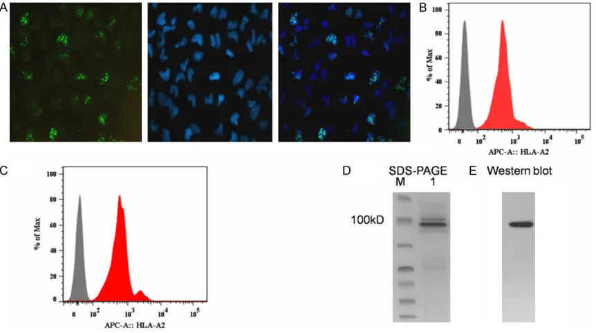
Whether Ki67, a promising molecular target in cancers [15, 16] and a novel immunogen [9], is expressed in OS is still unclear. Immunofluo- rescence assay was conducted to address it.
[image:4.612.93.524.73.314.2]study. Furthermore, we identified GP96 protein by SDS-PAGE and immunoblotting analysis (Figure 1D and 1E). Taken together, both Ki67 and HLA-A2 were expressed on U2OS cell line and PBMCs were found to have high HLA-A2 expression.
DCs maturation and related cytokines re-leased
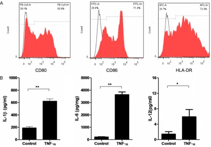
DCs are professional APC with significant phe-notypic heterogeneity and functional plasticity. DCs play important roles in initiating effective adaptive immune responses for elimination of invading pathogens [18]. On Day 7, cultured HLA-A2+ monocytes-derived DCs surface ma-rker CD80 (60.9%), CD86 (71.4%), HLA-DR (73.3%) were detected by FACS as shown in
Figure 2A. Moreover, pro-inflammatory cyto-kines expression significantly increased, such as IL-1β, IL-6, and IL-12 in DCs supernatant
(Figure 2B) compared with untreated DCs (no TNF-α) supernatant. The results suggested DC maturation.
Perforin secretion
As DCs maturation, they were loaded with Ki67280-288 plus GP96 (DCKi67280-288 plus GP96), Ki67280-288 (DCKi67280-288), GP96 (DCGP96), or PBS. The loading DCs were then cocultured with HLA-A2+ lymphocytes for 7 d, which was repe- ated 2 times. Perforin secretion from the lym-phocytes was analyzed by FACS. As shown in
[image:5.612.92.523.71.372.2]T-cells, which suggested that highly secret-
[image:6.612.93.522.72.320.2]ed perforin in lymphocytes cocultured with DCT-cells, particularly CD8Ki67280-288 plus GP96 group was perhaps due to + T-cells. Figure 3.Activity of peptide-specific lymphocytes with mature DCs loading peptides. A. Intracellular perforin produc-tion was measured from lymphocytes, which were cocultured with DCKi67280-288 plus GP96, DCKi67280-288, DCGP96 or PBS respectively. B. The percentages of perforin production in cocultured lymphocytes. Values are presented as means ± SD. The data shown are the representative of three experiments. *P<0.05.
[image:6.612.93.523.394.585.2]Cytotoxicity assay and IFN-γ production in vitro
DCKi67280-288 plus GP96 may promote lymphocytes, especially T-cell maturation (perforin as a matu-ration marker). To test whether co-cultured lym-phocytes was against human OS cell line-U2OS in vitro, the U2OS cell line was employed as tar-get cell to address it. Stimulated lymphocytes were used in the cytotoxicity assay against U2OS targets. Lymphocytes from DCKi67280-288 stimulated exhibited significantly higher cyto-toxicity against U2OS cells than DCGP96 and PBS treatment U2OS groups. Notably, cytotoxicity was further enhanced as lymphocytes from DCKi67280-288 plus GP96 group (Figure 4A). IFN-γ is an
[image:7.612.95.521.74.207.2]GP96 in vivo, murine OS model was used. C57BL/6-Tg (HLA-A2.1) mice, which were trans-genic human HLA-A2 gene (Figure 5A), were immunized with Ki67280-288 plus GP96, Ki67 280-288, GP96, or PBS, 3 times with 1 wk intervals, and then, the mice were challenged with 1 × 10⁷ human OS cell line U2OS two weeks after the last immunization and were monitored tumor size for 30 days. Mice immunized with Ki67280-288 plus GP96 group and Ki67280-288 group had significant reduction in tumor grow- th compared with mice immunized with PBS group (Figure 5B). Moreover, immunization with Ki67280-288 plus GP96 group further reduced tumor growth compared with immunization Figure 5.Anti-tumor response of Ki67 peptide-based vaccination in vivo. A. The phenotype of C57BL/6-Tg (HLA-A2.1) mice which were transgenic human HLA-A2 gene was determined by flow cytometry. B. Tumor growth curves of osteosarcoma model. Mice were immunized with Ki67280-288 plus GP96, Ki67280-288, GP96, or PBS, 3 times with
1-wk intervals, and then, the mice were challenged with 1×10⁷ human osteosarcoma cell line-U2OS, 2 wk after the
last immunization. The tumor volumes were monitored every 3 days. The experiments were performed with five to seven mice per group. The assays were done in triples. Values are presented as means ± SEM. The data shown are the representative of three experiments *P<0.05.
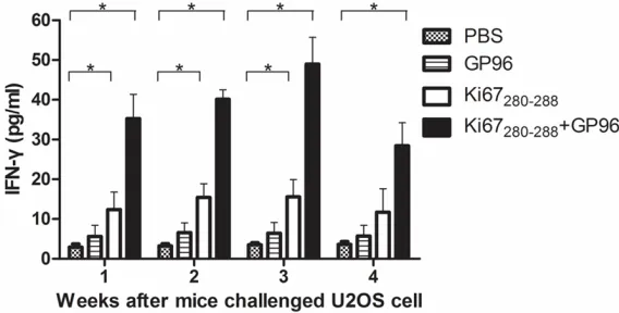
Figure 6.Concentrations of IFN-γ in tumor-burdened C57BL/6-Tg (HLA-A2.1) mice. Serum IFN-γ obtained from peripheral blood 1 week, 2 weeks, 3 weeks and 4 weeks after challenged with U2OS cells were determined by ELISA. Values are presented as means ± SEM. The data shown are the representa-tive of three experiments. *P<0.05, n=5.
essential cytokine for immu-nity against cancer and patho-gens. We examined lympho-cytes responses by in vitro stimulation with U2OS cell line and supernatant IFN-γ by ELISA. IFN-γ was significantly higher in lymphocytes from DCKi67280-288 plus GP96 group than the other groups (P<0.05) (Figure4B).
Ki67280-288 plus GP96 vaccine
induced osteosarcoma pro-tection in vivo
[image:7.612.91.375.312.456.2]with Ki67280-288 group. Serum IFN-γ, also, obtained from the mice’ peripheral blood 1 week, 2 weeks, 3 weeks and 4 weeks after challenged with U2OS cells were determined by ELISA. During the period, the serum concentra-tions IFN-γ increase significantly in most mice. One week after last challenge, IFN-γ of Ki67 280-288 group was significantly higher than other groups except Ki67280-288 plus GP96 group, reaching the highest in the 2nd week and decreased thereafter, while IFN-γ of Ki67280-288 plus GP96 group witnessed a continuous ele-vation and reached almost the peak in the 3rd week but interestingly decreased in the 4th week (Figure 6).
Discussion
Immunotherapy has become an attractive ther-apeutic modality for cancer, which offers poten-tially targeted therapy with fewer adverse effects compared with conventional therapy [19]. Tumor associated antigens (TAAs) that can be targeted by CTL are fundamental con-cepts behind developing current immunothera-py. Three characteristics are required for an ideal TAA to elicit effective and safe T-cell-mediated anti-tumor immunity in cancer pa- tients [20]: 1) Cancer-specific expression of TAA. An ideal TAA must be overexpressed in tumors, but not or low expressed on normal cells, such as cancer-testis antigen HER-2/neu, Ki67. 2) Oncogenic characteristics of TAA, which is involved in oncogenesis, are consid-ered to be barely lost in the process of tumor progression, such as p53, Ki67. 3) Immuno- genicity of TAA is required. Immune responses could be evaluated by detection of TAA-specific IgG or TAA-specific T-cell responses, such as IFN-γ, TNF-α, and IL-2 secretion. TAA-peptide-based immunotherapy is a promising approach to successfully induce anti-tumor immune responses and prolong survival in patients with various cancers [21-24]. However, without suit-able adjuvants, a small synthetic peptide or a recombinant protein can elicit only a weak CTL response in this process. For instance, Rosenberg et al. [25] reported that the clinical effect of vaccination of TAA-derived CTL-epitope peptides (short peptides) alone was limited in a very small fraction of advanced melanoma patients. Therefore, antigens need to be com-bined with an efficient adjuvant for use in humans.
HSPs have been demonstrated to act as po- tent adjuvants in immunotherapy of malignant tumors [26] and infectious diseases [27]. GP96, as a HSP, has emerged as an attractive adjuvant component for vaccines. GP96, the major chaperon in the lumen of the endoplas-mic reticulum involved in cross presentation of peptides to MHC class I and class II molecules, activates specific CD8+ and CD4+ T-cells [28, 29]. Moreover, GP96 can activate dendritic cells and macrophages through induction of proinflammatory cytokines via interaction with a subset of toll-like receptors [30]. Previous reports showed that GP96 or its N-terminal domain had an adjuvant function in tumor [31] and virus [32] specific CTL and humoral immune responses. HLA-A*0201 is an important HLA class I allele in Asian population. In our previ-ous study, Ki67280-288 peptide, which was identi-fied to be well recognized by CD8+ T-cells in can-cer patients expressing HLA-A*0201 mole-cules, was shown to be targets for HLA-A*0201-restricted CD8+ T-cells that produced generous IFN-γ.
In the current study, HLA-A*0201 expression, firstly, was assessed with both PBMCs of healthy donors and OS cell line-U2OS. As expected, HLA-A*0201 was highly expressed in U2OS cell line and PBMCs of partial volunteers that were pooled together upon identification of HLA-A*0201+ (Figure 1B and 1C). As a TAA, Ki67 was confirmed overexpressed in U2OS cell line by immunofluorescence assay in this study (Figure 1A). Our result suggested that the U2OS cell line, as an OS model, could be fur-ther studied in our system. Our hypothesis was that DCs maturation could facilitate Ki67280-288 peptide presentation. Thus, human monocyte derived DC was cultured in vitro with GM-CSF and IL-4. On Day 5, TNF-α instead of LPS was added for DC maturation. Immunophenotype of maturation marker was up-regulated, such as CD80, CD86, and HLA-DR (Figure 2A) on Day 7. Moreover, pro-inflammatory cytokines, which were produced by DC maturation, produced IL-1β, IL-6, and IL-12 which were enhanced on Day 7 (Figure 2B).
enhance-ment of the function of CTL responses, perforin (Figure 3A) and IFN-γ upregulation (Figure 4B). Strikingly perforin (Figure 3A) and IFN-γ upregu-lation (Figure 4B) in DCKi67280-288 plus GP96 group was significantly higher compared with those in DCKi67280-288 group. Notably, lymphocytes form DC loading Ki67280-288 peptide displayed signifi-cantly high cytotoxicity against U2OS in vitro, which was future enhanced as lymphocytes from DCKi67280-288 plus GP96 group (Figure 4A). The results are consistent with the findings that C-terminal fusion of GP96 to Her2 could en- hance efficacy of Her2 DNA vaccine [33]. Furthermore, Leila et al. [34] found that an N-terminal fusion of GP96 fragment plus HCV vaccine enhanced HCV-specific CTL immune responses. However, both Pakravan and Leila performed assay with DNA vaccine. In our experiments, we directly employed DC loading GP96 protein and Ki67280-288 peptide as a vac-cine. Overall, enhanced immune responses observed for an immunogen plus GP96 were consistent with others. Remarkably, in prophy-laxis transgenic mouse model, mice immunized with Ki67280-288 peptide plus with GP96 group revealed significant tumor reduction than other groups (Figure 5B). Mounting data showed that T cell-mediated tumor rejection depend not only upon perforin, granzyme and Fas/FasL but on some soluble cytokines such as IFN-γ [35]. During our treatment in vivo, IFN-γ production of Ki67280-288 peptide plus GP96 group is the highest than that of other groups, suggesting they promote DC-primed T cell polarization to Th1 (Figure 6). Ki67280-288 vaccinated mice as well as has high level IFN-γ. However, the pre-cise mechanism of protection effect in vivo, which our efforts will focus on, should be fur-ther investigated.
In conclusion, we utilized Ki67280-288 peptide plus with adjuvant GP96 vaccination to eluci-date effects and possible mechanisms involved in anti-tumor immune responses both in vivo and in vitro. Our results showed that Ki67280-288 plus GP96 was an efficient agent for OS immu-notherapy in prophylaxis transgenic mouse model, which perhaps enhanced tumor-specific CTL responses (IFN-γ production). The results showed promotion of IFN-γ and perforin pro-duction of lymphocytes and enhanced cytotox-icity against U2OS in vitro. Our results warrant future studies for the development of an
effec-tive cancer vaccine based on the current strategies.
Acknowledgements
This study was supported by grants from National Foundation of Nature Science of China (81202015), the Science Foundation of Xuzhou Science & Technology bureau (KC15J0060, KC15SX009, XM12B025, KC14- SH087).
Disclosure of conflict of interest
None.
Address correspondence to: Dr. Tie Xu, Institute of Emergency & Rescue Medicine, Xuzhou Medical College, Xuzhou 221002, China. E-mail: xutie889@ 163.com
References
[1] Mirabello L, Troisi RJ and Savage SA. Osteosarcoma incidence and survival rates from 1973 to 2004: data from the Surveillance, Epidemiology, and End Results Program. Cancer 2009; 115: 1531-1543.
[2] Moore DD and Luu HH. Osteosarcoma. Cancer Treat Res 2014; 162: 65-92.
[3] Goldsby R, Burke C, Nagarajan R, Zhou T, Chen Z, Marina N, Friedman D, Neglia J, Chuba P and Bhatia S. Second solid malignancies among children, adolescents, and young ad- ults diagnosed with malignant bone tumors af-ter 1976: follow-up of a Children’s Oncology Group cohort. Cancer 2008; 113: 2597-2604. [4] Bielack SS, Carrle D, Hardes J, Schuck A and
Paulussen M. Bone tumors in adolescents and young adults. Curr Treat Options Oncol 2008; 9: 67-80.
[5] Lu W, Qiu L, Yan Z, Lin Z, Cao M, Hu C, Wang Z, Wang J, Yu Y, Cheng X, Cao P and Li R. Cytotoxic T cell responses are enhanced by antigen de-sign involving the presentation of MUC1 pep-tide on cholera toxin B subunit. Oncotarget 2015; 6: 34537-48.
[6] Derouazi M, Di Berardino-Besson W, Belnoue E, Hoepner S, Walther R, Benkhoucha M, Teta P, Dufour Y, Yacoub Maroun C, Salazar AM, Martinvalet D, Dietrich PY and Walker PR. Novel Cell-penetrating peptide-based vaccine induces robust CD4+ and CD8+ T cell-mediat-ed antitumor immunity. Cancer Res 2015; 75: 3020-3031.
[8] Gerdes J, Schwab U, Lemke H and Stein H. Production of a mouse monoclonal antibody reactive with a human nuclear antigen associ-ated with cell proliferation. Int J Cancer 1983; 31: 13-20.
[9] Xu W, Li HZ, Liu JJ, Guo Z, Zhang BF, Chen FF, Pei DS and Zheng JN. Identification of HLA-A*0201-restricted cytotoxic T lymphocyte epit-ope from proliferating cell nuclear antigen. Tumour Biol 2011; 32: 63-69.
[10] Zhao R, Davey M, Hsu YC, Kaplanek P, Tong A, Parsons AB, Krogan N, Cagney G, Mai D, Greenblatt J, Boone C, Emili A and Houry WA. Navigating the chaperone network: an integra-tive map of physical and genetic interactions mediated by the hsp90 chaperone. Cell 2005; 120: 715-727.
[11] Taipale M, Krykbaeva I, Koeva M, Kayatekin C, Westover KD, Karras GI and Lindquist S. Quantitative analysis of HSP90-client interac-tions reveals principles of substrate recogni-tion. Cell 2012; 150: 987-1001.
[12] Ju Y, Fan H, Liu J, Hu J, Li X, Li C, Chen L, Gao Q, Gao GF and Meng S. Heat shock protein gp96 adjuvant induces T cell responses and cross-protection to a split influenza vaccine. Vaccine 2014; 32: 2703-2711.
[13] Crane CA, Han SJ, Ahn B, Oehlke J, Kivett V, Fedoroff A, Butowski N, Chang SM, Clarke J, Berger MS, McDermott MW, Prados MD and Parsa AT. Individual patient-specific immunity against high-grade glioma after vaccination with autologous tumor derived peptides bound to the 96 KD chaperone protein. Clin Cancer Res 2013; 19: 205-214.
[14] Guo QY, Yuan M, Peng J, Cui XM, Song G, Sui X and Lu SB. Antitumor activity of mixed heat shock protein/peptide vaccine and cyclophos-phamide plus interleukin-12 in mice sarcoma. J Exp Clin Cancer Res 2011; 30: 24.
[15] Li XQ, Pei DS, Qian GW, Yin XX, Cheng Q, Li LT, Li HZ and Zheng JN. The effect of methylat- ed oligonucleotide targeting Ki-67 gene in hu-man 786-0 renal carcinoma cells. Tumour Biol 2011; 32: 863-872.
[16] Li LT, Jiang G, Chen Q and Zheng JN. Ki67 is a promising molecular target in the diagnosis of cancer (review). Mol Med Rep 2015; 11: 1566-1572.
[17] Li Y, Zhou L, Li Y, Zhang J, Guo B, Meng G, Chen X, Zheng Q, Zhang L, Zhang M and Wang L. Identification of autoreactive CD8+ T cell re-sponses targeting chromogranin A in human-ized NOD mice and type 1 diabetes patients. Clin Immunol 2015; 159: 63-71.
[18] Liu J and Cao X. Regulatory dendritic cells in autoimmunity: A comprehensive review. J Autoimmun 2015; 63: 1-12.
[19] Couzin-Frankel J. Breakthrough of the year 2013. Cancer immunotherapy. Science 2013; 342: 1432-1433.
[20] Nishimura Y, Tomita Y, Yuno A, Yoshitake Y and Shinohara M. Cancer immunotherapy using novel tumor-associated antigenic peptides identified by genome-wide cDNA microarray analyses. Cancer Sci 2015; 106: 505-511. [21] Shariat S, Badiee A, Jalali SA, Mansourian M,
Yazdani M, Mortazavi SA and Jaafari MR. P5 HER2/neu-derived peptide conjugated to lipo-somes containing MPL adjuvant as an effec-tive prophylactic vaccine formulation for breast cancer. Cancer Lett 2014; 355: 54-60. [22] Kumari S, Walchli S, Fallang LE, Yang W,
Lund-Johansen F, Schumacher TN and Olweus J. Alloreactive cytotoxic T cells provide means to decipher the immunopeptidome and reveal a plethora of tumor-associated self-epitopes. Proc Natl Acad Sci U S A 2014; 111: 403-408. [23] Shen H, Shao HW, Chen XH, Wu FL, Wang H,
Huang ZL, Shen J, Wang T, Zhang WF and Huang SL. Identification of a novel HLA-A2-restricted mutated Survivin epitope and induc-tion of specific anti-HCC CTLs that could effec-tively cross-recognize wild-type Survivin anti-gen. Cancer Immunol Immunother 2013; 62: 393-403.
[24] Tomita Y, Imai K, Senju S, Irie A, Inoue M, Hayashida Y, Shiraishi K, Mori T, Daigo Y, Tsunoda T, Ito T, Nomori H, Nakamura Y, Kohrogi H and Nishimura Y. A novel tumor-as-sociated antigen, cell division cycle 45-like can induce cytotoxic T-lymphocytes reactive to tu-mor cells. Cancer Sci 2011; 102: 697-705. [25] Rosenberg SA, Yang JC and Restifo NP. Cancer
immunotherapy: moving beyond current vac-cines. Nat Med 2004; 10: 909-915.
[26] Bolhassani A and Rafati S. Heat-shock pro-teins as powerful weapons in vaccine develop-ment. Expert Rev Vaccines 2008; 7: 1185-1199.
[27] Wood CG and Mulders P. Vitespen: a preclini-cal and clinipreclini-cal review. Future Oncol 2009; 5: 763-774.
[28] Doody AD, Kovalchin JT, Mihalyo MA, Hagymasi AT, Drake CG and Adler AJ. Glycoprotein 96 can chaperone both MHC class I- and class II-restricted epitopes for in vivo presentation, but selectively primes CD8+ T cell effector func-tion. J Immunol 2004; 172: 6087-6092. [29] Javid B, MacAry PA and Lehner PJ. Structure
and function: heat shock proteins and adap-tive immunity. J Immunol 2007; 179: 2035-2040.
[30] Yang Y, Liu B, Dai J, Srivastava PK, Zammit DJ, Lefrancois L and Li Z. Heat shock protein gp96 is a master chaperone for toll-like receptors and is important in the innate function of mac-rophages. Immunity 2007; 26: 215-226. [31] Baker-LePain JC, Sarzotti M, Fields TA, Li CY
tissue nonrestricted tumor suppression. J Exp Med 2002; 196: 1447-1459.
[32] Li H, Zhou M, Han J, Zhu X, Dong T, Gao GF and Tien P. Generation of murine CTL by a hepatitis B virus-specific peptide and evaluation of the adjuvant effect of heat shock protein glycopro-tein 96 and its terminal fragments. J Immunol 2005; 174: 195-204.
[33] Pakravan N, Hashemi SM and Hassan ZM. Adjuvant activity of GP96 C-terminal domain towards Her2/neu DNA vaccine is fusion direc-tion-dependent. Cell Stress Chaperones 2011; 16: 41-48.
[34] Pishraft-Sabet L, Kosinska AD, Rafati S, Bolhassani A, Taheri T, Memarnejadian A, Alavian SM, Roggendorf M and Samimi-Rad K. Enhancement of HCV polytope DNA vaccine ef-ficacy by fusion to an N-terminal fragment of heat shock protein gp96. Arch Virol 2015; 160: 141-152.